Embed Size (px)
Citation preview
NEW ASPECTS IN THE BIOLOGY OF FISH PATHOGEN Photobacterium damselae subsp. piscicida:
PILI, MOTILITY AND ADHERENCE TO SOLID SURFACES
Ana Franco-González-de Canales1, Sara Remuzgo-Martínez1, María Lázaro-Díez1, D. Padilla2, B. Vega2, R. Agregán-Pérez3, Carmen Lobo4, Inés García de la Banda4, JM. Icardo5, F. Acosta2, José Ramos-Vivas1
1Laboratorio de Microbiología, Instituto de Investigación Valdecilla-IDIVAL, Santander, Cantabria, Spain. E-mail: 2Instituto Universitario de Sanidad Animal, Universidad de Las Palmas de Gran Canaria, Arucas, Spain.
3Departamento de Química Analítica y Alimentaria, Facultad de Ciencias, Universidad de Vigo, Ourense, Spain. 4 Instituto Español de Oceanografía IEO, Santander, Spain
5Departamento de Anatomía y Biología Celular, Universidad de Cantabria, Santander, Cantabria, Spain.
We have shown that Phdp, a facultative intracellular fish pathogen, expresses pilus-like surface structures resembling in several aspects other bacteria surface appendages. To our knowledge, such appendages have not been described previously in this species. Although historically Phpd is described as non motile, the strains used in this study migrated in the medium-plastic interface, referred to as twitching motility. We report here that Phdp forms biofilms on submerged surfaces and not at the air-liquid interface on microtiter plates. Biofilm production was found to be strongly dependent on incubation time and culture medium, as well as on the strain used. Microscopic examination of biofilms by SEM and CLSM revealed that Phdp displays extensive cellular chaining and cell elongation during biofilm formation in vitro. Based on our results, standardized analyses of Phdp surface appendages, biofilms, motility and their impact on Phdp survival, ecology and pathobiology are now more feasible, and will help to open up new research areas in several fields of the ecology and the pathogenesis of this important fish pathogen.
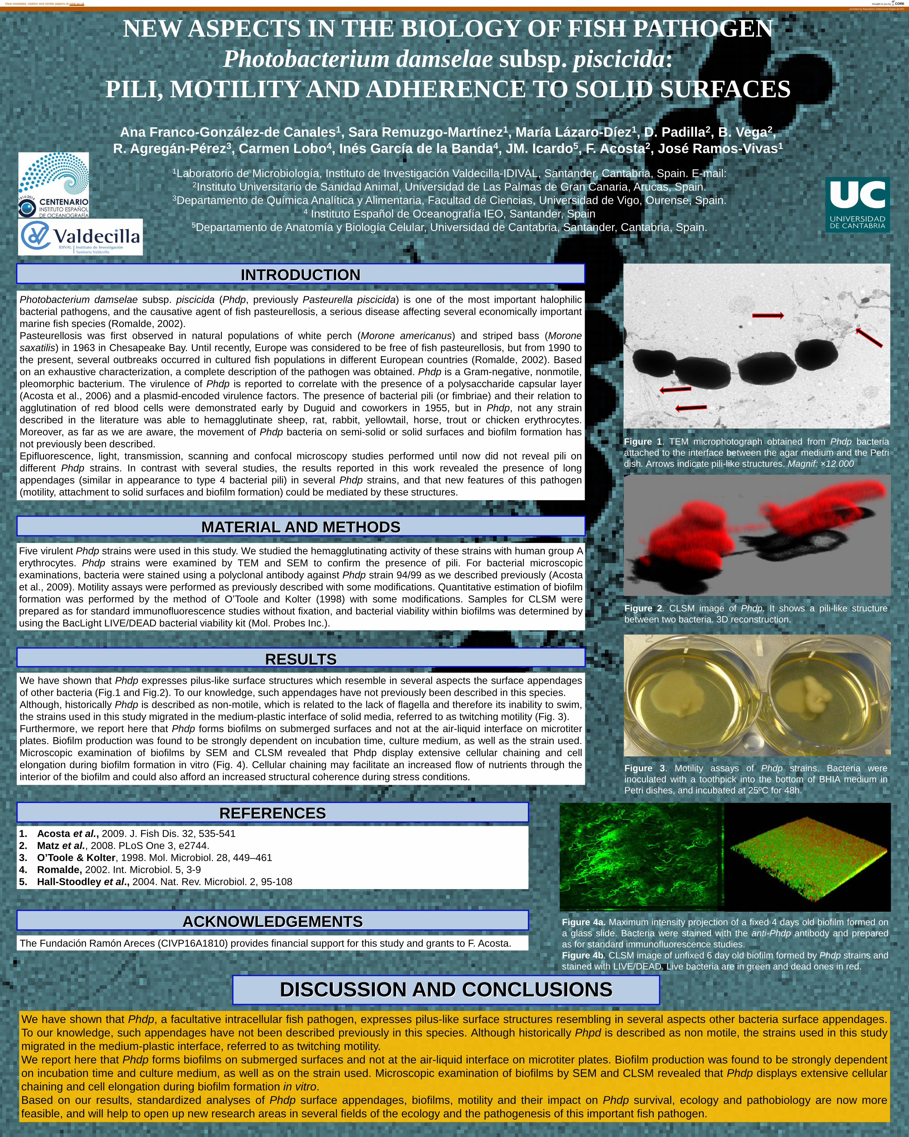
Figure 2. CLSM image of Phdp. It shows a pili-like structure between two bacteria. 3D reconstruction.
Figure 1. TEM microphotograph obtained from Phdp bacteria attached to the interface between the agar medium and the Petri dish. Arrows indicate pili-like structures. Magnif: ×12.000
A
Photobacterium damselae subsp. piscicida (Phdp, previously Pasteurella piscicida) is one of the most important halophilic bacterial pathogens, and the causative agent of fish pasteurellosis, a serious disease affecting several economically important marine fish species (Romalde, 2002). Pasteurellosis was first observed in natural populations of white perch (Morone americanus) and striped bass (Morone saxatilis) in 1963 in Chesapeake Bay. Until recently, Europe was considered to be free of fish pasteurellosis, but from 1990 to the present, several outbreaks occurred in cultured fish populations in different European countries (Romalde, 2002). Based on an exhaustive characterization, a complete description of the pathogen was obtained. Phdp is a Gram-negative, nonmotile, pleomorphic bacterium. The virulence of Phdp is reported to correlate with the presence of a polysaccharide capsular layer (Acosta et al., 2006) and a plasmid-encoded virulence factors. The presence of bacterial pili (or fimbriae) and their relation to agglutination of red blood cells were demonstrated early by Duguid and coworkers in 1955, but in Phdp, not any strain described in the literature was able to hemagglutinate sheep, rat, rabbit, yellowtail, horse, trout or chicken erythrocytes. Moreover, as far as we are aware, the movement of Phdp bacteria on semi-solid or solid surfaces and biofilm formation has not previously been described. Epifluorescence, light, transmission, scanning and confocal microscopy studies performed until now did not reveal pili on different Phdp strains. In contrast with several studies, the results reported in this work revealed the presence of long appendages (similar in appearance to type 4 bacterial pili) in several Phdp strains, and that new features of this pathogen (motility, attachment to solid surfaces and biofilm formation) could be mediated by these structures.
INTRODUCTION
Five virulent Phdp strains were used in this study. We studied the hemagglutinating activity of these strains with human group A erythrocytes. Phdp strains were examined by TEM and SEM to confirm the presence of pili. For bacterial microscopic examinations, bacteria were stained using a polyclonal antibody against Phdp strain 94/99 as we described previously (Acosta et al., 2009). Motility assays were performed as previously described with some modifications. Quantitative estimation of biofilm formation was performed by the method of O’Toole and Kolter (1998) with some modifications. Samples for CLSM were prepared as for standard immunofluorescence studies without fixation, and bacterial viability within biofilms was determined by using the BacLight LIVE/DEAD bacterial viability kit (Mol. Probes Inc.).
We have shown that Phdp expresses pilus-like surface structures which resemble in several aspects the surface appendages of other bacteria (Fig.1 and Fig.2). To our knowledge, such appendages have not previously been described in this species. Although, historically Phdp is described as non-motile, which is related to the lack of flagella and therefore its inability to swim, the strains used in this study migrated in the medium-plastic interface of solid media, referred to as twitching motility (Fig. 3). Furthermore, we report here that Phdp forms biofilms on submerged surfaces and not at the air-liquid interface on microtiter plates. Biofilm production was found to be strongly dependent on incubation time, culture medium, as well as the strain used. Microscopic examination of biofilms by SEM and CLSM revealed that Phdp display extensive cellular chaining and cell elongation during biofilm formation in vitro (Fig. 4). Cellular chaining may facilitate an increased flow of nutrients through the interior of the biofilm and could also afford an increased structural coherence during stress conditions.
RESULTS
DISCUSSION AND CONCLUSIONS
1. Acosta et al., 2009. J. Fish Dis. 32, 535-541 2. Matz et al., 2008. PLoS One 3, e2744. 3. O’Toole & Kolter, 1998. Mol. Microbiol. 28, 449–461 4. Romalde, 2002. Int. Microbiol. 5, 3-9 5. Hall-Stoodley et al., 2004. Nat. Rev. Microbiol. 2, 95-108
REFERENCES
Figure 3. Motility assays of Phdp strains. Bacteria were inoculated with a toothpick into the bottom of BHIA medium in Petri dishes, and incubated at 25ºC for 48h.
ACKNOWLEDGEMENTS The Fundación Ramón Areces (CIVP16A1810) provides financial support for this study and grants to F. Acosta.
Figure 4a. Maximum intensity projection of a fixed 4 days old biofilm formed on a glass slide. Bacteria were stained with the anti-Phdp antibody and prepared as for standard immunofluorescence studies. Figure 4b. CLSM image of unfixed 6 day old biofilm formed by Phdp strains and stained with LIVE/DEAD. Live bacteria are in green and dead ones in red.
MATERIAL AND METHODS
brought to you by COREView metadata, citation and similar papers at core.ac.uk
provided by Repositorio Institucional Digital del IEO


![35762-76-6 · Web view[22] K.W. Thomulka, D.J. McGee, J.H. Lange, Use of the bioluminescent bacterium Photobacterium phosphoreum to detect potentially biohazardous materials in …](https://img.pdfslide.us/doc/110x75/5f786b7e76e4934da52d1856/35762-76-6-web-view-22-kw-thomulka-dj-mcgee-jh-lange-use-of-the-bioluminescent.jpg)