Embed Size (px)
Citation preview
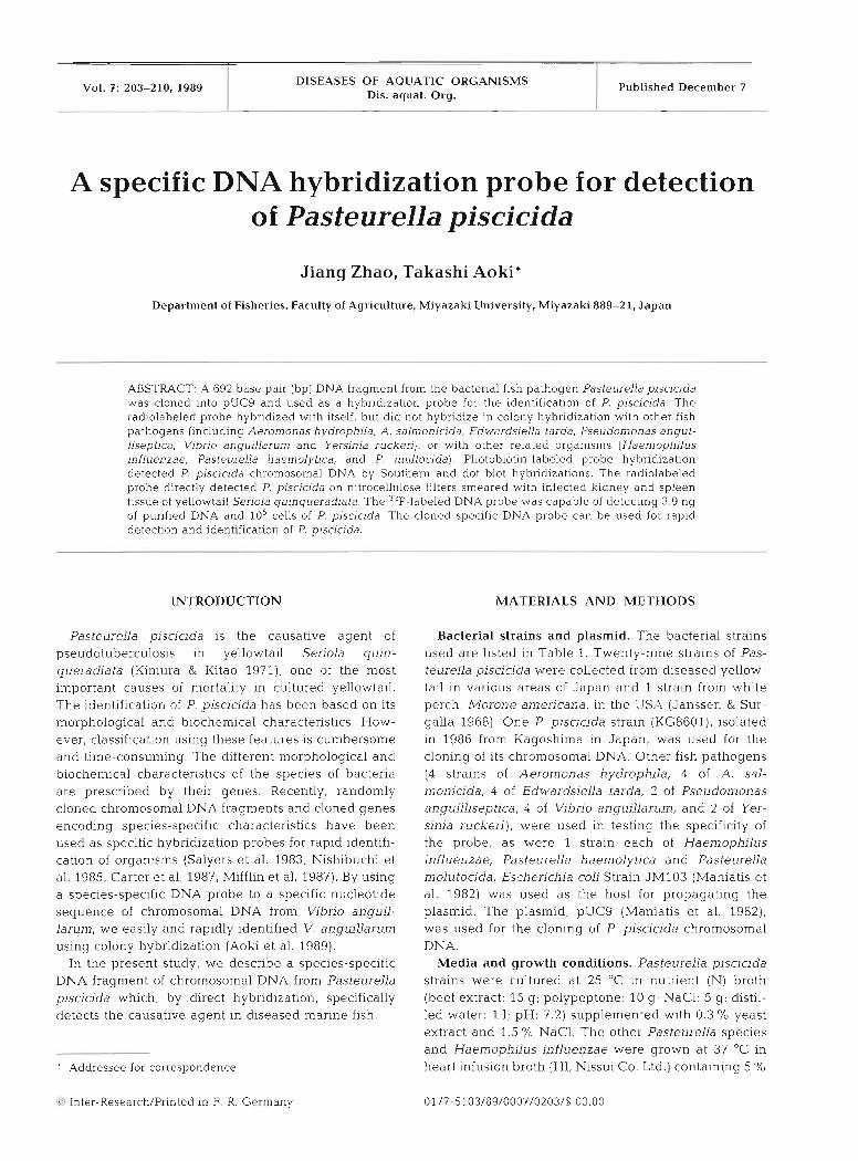
Vol. 7: 203-210, 1989 DISEASES OF AQUATIC ORGANISMS
Dis. aquat. Org. Published December 7
A specific DNA hybridization probe for detection of Pasteurella piscicida
Jiang Zhao, Takashi Aoki*
Department of Fisheries. Faculty of Agriculture, Miyazaki University, Miyazaki 889-21, Japan
ABSTRACT: A 692 base pair (bp) DNA fragment from the bacterial fish pathogen Pasteurella piscicida was cloned into pUC9 and used as a hybridization probe for the identification of P, piscicida. The radiolabeled probe hybridized with itself, but did not hybridize in colony hybridization with other fish pathogens (including Aeromonas hpdrophila, A. salmonicida, Edwardsiella tarda, Pseudomonas angul- liseptica, Vibrio anguillarum and Yersinia ruckeri), or with other related organisms (Haen~ophi lus influenzae, Pasteurella haemolytica, and P. multocida). Photobiotin-labeled probe hybridization detected P. piscicida chromosomal DNA by Southern and dot blot hybridizations. The radiolabeled probe directly detected P. piscicida on nitrocellulose filters smeared with infected kidney and spleen tissue of yellowtail Seriola quinqueradiata. The 3"P-labeled DNA probe was capable of detect~ng 3.9 ng of purified DNA and lo5 cells of P. piscicida. The cloned specific DNA probe can be used for rapid detection and identification of P. piscicida.
INTRODUCTION
Pasteurella piscicida is the causative agent of pseudotuberculosis in yellowtail Serjola quin- queradiata (Kimura & Kitao 1971), one of the most important causes of mortality in cultured yellowtail. The identification of P. pisciada has been based on its morphological and biochemical characteristics. How- ever, classification using these features is cumbersome and time-consuming. The different illorphological and biochemical characteristics of the species of bacteria are prescribed by their genes. Recently, randomly cloned chromosomal DNA fragments and cloned genes encoding species-specific characteristics have been used as specific hybridization probes for rapid identifi- cation of organisms (Salyers et al. 1983, N~shibuchi et al. 1985, Carter et al. 1987, Mifflin et al. 1987). By using a species-specific DNA probe to a specific nucleotide sequence of chromosomal DNA from Vibrio anguil- larun~, we easily and rapidly identified V. anguillarunl using colony hybridization (Aoki e t al. 1989).
In the present study, we describe a species-specific DNA fragment of chromosomal DNA from Pasteurella piscicida which, by direct hybridization, specifically detects the causative agent in diseased marine fish.
Addressee for correspondence
Inter-Research/Printed in F. R. Germany
MATERIALS AND METHODS
Bacterial strains and plasmid. The bacterial strains used are listed in Table 1. Twenty-nine strains of Pas- teurella piscicida were collected from diseased yellow- tail in various areas of Japan and 1 strain from white perch, Morone anlericana, in the U S A (Janssen & Sur- galla 1968). One P, piscicida strain (KG8601), isolated in 1986 from Kagoshima in Japan, was used for the cloning of its chromosomal DNA. Other fish pathogens (4 strains of Aeromonas hydrophila, 4 of A. sal- monicida, 4 of Edwardsiella tarda, 2 of Pseudomonas anguilliseptica, 4 of Vibl-io anguillarum, and 2 of Yer- sinia ruckeri), were used in testing the specificity of the probe, as were 1 strain each of Haemophilus influenzae, Pasteurella haemolytica and Pasteurella molutocida. Escherichia coli Strain JM103 (Maniatis et al. 1982) was used as the host for propagating the plasmid. The plasmid, pUC9 (Maniatis et al. 1982), was used for the cloning of P. piscicida chromosomal D N A .
Media and growth conditions. Pasteurella pisclcida strains were cultured at 25 "C in nutrient (N) broth (beef extract: 15 g ; polypeptone: 10 g ; NaC1: 5 g ; distil- led water: 1 1; pH: 7.2) supplemented with 0.3 % yeast extract and 1.5 % NaC1. The other Pasteurella species and Haemophilus influenzae were grown at 37 "C in heart infusion broth (HI, Nissui Co. Ltd.) containing 5 O/o
204 Dis. aquat. Org. 7: 203-210, 1989
Eel
Brooktrout
Amago
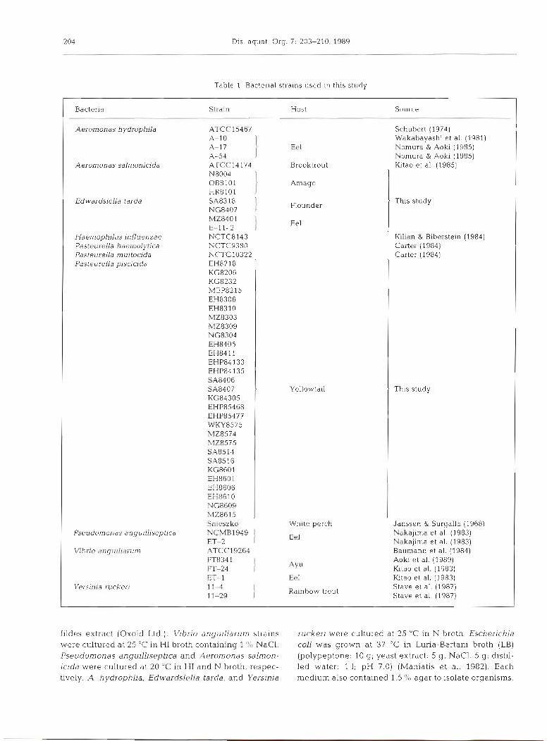
Table l . Bacterial strains used i n this study
Bacterid Slrdin Hvst Source
Aeromonas h ydrophila ATCC15467 Schubert (19741
1;; ] Wakabayashi et al. (1981) Nomura & Aoki (1985)
A-54 Nomura & Aoki (1985) Aeron~onas salmonicida ATCC14 174 l t a o et al. (1985)
N8004 OB8101 HR8101
Edwardslella tarda SA83 18 l t Thls study
NG8407 M2840 1 E-l 1-2
Haemophilus influenzae NCTC8143 l
Kilian & Blberste~n (1984) Fasteul-ella haen~olytica rJCTC338G Carter (1984)
Flounder
Eel
Pasteurella multocida NCTC10322 Pasteurella pisac~da El-18218
KG8206 KG8232 MEP8215 EH8306 EH8310 M28303 M28309 NG8304 EH8405 EH84 11 EHP84133 EHP84 135 SA8406 SA8407 KG84305 EHP85468 EHP85477 WKY8575 NZ8574 MZ8575 SA8514 SA85 16 KG8601 EH860 1 EH8606 EH86 10 NG8609 M286 15 Snieszko
Yellowtail
Carter (1984)
This study
Janssen & Surgalla (1968) Whlte perch
Eel Pseudornonas angullliseptica NCMB1949 l Nakajima et al. (1983)
ET-2 Nakajima e t al. (1983) Vibrio anguillarun~ ATCC 19264 Baumann et al. (1984)
PT834 1 Aoki e t al. (1989) PT-24 } AYU Kitao e t al. (1983) ET- l Eel h t a o et al. (1983)
Yersinla rucker) 1 1 4 I Rainbow trout
Stave et al. ( 1 987) 11-29 I Stave et al. (1987)
fildes extract (Oxoid Ltd.). Vibrio anyr~illarum strains ruckeri were cultured at 25 "C in N broth. Escherichia were cultured at 25 "C in HI broth containing l '?L NaC1, col; was grown at 37 "C in Luria-Bertani broth (LB) Pseudomonas anguilliseptjca and Aeromonas salmon- (polypeptone: 10 g; yeast extract: 5 g; NaCI. 5 g ; distil- icida were cultured at 20 'C in HI and N broth, respec- led water: l l ; pH 7.0) (Maniatis et al. 1982). Each tively. A. hydrophila, Edwardsiella tarda, and Yersinja medium also contained 1.5 O/O agar to isolate organisms.
Zhao & Aoki: A DNA probe for PasteureUa pksacida 205
Isolation of DNA. The chromosomal DNA of Pas- teurella piscjcida, Strain KG8601, and the fish-patho- gens Aeromonas hydrophila A-10, A. saln~onicjda HTCC14174, and Vibrio anguillarum PT8341 were iso- lated by previously reported methods (Silhavy 1984). Plasmid DNA was prepared by the alkaline lysis method of Birnboim & Doly (1979). Both isolated chromosomal DNA and plasmid DNA were purified by centrifugation to equilibrium in cesium chloride- ethidium bromide gradients.
Molecular cloning of DNA probe. The chromosomal DNA of Pasteurella piscicida was digested with Hind 111. The vector plasmid, pUC9, was also digested with the same enzyme and then dephosphorylated with bacterial phosphatase (Takara Shuzo Co. Ltd, Kyoto, Japan). The digested DNA was mixed together and ligated with T4 ligase (Nippon Gene Co. Ltd, Osaka, Japan). The recombinant plasmids were transformed to Escherichia coli JM103, and transformants selected on L-agar (Maniatis et al. 1982) supplemented with 50 yg ml- ' ampicillin and X-gal (5-bromo-4-chloro-3-indolyl- P-D-galactopyranoside).
Labeling of DNA probe. A cloned DNA fragment with a molecular weight of about 0.7 kilobase (kb) was selected, at random, for use as the probe, and was isotopically labeled with (CV-~*P)~CTP (New England Nuclear Corp., Boston, USA) by nick translation (Rigby et al. 1977).
The DNA probe was labeled with photobiotin (Biotechnology Research Enterprises S.A. Pty Ltd), and the Blu GENE nonradioactive DNA detection system (Bethesda Research Laboratories, USA) was used. Probe DNA was mixed with equal volumes of photo- biotin acetate in an Eppendorf tube, irradiated for 20 min with a 400W lamp from a distance of 10 cm, and diluted to 100 p1 with TE buffer (10 mM Tris HCl, 1mM EDTA, pH 8.0) followed by 100 111 2-butanol. The mix- ture was centrifuged and the 2-butanol phase dis- carded. Photobiotin-labeled DNA was purified by 3 successive ethanol precipitations, and finally resus- pended in TE buffer and stored at -20 'C.
Southern and dot blot hybridization. For Southern blot hybridization, chromosomal DNAs of Pasteurella pjscicida, Aeromonas hydrophila, A, salmonicida, and Vibrio anguillarum were cleaved by Hind 111, and elec- trophoresed in 0.8 '10 agarose gel at 80 mA for ca 3 h. Electrophoresed gels containing digested DNAs were transferred to nitrocellulose filters (0.45 pm, Schleicher & Schuell Inc., FRG) using a transfer pyramid for Southern blotting (Southern 1975).
For dot blot hybridization, chromosomal DNA from Pasteurella piscicida was denatured by heating at 90 "C for 5 min and diluted from 125 pg to 1.95 pg ml-' with 2 X SSC (1 X SSC = 0.15 M NaCl + 0.015 M sodium citrate, pH 7.0). Samples of 250, 125, 62.5, 31.3, 15.7,
7.8, and 3.9 ng of DNA were blotted on nitrocellulose filters.
Hybridization with 32P labeled DNA probe was car- ried out on filters at 65 "C for 36 h in 4 x SSC containing 10 X Denhardt's solution ( l '10 Ficoll, 1 % polyvinylpyr- rolidone, 1 % bovine serum albumin), 0.5 % sodium dodecyl sulfate (SDS), and 1.5 mg ml-' heat-denatured salmon-sperm DNA. Filters were washed with 2 X SSC + 0.1 % SDS, dried, and subjected to autoradiography. Hybridization with the photobiotin-labeled probe was carried out at 42 "C for 12 to 18 h in a hybridization solution containing 45 % formamide, 5 X SSC, l x Denhardt's solution, 25 mM sodium phosphate (pH 6.5), 5 YO dextran sulfate, and 0.2 mg ml-' heat-denatured salmon-sperm. After hybridization, the probe-target DNA hybrid was detected with conjugate dyes of the B1.u GENE (Bethesda Research Laboratories) strep- ta.uidin-alkaline phosphatase.
Colony hybridization. The organisms were inocu- lated onto autoclaved, gridded nitrocellulose filters (Toyo Roshi Co. Ltd, Japan) overlayed on an agar plate. After incubation, the filter was removed from the plate and treated with NaOH, neutralized, baked, and hy- bridized with probe DNA. Hybridization reactions were carried out by the method of Grunstein & Hog- ness (1975).
The minimum number of Pasteurella piscicida (Strain KG8601) cells detected was determined by hybridi.za- tion with the same probe. Bacterial cells diluted 10-fold from 10"o 10' were spotted on a nitrocellulose filter. DNA hybridization was thcn performed as described by Grunstein & Hogness (1975).
Detection of Pasteurella piscicida in fish. Nineteen diseased and 3 healthy Seriola quinqueradiata were obtained from an experimental fishery station. These fish were dissected and examined to determine whether nodules of kidney and spleen, characteristic of the pseudotuberculosis, were present in the kidney and spleen of fish. Kidney and spleen tissue from each fish was sampled using a platinum loop and smeared onto nitrocellulose filters. Hybridization was then carried out with the same probe and by the same method as used in the colony hybridization procedure. At the same time, the causative agent was isolated on agar media and identified.
Restriction sites and nucleotide sequence of the probe. A cloned DNA fragment of Pasteurella pis- cicida, which was chosen at random and used as the probe, was digested by various endonucleases: AvaII, BamHI, EcoRI, HapII, HincII, HindIII, HinfI, HpaI, RsaI, San, Sau3A1, SmaI, TaqI, and Xhol (Takara Shuzo Co. Ltd, Nippon Gene Co. Ltd, Japan). Nucleotide sequences of this cloned DNA fragment were deter- mined using pUC9 by the dideoxy method of Hattori et al. (1985).
206 Dis, aquai. &g. 7: 203-210. 1989
Panel 1
A B C D E
Panel 2
C D E
RESULTS
Screening of DNA probe
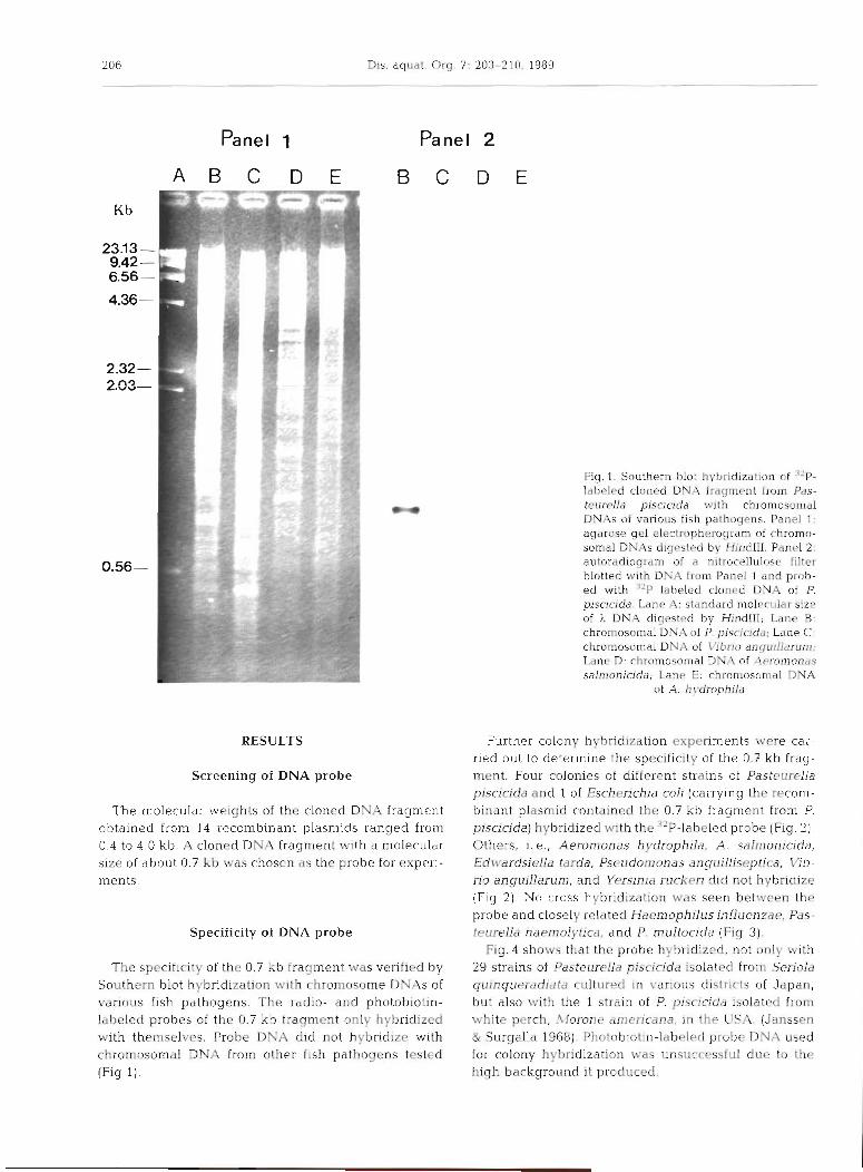
The molecular weights of the cloned DNA fragment obtained from 14 recombinant plasmids ranged from 0.4 to 4.0 kb. A cloned DNA fragment with a molecular size of about 0.7 kb was chosen as the probe for experi- ments.
Specificity of DNA probe
The specificity of the 0.7 kb fragment was verified by Southern blot hybridization with chromosome DNAs of various fish pathogens. The radio- and photobiotin- labeled probes of the 0.7 kb fragment only hybridized with themselves. Probe DNA did not hybridize with chromosomal DNA from other fish pathogens tested (Fig 1).
Fig. 1. Southern blot hybridization of "'P- Iabeled cloned DNA fragment from Pas- teurella pisciada with chromosomal DNAs of various fish pathogens. Panel 1: agarose gel electropherogram ol chromo- somal DNAs d~gested by HindlII. Panel 2. autoracfiogtam of a n~trocellulose filter blotted with DNA from Panel 1 and prob- ed wlth 'IP labeled cloned DNA of P p~snada. Lane A. standard molecular size of X DNA digested by HmdIII, Lane B chromosomal DNA of P. p~scicida: Lane C; chromosomal DNA of Vibno angulllarum; Lane D. chromosomal DNA of Aeromonas salmonicida; Lane E: chromosomal DNA
of A. h ydrophild
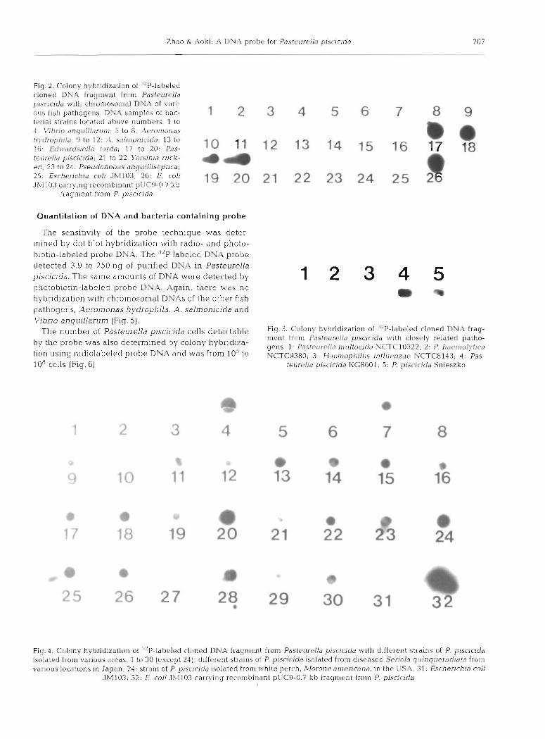
Further colony hybridization experiments were car- ried out to determine the speciflcity of the 0.7 kb frag- ment. Four colonies of different strains of Pasteurella piscicida and l of Escherichia coli (carrying the recom- binant plasmid contained the 0.7 kb fragment from P. piscicida) hybridized with the 32P-labeled probe (Fig. 2) . Others, i .e . , Aeromonas hydrophila, A. saln~onicida, Edwardsiella tarda, Pseudornonas anguilliseptica, W b - rio anguillarum, and Yersinia rrlckeri did not hybridize (Fig. 2) . No cross hybridization was seen between the probe and closely related Haemophilus influenzae, Pas- teurella haemolytica, and P. nlultocida (Fig. 3).
Fig. 4 shows that the probe hybridized, not only with 29 strains of Pasteurella piscicida isolated from Seriola quinqueradiatil cultured in various districts of Japan, but also with the 1 strain of P. piscicida isolated from white perch, A.lorone americana, in the U S A . (Janssen & Surgalla 1968). Photobiotin-labeled probe DNA used for colony hybridization was unsuccessful due to the high background it produced.
Zhao & .\oki A DNA probe for Pasteurrll~r p ~ s c i r ~ d a 207
Fig. 2. Colony hybridization of "P-labeled cloned D N A fragment from Pasl~[lrrl ld ~x.sr.icida with chron~osomdl DNA of varl- ou5 fish pathogens. D Y A s a n i p l ~ \ of t ~ a c - t r r ~ a l stralns located above numbers. 1 to
1 2 3 4 5 6 7 8 9 3 . \'ihno anguillerun~: 5 to $ 3 : ..'I~rornonas l~!.droph~l~j: 9 to 12: ,,l. si11nionic.ida; 13 to 16: Erlni~rd.siello tilrda; 17 to 20. P&s-
m a 10 11 12 13 14 15 16 17 78
te~1rc8lla piscicida; 21 to 22 Y'ersinia ruck- eri; 23 to 24: Pseudononils anguillisept~ca;
4 4 25: Escherichia coli Jh1103; 2G: E coli 19 2 0 2 1 2 2 23 24 2 5 2 JM103 carrying recombinant pLlCA-0.7 kb
a fragment from P. piscicida
Quantitation of DNA and bacteria containing probe
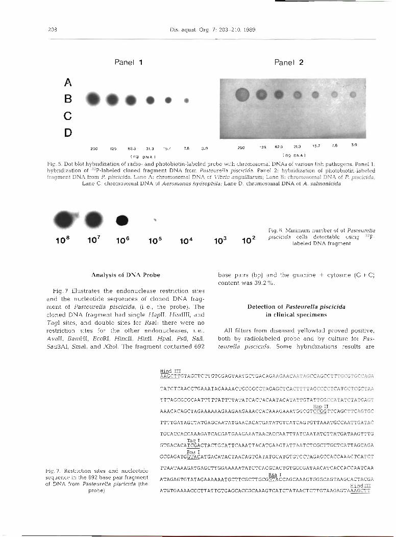
The sensitivity of the probe technique was deter- mined by dot blot hybridization with radio- and photo- biotin-labeled probe DNA. The 32P labeled DN.4 probe detected 3.9 to 250 ng of purified DNlZ in Pasteurella piscicida. The same amounts of DNA were detected by photobiotin-labeled probe DNA. Again, there was no hybridization with chromosomal DN.4s of the other fish pathogens, Aerornonas hydr-ophila, A. salnionicida and Vibrio anguillal-un~ (Fig. 5).
nulnber of P ~ ~ ~ ~ ~ ~ ~ ~ I ~ plscicida cells detectable Fig. 3. Colony hybridization of '-'P-lilbclcd cloned D N A frag- Inenl I r r jm P,isteumlla piscicitiii with closcly related patho-
by the probe determl"ed by hybridi7.a- gcns. 1: Pai!r~#rrrIlc, ~ n r ~ l t o c i d ~ i NCTC:10322, 2 . 1'. haenlolytlrd tion 'using radiolabeled probe DNA and was from 10'' to NCTC9380; 3: Ffdenrophilus inf lurnzi~e NCTC8143; 4: Pas- 10"ells (Fig. 6) . teurc!ld pisrldda KG8601; 5: P. piqcic.~cia Snleszko
Fig. 4. Colony hybridization 01 '"P-labeled cloned DNA fragment from Pastetirelld piscicida with d~fferent strains of P. piscicida isolated from various areas. 1 to 30 (except 23): different strains of P. pjscicida isolated from diseased Sei-iola q l~inqr~eradia ta from various locat~ons In Japan: 24: straln of P. p~sclcida isolated from while perch, A?orone aniericana, In the [:SA; 31: Eschenchia col1
JM103; 32: L: coli 3x1103 casrvlng rrcombinant pLlC9-0.7 kb fragnicnt from I? pisclrrda
208 Dis. aquat. Org 7: 203-210, 1989
Panel 1 Panel 2
250 125 82.5 31.3 15.7 7.8 3.9 250 125 6 2 5 31.3 15.7
(ng D N A ) ( n g D N A )
Fig.5. Dot blot hybndizatiofl of radio- and photobiom-labeled probe w t h chromosomal DNAs of varlous fish pathogens, Panel 1: hybridization of 3aP-labeled doned fragment DNA fmm Pasteurella p~scicida Panel 2: hybridization of pho-tobiotin-lakled fragment DNA born P, piscfcida. kule A: chromosod D N A cf Lfi/:br:o anguiIIanun, Lane B: chinosofftal DNA of P. plsciuda,
Lane C: chromosomal DNA of Aerornonas hydrophila; Lane D: chromosornaI DNA of A. s&nonMde
Fig. R. Minimum number of of Pasteurella
102 plscicida cells detectable using 3'P-
labeled DNA fragment
Analysis of DNA Probe base pairs (bp) and the guanine + cytosine (G + C) content was 39.2%.
Fig. 7 illustrates the endonuclease restriction sites and the nucleotide sequences of cloned DNA frag- ment of Pasteurella piscicida, ( i .e . , the probe). The Detection of Pasteurella piscicida cloned DNA fragment had single HapII, HindIII, and in clinical specimens Tag1 sites, and double sites for RsaI; there were no restriction sites for the other endonucleases, i. e., All filters from diseased yellowtail proved positive, AvaII. BamHI, EcoRI, HincII, HinfI, HpaI, PstI, San, both by radiolabeled probe and by culture for Pas- SauSAI, SmaI, and XhoI. The fragment contained 692 teurella piscicida. Some hybridizations results are
Hind m KGCTTGTAGCTCTTGTGGAGTAATGCTGACAGAAGAAC~
TATCTCAACCTGAAATAGAAAACTGCGGCCTAGAGCTCACTTTTAGCCCCTCATGCTCGCTAA
TTTAGCGGGCAATTTTTATTTTATATCACTACAATACATATTGTATTGGCCATATCTATGAGT Ha rf
A A A c A c A G c T A G A A A A A A G A A G A A G A A A c c A c A A A G A A A T G G c G T c ~ T T c A G c T T c A G T G c
TTTTGATAGCTATGAGCAATATGAACACATGATATGTCATCAGTGTTAAATGCCAATTGATAC
TGCACCACCAAAGATCACGATGAAGAAATAACACCAATTTATCAATATCTTATGATAAGTTTG
GTGACACAEACTACTGCATTCAAATTACATGAAGTATTAATCTCGCTTGCTCATTAGCAGA RsaI
GCGAGATGG~CATGACATACTAACAGTGATATGCATGTGT~~TAGAG~~A~~AAA~T~AT~T
Fia. 7. Restriction sites and nucleotide TTAATAAAGATGAGCTTGGAAAAATATCTCACGCACTGTGGCGATAACATCACCACCAATCAA - RsaI sequence in the 692 base pair fragment ATAGAGTGTATAGAAAAAATGCTTCGCTTGCGGTACCAGCAAAGTGGGCAGTAAGCAmACGA of DNA from Pasteurella piscicida (the H i n d I I I
probe) ATGTGAAAACCCTTATTGTGAGCACCGCAAAGTCATCTATAACTCTTGTAAGAGTA~CTT
Zhao & Aoki: A DNA probe for Pasteurella piscjcida 209
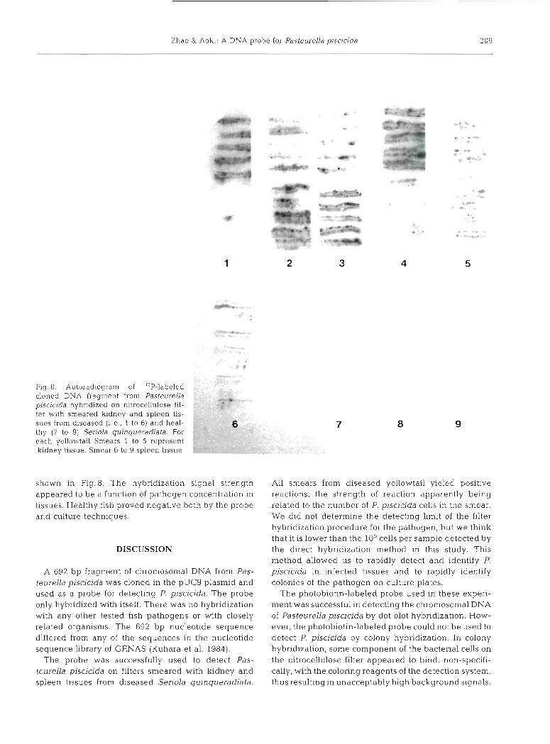
Fig 8 Autoradiogram of "P-labeled cloned DNA fragmeht from Pasteurella p~sactda hybndized on ruttocellulose 111- ter with s m e a r e d krdney and spleen tis- sues from diseased [i.e.. 1 to 6) and heal- thy (7 to 9) Serida quinquexadiata. For each yellowtail Smears 1 to 5 represent kidney t w e , Smear 6 to 9 spleen tissue
shown in Fig. 8. The hybridization signal strength appeared to be a function of pathogen concentration in tissues. Healthy fish proved negative both by the probe and culture techniques.
DISCUSSION
A 692 bp fragment of chromosomal DNA from Pas- teurella piscicida was cloned in the pUC9 plasmid and used as a probe for detecting P. piscicida. The probe only hybndized with itself. There was no hybridization with any other tested fish pathogens or with closely related organisms. The 692 bp nucleotide sequence differed from any of the sequences in the nucleotide sequence library of GENAS (Kuhara et al. 1984).
The probe was successfully used to detect Pas- teurella piscjcida on filters smeared with kidney and spleen tissues from diseased Seriola quinqueradiata.
All smears from diseased yellowtail yieled positive reactions, the strength of reaction apparently being related to the number of P. piscicjda cells in the smear. We did not determine the detecting limit of the filter hybridization procedure for the pathogen, but we think that it is lower than the 10%ells per sample detected by the dlrect hybridization method in this study. This method allowed us to rapidly detect and identify P. piscicjda in infected tissues and to rapidly identify colonies of the pathogen on culture plates.
The photobiotin-labeled probe used in these experi- ment was successful in detecting the chromosomal DNA of Pasteurella piscicida by dot blot hybridization. How- ever, the photobiotin-labeled probe could not be used to detect P. piscicida by colony hybridization. In colony hybridization, some component of the bacterial cells on the nitrocellulose filter appeared to bind, non-specifi- cally, with the coloring reagents of the detection system, thus resulting in unacceptably high background signals.
210 Dis. aquat. Org. 7. 203-210, 1989
The colony hybridization technique is more easily and quickly applied than the dot blot hybridization method. However, radiolabeled probes are generally unacceptable for routine clinical use and further development of detection procedures based on non- lsotope labellng is, therefore, still required.
Acknowledgements. This research was supported in part by a Grant-in-Aid from the Ministry of Education. Science and Culture (625660199) of Japan. We thank Professor Tadatoshi Kitao for his useful discussions and valuable suggestions.
LITERATURE CITED
Aoki, T S. Egusa, K. Kimura, Watanabe, T (1971). Detection oi Ii factors in naturaiiy occurring Aeromonas saimoniciaa strains. Appl. Microbial. 22: 716-717
Aoki, T. , Hirono, I . , Castro, T D., Kitao, T (1989) Rapid identification of Vibrio anguillarum by colony hybridiza- tion. J. appl. Ichthyol. 5: 67-73
Baumann, P., Farniss, A. L., Lee, Lee J. V (1984). Genus I, Vibr~o Pacin~ 1854. 411 Bergey's manual of systemahc bacteriology, Vol. I. Williams & Wilkins, Baltimore London. p. 518-538
Birnbo~m. H. C., Doly. J. (1979). A rapid alkaline extraction procedure for screening recombinant plasmid DNA. Nuc- leic Acids Res. 7: 1513-1523
Carter, G. L., Towner, K. J., Slack, R. C. (1987). Rapid detec- tion of a specific trimethoprim resistance gene using a biotinylated DNA probe. J . Antimicrob. Chemother. 20- 335-34 1
Carter, R. R. (1984). Genus I. Pasteurella Trevisan 1887, 94, Nom. cons. Opin. 13, Jud. Comm. 1954. 153. Bergey's manual of systemat~c bacteriology, Vol. I. Williams & WII- kins, Baltimore, London, p. 552-557
Grunstein, M., Hogness, D. S. (1975). Colony hybridization: a method for the isolation of cloned DNAs that contain a specific gene. Proc. natn. Acad. Sci. U.S.A. 72: 3961-3965
Hattori, M., Hidaka, S., Sakaki, Y (1985). Sequence analysis of a KpnI family member near the 3 ' end of human ci- globin gene. Nucleic Acids Res. 13: 7813-7827
Janssen, W. A., Surgalla, M. J . (1968). Morphology, physiol- ogy, and serology of a Pasteurella species pathogenic for white perch. J. Bact. 96: 1906-1910
Kilian, M,, Biberstein, E. L. (1984). Genus 11. Haemophilus Winslow, Broadhurst, Buchanan, Krumw~ede, Rogers and Smith 1917, 561, Bergey's manual of systematic bac- teriology, Vol. I. Williams & Wilkins, Baltimore, London, p. 558-569
Kimura. M,, Kitao, T. (1971). On the etiological agent of bacterial tuberculoidosis of Seriola. Fish Pathol. 6: 8-14
I t a o , T., Aoki, T., Fukudome. M. , Kawano, K., Wada, Yo.
Responsible Subject Editor Dr T Evelyn, Nanaimo, B.C. Canada
Mizuno, Y. (1983). Serotyping of Vibrio anguiflarum isolated from diseased freshwater f ~ s h in Japan. J. Fish Dis. 6: 175-181
I t a o , T., Yoshida, T., Aoki, T., and Fukudon~e, M. (1985). Characterization of an atypical Aerornorlas salrnonicida strain causing epizootic ulcer disease in cultured eel. Fish Pathol. 20: 107-1 14
Kuhara, S.. Matsuo. F., Futamura, S., Fujita, A., Shinohara, T.. Takagi, T., Sakaki, Y (1984). GENAS: a database system for nucleic acid sequence analysis. Nucleic Acids Res. 12: 89-99
Maniatis, T., Fritsch, E. F., Sambrook, J . (1982). Molecular cloning. A laboratory manual. Cold Spr~ng Harbor Laboratory, Cold Spring Harbor, N. Y
Mifflin, T. E . , Bowden, J . , Lovell, M. A. , Bruns, D. E., Hayden, F. G., Groschel, D. H. M,, Savory, J. (1987). Comparison of radioactive ( 3 2 ~ and "S) and biotinylated probes for detec- tion of cytomegalovirus DNA. Clin. Biochem. 20: 231-235
Naitajima, K., iviuroga, K., Hancock, R. E. W. j1983j. Compari- son of fatty acid, protein, and serological properties distin- guishing outer membranes of Pseudornonas anguillisep- tica strains from those of fish pathogens and other pseudomonads. Int. J . system. Bact. 33: 1-8
Nishibuchi, M., Ishibashi, M., Takeda, Y., Kaper, J . B. (1985). Detect~on of the thermostable direct hemolysin gene and related DNA sequences in Vihrio parahaernolyticus and other Vibrio species by the DNA colony hybridization test. Infect~on Immunity 49 481-486
Nomura, J., Aoki, T (1985). Morphological analysis of lipopolysaccharide from gram-negative fish pathogenic bacteria. Fish Pathol. 20: 193-197
R~gby. P. W. J., Dieckmann. M. Rhodes, C., Berg, P. (1977). Labeling deoxyribonucleic acid to high specific activity in vitro by nick translat~on, with DNA polymerase I. J. molec. Biol. 113: 237-251
Salyers, A. A., Lynn, S. P,, Gardner, J. (19831, Use of randomly cloned DNA fragments for idenhfication of Bacteroides thetaiotaornicron. J. Bact. 154: 287-293
Schubert. R. H. W. (1974). Genus 11. Aeromonas Kluyver and van Niel 1936, 398 Bergey's manual of determinative bacteriology. William & Wilkins Co., Baltimore, p. 345-348
Silhavy, T. J. (1984). Experiments with gene fusions. Cold Spring Harbor Laboratory, Cold Sprlng Harbor, N. Y., p. 137-139
Southern, E. M. (1975). Detection of specific sequences among DNA fragments separated by electrophoresis. J . molec. Biol. 98: 503-507
Stave, J . W., Cook, T M., Roberson, B. S. (1987). Chemilumi- nescent responses of striped bass, Morone saxafiiis (Wal- baum), phagocytes to strains of Yersinia ruckeri. J. Fish Dis. 10: 1-10
Wakabayashi, H., Kanai K . , Hsh Ta-C., Egusa. S. (1981). Pathogenic activities of Aeromonas hydrophila biovar hy- drophila (Chester) Popoff and Veron, 1976 to f~shes. Fish Pathol 15: 319-325
Manuscript first received: November 4. 1988 Revised version accepted: September 27, 1989