Embed Size (px)
Citation preview
Jour
nal o
f Cel
l Sci
ence
RESEARCH ARTICLE
Phosphorylation of the retinoic acid receptor RARc2 is crucial forthe neuronal differentiation of mouse embryonic stem cells
Ziad Al Tanoury, Samia Gaouar, Aleksandr Piskunov*, Tao Ye, Sylvia Urban, Bernard Jost, Celine Keime,Irwin Davidson, Andree Dierich and Cecile Rochette-Egly`
ABSTRACT
Retinoic acid (RA) plays key roles in cell differentiation and growth
arrest by activating nuclear RA receptors (RARs) (a, b and c),
which are ligand-dependent transcription factors. RARs are also
phosphorylated in response to RA. Here, we investigated the in vivo
relevance of the phosphorylation of RARs during RA-induced
neuronal differentiation of mouse embryonic stem cells (mESCs).
Using ESCs where the genes encoding each RAR subtype had been
inactivated, and stable rescue lines expressing RARs mutated in
phospho-acceptor sites, we show that RA-induced neuronal
differentiation involves RARc2 and requires RARc2 phosphorylation.
By gene expression profiling, we found that the phosphorylated form
of RARc2 regulates a small subset of genes through binding an
unusual RA response element consisting of two direct repeats with a
seven-base-pair spacer. These new findings suggest an important
role for RARc phosphorylation during cell differentiation and pave the
way for further investigations during embryonic development.
KEY WORDS: Retinoic acid, RA, RAR, Nuclear receptor,
Phosphorylation, Transcription, Differentiation
INTRODUCTIONRetinoic acid (RA), the active metabolite of vitamin A, regulates
multiple biological processes and plays key roles in embryonic
development through the regulation of cell growth and
differentiation (Samarut and Rochette-Egly, 2012). RA exerts
its effects through nuclear RA receptors (RARs), which are
ligand-regulated transcription factors with a well-defined domain
organization and structure, consisting of a variable N-terminal
domain (NTD), and two well-structured and conserved domains,
a central DNA-binding domain (DBD) and a C-terminal ligand-
binding domain (LBD) (Bastien and Rochette-Egly, 2004;
Rochette-Egly and Germain, 2009).
The RA signalling system is highly complex as it comprises
three subtypes (RARa, RARb and RARc), and for each subtype
there are at least two isoforms, differing in their NTD (Chambon,
1996). Moreover, RARs act as heterodimers with another family
of nuclear receptors, the retinoid X receptors (RXRs) (Germain
et al., 2006). According to the canonical model, RXR–RAR
heterodimers control the expression of their target genes involved
in cell growth and differentiation through binding to specific and
polymorphic RA response elements (RAREs) located in their
regulatory regions (Moutier et al., 2012) and through the dynamic
association and dissociation of coregulator complexes (Rosenfeld
et al., 2006).
During the past decade, this scenario became more complicated
with the discovery that RA also has non-genomic effects and
induces the rapid activation of the MAPK pathway (Piskunov and
Rochette-Egly, 2012; Stavridis et al., 2010), and that RARs are
phosphoproteins (Al Tanoury et al., 2013b; Rochette-Egly, 2003).
Indeed, we have shown that the RA-activated kinases induce RARs
phosphorylation at a serine residue located in the NTD (Bastien
et al., 2000; Rochette-Egly et al., 1997). Interestingly, this serine
residue is conserved between RARs and during evolution (Samarut
et al., 2011), emphasizing that phosphorylation of RARs might be
important in RA signalling. However, the in vivo relevance of the
RAR phosphorylation still remains to be defined, especially in the
context of development. As cell differentiation is one of the most
crucial steps during development, mouse embryonic stem cells
(mESCs) (Gudas and Wagner, 2011) provide an interesting model
to study the influence of RAR phosphorylation. Indeed, ESCs are
pluripotent cells that self-renew indefinitely and can differentiate
in vitro into a large variety of cell types (Wilson et al., 2009), such
as neuronal cells, in response to RA (Bibel et al., 2007).
Here, by using mESCs specifically lacking each RAR subtype
and stable rescue cell lines expressing RAR phosphomutants, we
demonstrate that their neuronal differentiation crucially involves
the phosphorylated form of RARc2. Finally, genome-wide RNA-
seq experiments identify direct target genes whose expression is
controlled by phosphorylated RARc2. These results suggest that
there is an important role for RAR phosphorylation in RA
signalling during cell differentiation, and pave the way for further
investigations during embryonic and tissue development.
RESULTSRARc2 mediates the RA-induced neuronal differentiation ofmESCsWhen cultured as cellular aggregates, treated with RA and then
dissociated and plated according to the protocol of Bibel et al.
(Bibel et al., 2007; Bibel et al., 2004), pluripotent mESCs first
adopted a spindle-shape morphology characteristic of neural
progenitors (Fig. 1A, panel b) (Bibel et al., 2004). Then within 2
days these progenitors gave rise to glutamergic neuronal cells
(Fig. 1A, panels c and d) and formed a dense axon network
expressing class III b-tubulin (Tuj1) by 7–9 days (Fig. 1B).
Concomitantly, the markers for pluripotency [Oct4 (also known
as Pou5f1) and Nanog] were downregulated, whereas those for
neuronal differentiation (Pax6) were increased (Fig. 1C,D).
IGBMC (Institut de Genetique et de Biologie Moleculaire et Cellulaire), INSERM,U596, CNRS, UMR7104, Universite de Strasbourg, 1 rue Laurent Fries, BP 10142,67404 Illkirch Cedex, France.*Present address: International Biotechnology Center ‘Generium’ 601125Volgynsky, Russia.
`Author for correspondence ([email protected])
Received 7 November 2013; Accepted 1 February 2014
� 2014. Published by The Company of Biologists Ltd | Journal of Cell Science (2014) 127, 2095–2105 doi:10.1242/jcs.145979
2095

Jour
nal o
f Cel
l Sci
ence
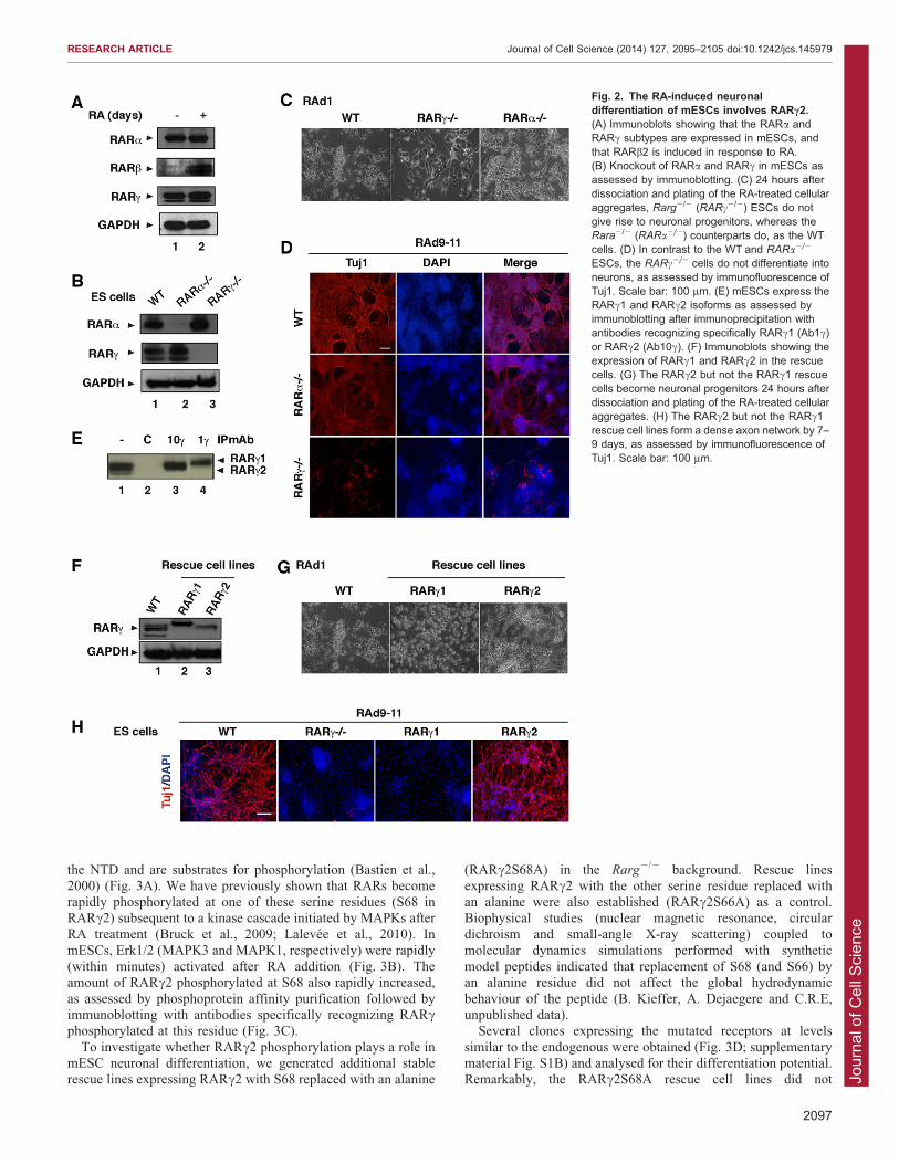
As this entire process is initiated by activation of RARs, we
investigated which specific RAR subtype (a, b or c) is required todrive differentiation. ESCs constitutively expressed the RARaand RARc proteins and their levels were not significantlyaffected upon RA treatment (Fig. 2A). In contrast, the RARbprotein was hardly detectable but was induced in response to RA(Fig. 2A). Such data suggest that the RARa and/or RARcsubtypes might be the primary RA targets for mediating the initial
phase of neuronal differentiation. To investigate the role of thesereceptors in the RA-driven neuronal differentiation of mESCs, weused cell lines in which the Rara or Rarg genes (encoding RARaand RARc, respectively) were disrupted by homologousrecombination (Lohnes et al., 1993; Lufkin et al., 1993)(Fig. 2B). Our results show that, like wild-type (WT) ESCs,cells lacking RARa became neuronal progenitors (Fig. 2C) that
gave rise to neurons forming a dense axon network (Fig. 2D). Incontrast, cells lacking RARc did not differentiate into cells of theneuronal lineage. They did not become neuronal progenitors
(Fig. 2C) and axon structures did not appear even up to 15 daysafter RA addition (Fig. 2D). These results indicate that RARc isessential for the RA-induced commitment of mESCs into
neuronal precursors that give rise to neurons. They also indicatethat RARa and RARb cannot functionally compensate for loss ofRARc during this process.
Seven murine RARc isoforms (mRARc1 to mRARc7) havebeen characterized so far, generated by alternative splicing of atleast seven exons and differing in their NTD (Kastner et al.,
1990). In mESCs, RARc1 and RARc2 are the predominant
isoforms, whereas the others (RARc3 to RARc7) have not beendetected (Kastner et al., 1990). Moreover, both RARc1 andRARc2 can be detected at the protein level in mESCs as assessedby immunoblotting after immunoprecipitation with antibodies
recognizing specifically each isoform (Bastien et al., 2000)(Fig. 2E).
To investigate the role played by each isoform in the RA-
induced neuronal differentiation of mESCs, stable rescue linesexpressing the RARc2 or RARc1 proteins were established fromRarg2/2 cells. Several clones were obtained for each ‘rescue’
transgene, and these expressed RARc1 or RARc2 at levelssimilar to the endogenous receptors (Fig. 2F; supplementarymaterial Fig. S1A). We investigated the ability of the RARc1and RARc2 rescue lines to differentiate in response to
RA. Remarkably, the RARc2 rescue lines became neuronalprogenitors (Fig. 2G) that gave rise to neurons forming a denseaxon network (Fig. 2H). In contrast, the RARc1-expressing lines
did not give spindle-shaped neuronal progenitors (Fig. 2G) norany axon structures (Fig. 2H). These results suggest that theRARc2 isoform mediates the effects of RA for the commitment
of mESCs into the neuronal lineage.
RARc2 phosphorylation is required for the neuraldifferentiation of mESCsRARs are phosphoproteins and RARc2 comprises two serine (S)residues (S66 and S68) that are located in a proline-rich motif of
Fig. 1. Neuronal differentiation ofmESCs in response to RA.(A) Morphology of mESCs at differentstages of their differentiation after RAaddition. (a) RA-treated cellular aggregatesbefore their dissociation. (b) Progenitors24 hours after plating dissociated cellularaggregates. (c,d) Neurons, 7–9 days afterplating. A dense axon network has formed.(B) Neuronal differentiation of mESCs asassessed by immunofluorescence analysisof class III b tubulin (Tuj1) at 11 days afterRA treatment (7 days after plating of thedissociated cellular aggregates).(C) Immunoblotting analysis of the stemcell pluripotency markers (Oct4 and Nanog)4 days after RA addition. (D) RT-qPCRanalysis of the downregulation of Oct4 andupregulation of the differentiation markerPax6 after RA addition. The results are themean6s.e.m. of triplicates from twoexperiments.
RESEARCH ARTICLE Journal of Cell Science (2014) 127, 2095–2105 doi:10.1242/jcs.145979
2096
Jour
nal o
f Cel
l Sci
ence
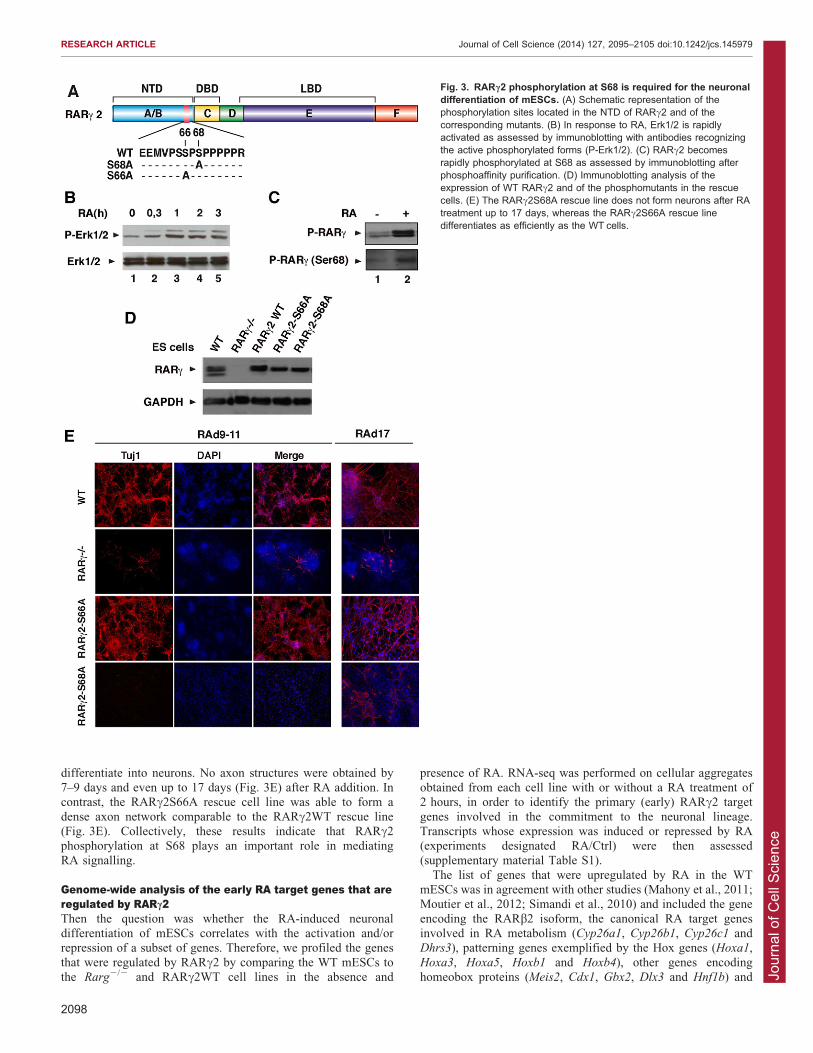
the NTD and are substrates for phosphorylation (Bastien et al.,2000) (Fig. 3A). We have previously shown that RARs become
rapidly phosphorylated at one of these serine residues (S68 inRARc2) subsequent to a kinase cascade initiated by MAPKs afterRA treatment (Bruck et al., 2009; Lalevee et al., 2010). In
mESCs, Erk1/2 (MAPK3 and MAPK1, respectively) were rapidly(within minutes) activated after RA addition (Fig. 3B). Theamount of RARc2 phosphorylated at S68 also rapidly increased,
as assessed by phosphoprotein affinity purification followed byimmunoblotting with antibodies specifically recognizing RARcphosphorylated at this residue (Fig. 3C).
To investigate whether RARc2 phosphorylation plays a role inmESC neuronal differentiation, we generated additional stablerescue lines expressing RARc2 with S68 replaced with an alanine
(RARc2S68A) in the Rarg2/2 background. Rescue linesexpressing RARc2 with the other serine residue replaced with
an alanine were also established (RARc2S66A) as a control.Biophysical studies (nuclear magnetic resonance, circulardichroism and small-angle X-ray scattering) coupled to
molecular dynamics simulations performed with syntheticmodel peptides indicated that replacement of S68 (and S66) byan alanine residue did not affect the global hydrodynamic
behaviour of the peptide (B. Kieffer, A. Dejaegere and C.R.E,unpublished data).
Several clones expressing the mutated receptors at levels
similar to the endogenous were obtained (Fig. 3D; supplementarymaterial Fig. S1B) and analysed for their differentiation potential.Remarkably, the RARc2S68A rescue cell lines did not
Fig. 2. The RA-induced neuronaldifferentiation of mESCs involves RARc2.(A) Immunoblots showing that the RARa andRARc subtypes are expressed in mESCs, andthat RARb2 is induced in response to RA.(B) Knockout of RARa and RARc in mESCs asassessed by immunoblotting. (C) 24 hours afterdissociation and plating of the RA-treated cellularaggregates, Rarg2/2 (RARc2/2) ESCs do notgive rise to neuronal progenitors, whereas theRara2/2 (RARa2/2) counterparts do, as the WTcells. (D) In contrast to the WT and RARa2/2
ESCs, the RARc2/2 cells do not differentiate intoneurons, as assessed by immunofluorescence ofTuj1. Scale bar: 100 mm. (E) mESCs express theRARc1 and RARc2 isoforms as assessed byimmunoblotting after immunoprecipitation withantibodies recognizing specifically RARc1 (Ab1c)or RARc2 (Ab10c). (F) Immunoblots showing theexpression of RARc1 and RARc2 in the rescuecells. (G) The RARc2 but not the RARc1 rescuecells become neuronal progenitors 24 hours afterdissociation and plating of the RA-treated cellularaggregates. (H) The RARc2 but not the RARc1rescue cell lines form a dense axon network by 7–9 days, as assessed by immunofluorescence ofTuj1. Scale bar: 100 mm.
RESEARCH ARTICLE Journal of Cell Science (2014) 127, 2095–2105 doi:10.1242/jcs.145979
2097
Jour
nal o
f Cel
l Sci
ence
differentiate into neurons. No axon structures were obtained by7–9 days and even up to 17 days (Fig. 3E) after RA addition. Incontrast, the RARc2S66A rescue cell line was able to form a
dense axon network comparable to the RARc2WT rescue line(Fig. 3E). Collectively, these results indicate that RARc2phosphorylation at S68 plays an important role in mediatingRA signalling.
Genome-wide analysis of the early RA target genes that areregulated by RARc2Then the question was whether the RA-induced neuronaldifferentiation of mESCs correlates with the activation and/orrepression of a subset of genes. Therefore, we profiled the genes
that were regulated by RARc2 by comparing the WT mESCs tothe Rarg2/2 and RARc2WT cell lines in the absence and
presence of RA. RNA-seq was performed on cellular aggregatesobtained from each cell line with or without a RA treatment of2 hours, in order to identify the primary (early) RARc2 target
genes involved in the commitment to the neuronal lineage.Transcripts whose expression was induced or repressed by RA(experiments designated RA/Ctrl) were then assessed(supplementary material Table S1).
The list of genes that were upregulated by RA in the WTmESCs was in agreement with other studies (Mahony et al., 2011;Moutier et al., 2012; Simandi et al., 2010) and included the gene
encoding the RARb2 isoform, the canonical RA target genesinvolved in RA metabolism (Cyp26a1, Cyp26b1, Cyp26c1 andDhrs3), patterning genes exemplified by the Hox genes (Hoxa1,
Hoxa3, Hoxa5, Hoxb1 and Hoxb4), other genes encodinghomeobox proteins (Meis2, Cdx1, Gbx2, Dlx3 and Hnf1b) and
Fig. 3. RARc2 phosphorylation at S68 is required for the neuronaldifferentiation of mESCs. (A) Schematic representation of thephosphorylation sites located in the NTD of RARc2 and of thecorresponding mutants. (B) In response to RA, Erk1/2 is rapidlyactivated as assessed by immunoblotting with antibodies recognizingthe active phosphorylated forms (P-Erk1/2). (C) RARc2 becomesrapidly phosphorylated at S68 as assessed by immunoblotting afterphosphoaffinity purification. (D) Immunoblotting analysis of theexpression of WT RARc2 and of the phosphomutants in the rescuecells. (E) The RARc2S68A rescue line does not form neurons after RAtreatment up to 17 days, whereas the RARc2S66A rescue linedifferentiates as efficiently as the WT cells.
RESEARCH ARTICLE Journal of Cell Science (2014) 127, 2095–2105 doi:10.1242/jcs.145979
2098
Jour
nal o
f Cel
l Sci
ence
genes with a wide variety of functions such as Lefty1, Arg1 andStra8. Only a few genes were downregulated, and included the
Otx2 gene, also in agreement with other studies.To assess which of these genes are specifically regulated by
RARc2 in the presence of RA, the results obtained with the WT
cells were cross-referenced with those obtained with the Rarg2/2
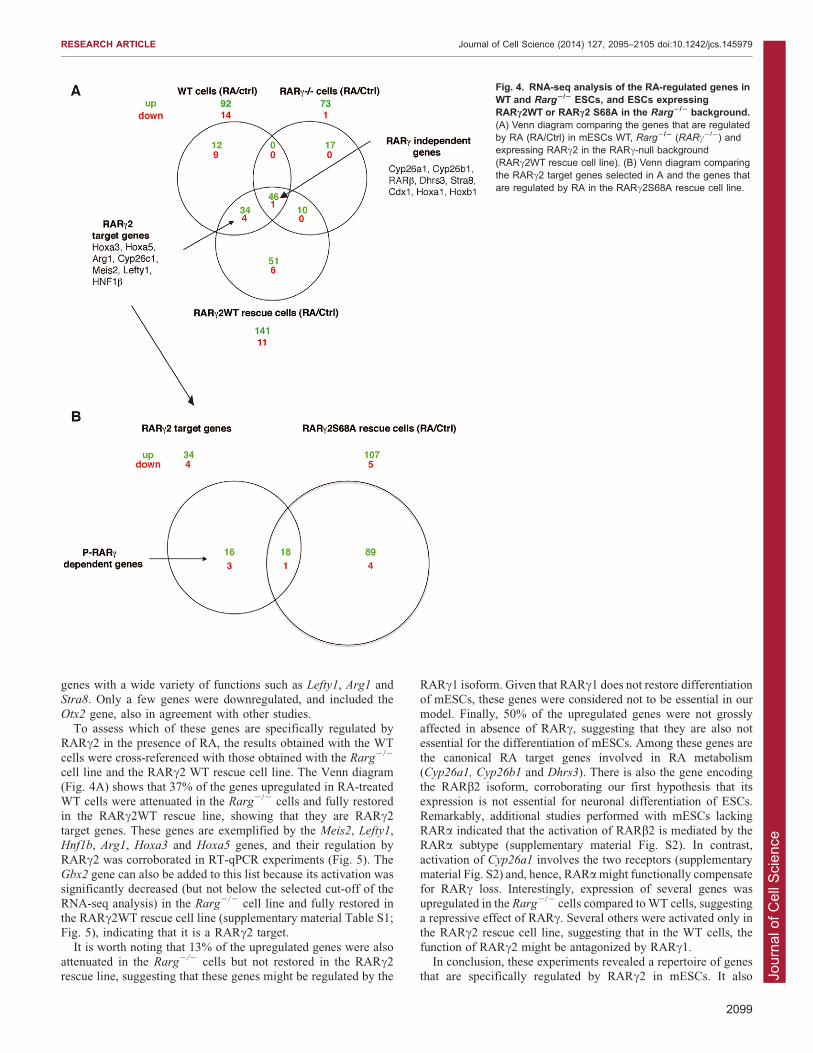
cell line and the RARc2 WT rescue cell line. The Venn diagram(Fig. 4A) shows that 37% of the genes upregulated in RA-treatedWT cells were attenuated in the Rarg2/2 cells and fully restored
in the RARc2WT rescue line, showing that they are RARc2target genes. These genes are exemplified by the Meis2, Lefty1,Hnf1b, Arg1, Hoxa3 and Hoxa5 genes, and their regulation by
RARc2 was corroborated in RT-qPCR experiments (Fig. 5). TheGbx2 gene can also be added to this list because its activation wassignificantly decreased (but not below the selected cut-off of the
RNA-seq analysis) in the Rarg2/2 cell line and fully restored inthe RARc2WT rescue cell line (supplementary material Table S1;Fig. 5), indicating that it is a RARc2 target.
It is worth noting that 13% of the upregulated genes were alsoattenuated in the Rarg2/2 cells but not restored in the RARc2rescue line, suggesting that these genes might be regulated by the
RARc1 isoform. Given that RARc1 does not restore differentiationof mESCs, these genes were considered not to be essential in our
model. Finally, 50% of the upregulated genes were not grosslyaffected in absence of RARc, suggesting that they are also notessential for the differentiation of mESCs. Among these genes are
the canonical RA target genes involved in RA metabolism(Cyp26a1, Cyp26b1 and Dhrs3). There is also the gene encodingthe RARb2 isoform, corroborating our first hypothesis that itsexpression is not essential for neuronal differentiation of ESCs.
Remarkably, additional studies performed with mESCs lackingRARa indicated that the activation of RARb2 is mediated by theRARa subtype (supplementary material Fig. S2). In contrast,
activation of Cyp26a1 involves the two receptors (supplementarymaterial Fig. S2) and, hence, RARa might functionally compensatefor RARc loss. Interestingly, expression of several genes was
upregulated in the Rarg2/2 cells compared to WT cells, suggestinga repressive effect of RARc. Several others were activated only inthe RARc2 rescue cell line, suggesting that in the WT cells, the
function of RARc2 might be antagonized by RARc1.In conclusion, these experiments revealed a repertoire of genes
that are specifically regulated by RARc2 in mESCs. It also
Fig. 4. RNA-seq analysis of the RA-regulated genes inWT and Rarg2/2 ESCs, and ESCs expressingRARc2WT or RARc2 S68A in the Rarg2/2 background.(A) Venn diagram comparing the genes that are regulatedby RA (RA/Ctrl) in mESCs WT, Rarg2/2 (RARc2/2) andexpressing RARc2 in the RARc-null background(RARc2WT rescue cell line). (B) Venn diagram comparingthe RARc2 target genes selected in A and the genes thatare regulated by RA in the RARc2S68A rescue cell line.
RESEARCH ARTICLE Journal of Cell Science (2014) 127, 2095–2105 doi:10.1242/jcs.145979
2099
Jour
nal o
f Cel
l Sci
ence
highlighted the complexity of the regulation of the other RAtarget genes.
The phosphorylated form of RARc2 regulates a subset ofgenesGiven that RARc2 phosphorylation is required for the neuronaldifferentiation of mESCs, we investigated the genes that arecontrolled by the phosphorylated form of this receptor. Cellular
aggregates obtained from mESCs expressing RARc2S68A weretreated with RA for 2 hours as above and gene expressionassessed by RNA-seq (supplementary material Table S1). The list
of genes with altered expression was compared with the list ofRARc2 target genes generated above. The Venn diagram(Fig. 4B) shows that ,50% of RARc2-target genes are notexpressed in the RARc2S68A recue cell line, suggesting that their
activation is controlled by the phosphorylation of RARc2. Thesegenes include Lefty1 (left right determination factor 1) and thehomeobox gene Hnf1b (hepatocyte nuclear factor 1 homeobox B)
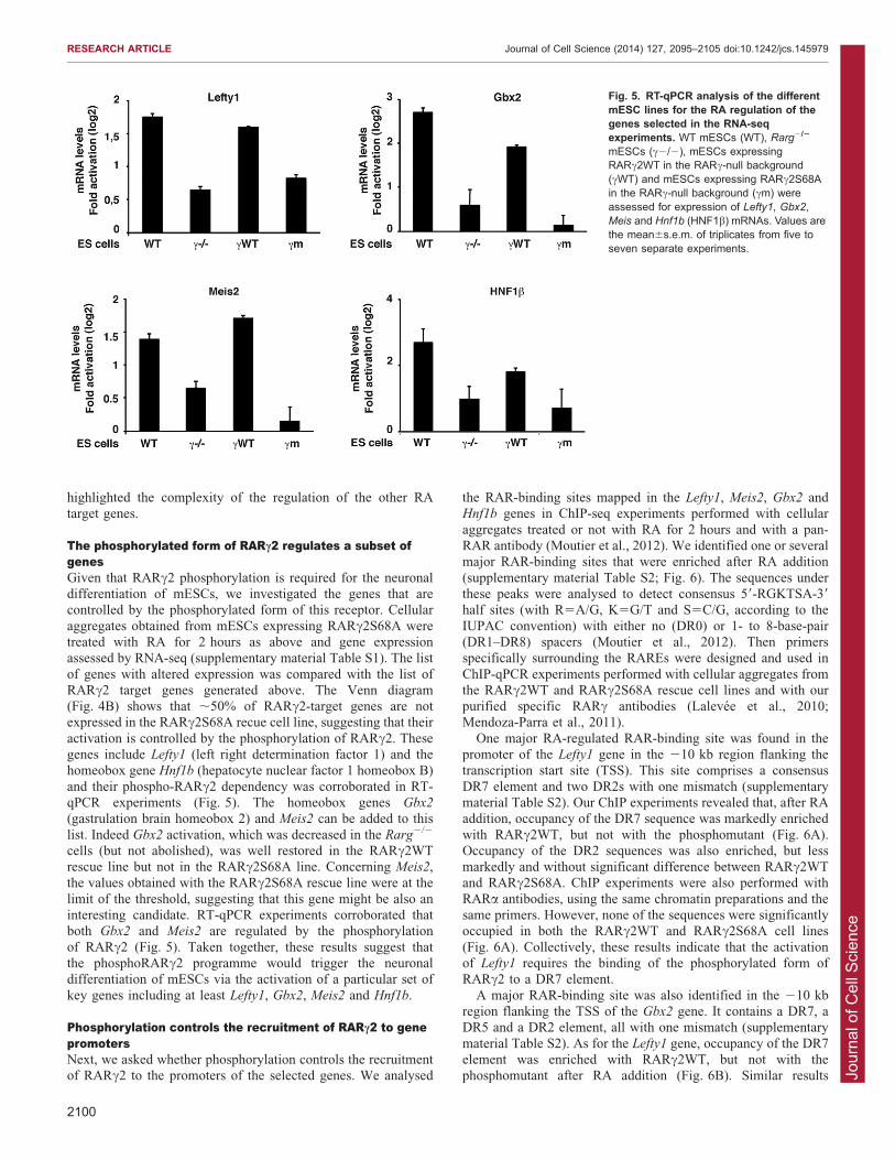
and their phospho-RARc2 dependency was corroborated in RT-qPCR experiments (Fig. 5). The homeobox genes Gbx2
(gastrulation brain homeobox 2) and Meis2 can be added to this
list. Indeed Gbx2 activation, which was decreased in the Rarg2/2
cells (but not abolished), was well restored in the RARc2WTrescue line but not in the RARc2S68A line. Concerning Meis2,the values obtained with the RARc2S68A rescue line were at the
limit of the threshold, suggesting that this gene might be also aninteresting candidate. RT-qPCR experiments corroborated thatboth Gbx2 and Meis2 are regulated by the phosphorylation
of RARc2 (Fig. 5). Taken together, these results suggest thatthe phosphoRARc2 programme would trigger the neuronaldifferentiation of mESCs via the activation of a particular set of
key genes including at least Lefty1, Gbx2, Meis2 and Hnf1b.
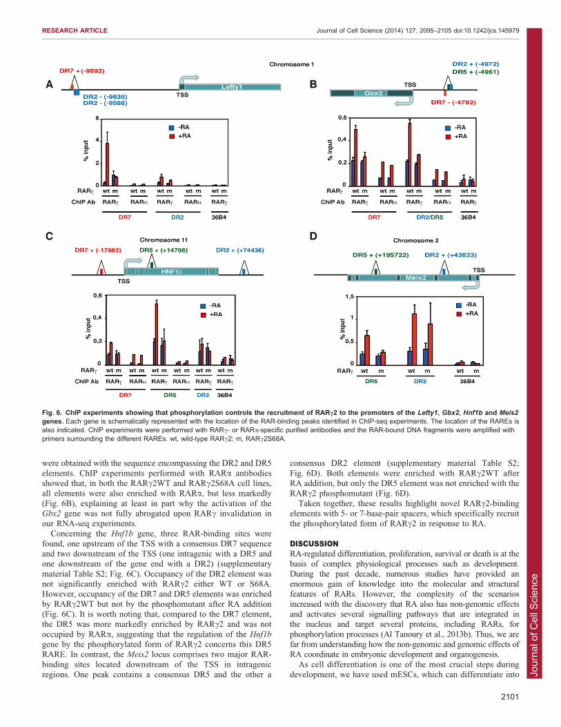
Phosphorylation controls the recruitment of RARc2 to genepromotersNext, we asked whether phosphorylation controls the recruitmentof RARc2 to the promoters of the selected genes. We analysed
the RAR-binding sites mapped in the Lefty1, Meis2, Gbx2 andHnf1b genes in ChIP-seq experiments performed with cellular
aggregates treated or not with RA for 2 hours and with a pan-RAR antibody (Moutier et al., 2012). We identified one or severalmajor RAR-binding sites that were enriched after RA addition
(supplementary material Table S2; Fig. 6). The sequences underthese peaks were analysed to detect consensus 59-RGKTSA-39
half sites (with R5A/G, K5G/T and S5C/G, according to the
IUPAC convention) with either no (DR0) or 1- to 8-base-pair(DR1–DR8) spacers (Moutier et al., 2012). Then primersspecifically surrounding the RAREs were designed and used in
ChIP-qPCR experiments performed with cellular aggregates fromthe RARc2WT and RARc2S68A rescue cell lines and with ourpurified specific RARc antibodies (Lalevee et al., 2010;Mendoza-Parra et al., 2011).
One major RA-regulated RAR-binding site was found in thepromoter of the Lefty1 gene in the 210 kb region flanking thetranscription start site (TSS). This site comprises a consensus
DR7 element and two DR2s with one mismatch (supplementarymaterial Table S2). Our ChIP experiments revealed that, after RAaddition, occupancy of the DR7 sequence was markedly enriched
with RARc2WT, but not with the phosphomutant (Fig. 6A).Occupancy of the DR2 sequences was also enriched, but lessmarkedly and without significant difference between RARc2WTand RARc2S68A. ChIP experiments were also performed with
RARa antibodies, using the same chromatin preparations and thesame primers. However, none of the sequences were significantlyoccupied in both the RARc2WT and RARc2S68A cell lines
(Fig. 6A). Collectively, these results indicate that the activationof Lefty1 requires the binding of the phosphorylated form ofRARc2 to a DR7 element.
A major RAR-binding site was also identified in the 210 kbregion flanking the TSS of the Gbx2 gene. It contains a DR7, aDR5 and a DR2 element, all with one mismatch (supplementary
material Table S2). As for the Lefty1 gene, occupancy of the DR7element was enriched with RARc2WT, but not with thephosphomutant after RA addition (Fig. 6B). Similar results
Fig. 5. RT-qPCR analysis of the differentmESC lines for the RA regulation of thegenes selected in the RNA-seqexperiments. WT mESCs (WT), Rarg2/2
mESCs (c2/2), mESCs expressingRARc2WT in the RARc-null background(cWT) and mESCs expressing RARc2S68Ain the RARc-null background (cm) wereassessed for expression of Lefty1, Gbx2,Meis and Hnf1b (HNF1b) mRNAs. Values arethe mean6s.e.m. of triplicates from five toseven separate experiments.
RESEARCH ARTICLE Journal of Cell Science (2014) 127, 2095–2105 doi:10.1242/jcs.145979
2100
Jour
nal o
f Cel
l Sci
ence
were obtained with the sequence encompassing the DR2 and DR5
elements. ChIP experiments performed with RARa antibodiesshowed that, in both the RARc2WT and RARc2S68A cell lines,all elements were also enriched with RARa, but less markedly
(Fig. 6B), explaining at least in part why the activation of theGbx2 gene was not fully abrogated upon RARc invalidation inour RNA-seq experiments.
Concerning the Hnf1b gene, three RAR-binding sites werefound, one upstream of the TSS with a consensus DR7 sequenceand two downstream of the TSS (one intragenic with a DR5 andone downstream of the gene end with a DR2) (supplementary
material Table S2; Fig. 6C). Occupancy of the DR2 element wasnot significantly enriched with RARc2 either WT or S68A.However, occupancy of the DR7 and DR5 elements was enriched
by RARc2WT but not by the phosphomutant after RA addition(Fig. 6C). It is worth noting that, compared to the DR7 element,the DR5 was more markedly enriched by RARc2 and was not
occupied by RARa, suggesting that the regulation of the Hnf1b
gene by the phosphorylated form of RARc2 concerns this DR5RARE. In contrast, the Meis2 locus comprises two major RAR-
binding sites located downstream of the TSS in intragenicregions. One peak contains a consensus DR5 and the other a
consensus DR2 element (supplementary material Table S2;
Fig. 6D). Both elements were enriched with RARc2WT afterRA addition, but only the DR5 element was not enriched with theRARc2 phosphomutant (Fig. 6D).
Taken together, these results highlight novel RARc2-bindingelements with 5- or 7-base-pair spacers, which specifically recruitthe phosphorylated form of RARc2 in response to RA.
DISCUSSIONRA-regulated differentiation, proliferation, survival or death is at thebasis of complex physiological processes such as development.
During the past decade, numerous studies have provided anenormous gain of knowledge into the molecular and structuralfeatures of RARs. However, the complexity of the scenarios
increased with the discovery that RA also has non-genomic effectsand activates several signalling pathways that are integrated inthe nucleus and target several proteins, including RARs, for
phosphorylation processes (Al Tanoury et al., 2013b). Thus, we arefar from understanding how the non-genomic and genomic effects ofRA coordinate in embryonic development and organogenesis.
As cell differentiation is one of the most crucial steps duringdevelopment, we have used mESCs, which can differentiate into
Fig. 6. ChIP experiments showing that phosphorylation controls the recruitment of RARc2 to the promoters of the Lefty1, Gbx2, Hnf1b and Meis2
genes. Each gene is schematically represented with the location of the RAR-binding peaks identified in ChIP-seq experiments. The location of the RAREs isalso indicated. ChIP experiments were performed with RARc- or RARa-specific purified antibodies and the RAR-bound DNA fragments were amplified withprimers surrounding the different RAREs. wt, wild-type RARc2; m, RARc2S68A.
RESEARCH ARTICLE Journal of Cell Science (2014) 127, 2095–2105 doi:10.1242/jcs.145979
2101
Jour
nal o
f Cel
l Sci
ence
a large variety of cell types, such as neuronal cells, in response toRA. However, whether a particular RAR subtype was involved in
the differentiation of these cells was still undefined. Moreover,the relevance of RAR phosphorylation had also not beenaddressed. Therefore, we investigated which RAR subtype isinvolved in neuronal differentiation of mESCs and whether their
phosphorylation is required. With that aim, we took advantageof mESCs in which the different RARs have been inactivatedand used them to generate rescue lines expressing RAR
phosphomutants.The first novel finding of this study is that RARc, and more
precisely the RARc2 isoform, which is the predominant RARcisoform in embryonic stem cells (Kastner et al., 1990), is requiredfor the RA-induced generation of neuronal progenitors, whichthen give rise to neurons forming a dense axon network. An
additional important observation is the identification of a subsetof early RA target genes involved in development whoseregulation is lost in the RARc-knockout cells, but well re-established in the RARc2 rescue line. Because these genes are
activated as early as 2 hours after RA addition to the cellaggregates, one can hypothesize that they are involved in thecommitment of ESC to the neuronal lineage. A recent study
conducted by Kashyap et al. (Kashyap et al., 2013) has alsoshown that inactivation of RARc is associated with a reducedexpression of several genes. However, the cells were not analysed
under the same conditions as here and there was no correlationwith neuronal differentiation.
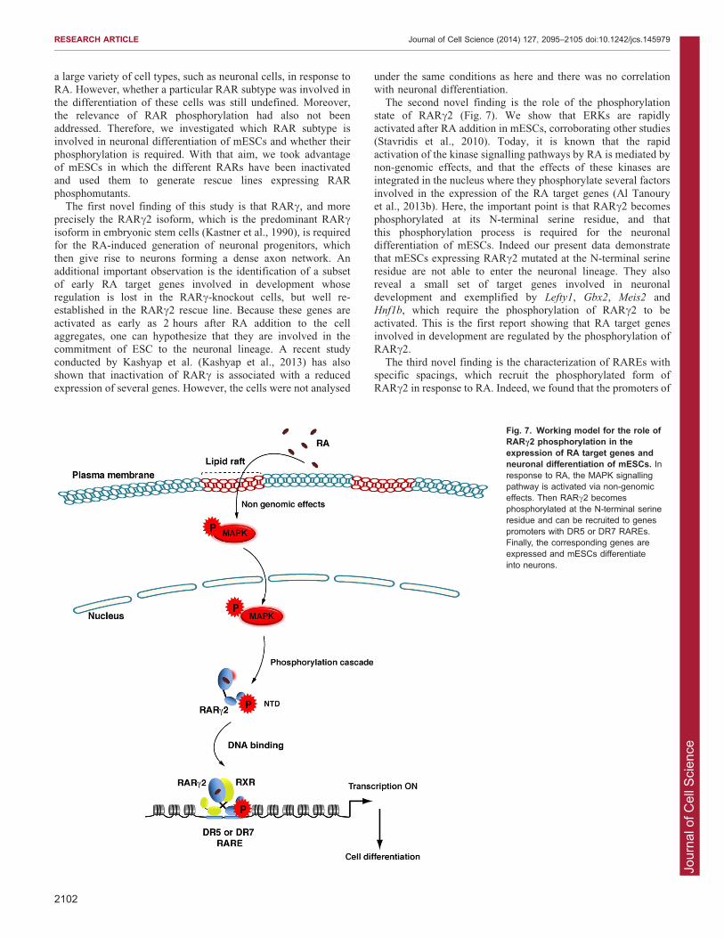
The second novel finding is the role of the phosphorylationstate of RARc2 (Fig. 7). We show that ERKs are rapidlyactivated after RA addition in mESCs, corroborating other studies(Stavridis et al., 2010). Today, it is known that the rapid
activation of the kinase signalling pathways by RA is mediated bynon-genomic effects, and that the effects of these kinases areintegrated in the nucleus where they phosphorylate several factors
involved in the expression of the RA target genes (Al Tanouryet al., 2013b). Here, the important point is that RARc2 becomesphosphorylated at its N-terminal serine residue, and that
this phosphorylation process is required for the neuronaldifferentiation of mESCs. Indeed our present data demonstratethat mESCs expressing RARc2 mutated at the N-terminal serine
residue are not able to enter the neuronal lineage. They alsoreveal a small set of target genes involved in neuronaldevelopment and exemplified by Lefty1, Gbx2, Meis2 andHnf1b, which require the phosphorylation of RARc2 to be
activated. This is the first report showing that RA target genesinvolved in development are regulated by the phosphorylation ofRARc2.
The third novel finding is the characterization of RAREs withspecific spacings, which recruit the phosphorylated form ofRARc2 in response to RA. Indeed, we found that the promoters of
Fig. 7. Working model for the role ofRARc2 phosphorylation in theexpression of RA target genes andneuronal differentiation of mESCs. Inresponse to RA, the MAPK signallingpathway is activated via non-genomiceffects. Then RARc2 becomesphosphorylated at the N-terminal serineresidue and can be recruited to genespromoters with DR5 or DR7 RAREs.Finally, the corresponding genes areexpressed and mESCs differentiateinto neurons.
RESEARCH ARTICLE Journal of Cell Science (2014) 127, 2095–2105 doi:10.1242/jcs.145979
2102

Jour
nal o
f Cel
l Sci
ence
the above genes targeted by phosphoRARc2 depict not onlycanonical DR2 and DR5 response elements but also atypical DR7
elements, and that only the DR5 and DR7 elements recruitspecifically the phosphorylated form of RARc2 in response toRA. The next challenge will be to determine how phosphorylationof the N-terminal serine residue, which is located in the vicinity
of the DBD, regulates the binding of RARc2 only to responseelements with DR5 or DR7 spacing. However, it is worth pointingout that the spacing of the response elements directs the
architecture of the DNA-bound RXR–RAR heterodimers(Brelivet et al., 2012; Rochel et al., 2011). Becausephosphorylation of the N-terminal serine residue induces the
dissociation of partners with SH3 domains (Lalevee et al., 2010),preliminary studies have suggested that the interaction of the non-phosphorylated form with SH3 proteins would impede the
binding of RARc2 only to DRs with specific spacing (DR5 andDR7), whereas the phosphorylated form (without SH3 partners)would be recruited to these DRs in response to RA (B. Kiefferand C.R.-E., unpublished results).
It is important to note that the DR7 elements show a rather lowfrequency (Moutier et al., 2012) among the RAR-occupied sites,suggesting that they would control the expression of a small set of
genes involved in the commitment of pluripotent cells to specificlineages. Here, we have defined some of these genes, i.e. theLefty1, Gbx2 and Hnfb1 genes, and we show that they belong to
an early phosphoRARc2-regulated gene programme and arecrucial for the loss of pluripotency and for triggering the neuronaldifferentiation of mESCs. Remarkably, comparison of our
findings with those reported for other cell types highlights thatLefty1 and Gbx2, which are also known as the ‘stimulated by RA’Stra3 and Stra7 genes, respectively (Chazaud et al., 1996; Oulad-Abdelghani et al., 1998), are involved in early neural
development (Li et al., 2009; Smith et al., 2008; Sunmonuet al., 2011) and, thus, are also activated in the P19 cell line thatdifferentiates into neurons in response to RA. However, they are
absent from the lists of RA-activated genes in cells with otherfeatures, such as mouse embryo carcinoma cells (F9 cell line)(Lalevee et al., 2011; Mendoza-Parra et al., 2011; Su and Gudas,
2008), MCF7 cells (Hua et al., 2009) and mouse embryonicfibroblasts (Al Tanoury et al., 2013a). Collectively such datahighlight the importance of these genes for commitment to theneuronal lineage. Concerning the Hnf1b and Meis2 (also known
as Stra10; Oulad-Abdelghani et al., 1997) genes, they are alsoinvolved in neuronal differentiation, but are not restricted tothis cell type because they are expressed in F9 cells and
other developing tissues (Mendoza-Parra et al., 2011; Oulad-Abdelghani et al., 1997). Nevertheless, all these data suggest thatRARc2 phosphorylation would control neuronal differentiation
via the activation of target genes with specific DR7 and/or DR5element combinations.
In attempts to validate the role of the early phosphoRARc2-
regulated genes in the neuronal differentiation of mESCs, wemonitored the effects of shRNA-mediated Gbx2 and Meis2
knockdown on the appearance of axons after RA treatment. Infact, despite the efficiency of the knockdown, the neuronal
differentiation of the cells was not significantly affected. Thusknockdown of one of these transcription factors alone is notsufficient to abolish the differentiation programme. Therefore,
either the residual levels of expression are sufficient to sustaindifferentiation, or the genes act together to establish a sub-programme of interconnected regulatory networks that are not
strongly perturbed by the loss of only one of these genes. Further
studies involving simultaneous knockdown of several or all ofthese genes will be required to answer this question. It is also
important to note that almost 50% of the RA-induced genes arestill normally activated in the RARc-knockout cells althoughthese cells do not differentiate into neurons. This again highlightsthe crucial role played by the small subset of genes specifically
induced by the RARc2 isoform.In conclusion, we provide several lines of evidence for a
function of RARc2 phosphorylation in positively regulating
neuronal differentiation (Fig. 7). It would be interesting toextrapolate this work to other RA responsive systems.Ultimately, one can predict specific sets of genes with DR5
and/or DR7 elements that could be controlled by phosphorylationof RARs, depending on the feature of the cells.
MATERIALS AND METHODSPlasmidsAll constructs containing the RARc receptors were cloned into the pCA
vector, which is driven by a CAG early promoter, coupled to
hygromycin/neomycin resistance. mRARc2 and mRARc1 were isolated
as XhoI/BamH1 fragments from the pSG5 constructs (Bastien et al.,
2000) and subcloned in the same sites of pCA. mRARc2S66A and
mRARc2S68A in pCA were constructed by PCR amplification reactions.
Internal oligonucleotides used in the PCR reaction encoded alanine (A)
instead of serine (S) residues at positions 66 or 68.
AntibodiesRabbit polyclonal antibodies recognizing RARa [RPa(F)], RARb[RPb(F) and RARc [RPc(F)] were as described previously (Bastien
et al., 2000; Bruck et al., 2009; Rochette-Egly et al., 1992). RPc(F)
was purified on sulfoLink gel columns (Pierce Chemical) coupled to
the corresponding immunizing peptide (Lalevee et al., 2010). Mouse
monoclonal antibodies specifically recognizing RARc phosphorylated at
position S68 were as previously described (Lalevee et al., 2010), as were
mouse monoclonal antibodies recognizing specifically the RARc1
[Ab1c(A1)] and RARc2 [Ab10c(A2)] isoforms (Bastien et al., 2000).
Rabbit polyclonal antibodies against GAPDH were from Sigma-Aldrich
and those against Pax6, Oct4 and Nanog were from Abcam Ltd Mouse
monoclonal antibodies recognizing neuronal class III b-tubulin (Tuj1)
were from Eurogentec France. The antibodies against ERKs and their
active phosphorylated forms were from Santa Cruz Biotechnology.
Mouse ESC lines and culture conditionsMouse ESCs (clone D4), derived from the 129 sub-strain were as
previously described, as were the Rarg2/2 (clone AA71) and Rara2/2
(clone KC25) cell lines (Lohnes et al., 1993; Lufkin et al., 1993). To
establish the rescue lines, the RARc2WT, RARc2S66A or RARc2S68A
constructs were introduced into the Rarg2/2 cells by LipofectamineH2000 transfection (Invitrogen). The stable rescue lines were selected with
G418 (350 mg/ml) or hygromycin B (100 mg/ml) for 1 week and analysed
for the presence of the transgene by qPCR and western blotting.
All cell lines were grown on inactivated mouse embryonic fibroblast
feeder cells in the presence of LIF under standard conditions [DMEM
supplemented with Glut AmaxTM-I (Fischer Scientific SAS), 15% FCS,
non essential amino acids and b-mercaptoethanol]. Then cells were
differentiated into neurons according to the protocol of Bibel et al. (Bibel
et al., 2007). In brief, cells were trypsinized, plated on non-adhesive
bacteriological Greiner Petri dishes (46106 cells) in 15 ml CA (cellular
aggregates) medium (DMEM supplemented with GlutaMAXTM-I, 10%
FCS, non essential amino acids and b-mercaptoethanol) and aggregated
for 8 days with a medium change every 2 days. At day four, all-trans RA
(2 mM) (Sigma-Aldrich Chimie SARL) was added and 4 days later, the
cellular aggregates were washed with PBS, dissociated with trypsin,
suspended in N2 medium [DMEM/Ham-F12, BSA and N2H (Fischer
Scientific SAS)], and plated on culture dishes precoated with PORN
(Poly-DL-ornithine hydrobromide, Sigma-Aldrich, Chimie SARL) and
laminin (Roche Diagnostics). After 1 day, the N2 medium was changed
RESEARCH ARTICLE Journal of Cell Science (2014) 127, 2095–2105 doi:10.1242/jcs.145979
2103

Jour
nal o
f Cel
l Sci
ence
and 2 days later replaced by neurobasal medium supplemented with B27H(Fisher Scientific).
Immunoblotting, immunoprecipitation andimmunofluorescence assaysExtract preparation and immunoblotting were as described previously
(Bour et al., 2005). Immunoprecipitation was performed with mouse
monoclonal antibodies immobilized on DynabeadsH Protein A/G
(Invitrogen). For immunofluorescence assays, cells were grown on Lab-
TekH glass chamber slides (Thermoscientific), fixed in 4% formaldehyde
(PFA)-PBS (20 min), permeabilized with 0.1% Triton X-100 (15 min) and
blocked with 3% non-immune serum in PBS (30 min). Then, cells were
incubated with primary antibodies, followed by Alexa-Fluor-448- or
Alexa-Fluor-555-conjugated secondary antibodies (Invitrogen). Nuclei
were counterstained with DAPI (Sigma-Aldrich). Cells were analysed by
fluorescence microscopy using a LEICA DMRX microscope equipped
with a LEICA True Confocal Scanner TCS SP.
Detection of phosphorylated RARc
Cell extracts were applied to phosphoprotein purification columns
(Qiagen). Column eluates containing protein peaks were concentrated and
analysed by immunoblotting as previously described (Bruck et al., 2009).
RNA extraction and RT-qPCRTotal RNA was extracted from cellular aggregates grown for 4 days in
the absence of RA and then treated with RA for 2–6 hours. Aliquots were
subjected to RT-qPCR as described (Bruck et al., 2009). Transcripts were
normalized according to the housekeeping gene GAPDH. Primer
amplification and specificity were verified on DNA serial dilutions.
Primer sequences are available from the corresponding author upon
request.
High-throughput mRNA sequencingAfter isolation of total RNA, a library of template molecules suitable for
high-throughput DNA sequencing (RNA-Seq) was created following the
Illumina ‘Truseq RNA sample prep v2’ protocol with some
modifications. Briefly, mRNA was purified from 2 mg total RNA using
oligo-dT magnetic beads and fragmented using divalent cations (94 C,
8 minutes). The cleaved mRNA fragments were reverse-transcribed to
cDNA using random primers and then the second cDNA strand was
synthesized using Polymerase I and RNase H. The next steps of RNA-
Seq library preparation were performed in a fully automated system using
SPRIworks Fragment Library System I kit (ref A84803, Beckman
Coulter, Inc.) with the SPRI-TE instrument (Beckman Coulter, Inc.).
Briefly, in this system, double-stranded cDNA fragments were blunted,
phosphorylated and ligated to indexed adapter dimers, and fragments in
the range of ,200–400 bp were selected. Finally, the library was
amplified by PCR [30 s at 98 C (10 s at 98 C, 30 s at 60 C, 30 s at
72 C)612 cycles; 5 min at 72 C] and the surplus PCR primers were
removed using AMPure beads (Agencourt Biosciences Corporation) with
the Biomek 3000 instrument (Beckman Coulter, Inc.). DNA libraries
were checked for quality and quantified using 2100 Bioanalyzer
(Agilent). The libraries were loaded in the flow cell at 11 pM
concentration and clusters were generated and sequenced in the
Illumina Hiseq2000 as single-end 50-base reads.
Image analysis and base calling were performed using CASAVA
v1.8.2 sequence reads were mapped onto the mm9 assembly of the mouse
genome by using Tophat v1.4.1 (Trapnell et al., 2009) and the bowtie
v0.12.7 aligner. Only uniquely aligned reads have been retained for
further analyses. Gene expression was quantified using HTSeq v0.5.3p3
(Anders and Huber, 2010) and gene annotations from Ensembl release
66. Read counts were normalized across libraries with the method
proposed by Anders and Huber (Anders and Huber, 2010). Comparisons
of interest were performed using the statistical method proposed by
Anders and Huber (Anders and Huber, 2010) implemented in the DESeq
v.1.6.1 Bioconductor package. P-values are adjusted for multiple testing
by using the Benjamini and Hochberg (Benjamini and Hochberg, 1995)
method. Only genes with |log2 fold-change|.1 or ,21 and an adjusted
P,0.05 were considered. Functional analyses of these genes were
performed using the Manteia program (http://manteia.igbmc.fr).
Motif researchThe gene regions located 610 kb from gene limits (Ensembl release 63)
were analysed using regular expression search to detect perfect consensus
59-RGKTSA-39 half sites with different spacing. The potential RAR-
binding elements were aligned on the same strand to ensure the sense and
antisense matches gave homogeneous positions.
Chromatin immunoprecipitation experimentsCellular aggregates were treated with RA for 45 minutes and chromatin
immunoprecipitation (ChIP) experiments were performed as previously
described (Bruck et al., 2009). Control ChIP were performed without
antibodies, and RARc was immunoprecipitated with purified RPc(F)
immobilized on DynabeadsH Protein A (Invitrogen). RARa was also
immunoprecipitated as previously described (Bruck et al., 2009).
Immunoprecipitated DNA was amplified by PCR primers designed
using Primer3 software (Rozen and Skaletsky, 2000), which are available
upon request. Occupancy of the promoters was calculated by normalizing
the PCR signals from the immunoprecipitated samples to the signals
obtained from the input DNA.
AcknowledgementsWe are grateful to Jean-Marie Garnier for the RARc constructs and to M. OuladAbdelghani (IGBMC) for the mouse monoclonal antibodies. Special thanks toMarie Hestin, Regis Lutzing and the cell culture facilities for help.
Competing interestsThe authors declare no competing interests.
Author contributionsZ.A.T. and A.P. devised and undertook all experimental work and analyzed the data.A.D. generated the KO cell lines. S.G. performed the RT-qPCR and ChIPexperiments. S.U. and I.D. performed and analyzed the ChIP-seq experiments. B.J.performed the RNA-seq experiments; T.Y. and C.K. performed the bioinformaticanalysis of the results. C.R.E. analyzed the data and wrote the paper.
FundingThis work was supported by the Centre national de la recherche scientifique(CNRS); the Institut national de la sante et de la recherche medicale (INSERM); theAgence Nationale pour la Recherche [grant numbers ANR-05-BLAN-0390-02,ANR-09-BLAN-0297-01, ANR-SVS8-11-Rarescales]; the Association pour larecherche sur le Cancer [grant numbers ARC-07-1-3169 and SL220110603474];the Fondation pour la Recherche Medicale (FRM) [grant numberDEQ20090515423], and the Institut National du Cancer [grant numbers INCa-PL09-194, PL07-96099]. A.P. was supported by FRM and the Lady TATA MemorialTrust, Z.A.T. by INCA, and S.U. by the Ministere de la Recherche. I.D. is an ‘equipelabelisee’’ of the Ligue Nationale contre le Cancer. The Institut de Genetique et deBiologie Moleculaire et Cellulaire (IGBMC) sequencing facility is a member of the‘France Genomique’ consortium [grant number ANR10-INBS-09-08].
Supplementary materialSupplementary material available online athttp://jcs.biologists.org/lookup/suppl/doi:10.1242/jcs.145979/-/DC1
ReferencesAl Tanoury, Z., Piskunov, A. and Rochette-Egly, C. (2013). Vitamin A andretinoid signaling: genomic and nongenomic effects. J. Lipid Res. 54, 1761-1775.
Al Tanoury, Z., Piskunov, A., Andriamoratsiresy, D., Gaouar, S., Lutzing, R.,Ye, T., Jost, B., Keime, C. and Rochette-Egly, C. (2014). Genes involved incell adhesion and signaling: A new repertoire of Retinoic Acid Receptors targetgenes in mouse embryonic fibroblasts. J. Cell Sci. 127, 521-533.
Anders, S. and Huber, W. (2010). Differential expression analysis for sequencecount data. Genome Biol. 11, R106.
Bastien, J. and Rochette-Egly, C. (2004). Nuclear retinoid receptors and thetranscription of retinoid-target genes. Gene 328, 1-16.
Bastien, J., Adam-Stitah, S., Riedl, T., Egly, J. M., Chambon, P. and Rochette-Egly, C. (2000). TFIIH interacts with the retinoic acid receptor gamma andphosphorylates its AF-1-activating domain through cdk7. J. Biol. Chem. 275,21896-21904.
Benjamini, Y. and Hochberg, Y. (1995). Controlling the false discovery rate: apractical and powerful approach to multiple testing. J. R. Stat. Soc. 57B, 289-300.
RESEARCH ARTICLE Journal of Cell Science (2014) 127, 2095–2105 doi:10.1242/jcs.145979
2104
Jour
nal o
f Cel
l Sci
ence
Bibel, M., Richter, J., Schrenk, K., Tucker, K. L., Staiger, V., Korte, M., Goetz,M. and Barde, Y. A. (2004). Differentiation of mouse embryonic stem cells into adefined neuronal lineage. Nat. Neurosci. 7, 1003-1009.
Bibel, M., Richter, J., Lacroix, E. and Barde, Y. A. (2007). Generation of adefined and uniform population of CNS progenitors and neurons from mouseembryonic stem cells. Nat. Protoc. 2, 1034-1043.
Bour, G., Plassat, J. L., Bauer, A., Lalevee, S. and Rochette-Egly, C. (2005).Vinexin beta interacts with the non-phosphorylated AF-1 domain of retinoidreceptor gamma (RARgamma) and represses RARgamma-mediatedtranscription. J. Biol. Chem. 280, 17027-17037.
Brelivet, Y., Rochel, N. and Moras, D. (2012). Structural analysis of nuclearreceptors: from isolated domains to integral proteins. Mol. Cell. Endocrinol. 348,466-473.
Bruck, N., Vitoux, D., Ferry, C., Duong, V., Bauer, A., de The, H. and Rochette-Egly, C. (2009). A coordinated phosphorylation cascade initiated by p38MAPK/MSK1 directs RARalpha to target promoters. EMBO J. 28, 34-47.
Chambon, P. (1996). A decade of molecular biology of retinoic acid receptors.FASEB J. 10, 940-954.
Chazaud, C., Bouillet, P., Oulad-Abdelghani, M. and Dolle, P. (1996). Restrictedexpression of a novel retinoic acid responsive gene during limb bud dorsoventralpatterning and endochondral ossification. Dev. Genet. 19, 66-73.
Germain, P., Chambon, P., Eichele, G., Evans, R. M., Lazar, M. A., Leid, M., DeLera, A. R., Lotan, R., Mangelsdorf, D. J. and Gronemeyer, H. (2006).International Union of Pharmacology. LXIII. Retinoid X receptors. Pharmacol.Rev. 58, 760-772.
Gudas, L. J. and Wagner, J. A. (2011). Retinoids regulate stem celldifferentiation. J. Cell. Physiol. 226, 322-330.
Hua, S., Kittler, R. and White, K. P. (2009). Genomic antagonism betweenretinoic acid and estrogen signaling in breast cancer. Cell 137, 1259-1271.
Kashyap, V., Laursen, K. B., Brenet, F., Viale, A. J., Scandura, J. M. andGudas, L. J. (2013). RARc is essential for retinoic acid induced chromatinremodeling and transcriptional activation in embryonic stem cells. J. Cell Sci.126, 999-1008.
Kastner, P., Krust, A., Mendelsohn, C., Garnier, J. M., Zelent, A., Leroy, P.,Staub, A. and Chambon, P. (1990). Murine isoforms of retinoic acid receptorgamma with specific patterns of expression. Proc. Natl. Acad. Sci. USA 87,2700-2704.
Lalevee, S., Bour, G., Quinternet, M., Samarut, E., Kessler, P., Vitorino,M., Bruck, N., Delsuc, M. A., Vonesch, J. L., Kieffer, B. et al. (2010). Vinexinß,an atypical ‘‘sensor’’ of retinoic acid receptor gamma signaling: union andsequestration, separation, and phosphorylation. FASEB J. 24, 4523-4534.
Lalevee, S., Anno, Y. N., Chatagnon, A., Samarut, E., Poch, O., Laudet, V.,Benoit, G., Lecompte, O. and Rochette-Egly, C. (2011). Genome-wide in silicoidentification of new conserved and functional retinoic acid receptor responseelements (direct repeats separated by 5 bp). J. Biol. Chem. 286, 33322-33334.
Li, B., Kuriyama, S., Moreno, M. and Mayor, R. (2009). The posteriorizing geneGbx2 is a direct target of Wnt signalling and the earliest factor in neural crestinduction. Development 136, 3267-3278.
Lohnes, D., Kastner, P., Dierich, A., Mark, M., LeMeur, M. and Chambon, P.(1993). Function of retinoic acid receptor gamma in the mouse. Cell 73, 643-658.
Lufkin, T., Lohnes, D., Mark, M., Dierich, A., Gorry, P., Gaub,M. P., LeMeur, M. andChambon, P. (1993). High postnatal lethality and testis degeneration in retinoicacid receptor alpha mutant mice. Proc. Natl. Acad. Sci. USA 90, 7225-7229.
Mahony, S., Mazzoni, E. O., McCuine, S., Young, R. A., Wichterle, H. andGifford, D. K. (2011). Ligand-dependent dynamics of retinoic acid receptorbinding during early neurogenesis. Genome Biol. 12, R2.
Mendoza-Parra, M. A., Walia, M., Sankar, M. and Gronemeyer, H. (2011).Dissecting the retinoid-induced differentiation of F9 embryonal stem cells byintegrative genomics. Mol. Syst. Biol. 7, 538.
Moutier, E., Ye, T., Choukrallah, M. A., Urban, S., Osz, J., Chatagnon, A.,Delacroix, L., Langer, D., Rochel, N., Moras, D. et al. (2012). Retinoic acidreceptors recognize the mouse genome through binding elements with diversespacing and topology. J. Biol. Chem. 287, 26328-26341.
Oulad-Abdelghani, M., Chazaud, C., Bouillet, P., Sapin, V., Chambon, P. andDolle, P. (1997). Meis2, a novel mouse Pbx-related homeobox gene induced byretinoic acid during differentiation of P19 embryonal carcinoma cells. Dev. Dyn.210, 173-183.
Oulad-Abdelghani, M., Chazaud, C., Bouillet, P., Mattei, M. G., Dolle, P. andChambon, P. (1998). Stra3/lefty, a retinoic acid-inducible novel member of thetransforming growth factor-beta superfamily. Int. J. Dev. Biol. 42, 23-32.
Piskunov, A. and Rochette-Egly, C. (2012). A retinoic acid receptor RARa poolpresent in membrane lipid rafts forms complexes with G protein aQ to activatep38MAPK. Oncogene 31, 3333-3345.
Rochel, N., Ciesielski, F., Godet, J., Moman, E., Roessle, M., Peluso-Iltis, C.,Moulin, M., Haertlein, M., Callow, P., Mely, Y. et al. (2011). Commonarchitecture of nuclear receptor heterodimers on DNA direct repeat elementswith different spacings. Nat. Struct. Mol. Biol. 18, 564-570.
Rochette-Egly, C. (2003). Nuclear receptors: integration of multiple signallingpathways through phosphorylation. Cell. Signal. 15, 355-366.
Rochette-Egly, C. and Germain, P. (2009). Dynamic and combinatorial control ofgene expression by nuclear retinoic acid receptors (RARs). Nucl. Recept.Signal. 7, e005.
Rochette-Egly, C., Gaub, M. P., Lutz, Y., Ali, S., Scheuer, I. and Chambon, P.(1992). Retinoic acid receptor-beta: immunodetection and phosphorylation ontyrosine residues. Mol. Endocrinol. 6, 2197-2209.
Rochette-Egly, C., Adam, S., Rossignol, M., Egly, J. M. and Chambon, P. (1997).Stimulation of RAR alpha activation function AF-1 through binding to the generaltranscription factor TFIIH and phosphorylation by CDK7. Cell 90, 97-107.
Rosenfeld, M. G., Lunyak, V. V. and Glass, C. K. (2006). Sensors and signals: acoactivator/corepressor/epigenetic code for integrating signal-dependentprograms of transcriptional response. Genes Dev. 20, 1405-1428.
Rozen, S. and Skaletsky, H. (2000). Primer3 on the WWW for general users andfor biologist programmers. Methods Mol. Biol. 132, 365-386.
Samarut, E. and Rochette-Egly, C. (2012). Nuclear retinoic acid receptors:conductors of the retinoic acid symphony during development. Mol. Cell.Endocrinol. 348, 348-360.
Samarut, E., Amal, I., Markov, G. V., Stote, R., Dejaegere, A., Laudet, V. andRochette-Egly, C. (2011). Evolution of nuclear retinoic acid receptor alpha(RARa) phosphorylation sites. Serine gain provides fine-tuned regulation. Mol.Biol. Evol. 28, 2125-2137.
Simandi, Z., Balint, B. L., Poliska, S., Ruhl, R. and Nagy, L. (2010). Activation ofretinoic acid receptor signaling coordinates lineage commitment ofspontaneously differentiating mouse embryonic stem cells in embryoid bodies.FEBS Lett. 584, 3123-3130.
Smith, J. R., Vallier, L., Lupo, G., Alexander, M., Harris, W. A. and Pedersen,R. A. (2008). Inhibition of Activin/Nodal signaling promotes specification ofhuman embryonic stem cells into neuroectoderm. Dev. Biol. 313, 107-117.
Stavridis, M. P., Collins, B. J. and Storey, K. G. (2010). Retinoic acidorchestrates fibroblast growth factor signalling to drive embryonic stem celldifferentiation. Development 137, 881-890.
Su, D. and Gudas, L. J. (2008). Gene expression profiling elucidates a specificrole for RARgamma in the retinoic acid-induced differentiation of F9teratocarcinoma stem cells. Biochem. Pharmacol. 75, 1129-1160.
Sunmonu, N. A., Li, K., Guo, Q. and Li, J. Y. (2011). Gbx2 and Fgf8 aresequentially required for formation of the midbrain-hindbrain compartmentboundary. Development 138, 725-734.
Trapnell, C., Pachter, L. and Salzberg, S. L. (2009). TopHat: discovering splicejunctions with RNA-Seq. Bioinformatics 25, 1105-1111.
Wilson, V., Olivera-Martinez, I. and Storey, K. G. (2009). Stem cells, signals andvertebrate body axis extension. Development 136, 1591-1604.
RESEARCH ARTICLE Journal of Cell Science (2014) 127, 2095–2105 doi:10.1242/jcs.145979
2105