Embed Size (px)
Citation preview
Phosphorylation of Sli15 by Ipl1 Is Important for ProperCPC Localization and Chromosome Stability inSaccharomyces cerevisiaeVasso Makrantoni1¤a, Stephen J. Corbishley1, Najma Rachidi1¤b, Nicholas A. Morrice2¤c,
David A. Robinson3, Michael J. R. Stark1*
1 Centre for Gene Regulation and Expression, College of Life Sciences, University of Dundee, Scotland, United Kingdom, 2 MRC Protein Phosphorylation Unit, College of
Life Sciences, University of Dundee, Scotland, United Kingdom, 3 Division of Biological Chemistry and Drug Discovery, College of Life Sciences, University of Dundee,
Scotland, United Kingdom
Abstract
The chromosomal passenger complex (CPC) is a key regulator of eukaryotic cell division, consisting of the protein kinaseAurora B/Ipl1 in association with its activator (INCENP/Sli15) and two additional proteins (Survivin/Bir1 and Borealin/Nbl1).Here we have identified multiple sites of CPC autophosphorylation on yeast Sli15 that are located within its centralmicrotubule-binding domain and examined the functional significance of their phosphorylation by Ipl1 through mutation ofthese sites, either to non-phosphorylatable alanine (sli15-20A) or to acidic residues to mimic constitutive phosphorylation(sli15-20D). Both mutant sli15 alleles confer chromosome instability, but this is mediated neither by changes in the capacityof Sli15 to activate Ipl1 kinase nor by decreased efficiency of chromosome biorientation, a key process in cell division thatrequires CPC function. Instead, we find that mimicking constitutive phosphorylation of Sli15 on the Ipl1 phosphorylationsites causes delocalization of the CPC in metaphase, whereas blocking phosphorylation of Sli15 on the Ipl1 sites drivesexcessive localization of Sli15 to the mitotic spindle in pre-anaphase cells. Consistent with these results, direct interaction ofSli15 with microtubules in vitro is greatly reduced either following phosphorylation by Ipl1 or when constitutivephosphorylation at the Ipl1-dependent phosphorylation sites is mimicked by aspartate or glutamate substitutions.Furthermore, we find that mimicking Ipl1 phosphorylation of Sli15 interferes with the ‘tension checkpoint’ – the CPC-dependent mechanism through which cells activate the spindle assembly checkpoint to delay anaphase in the absence oftension on kinetochore-microtubule attachments. Ipl1-dependent phosphorylation of Sli15 therefore inhibits its associationwith microtubules both in vivo and in vitro and may negatively regulate the tension checkpoint mechanism.
Citation: Makrantoni V, Corbishley SJ, Rachidi N, Morrice NA, Robinson DA, et al. (2014) Phosphorylation of Sli15 by Ipl1 Is Important for Proper CPC Localizationand Chromosome Stability in Saccharomyces cerevisiae. PLoS ONE 9(2): e89399. doi:10.1371/journal.pone.0089399
Editor: Barbara G. Mellone, University of Connecticut, Storrs, United States of America
Received November 29, 2013; Accepted January 19, 2014; Published February 18, 2014
Copyright: � 2014 Makrantoni et al. This is an open-access article distributed under the terms of the Creative Commons Attribution License, which permitsunrestricted use, distribution, and reproduction in any medium, provided the original author and source are credited.
Funding: This research was supported by Project Grant BB/G003440/1 from the Biotechnology and Biological Sciences Research Council awarded to MichaelStark. We also gratefully acknowledge support from the Wellcome Trust (083524/Z/07/Z). The funders had no role in study design, data collection and analysis,decision to publish, or preparation of the manuscript.
Competing Interests: The authors have declared that no competing interests exist.
* E-mail: [email protected]
¤a Current address: Wellcome Trust Centre for Cell Biology, Michael Swann Building, The King’s Buildings, University of Edinburgh, Edinburgh, Scotland, UnitedKingdom¤b Current address: Unite de Parasitology Moleculaire et Signalisation, Institut Pasteur, Paris, France¤c Current address: The Beatson Institute for Cancer Research, Garscube Estate, Bearsden, Glasgow, Scotland, United Kingdom
Introduction
The chromosomal passenger complex (CPC) has emerged over
the past decade as a critical and conserved regulator of eukaryotic
cell division, with key roles in promoting chromosome bi-
orientation, in the spindle assembly checkpoint, in spindle
disassembly during anaphase and as a regulator of cytokinesis
[1]. The CPC consists of a protein kinase (Aurora B/Ipl1) in
association with three other proteins (INCENP/Sli15, Survivin/
Bir1 and Borealin/Nbl1) and in metaphase is localized to
centromeres, where it promotes chromosome bi-orientation on
the mitotic spindle. This is thought to involve destabilization of
incorrect microtubule-kinetochore connections through phosphor-
ylation of Aurora B/Ipl1 targets at the kinetochore [2,3], allowing
turnover of connections until chromosomes achieve amphitelic
attachment, which is most likely signaled by sister chromatids
coming under tension from the kinetochore microtubules once
they are correctly attached. Kinetochore localization of the CPC is
promoted by Sgo1, which recognizes histone H2A following
phosphorylation of its C-terminal tail by Bub1 [4], and by haspin
kinase, which phosphorylates histone H3 on Thr-3 to create a
binding site for Survivin [5,6,7]. However, in budding yeast the
role of these mechanisms is less clear-cut [8,9,10] and direct
interaction between Bir1 and the inner kinetochore protein Ndc10
may be utilized to target the CPC [11,12].
During anaphase, the CPC undergoes dynamic relocalization to
the spindle mid-zone [1], promoting spindle disassembly and
cytokinesis. In yeast, the kinetochore protein Dam1 is a key Ipl1
substrate that mediates its role in chromosome bi-orientation
[13,14], while phosphorylation by Ipl1 of proteins such as Bim1
PLOS ONE | www.plosone.org 1 February 2014 | Volume 9 | Issue 2 | e89399

and She1 in anaphase are important for promoting spindle
disassembly [15,16]. Thus Ipl1-dependent phosphorylation of
Bim1, a microtubule-stabilizing protein related to EB1 [17],
promotes unloading of Bim1 from the spindle microtubules
[15,16], while phosphorylation of She1 appears to promote its
activity as a spindle disassembly factor [15]. The dramatic
relocalization of the CPC as cells enter anaphase is related to
changes in the activity of cyclin-dependent kinases and their
opposing phosphatases: INCENP/Sli15 is phosphorylated by
cyclin-dependent kinase 1 (Cdk1) and this inhibits its interaction
with the spindle [18,19], while removal of Cdk1-dependent
phosphorylation (in the case of Sli15 by Cdc14 phosphatase) is a
key element driving interaction of the yeast CPC with the spindle
mid-zone in anaphase [18]. Relocalization of the CPC to the
spindle mid-zone is also important for preventing re-engagement
of the spindle assembly checkpoint during anaphase when tension
on microtubule-kinetochore attachments is reduced [20,21]
following loss of sister chromatid cohesion.
INCENP/Sli15 consists of three domains: the N-terminal
region that mediates association with Bir1 and Nbl1, the central
domain that binds microtubules and the C-terminal domain or
IN-box that is involved in binding to and activating Aurora B/Ipl1
protein kinase [22,23]. Sli15 is itself a substrate for phosphoryla-
tion by Ipl1, becoming phosphorylated by the kinase during in vitro
protein kinase assays [23], while in higher eukaryotes phosphor-
ylation of INCENP by Aurora B on a C-terminal Thr-Ser-Ser
motif contributes to full activation of Aurora B [24,25]. To
examine whether Ipl1-dependent phosphorylation of Sli15 is
relevant for CPC function or localization, we set out to identify the
Ipl1-dependent phosphorylation sites and to examine the potential
role that phosphorylation might play. A recent study in which
predicted Ipl1 phosphorylation sites in Sli15 were changed to non-
phosphorylatable alanine showed that in addition to Cdk1-
dependent phosphorylation, Ipl1-dependent phosphorylation of
Sli15 is also likely to regulate CPC interaction with the spindle and
that in cooperation with Cdk1, Ipl1 phosphorylation of Sli15 helps
to ensure appropriate microtubule dynamics at different stages in
the cell cycle [26]. Here we report the identification of 14 sites in
Sli15 that are phosphorylated directly by Ipl1 and confirm that
Ipl1-dependent phosphorylation of Sli15 regulates CPC associa-
tion with spindle microtubules. By mutating SLI15 to encode a
protein in which constitutive phosphorylation of Sli15 is mimicked,
we demonstrate that phosphorylation in vivo is likely to limit the
interaction of the CPC with the spindle, and show that
phosphorylation of Sli15 or the acidic substitutions that mimic
constitutive phosphorylation block the direct binding of Sli15 to
microtubules in a novel in vitro binding assay. Furthermore, we find
that mimicking constitutive phosphorylation of Sli15 on its Ipl1
phosphorylation sites interferes with the cell’s ability to mount a
spindle assembly checkpoint response specifically to reduced
tension on sister kinetochores (the ‘tension checkpoint’). Thus in
addition to affecting the spindle association of the CPC, Ipl1-
dependent phosphorylation of Sli15 may act as a negative
regulator of the tension checkpoint response.
Materials and Methods
Yeast strains and general methodsBasic yeast methods, growth media, and routine recombinant
DNA methodology were performed as previously described
[27,28]. All yeast strains used in this study (Table 1) are derivatives
of W303-1a [29] and have the following markers unless indicated
to the contrary: ade2-1 his3-11, 15 leu2-3, 112 trp1-1 ura3-1 can1-
100 ssd1-d2 Gal+. Plasmids used or generated in this work are
summarized in Table 2. Relative growth, temperature sensitivity
and benomyl sensitivity were assessed by adjusting overnight
cultures of strains to OD600 0.1 and then spotting 5 ml of undiluted
and 10-fold serial dilutions onto YPAD agar plates or plates
containing 10, 11 or 12.5 mg/ml benomyl, followed by growth for
2 days at the indicated temperatures. Chromosome loss was
examined by monitoring the loss of a chromosome III fragment
(CFIII) carrying URA3 and SUP11 [30] in a colony-sectoring
assay, where the loss of CFIII (and hence SUP11) leads to a red
colony color due to the failure to suppress the ade2-1 mutation.
sli15 mutant allelesFor generating strains with mutant copies of SLI15, a 2.55 kb
BamHI-XhoI fragment carrying SLI15 together with 300 bp of
upstream and downstream flanking sequence was first excised
from pCJ145 and inserted into pRS303. The sli15-20A plasmid
was created from pRS303-SLI15 by sequential site-directed
mutagenesis using different primers on pRS303-SLI15 with the
QuikChange Site-Directed Mutagenesis Kit (Stratagene, La Jolla,
CA) according to the manufacturer’s instructions. The mutated
plasmid was verified by DNA sequencing, linearized with BsiWI
and integrated in single copy at one of the his3 loci in VMYD16
(SLI15/sli15D::KanMX6). Following sporulation and tetrad dissec-
tion, the required haploid sli15D::KanMX6 his3::sli15-20A::HIS3
strains were generated. Control strains expressing wild-type SLI15
were similarly generated using pRS303-SLI15. For creating the
sli15-20D plasmid, a 1479 bp fragment of SLI15 encoding 20
substitutions to glutamate or aspartate was synthesized (DNA 2.0,
Menlo Park, California) and subcloned as an NcoI-MfeI restriction
fragment in place of the equivalent wild-type fragment in pRS303-
SLI15, generating pRS303-SLI15-20D. pRS303-SLI15-20D was
integrated in VMYD16 as above. Following tetrad dissection, high
levels of spore inviability were seen that were rescued following
transformation with pCJ145 (pRS316-SLI15). sli15D::KanMX6
his3::sli15-20D::HIS3 strains were therefore generated by sporula-
tion and tetrad dissection of the pCJ145-containing strain,
followed by two rounds of selection on 5-fluoroorotic acid to evict
the plasmid.
Spindle assembly checkpoint analysisWhole-cell extracts were made and immunoblotted as previ-
ously described [31]. Briefly, for synchronization of cells in G1,
1.25 mg/ml a-factor was used. To prevent cells from entering the
next cell cycle after release from a-factor arrest, a-factor was
added back (7.5 mg/ml) when small buds appeared in the majority
(.80%) of cells. Nocodazole was used at 30 mg/ml. pGAL-SCC1
shut-off was performed as previously described [31]. Cell cycle
progression was studied by monitoring budding and Pds1 levels in
strains expressing PDS1-myc18. For lysing cells sequential NaOH
and trichloroacetic acid treatment was used [32]. Pds1-myc18 was
detected by Western blotting with an anti-myc antibody (c-myc A-
14; sc-789; Santa Cruz Biotechnology). Cdc28 was detected using
an anti-Cdc28 antibody from Santa Cruz Biotechnology (sc-6709).
GFP was detected with anti-GFP (11814460001; Roche).
MicroscopyThe time-lapse fluorescence microscopy of live cells for
monitoring chromosome biorientation was performed as previ-
ously described [31]. Briefly, time-lapse images of cells immobi-
lized on agarose pads released from G1 (a-factor) to metaphase
arrest (Cdc20 depletion) were collected for 4 min using a
DeltaVision RT microscope (Applied Precision), a UplanSApo
1006objective lens (numeric aperture 1.40; Olympus), SoftWoRx
software (Applied Precision), and a CoolSnap HQ (Photometrics)
Ipl1-Dependent Phosphorylation of Sli15
PLOS ONE | www.plosone.org 2 February 2014 | Volume 9 | Issue 2 | e89399

charge-coupled device camera. Seven Z sections (0.3 mm apart)
were acquired and subsequently deconvolved, projected as two-
dimensional images, and analyzed with SoftWoRx software.
VENUS and GFP fluorescent protein signals were visualized with
a JP3 filter set, whereas for GFP and mCherry fluorescent proteins
an ET filter set was used. Sister CEN5 centromeres that remained
unseparated at one end of the metaphase spindle were scored as
mono-oriented, whereas sister CEN5 centromeres that showed
dynamic separation and reassociation were scored as bioriented.
At least 100 cells were analyzed for all reported experiments.
Quantification of Sli15-EGFP fluorescence was performed with
Volocity software (PerkinElmer). Statistical analysis of chromo-
some biorientation data was carried out using Fisher’s exact test.
Statistical analysis of all other microscopy data made use of the
Mann-Whitney U test. All P values are two tailed.
Protein expression and purificationRecombinant GST-Ipl1, GST-Sli15, Ipl1-His6 and GST-Dam1
were expressed and purified essentially as described previously
[33,34]. To prepare wild-type GST-Sli15-His6, GST-Sli15-20A-
His6 and GST-Sli15-20D-His6, E. coli BL21(DE3) cells trans-
formed with the appropriate construct were induced with 100 mM
IPTG at OD600 = 0.6 for 12–16 h at 22uC. Cells were harvested
by centrifugation at 5000 rpm, 4uC. Bacterial pellets were washed
once with chilled PBS and resuspended in Lysis Buffer (50 mM
Bis-Tris propane pH 7.5, 300 mM KCl, 1% NP40, 1 mM EDTA,
1 mg/ml lysozyme, 10 mg/ml DNase, 5 mM DTT and Roche
Complete Protease Inhibitor Cocktail). Sonicated lysates were
cleared by centrifugation at 40,000 rpm for 30–45 min in a
Beckman Ti45 rotor at 4uC. Supernatants were filtered with
0.2 mm filters and incubated with 1 ml of Glutathione Sepharose 4
Fast Flow (GE Healthcare) per liter of bacterial culture. Beads
Table 1. Yeast Strains.
Strainsa Genotype Source
K699 MATa Kim Nasmyth
SJC591 MATa sli15D::KanMX6 his3::SLI15-S20A-EGFP::HIS3 trp1::pTEF1-mCherry-TUB1::TRP1 cdc20::pMET-CDC20::TRP1 This study
SJC594 MATa ipl1(S50A,S76A)::HIS3MX6 sli15D::KANMX6 his3::sli15(S335A)::HIS3 This study
SJC597 MATa ipl1(S50A,S76A)::HIS3MX6 sli15D::KANMX6 his3::sli15(20A)::HIS3 This study
SJC600 MATa ipl1(S50E,S76E)::HIS3MX6 sli15D::KANMX6 his3::sli15(S335D)::HIS3 This study
SJC603 MATa ipl1(S50E,S76E)::HIS3MX6 sli15D::KANMX6 his3::sli15(20D)::HIS3 This study
SJC641 MATa ipl1(S50A,S76A)::HIS3MX6 sli15D::KANMX6 his3::SLI15::HIS3 This study
SJC644 MATa ipl1(S50E,S76E)::HIS3MX6 sli15D::KANMX6 his3::SLI15::HIS3 This study
SJC649 ipl1(S50A,S76A)::HIS3MX6 sli15D::KANMX6 his3::sli15(20A,S335A)::HIS3 This study
SJC655 ipl1(S50E,S76E)::HIS3MX6 sli15D::KANMX6 his3::sli15(20D,S335D)::HIS3 This study
VMY30 MATa sli15D::KANMX6 his3::SLI15::HIS3 This study
VMY92 MATa CFIII (CEN3.L.YPH278) URA3 SUP11 sli15D::KANMX6 his3::SLI15::HIS3 This study
VMY148 MATa sli15D::KANMX6 his3::SLI15-S20A::HIS3 This study
VMY157 MATa CFIII (CEN3.L.YPH278) URA3 SUP11 sli15D::KANMX6 his3::SLI15-S20A::HIS3 This study
VMY162 MATa sli15D::KANMX6 his3::SLI15-S20A::HIS3 pds1::PDS1-myc18::LEU2 This study
VMY166 MATa pds1::PDS1-myc18::LEU2 scc1D::TRP1 leu2::LEU2::pGAL-SCC1 sli15D::KANMX6 his3::SLI15-S20A::HIS3 This study
VMY187 MATa sli15D::KANMX6 his3::SLI15-S20D::HIS3 This study
VMY191 MATa sli15D::KANMX6 his3::SLI15-S20D::HIS3 pds1::PDS1-myc18::LEU2 This study
VMY194 MATa sli15D::KANMX6 his3::SLI15::HIS3 PDS1-myc13::NatRMX6 This study
VMY222 MATa pds1::PDS1-myc18::LEU2 scc1D::TRP1 leu2::LEU2::pGAL-SCC1 sli15D::KANMX6 his3::SLI15::HIS3 This study
VMY224 MATa CFIII (CEN3.L.YPH278) URA3 SUP11 sli15D::KANMX6 his3::SLI15-S20D::HIS3 This study
VMY316 MATa sli15D::KANMX6 his3::SLI15::HIS3 cdc20::pMET-CDC20::TRP1 CEN5-tetO336::HIS3 leu2::tetR-GFP::LEU2 ura3::VENUS-TUB1::URA3 This study
VMY318 MATa sli15D::KANMX6 his3::SLI15-S20A::HIS3 cdc20::pMET-CDC20::TRP1 CEN5-tetO336::HIS3 leu2::tetR-GFP::LEU2 ura3::VENUS-TUB1::URA3
This study
VMY320 MATa sli15D::KANMX6 his3::SLI15-S20D::HIS3 cdc20::pMET-CDC20::TRP1 CEN5-tetO336::HIS3 leu2::tetR-GFP::LEU2 ura3::VENUS-TUB1::URA3
This study
VMY356 MATa pds1::PDS1-myc18::LEU2 scc1D::TRP1 leu2::LEU2::pGAL-SCC1 sli15D::KANMX6 his3::SLI15-S20D::HIS3 This study
VMY357 MATa sli15D::KANMX6 his3::SLI15-EGFP::HIS3 trp1::pTEF1-mCherry-TUB1::TRP1 cdc20::pMET-CDC20::TRP1 This study
VMY361 MATa sli15D::KANMX6 his3::SLI15-S335D::HIS3 This study
VMY363 MATa sli15D::KANMX6 his3::SLI15-S335A::HIS3 This study
VMY365 MATa sli15D::KANMX6 his3::SLI15-S20A-S335A::HIS3 This study
VMY367 MATa sli15D::KANMX6 his3::SLI15-S20D-S335D::HIS3 This study
VMY375 MATa sli15D::KANMX6 his3::SLI15-S20D-EGFP::HIS3 trp1::pTEF1-mCherry-TUB1::TRP1 cdc20::pMET-CDC20::TRP1 This study
VMYD16 MATa/MATa SLI15/sli15D::KANMX6 This study
aAll strains are in the W303 background: ade2-1 his3-11,15 leu2-3,112 trp1-1 ura3-1 can1-100 ssd1-d2 Gal+.doi:10.1371/journal.pone.0089399.t001
Ipl1-Dependent Phosphorylation of Sli15
PLOS ONE | www.plosone.org 3 February 2014 | Volume 9 | Issue 2 | e89399

were prewashed with PBS (10 mM Na2HPO4, 1.8 mM KH2PO4,
137 mM NaCl, 2.7 mM KCl) and equilibrated with Lysis Buffer.
After incubation for 1 h at 4uC, beads were transferred to a 10 ml
Econo-Pac column (BioRad), washed with five column volumes of
Lysis Buffer containing 1 M KCl prior to elution in 1 ml fractions
with 40 mM reduced glutathione (Sigma-Aldrich), 100 mM Tris-
HCl (pH 8.0), 300 mM NaCl, 10%(v/v) glycerol. Elution fractions
were assessed by SDS-PAGE and appropriate fractions pooled and
dialyzed in 2 L Buffer I (50 mM Bis-Tris propane pH 7.5,
300 mM KCl, 10%(v/v) glycerol, 5 mM b-mercaptoethanol,
30 mM imidazole) overnight. Dialyzed protein solutions were
diluted to 10 ml in Buffer I, and incubated with 200 ml of
equilibrated Ni-NTA slurry (Qiagen) for 2 h at 4uC with mixing
by rotation. The beads were transferred to a 10 ml Econo-Pac
column (BioRad), washed with three column volumes Buffer I
containing 1 M KCl, followed by a two column volume wash in
Buffer I. The bound proteins were eluted by the addition of 0.2 ml
Buffer I containing 250 mM imidazole (Sigma-Aldrich). Eluted
fractions were assessed by SDS-PAGE and appropriate fractions
dialyzed in 2 L 50 mM Bis-Tris propane (pH 6.8), 150 mM KCl,
100 mM imidazole, 20%(v/v) glycerol and 5 mM b-mercapto-
ethanol. Aliquots of 200 ml were snap-frozen and stored at 280uC.
Phosphorylation site mappingGST-Ipl1 kinase (,2 mg) and GST-Sli15 (,5 mg) prepared as
described previously [33] were incubated for 30 min at 30uC in
buffer containing 50 mM Tris-HCl (pH 7.5), 0.1% 2-mercapto-
ethanol, 0.1 mM EGTA, 10 mM MgCl2 and 100 mM [c-32P]ATP
(5000 cpm/pmol) in a total reaction volume of 200 ml. The
reaction was stopped by adding SDS, dithiothreitol (DTT) and
Sample Buffer (Invitrogen) to give final concentrations of 1% SDS,
10 mM DTT and 16 Sample Buffer, then the samples were
heated at 70uC for 5 min and separated by SDS-PAGE on 10%
polyacrylamide gels, stained with Colloidal Coomassie (Invitrogen)
and phosphoprotein localized by autoradiography. The 32P
labeled proteins were excised and the gel pieces were digested
with either endoproteinase Lys-C or with trypsin, then peptides
separated by HPLC and analyzed by MALDI-TOF-TOF mass
spectrometry using a Biosystems 4700 Proteomics Analyzer and
Edman degradation as described previously [35].
Protein kinase assaysProtein kinase assays using recombinant GST-Sli15-His6
(0.06 mg) and Ipl1-His6 (0.3 mg) were carried out in 20 ml reactions
with [c-32P]ATP (.800 cpm/pmol) essentially as previously
described [33,34], using either 5 mg GST-Dam1, 1 mg histone
H3.3 (kind gift from Professor Tom Owen-Hughes University of
Dundee, UK) or 1 mg Myelin Basic Protein (MBP) as substrates.
For two-step reactions involving a pre-incubation step GST-Ipl1
(0.6 mg/20 ml) and GST-Sli15 (0.5 mg/20 ml) were used and
reactions were scaled up appropriately to allow for analysis of
multiple samples. Microcystin (a kind gift from Professor C.
MacKintosh, University of Dundee) was used at 1 mM and
recombinant human protein phosphatase 1 (PP1c) at 0.05 mg per
assay. Phosphorylated proteins were separated by SDS-PAGE,
detected by autoradiography and quantitated by liquid scintilla-
tion counting after excising the radiolabelled bands.
Measurement of Sli15 and microtubule binding affinityusing Biolayer Interferometry
Tubulin (250 mg) purified from bovine brain (TL238A; Cyto-
skeleton, Inc.) was mixed with 20 mg biotin labeled tubulin
(T333P; Cytoskeleton, Inc.) in ice-cold PME buffer (80 mM Pipes-
KOH pH 6.8, 1 mM MgCl2, 1 mM EGTA) containing 1 mM
GTP (BST06-001; Cytoskeleon, Inc) and 1 mM DTT was thawed
and centrifuged to remove insoluble protein for 5 min at
42,000 rpm using a Beckman TLA100 rotor at 4uC. To assemble
microtubules, the reaction was incubated at 37uC for 30 min while
taxol (TXD01; Cytoskeleton, Inc.) was added in a stepwise manner
to a final concentration of 20 mM to stabilize microtubules.
Microtubules were then centrifuged at 50,000 rpm for 5 min at
30uC and the pellet was resuspended in warm PME buffer
containing 1 mM GTP, 1 mM DTT and 20 mM taxol to a final
concentration of 5 mM polymerized tubulin.
Binding assays were carried out by biolayer interferometry at
25uC in solid black 96-well plates (Greiner Bio-One) using an
Octet Red384 (ForteBio, Inc.). Streptavidin-coated biosensors (SA
biosensors, ForteBio, Inc.) were equilibrated in Buffer A (80 mM
Pipes-KOH pH 6.8, 1 mM MgCl2, 1 mM EGTA, 20 mM taxol),
then loaded with 5 mM biotinylated polymerized tubulin (MTs).
Unbound MTs were removed in a 5 min wash with Buffer A,
followed by a subsequent wash with Buffer A containing 10 mM
biocytin (Tocris) to block free streptavidin sites. Biocytin was
removed in a 5 min wash step with Buffer A and the system was
equilibrated for 5 min with Buffer B (50 mM Bis-Tris propane
pH 6.8, 150 mM KCl, 20%(v/v) glycerol, 5 mM b-mercaptoeth-
anol, 100 mM imidazole, 1 mg/ml BSA, 20 mM taxol). A parallel
set of SA biosensors was blocked with biocytin to act as a reference
surface to correct for non-specific binding of protein to the
biosensors. Serial dilutions of GST-Sli15-His6, GST-Sli15-20A-
His6, GST-Sli15-20D-His6 and GST control were allowed to
associate in Buffer B for 5 min and dissociation from MTs was
monitored for 10 min. Data were processed using ForteBio Data
Analysis Software 7.0 to determine binding. In the kinase assay
reaction, Ipl1-His6 was incubated with GST-Sli15 in the presence
and absence of unlabeled 10 mM ATP for 30 min at 30uC as
previously described [33] and subsequently association and
dissociation from microtubules was monitored as described above.
Table 2. Plasmids.
Plasmid Description Source
pCJ145 pRS316-SLI15 Gislene Pereira
pMS53 pGEX-2T-SLI15 This study
pMS294 pGEX-2T-DAM1 Patrick Keating [34]
pMS295 pET28a-Ipl1- His6 This study
pMS84 pJ201-sli15-20D This study
pMS85 pRS303-sli15-S20D This study
pMS86 pRS303-sli15-S20A This study
pMS218 pRS303-SLI15 This study
pMS176 pGEX-6P-GST-SLI15-His6 This study
pMS177 pGEX-6P-GST-sli15-S20A-His6 This study
pMS178 pGEX-6P-GST-sli15-S20D-His6 This study
pMS226 pRS303-sli15-S335D This study
pMS227 pRS303-sli15-S335A This study
pMS228 pRS303-sli15-S20A, S335A This study
pMS229 pRS303-sli15-S20D, S335D This study
pMS230 pRS303-SLI15-EGFP This study
pMS231 pRS303-sli15-S20A-EGFP This study
pMS232 pRS303-sli15-S20D-EGFP This study
doi:10.1371/journal.pone.0089399.t002
Ipl1-Dependent Phosphorylation of Sli15
PLOS ONE | www.plosone.org 4 February 2014 | Volume 9 | Issue 2 | e89399

Results
Identification of Ipl1 phosphorylation sites in Sli15Since Sli15 rapidly becomes phosphorylated by Ipl1 during in
vitro protein kinase assays [23], we undertook the identification of
those sites in Sli15 that are directly phosphorylated by Ipl1 so that
their potential role in CPC function could be tested. Following an
in vitro phosphorylation reaction, radiolabelled phosphopeptides
were separated by HPLC, identified by mass spectrometry and the
phosphorylation sites in each established following Edman
degradation. In this way fourteen phosphorylation sites were
found, of which all but one were identified with high confidence
(Table 3). Except for the three sites closest to the C-terminus of
Sli15, all of these phosphorylation sites are located within the
central domain of Sli15 (Figure 1A) that is involved in binding to
microtubules [23] and is also required for chromosome biorienta-
tion on the mitotic spindle [36]. Sli15 contains 17 matches to the
consensus motif [KR]-X-[ST]-[ILVST] proposed for Aurora B/
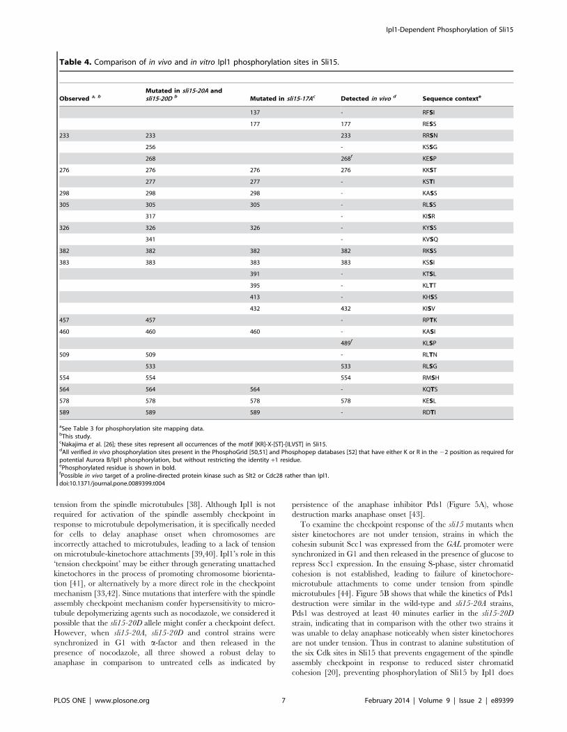
Ipl1 phosphorylation by Cheeseman et al. [13]. Ten of our mapped
phosphorylation sites match this consensus (Table 4), while a
further two of our mapped sites have asparagine in the +1 position,
which was found to be the third most favored +1 residue in a
recent large study of Aurora B-dependent phosphorylation [37].
All 14 sites have a basic residue in the –2 position that is a
hallmark of Aurora B/Ipl1 phosphorylation sites (Table 4).
Phosphopeptides corresponding to six of our mapped sites have
also been identified in vivo in large-scale phosphoproteomics studies
(Table 4); our work therefore strengthens the link between Ipl1
and these in vivo sites by demonstrating that Ipl1 is capable of
phosphorylating them.
To investigate the possible functional significance of Sli15
phosphorylation by Ipl1 we mutated all 14 mapped sites to non-
phosphorylatable alanine residues, together with six additional
sites close to mapped sites that also conformed to the Ipl1
consensus motif of Cheeseman et al. [13], two of which are known
to be phosphorylated in vivo (Table 4). This allele (encoding Sli15
with 20 alanine substitutions) was termed sli15-20A. A corre-
sponding phosphomimic allele (sli15-20D) was also generated in
which the serine and threonine residues at these twenty sites were
replaced with aspartate or glutamate respectively. Detection of the
wild-type and mutant versions of Sli15 following EGFP-tagging
indicated that all three proteins were present at similar levels in
asynchronously growing cells, and also in metaphase-arrested or
anaphase cells, confirming that neither the sli15-20A nor sli15-20D
alleles conferred a significant change in the level of Sli15 in
comparison to the wild-type protein (data not shown). Despite the
multiple amino acid replacements, yeast cells relying on either
sli15-20A or sli15-20D as their sole source of Sli15 grew normally
at 26uC and 37uC, indicating that both mutant proteins can
provide sufficient Sli15 function to support normal rates of
proliferation (Figure 1B). However, in contrast to sli15-20A, the
sli15-20D allele conferred considerable hypersensitivity to the
microtubule depolymerizing drug benomyl (Figure 1B).
Sli15 phosphorylation is not required for Ipl1 kinaseactivation
We next compared the ability of Sli15, Sli15-20A and Sli15-
20D to promote the protein kinase activity of Ipl1 in an in vitro
assay. Since activation is primarily a function of the conserved C-
terminal IN-box [23] whereas the phosphorylation sites are all
located upstream of this region, it was not anticipated that
mutation of the phosphorylation sites would affect activation of
Ipl1. Figure 2 shows that wild-type Sli15, Sli15-20A and Sli15-20D
were each able to promote similar levels of phosphorylation by
Table 3. Identification of Ipl1 phosphorylation sites on Sli15 following in vitro phosphorylation.
Enzyme Mode am/zb
observedm/zb
theoreticalLocation inSli15 Sequencec
Number ofphosphates
Phosphorylatedresidues
Lys-C R 2870.37 2870.50 218–242 (K)VKPPPNSGIARSQRRsNMFVPLPNK 1 Ser-233
Lys-C R 2887.09 2888.28 218–242 (K)VKPPPNSGIARSQRRsNmFVPLPNK 1 Ser-233
Lys-C L 3771.93 3769.96 276–310 (K)sTINSPAIRAVENSDTAGSTKAsSVFDRLSSIPTK 2 Ser-276, Ser-298
trypsin L 2828.54 2829.88 285–310 (R)AVENSDTAGSTKAsSVFDRLsSIPTK 2 Ser-298, Ser-305
trypsin R 2160.96 2160.96 319–338 (R)GNVGHKYsSSSIDLTGSPmK 1 Ser-326
Lys-C R 2501.10 2501.19 316–338 (K)ISRGNVGHKYsSSSIDLTGSPMK 1 Ser-326
Lys-C R 2517.02 2517.18 316–338 (K)ISRGNVGHKYsSSSIDLTGSPmK 1 Ser-326
Lys-C R 1594.72 1594.71 378–389 (K)NSRKssIPRFDK 2 Ser-382, Ser-383
Lys-C R 2698.98 2699.27 444–465 (K)NYYQSPVRGYLRPtKAsISPNK 2 Thr-457, Ser-460
trypsin R 1611.88 1611.83 452–465 (R)GYLRPTKAsISPNK 1 Ser-460
Lys-C L 2549.92 2550.79 505–525 (K)NYRLtNLQLLPPAEAERDDLK 1 Thr-509
Lys-C R 2549.15 2549.28 505–525 (K)NYRLtNLQLLPPAEAERDDLK 1 Thr-509
trypsin R 1336.65 1336.61 552–561 (K)RMsHLEQDLK 1 Ser-554
trypsin R 1352.64 1352.60 552–561 (K)RmsHLEQDLK 1 Ser-554
Lys-C R 1336.60 1336.61 552–561 (K)RMsHLEQDLK 1 Ser-554
Lys-C R 1352.58 1352.60 552–561 (K)RmsHLEQDLK 1 Ser-554
Lys-C R 1297.54 1297.55 562–571 (K)KQtSFSNDYK 1 Thr-564
Lys-C L 2643.22 2642.78 572–592 (K)DIRLKEsLAPFDNHVRDtINK 2 Ser-578, Thr-589
aL, linear mode; R, reflector mode.bm/z values are average [M+H]+ for linear mode and monoisotopic [M+H]+ for reflector mode.cs, phosphoserine; t, phosphothreonine; m, oxidized methionine; (K), (R), residue preceding trypsin/Lys-C cleavage site.doi:10.1371/journal.pone.0089399.t003
Ipl1-Dependent Phosphorylation of Sli15
PLOS ONE | www.plosone.org 5 February 2014 | Volume 9 | Issue 2 | e89399

Ipl1 of either Dam1 (Figure 2B) or histone H3 (Figure 2C),
indicating that phosphorylation of Sli15 by Ipl1 is not intrinsically
required for full activation of its kinase activity and consistent with
the established role of the IN-box. Since the central domain of
Sli15 has been shown to interact with Dam1 as well as with
microtubules [23], these results also indicate that phosphorylation
of Sli15 by Ipl1 is unlikely to affect the interaction between the
yeast CPC and its critical substrate at the kinetochore, since all
three GST-Sli15 fusions promoted Dam1 phosphorylation with
similar efficiency. Both Sli15-20A and Sli15-20D showed a major
reduction in phosphorylation in comparison with wild-type GST-
Sli15 in these assays, consistent with the notion that the 20 sites
that we have mutated encompass most if not all major sites of Ipl1
phosphorylation within Sli15.
To confirm that Sli15 phosphorylation by Ipl1 is not involved in
full kinase activation, we compared the ability of Sli15 that was
either phosphorylated or not phosphorylated on the Ipl1-
dependent sites to promote Ipl1-dependent phosphorylation of
an exogenous substrate. To generate these two conditions the
scheme shown in Figure 3A was used in which Ipl1, Sli15 and
protein phosphatase 1 (PP1) were first pre-incubated with
radiolabelled ATP either in the presence or absence of the PP1
inhibitor microcystin. In its presence, Sli15 became phosphory-
lated as expected, whereas in the absence of microcystin the active
PP1 opposed Sli15 phosphorylation, as indicated by lack of 32P
incorporation into Sli15 (Figure 3; lanes labelled P). At the end of
the pre-incubation, microcystin was added to the reaction without
inhibitor and then both reactions were supplemented with myelin
basic protein (MBP), which is a good substrate for Ipl1-Sli15. MBP
phosphorylation in both reactions progressed at essentially
identical rates, while the stable level of 32P incorporation into
Sli15 following pre-incubation with inhibitor indicates that it had
become maximally phosphorylated during the pre-incubation step
(Figure 3). Thus the initial phosphorylation state of Sli15 had
no significant influence on its ability to activate Ipl1 and is
therefore consistent with the ability of both Sli15-20A and Sli15-
20D to activate Ipl1 to a similar extent as wild-type Sli15 in vitro
(Figure 2).
Mutation of Ipl1 phosphorylation sites on Sli15 causeselevated levels of chromosome loss but does notcompromise chromosome bi-orientation
To investigate whether Ipl1-dependent phosphorylation of Sli15
plays a role in chromosome stability, chromosome loss rates were
measured using a colony-sectoring assay in wild-type, sli15-20A
and sli15-20D strains. As shown in Table 5, both sli15-20A and
sli15-20D led to an approximately 10-fold increase in chromosome
loss rate. Thus either blocking or constitutively mimicking
phosphorylation reduced the fidelity of chromosome transmission,
indicating that both mutant proteins are functionally compromised
in some way and suggesting that phosphorylation of Sli15 by Ipl1
plays a role in ensuring accurate chromosome segregation. A key
role of the CPC in yeast and other eukaryotes is in the
establishment of chromosome bi-orientation, and both ipl1 and
sli15 temperature-sensitive mutants show high levels of mono-
oriented chromosomes at their restrictive temperatures [2]. Thus
elevated chromosome loss in the sli15 mutants could result from
failure of the CPC to promote chromosome biorientation. We
therefore generated strains containing the sli15-20A or sli15-20D
alleles in which we could monitor chromosome biorientation
through the behavior of GFP-labelled sister CEN5s in cells arrested
in metaphase by Cdc20 depletion. Bioriented chromosomes show
dynamic splitting and reassociation of GFP-labelled sister centro-
meres in metaphase-arrested cells, whereas mono-oriented chro-
mosomes show a single, unresolved GFP locus at one end of the
metaphase spindle [31]. As shown in Figure 4, chromosome
biorientation was unaffected by either sli15-20A or sli15-20D,
occurring with the same efficiency as in the SLI15 wild-type
control strain. Thus inefficient chromosome biorientation cannot
account for the elevated rate of chromosome loss conferred by
either the sli15-20A or the sli15-20D allele.
Mimicking constitutive Ipl1 phosphorylation of Sli15compromises the spindle assembly checkpoint responseto kinetochores that are not under tension
The spindle assembly checkpoint monitors kinetochore-micro-
tubule interactions during chromosome alignment on the mitotic
spindle, delaying the onset of anaphase when chromosomes are
either unattached or mono-oriented and therefore not under
Figure 1. Characterization of the sli15-20A and sli15-20D alleles. (A) Schematic representation of Sli15 showing the 14 phosphorylation sitesmapped in vitro (black) and the six additional sites discussed in the text (blue) that were mutated in sli15-20A and sli15-20D. The microtubule-bindingregion (residues 227–559) [23] is shaded green and the conserved IN box (residues 626–698) is shaded blue. (B) Equivalent 10-fold dilutions of a wild-type strain (K699) and of sli15D::KanMX6 strains with either wild-type SLI15 (VMY30), sli15-20A (VMY148) or sli15-20D (VMY187) integrated at the his3locus were spotted onto YPAD agar in the presence of absence of benomyl at 11 mg/ml and grown for two days at 26uC or 37uC as indicated.doi:10.1371/journal.pone.0089399.g001
Ipl1-Dependent Phosphorylation of Sli15
PLOS ONE | www.plosone.org 6 February 2014 | Volume 9 | Issue 2 | e89399
tension from the spindle microtubules [38]. Although Ipl1 is not
required for activation of the spindle assembly checkpoint in
response to microtubule depolymerisation, it is specifically needed
for cells to delay anaphase onset when chromosomes are
incorrectly attached to microtubules, leading to a lack of tension
on microtubule-kinetochore attachments [39,40]. Ipl1’s role in this
‘tension checkpoint’ may be either through generating unattached
kinetochores in the process of promoting chromosome biorienta-
tion [41], or alternatively by a more direct role in the checkpoint
mechanism [33,42]. Since mutations that interfere with the spindle
assembly checkpoint mechanism confer hypersensitivity to micro-
tubule depolymerizing agents such as nocodazole, we considered it
possible that the sli15-20D allele might confer a checkpoint defect.
However, when sli15-20A, sli15-20D and control strains were
synchronized in G1 with a-factor and then released in the
presence of nocodazole, all three showed a robust delay to
anaphase in comparison to untreated cells as indicated by
persistence of the anaphase inhibitor Pds1 (Figure 5A), whose
destruction marks anaphase onset [43].
To examine the checkpoint response of the sli15 mutants when
sister kinetochores are not under tension, strains in which the
cohesin subunit Scc1 was expressed from the GAL promoter were
synchronized in G1 and then released in the presence of glucose to
repress Scc1 expression. In the ensuing S-phase, sister chromatid
cohesion is not established, leading to failure of kinetochore-
microtubule attachments to come under tension from spindle
microtubules [44]. Figure 5B shows that while the kinetics of Pds1
destruction were similar in the wild-type and sli15-20A strains,
Pds1 was destroyed at least 40 minutes earlier in the sli15-20D
strain, indicating that in comparison with the other two strains it
was unable to delay anaphase noticeably when sister kinetochores
are not under tension. Thus in contrast to alanine substitution of
the six Cdk sites in Sli15 that prevents engagement of the spindle
assembly checkpoint in response to reduced sister chromatid
cohesion [20], preventing phosphorylation of Sli15 by Ipl1 does
Table 4. Comparison of in vivo and in vitro Ipl1 phosphorylation sites in Sli15.
Observed a, bMutated in sli15-20A andsli15-20D b Mutated in sli15-17Ac Detected in vivo d Sequence contexte
137 - RFSI
177 177 RESS
233 233 233 RRSN
256 - KSSG
268 268f KESP
276 276 276 276 KKST
277 277 - KSTI
298 298 298 - KASS
305 305 305 - RLSS
317 - KISR
326 326 326 - KYSS
341 - KVSQ
382 382 382 382 RKSS
383 383 383 383 KSSI
391 - KTSL
395 - KLTT
413 - KHSS
432 432 KISV
457 457 - RPTK
460 460 460 - KASI
489f KLSP
509 509 - RLTN
533 533 RLSG
554 554 554 RMSH
564 564 564 - KQTS
578 578 578 578 KESL
589 589 589 - RDTI
aSee Table 3 for phosphorylation site mapping data.bThis study.cNakajima et al. [26]; these sites represent all occurrences of the motif [KR]-X-[ST]-[ILVST] in Sli15.dAll verified in vivo phosphorylation sites present in the PhosphoGrid [50,51] and Phosphopep databases [52] that have either K or R in the 22 position as required forpotential Aurora B/Ipl1 phosphorylation, but without restricting the identity +1 residue.ePhosphorylated residue is shown in bold.fPossible in vivo target of a proline-directed protein kinase such as Slt2 or Cdc28 rather than Ipl1.doi:10.1371/journal.pone.0089399.t004
Ipl1-Dependent Phosphorylation of Sli15
PLOS ONE | www.plosone.org 7 February 2014 | Volume 9 | Issue 2 | e89399

Figure 2. In vitro Ipl1-dependent phosphorylation promoted by wild-type and mutant versions of Sli15. (A) Coomassie-stained gelshowing recombinant GST-Sli15-His6 preparations used for in vitro phosphorylation and microtubule binding. (B) Phosphorylation of GST-Dam1 byIpl1 in the absence or presence of wild-type (Sli15) or mutant forms (Sli15-20A, Sli15-20D) of GST-Sli15-His6. Top panel, GST-Sli15-His6
phosphorylation (32P incorporation from [c-32P]ATP); middle panel, GST-Dam1 phosphorylation (32P incorporation from [c-32P]ATP); lower panel, GST-Dam1 detected by Coomassie staining. (C) Phosphorylation of H3.3 by Ipl1 in the absence or presence of wild-type or mutant forms of GST-Sli15-His6.Top panel, GST-Sli15-His6 phosphorylation (32P incorporation from [c-32P]ATP); middle panel, H3.3 phosphorylation (32P incorporation from[c-32P]ATP); lower panel, H3.3 detected by Coomassie staining. The histograms show mean relative specific activity (n = 3) of H3.3 (B) and GST-Dam1(C) phosphorylation by Ipl1 promoted by GST-Sli15-20A-His6 or GST-Sli15-20D-His6 in comparison with GST-Sli15-His6 (arbitrarily set to 1.0).doi:10.1371/journal.pone.0089399.g002
Figure 3. Ipl1 activation by Sli15 is independent of Sli15 phosphorylation. GST-Ipl1 (Ipl1), GST-Sli15 (Sli15), recombinant PP1c and[c-32P]ATP were pre-incubated for 20 min, a time that allowed maximal phosphorylation of Sli15 in the absence of PP1c, in either the presence orabsence of the PP1 inhibitor microcystin. A sample (P) was taken, then microcystin was added to the reaction that lacked the inhibitor and myelinbasic protein (MBP) added to both reactions, taking further samples over a 15-minute time course. All samples were separated by SDS-PAGE and bothMBP and Sli15 phosphorylation monitored by autoradiography. (A) Schematic showing the experimental design. (B) Autoradiograph. (C)Quantitation of Sli15 and MBP bands shown in (B) by liquid scintillation counting after excising the labelled bands.doi:10.1371/journal.pone.0089399.g003
Ipl1-Dependent Phosphorylation of Sli15
PLOS ONE | www.plosone.org 8 February 2014 | Volume 9 | Issue 2 | e89399

not appear to compromise the tension checkpoint response. In
contrast, though, the tension checkpoint is rendered ineffective by
mimicking constitutive Sli15 phosphorylation by Ipl1.
Sli15-20A and Sli15-20D show altered interaction withmicrotubules in vivo and in vitro
Since the Ipl1 phosphorylation sites in Sli15 are largely located
within the region known to interact with microtubules [23], we
next examined the localization of the wild-type and mutant Sli15
proteins in metaphase-arrested cells (Figure 6). As discussed above,
both the mutant Sli15 proteins were present at similar levels to the
wild-type protein. In the majority of metaphase-arrested cells,
Sli15-EGFP was largely evident as a cloud of fluorescence
surrounding the area of the metaphase spindle as previously
reported [18], but with some Sli15-EGFP coincident with the
spindle (Figure 6A). This was in contrast to the pattern shown by
Sli15-20A-EGFP, which was tightly focused on the spindle and
spindle poles in almost every metaphase arrested cell and which
lacked the delocalized fluorescence surrounding the spindle seen in
cells expressing wild-type Sli15-EGFP. This pattern of localization
is essentially identical to that seen either when analog-sensitive
Ipl1-as6 activity was inhibited or when an overlapping set of
predicted Ipl1 phosphorylation sites in Sli15 were mutated to
alanines (sli15-17A) [26]. In contrast, Sli15-20D-EGFP appeared
to be completely delocalized in the metaphase-arrested cells, with
greatly reduced levels associated with the spindle region in most
cells. Quantitation of EGFP fluorescence coincident with spindle
microtubules in all three strains confirmed these conclusions and
demonstrated that the increased spindle association of Sli15 in the
sli15-20A strain and the decreased levels in the sli15-20D strain
were both highly statistically significant (Figure 6B). Thus non-
phosphorylatable Sli15 showed increased metaphase spindle
localization while the phosphomimic mutant showed reduced
metaphase spindle localization, consistent with a role for Ipl1
phosphorylation of Sli15 in regulating its interaction with
microtubules. Our novel finding that mimicking constitutive
phosphorylation and blocking phosphorylation have opposite
effects on spindle association of Sli15 in vivo strengthens the notion
that its phosphorylation by Ipl1 regulates CPC localization and
emphasizes that Sli15-20D has distinct properties in comparison
with the non-phosphorylatable form.
Since the behavior of the mutant Sli15 proteins in vivo strongly
suggested that interaction of Sli15 with microtubules is affected by
Ipl1 phosphorylation, we next investigated the direct binding of
recombinant wild-type and mutant Sli15 proteins, prepared using
GST/His6 tandem affinity purification, to taxol-stabilized micro-
tubules. Using a novel assay based on biolayer interferometry,
both GST-Sli15 and GST-Sli15-20A bound to microtubules, with
GST-Sli15-20A showing greater binding over a range of
concentrations (Figure 7). Thus even in the absence of phosphor-
ylation, the wild-type protein binds microtubules less well than the
alanine substitution mutant, showing that removal of the 20 polar
side chains in the Sli15-20A microtubule domain can enhance
its affinity for microtubules in comparison with the wild-type,
non-phosphorylated protein. Most notably, however, binding of
Figure 4. Ipl1-dependent Sli15 phosphorylation is dispensablefor chromosome bi-orientation. Wild type SLI15 (VMY316), sli15-20A(VMY318) and sli15-20D (VMY320) cells containing CEN5-(tetO)336, tetR-GFP, Venus-TUB1 and pMET3-CDC20 were arrested in G1 with a-factor at26uC and then released to a metaphase block in rich medium(containing 2 mM methionine to deplete Cdc20) for 2.5 h. (A)Representative stills from time-lapse images of live cells. Bi-orientedchromosomes show dynamic splitting and reassociation of sister CEN5s.Green, CEN5 labeled with tetR-GFP; red, Venus-tubulin. (B) Quantifica-tion of chromosome bi-orientation in metaphase-arrested cells frommultiple time-lapse fields (n = number of cells scored in each category).doi:10.1371/journal.pone.0089399.g004
Table 5. sli15-20A and sli15-20D show elevated rates of chromosome loss.
Strain Red coloniesHalf-sectoredcolonies Total colonies
Chromosome loss rate percell division (6103)a Fold increase
wt 17 6 4160 1.45 -
sli15-20A 48 55 4140 13.4 9.3
sli15-20D 114 75 5088 15.1 10.4
aCalculated as [half-sectored]4([total]2[red]).doi:10.1371/journal.pone.0089399.t005
Ipl1-Dependent Phosphorylation of Sli15
PLOS ONE | www.plosone.org 9 February 2014 | Volume 9 | Issue 2 | e89399

Sli15-20D was virtually undetectable even at the highest protein
concentration used (500 nM). Thus the in vitro microtubule
binding properties of the recombinant Sli15 proteins mirrored
their behavior in vivo in metaphase-arrested cells and imply that the
addition of multiple, negatively charged groups to the Sli15
microtubule-binding domain disrupts its ability to bind microtu-
bules.
To confirm that the multiple amino acid substitutions in Sli15-
20D were mimicking phosphorylation of Sli15 by Ipl1, we next
compared the microtubule binding properties of GST-Sli15 in the
presence of Ipl1, after pre-incubation either alone or with addition
of ATP to allow Sli15 phosphorylation. Figure 7C shows that Ipl1
alone bound weakly to microtubules as expected from earlier
studies [23,45]. In the absence of ATP, GST-Sli15 and Ipl1
showed synergistic binding to microtubules. Strikingly, however,
incubation with ATP caused a strong reduction in binding,
indicating that phosphorylation of Sli15 by Ipl1 suppresses its
affinity for microtubules. However, the phosphorylated complex
still showed significant binding in comparison to Sli15-20D,
possibly because the stoichiometry of phosphorylation was not
maximal or because of additional effects of the Sli15-20D
mutations.
Sli15-20A and Sli15-20D show opposite patterns oflocalization along the anaphase spindle
We next examined the localization of the three Sli15 proteins in
cells allowed to progress from metaphase to anaphase (Figure 8).
In anaphase cells expressing wild-type Sli15-EGFP the protein
decorated the full length of the extended anaphase spindle in a
punctate manner. However, in the equivalent sli15-20A strain,
although Sli15 was clearly localized along the spindle, it frequently
appeared more abundant in the central region. In contrast,
Figure 5. Mimicking constitutive Ipl1-dependent phosphorylation of Sli15 interferes with the checkpoint response to reducedcohesion. (A) Wild-type SLI15 (VMY194), sli15-20A (VMY162), and sli15-20D (VMY191) strains expressing Pds1-myc18 were arrested in G1 with a-factorand synchronously released into YPD medium in the presence (+NOC) or absence (2NOC) of 30 mg/ml nocodazole. Samples were collected at theindicated times. Levels of Pds1-myc18 (Pds1) and Cdc28 (loading control) were monitored by immunoblotting using anti-myc and anti-Cdc28antibodies, respectively. (B) Wild-type pGAL-SCC1 SLI15 (VMY222), pGAL-SCC1 sli15-20A (VMY166) and pGAL-SCC1 sli15-20D (VMY356) cells expressingPds1-myc18 were arrested with a-factor for 2 h in medium containing galactose and then released in medium containing glucose to repress pGAL-SCC1. Pds1 and Cdc28 were monitored as described for panel A.doi:10.1371/journal.pone.0089399.g005
Ipl1-Dependent Phosphorylation of Sli15
PLOS ONE | www.plosone.org 10 February 2014 | Volume 9 | Issue 2 | e89399

Sli15-20D-EGFP appeared to be present at lower levels and was
frequently absent from the central spindle. We quantitated this
apparent difference by plotting the mean distribution of Sli15
fluorescence along multiple anaphase spindles from each of the
three strains, finding a significant focusing of Sli15 in the central
zone of the spindle in the sli15-20A spindles, compared with
depletion of Sli15 in the corresponding region of the sli15-20D
spindles (Figure 8). Thus in anaphase cells, Sli15-20A and Sli15-
20D showed opposite patterns of distribution along the elongated
spindle. Sli15-20D mimics constitutive phosphorylation of its Ipl1
sites, but is presumably dephosphorylated on its Cdk sites by
Cdc14 phosphatase during anaphase. The bias in localization of
Sli15-20D towards the poles of the anaphase spindle is reminiscent
of the pattern seen when Sli15 remains phosphorylated on its Cdk
sites (due to lack of Cdc14 phosphatase activity) but cannot be
phosphorylated on its Ipl1 sites [26]. Thus phosphorylation of
Sli15 may both reduce overall microtubule association and bias
Sli15 localization towards the poles and away from the mid-zone
of the anaphase spindle, with intermediate levels of phosphoryla-
tion on either the Ipl1 or Cdk sites being insufficient to prevent
association.
Strains lacking three distinct mechanisms for regulatingthe interaction of the CPC with spindle microtubules failto show an additive growth defect
Recent work has indicated that several mechanisms influence
the interaction of yeast CPC with microtubules including a weak,
direct interaction between microtubules and Ipl1 itself [23,45] that
we have confirmed in our work, binding via Bim1 that is
antagonized by Cdc28 phosphorylation on Ser-50 and Ser-76 in
Ipl1 [45], Cdc28 phosphorylation of Sli15 (principally on Ser-335)
that is antagonized by Cdc14 phosphatase as cells enter anaphase
[18] and phosphorylation of Sli15 by Ipl1 [26] as shown here.
Given the highly conserved nature of CPC relocalization to the
spindle in anaphase, interfering with each of these pathways
individually has surprisingly little impact on cell viability or
proliferation, indicating that these mechanisms might function in a
redundant manner to regulate CPC localization. We therefore
attempted to generate strains containing different combinations of
either alanine or phosphomimic substitution mutations affecting
the three phosphorylation-dependent mechanisms. However, all
combinations of mutations could be generated without any signs of
synthetic negative genetic interactions, both when the mutations
would be expected to drive premature microtubule binding or to
interfere with it (Figure 9). Thus while each of these mechanisms
leads to detectable phenotypes when perturbed, even in combi-
nation such perturbations still have limited consequences for
proliferation.
Discussion
Identification of Sli15 phosphorylation sitesIt has been known for some time that INCENP/Sli15 shows
Aurora B/Ipl1-dependent phosphorylation [24], but apart from
the implication of IN-box phosphorylation in full Aurora B kinase
activation [23], the role of Aurora B/Ipl1-dependent phosphor-
ylation elsewhere in INCENP/Sli15 has been unclear. Here we
have identified fourteen specific residues in Sli15 that are
phosphorylated in vitro by Ipl1, eleven of which fall within the
central domain of the protein that has been implicated in
microtubule binding, and all of which are upstream of the
conserved IN-box required for Ipl1 kinase activation. The
majority of the sites we have mapped fall within the accepted
consensus for Aurora B/Ipl1 phosphorylation [13], but two of the
sites we identified support the notion that asparagine is also a
preferred residue at +1, in keeping with a recent survey of Aurora
B phosphorylation sites [37], and the consensus for Aurora B/Ipl1
phosphorylation should therefore be adapted to include aspara-
gine at the +1 position i.e. [KR]-X-[ST]-[ILVSTN]. Six of the
sites we identified in vitro have been found to be phosphorylated in
vivo, and only two sites conforming to the accepted consensus that
are now known to be phosphorylated in vivo were not identified in
our study. Our work therefore strengthens the link between these
six in vivo phosphorylation sites and Ipl1 by demonstrating that the
kinase can phosphorylate them directly, and that it is therefore
likely to be the in vivo kinase. None of our phosphorylation sites are
Figure 6. Localization of Sli15, Sli15-20A and Sli15-20D inmetaphase-arrested cells. VMY357 (SLI15-EGFP), SJC591 (sli15-20A)and VMY375 (sli15-20D) cells containing mCherry-TUB1 and pMET3-CDC20 were arrested in G1 with a-factor at 26uC and then released to ametaphase block in rich medium for 2.5 h. (A) Representative images oflive cells arrested in metaphase. (B) Quantification of spindle associatedSli15-EGFP fluorescence in cells from multiple fields imaged 2.5–3.5 hfollowing a-factor release. The top and bottom of the boxes indicatethe 75th and 25th percentiles and the whiskers indicate the 10th and 90th
percentiles. Significance levels were determined using the Mann-Whitney U test of the null hypothesis that the medians were identical.SLI15 vs. sli15-20A: Mann-Whitney U = 36165, nSLI15 = 378, nsli15-20A = 387,p,0.001, two tailed. sli15-20D vs. SLI15: Mann-Whitney U = 15992, nsli15-
20D = 381, nSLI15 = 378, p,0.001, two tailed. Data shown are represen-tative of three separate experiments. AU, arbitrary units of fluorescence.doi:10.1371/journal.pone.0089399.g006
Ipl1-Dependent Phosphorylation of Sli15
PLOS ONE | www.plosone.org 11 February 2014 | Volume 9 | Issue 2 | e89399

located within the IN-box, consistent with our finding that non-
phosphorylatable Sli15 (Sli15-20A) can promote full activation of
Ipl1 in an in vitro protein kinase assay using Dam1 as a substrate.
Thus direct activation of Aurora B/Ipl1 via IN-box phosphory-
lation may not be a conserved feature of the CPC, consistent with
the apparent lack of conservation of the activatory phosphoryla-
tion sites in the IN-box region [23] in many lower eukaryotes.
Conversely, multiple consensus sites for Aurora B/Ipl1 are found
in the central domain of INCENP/Sli15 from a wide variety of
organisms including mouse, chicken, slime moulds and yeasts (not
shown), supporting the notion that phosphorylation of this region
by Aurora B/Ipl1 may represent a conserved feature of the CPC.
Sli15 phosphorylation by Ipl1 is not required forchromosome biorientation
Yeast cells relying on either non-phosphorylatable or phospho-
mimic alleles of SLI15 were fully viable, in contrast to cells in
which conditional mutations in either the SLI15 IN-box region
[36] or IPL1 causes lethality due to failed chromosome biorienta-
tion under restrictive conditions [2]. This implies that both sli15-
20A and sli15-20D strains should be capable of promoting efficient
chromosome biorientation. Consistently, we could detect no
significant change in the efficiency of chromosome biorientation
in either strain, in agreement with the properties of a similar non-
phosphorylatable sli15 mutant [26]. Our data therefore indicate
that constitutive phosphorylation of Sli15 on the Ipl1 sites is
Figure 7. Binding kinetic analysis of wild type and mutant versions of Sli15 to immobilized microtubules by biolayerinterferometry. A comparison of the binding abilities between recombinant GST-Sli15-His6 and its mutants to 5 mM taxol-stabilized, biotinylatedpolymerized tubulin, immobilized to streptavidin-coated biosensors is shown. (A) Binding results for five different concentrations of wild-type andmutant Sli15 (see inset) with equivalent concentrations of recombinant GST protein used as a control. Data shown are representative of threeseparate experiments. (B) Binding of wild type Sli15 and Ipl1 alone or in combination to taxol-stabilized, biotinylated polymerized tubulin with orwithout Sli15 phosphorylation by Ipl1 (+ATP); see inset for key. Data shown are representative of two separate experiments.doi:10.1371/journal.pone.0089399.g007
Ipl1-Dependent Phosphorylation of Sli15
PLOS ONE | www.plosone.org 12 February 2014 | Volume 9 | Issue 2 | e89399

unlikely to interfere with chromosome biorientation despite the
benomyl hypersensitivity of the sli15-20D strain. The robust spindle
association of Sli15-20A in pre-anaphase cells could potentially
reduce kinetochore-localized CPC, but efficient chromosome
biorientation in the sli15-20A strain indicates that Ipl1 can still gain
proper access to its substrates on incorrectly attached kinetochores.
Deletion of the entire N-terminal domain of Sli15, which mediates
its association with the other CPC components (Bir1 and Nbl1)
involved in centromere targeting, also drives Sli15 onto the pre-
anaphase spindle but has little or no effect on the efficiency of
chromosome biorientation or chromosome segregation [8]. While
this questions the importance of centromere targeting of the CPC at
least in yeast, it underlines the view that an abnormal CPC
association with the pre-anaphase spindle is not an obstacle to
achieving efficient chromosome biorientation.
Sli15 phosphorylation by Ipl1 affects its interaction withspindle microtubules
Phosphorylation of the central domain of Sli15 on its cyclin-
dependent kinase (Cdk) sites is known to regulate its interaction with
spindle microtubules, and alanine substitution of either just ser-335
or all six Cdk phosphorylation sites drives Sli15 onto the spindle
prematurely in metaphase-arrested cells [18]. Both our non-
phosphorylatable Sli15-20A protein and another similar Sli15
mutant [26] show strong localization to the metaphase spindle that
mimics the effect of inhibiting Ipl1 [26], indicating that phosphor-
ylation by Ipl1 is involved in restricting interaction of the CPC with
the spindle in pre-anaphase cells. Consistent with its reduced spindle
localization in vivo, Sli15-20D was completely defective in binding
microtubules in vitro whereas Sli15-20A and wild-type Sli15 bound
microtubules well. Binding of the wild-type protein was strongly
reduced following phosphorylation by Ipl1, indicating that phos-
phorylation of Sli15 by Ipl1 directly affects its affinity for
microtubules and confirming that addition of multiple negative
charges to the microtubule domain is inhibitory to binding. The
opposite behavior of Sli15-20A and Sli15-20D in relation to
microtubule association is therefore in contrast to alanine or
aspartate substitutions at the six Cdk phosphorylation sites in Sli15,
both of which conferred similar behavior [18]. This indicates that
Sli15-20D is not just behaving as non-phosphorylatable form of
Figure 8. Localization of Sli15, Sli15-20A and Sli15-20D in anaphase cells. Metaphase-arrested cells of strains VMY357 (SLI15-EGFP), SJC591(sli15-20A) and VMY375 (sli15-20D) prepared as in Figure 6 and allowed to progress into anaphase. (A) Representative images of anaphase cells fromeach strain (Sli15, Sli15-EGFP; tubulin, mCherry-Tub1). Bar, 3 mm. (B) Distribution of Sli15-EGFP along anaphase spindles in each strain. Sli15-EGFPfluorescence intensity along 12-16 anaphase spindles (6.0–12.6 mm) in each strain was measured on a normalized length scale of 0-100% andintensities sorted into 20 bins of increasing distance from the center of the spindle as shown, treating spindles as symmetrical. The mean fluorescenceintensities in each region are plotted (error bars indicate standard deviation of the sample). Background fluorescence (,100 arbitrary units; AU) wasnot subtracted but is indicated by the horizontal dashed line. The statistical significance of differences in the distribution of the Sli15-20A and Sli15-20D values in each bin was evaluated using the Mann-Whitney test (* = p,0.01; ** = p,0.001). The Sli15 wild-type profile was significantly differentfrom the other two profiles at all points.doi:10.1371/journal.pone.0089399.g008
Figure 9. Strains lacking three distinct mechanisms forcontrolling the interaction of the CPC with spindle microtu-bules fail to show an additive growth defect. Equivalent 10-foldserial dilutions of the indicated strains were spotted onto YPADmedium and YPAD medium containing the indicated concentrations ofbenomyl and then incubated for 2 days at 26uC. ipl1-2A and ipl1-2E arealleles in which Cdc28 phosphorylation sites Ser-50 and Ser-76 in Ipl1have been substituted by either alanine or glutamate residuesrespectively, while sli15-335A and sli15-335D have either alanine oraspartate substituted for the key serine residue in Sli15 that isphosphorylated by Cdc28 [18].doi:10.1371/journal.pone.0089399.g009
Ipl1-Dependent Phosphorylation of Sli15
PLOS ONE | www.plosone.org 13 February 2014 | Volume 9 | Issue 2 | e89399

Sli15 but that it has distinct properties conferred by the negatively-
charged side chains of the glutamate and aspartate residues. Thus
the poor in vitro microtubule binding properties of both Sli15-20D
and of phosphorylated wild-type Sli15 in comparison with the non-
phosphorylated protein or Sli15-20A support the contention that
Sli15-20D is mimicking the constitutively phosphorylated form of
the protein. Given that neither sli15-20A nor sli15-20D confer a
significant chromosome biorientation defect, it is likely that the
elevated chromosome loss rates observed in each mutant result from
altered behavior of the spindle microtubules resulting from
inappropriate levels of CPC association, although we cannot
exclude the possibility of a small defect in bi-orientation that was
below the limit of detection in our biorientation assay.
Since it is likely that Sli15-20A remains phosphorylated on its
Cdk sites, Cdk phosphorylation on its own may be insufficient to
block spindle association of the CPC. However, since Sli15-20D
retains some ability to interact with the anaphase spindle, albeit at
a reduced level, Ipl1 phosphorylation also appears insufficient to
block spindle association of the CPC in anaphase when the Cdk
sites have been dephosphorylated. Thus our data and those of
Nakajima et al. [26] support the notion of combinatorial regulation
of CPC localization by both Ipl1 and Cdk phosphorylation. While
phosphorylation at the Cdk sites in Sli15 is clearly regulated, it is
not clear to what extent phosphorylation of the Ipl1-dependent
sites may also be controlled. Sli15 bound to Ipl1 within the CPC
might be constitutively phosphorylated on its Ipl1 sites, providing a
constant level of phosphorylation to tune the basal microtubule
binding affinity of the complex and against which changes in
phosphorylation of the Cdk sites can push the affinity for
microtubules in one direction or the other. Ipl1 is clearly active
throughout anaphase [46], supporting the idea that Sli15 may
remain phosphorylated on its Ipl1 sites at this stage, but protein
phosphatases such as PP1, which is known to counteract Ipl1
function [41,47], may provide a means to antagonize CPC
autophosphorylation.
Association of the CPC with spindle microtubules is regulated in
multiple ways: binding via the interaction between Ipl1 and Bim1
is inhibited by Cdk phosphorylation on Ser-50 and Ser-76 [45],
while both Cdk and Ipl1-dependent phosphorylation of Sli15
antagonize its interaction with microtubules [18,26] as shown in
our study. It therefore seemed likely that these different
mechanisms would cooperate to regulate spindle association of
the CPC, since blocking any of these sets of regulatory
phosphorylations can drive premature spindle association of the
CPC. It is therefore remarkable that interfering with all three
pathways concurrently failed to confer any obvious additive defect.
Perhaps additional redundant pathways await discovery or else the
functions conferred by modulating spindle localization represent
fine tuning mechanisms, improving the efficiency of chromosome
segregation but not an essential component of it. Given the
conservation of CPC relocalization at the metaphase to anaphase
transition it is surprising that neither the conserved mechanisms
governing its interaction with centromeres [8] nor the multiple
regulatory pathways that control its spindle localization [18,26,45]
appear to be essential.
Sli15 phosphorylation by Ipl1 and the tension checkpointOur work has shown that in contrast to the sli15-20A mutant,
strains dependent on Sli15-20D fail to delay entry into anaphase in
response to reduced tension on kinetochore-microtubule attach-
ments – the ‘tension checkpoint’. However, both sli15 mutants
showed a normal metaphase arrest in response to microtubule
depolymerisation by nocodazole, indicating that the core check-
point machinery is functional. Ipl1 [39] and other components of
the CPC [31,48] are required specifically for the tension
checkpoint response and our data suggest that phosphorylation
of Sli15 by Ipl1 may negatively regulate the yeast cell’s ability to
sense tension on microtubule-kinetochore connections. Previous
work demonstrated that the CPC components Bir1 and Sli15 can
form a physical link in vitro between centromeric DNA and
microtubules that requires the central microtubule-binding
domain of Sli15 and that is inhibited by Ipl1 phosphorylation
[36]. This linkage was proposed to be a tension sensor for
activation of Ipl1 leading detachment of incorrect microtubule
attachments so as to promote chromosome biorientation, although
more recent work has favored models whereby tension regulates
biorientation via spatial separation of the CPC from its
kinetochore substrates [49]. Our finding that Sli15-20D confers
a profound defect in the tension checkpoint without preventing
efficient chromosome segregation is therefore consistent with a
role for Sli15 and its interaction with microtubules in tension
sensing, but in the context of spindle assembly checkpoint
signaling rather than the promotion of chromosome biorientation;
phosphorylation of Sli15 by Ipl1 may therefore act as a negative
regulator of the tension checkpoint.
Acknowledgments
Thanks are due to David Campbell for help with phosphorylation site
mapping, to Carol Mackintosh for providing microcystin, to Tom Owen-
Hughes for providing H3.3, to Gislene Pereira and Carsten Janke for
providing pCJ145, to Etsushi Kitamura for advice on microscopy and to
Adele Marston and Kevin Hardwick for critical comments. We are also
grateful to Lesley Clayton for advice on microtubule preparation.
Author Contributions
Conceived and designed the experiments: VM SJC NR DAR MJRS.
Performed the experiments: VM SJC NR NAM. Analyzed the data: VM
SJC NR NAM DAR MJRS. Contributed reagents/materials/analysis
tools: VM SJC NR MJRS DAR. Wrote the paper: VM MJRS.
References
1. Carmena M, Wheelock M, Funabiki H, Earnshaw WC (2012) The
chromosomal passenger complex (CPC): from easy rider to the godfather of
mitosis. Nat Rev Mol Cell Biol 13: 789–803.
2. Tanaka TU, Rachidi N, Janke C, Pereira G, Galova M, et al. (2002) Evidence
that the Ipl1-Sli15 (Aurora kinase-INCENP) complex promotes chromosome bi-
orientation by altering kinetochore-spindle pole connections. Cell 108: 317–329.
3. Tanaka TU, Stark MJ, Tanaka K (2005) Kinetochore capture and bi-orientation
on the mitotic spindle. Nat Rev Mol Cell Biol 6: 929–942.
4. Kawashima SA, Yamagishi Y, Honda T, Ishiguro K, Watanabe Y (2010)
Phosphorylation of H2A by Bub1 prevents chromosomal instability through
localizing shugoshin. Science 327: 172–177.
5. Kelly AE, Ghenoiu C, Xue JZ, Zierhut C, Kimura H, et al. (2010) Survivin
reads phosphorylated histone H3 threonine 3 to activate the mitotic kinase
Aurora B. Science 330: 235–239.
6. Wang F, Dai J, Daum JR, Niedzialkowska E, Banerjee B, et al. (2010) Histone
H3 Thr-3 phosphorylation by Haspin positions Aurora B at centromeres in
mitosis. Science 330: 231–235.
7. Yamagishi Y, Honda T, Tanno Y, Watanabe Y (2010) Two histone marks
establish the inner centromere and chromosome bi-orientation. Science 330:
239–243.
8. Campbell CS, Desai A (2013) Tension sensing by Aurora B kinase is
independent of survivin-based centromere localization. Nature 497: 118–121.
9. Panigada D, Grianti P, Nespoli A, Rotondo G, Gallo Castro D, et al. (2013)
Yeast haspin kinase regulates polarity cues necessary for mitotic spindle
positioning and Is required to tolerate mitotic arrest. Dev Cell 26: 483–495.
10. Storchova Z, Becker J, Talarek N, Kogelsberger S, Pellman D (2011) Bub1,
Sgo1 and Mps1 mediate a distinct pathway for chromosome bi-orientation in
budding yeast. Mol Biol Cell 22: 1473–1485.
Ipl1-Dependent Phosphorylation of Sli15
PLOS ONE | www.plosone.org 14 February 2014 | Volume 9 | Issue 2 | e89399

11. Cho US, Harrison SC (2012) Ndc10 is a platform for inner kinetochore assembly
in budding yeast. Nature Struct Mol Biol 19: 48–55.12. Yoon HJ, Carbon J (1999) Participation of Bir1p, a member of the inhibitor of
apoptosis family, in yeast chromosome segregation events. Proc Natl Acad Sci
USA 96: 13208–13213.13. Cheeseman IM, Anderson S, Jwa M, Green EM, Kang J, et al. (2002) Phospho-
regulation of kinetochore-microtubule attachments by the Aurora kinase Ipl1p.Cell 111: 163–172.
14. Tien JF, Umbreit NT, Gestaut DR, Franck AD, Cooper J, et al. (2010)
Cooperation of the Dam1 and Ndc80 kinetochore complexes enhancesmicrotubule coupling and is regulated by aurora B. J Cell Biol 189: 713–723.
15. Woodruff JB, Drubin DG, Barnes G (2010) Mitotic spindle disassembly occursvia distinct subprocesses driven by the anaphase-promoting complex, Aurora B
kinase, and kinesin-8. J Cell Biol 191: 795–808.16. Zimniak T, Stengl K, Mechtler K, Westermann S (2009) Phosphoregulation of
the budding yeast EB1 homologue Bim1p by Aurora/Ipl1p. J Cell Biol 186:
379–391.17. Vaughan KT (2005) TIP maker and TIP marker; EB1 as a master controller of
microtubule plus ends. J Cell Biol 171: 197–200.18. Pereira G, Schiebel E (2003) Separase regulates INCENP-Aurora B anaphase
spindle function through Cdc14. Science 302: 2120–2124.
19. Hummer S, Mayer TU (2009) Cdk1 negatively regulates midzone localization ofthe mitotic kinesin Mklp2 and the chromosomal passenger complex. Curr Biol
19: 607–612.20. Mirchenko L, Uhlmann F (2010) Sli15(INCENP) Dephosphorylation prevents
mitotic checkpoint reengagement due to loss of tension at anaphase onset. CurrBiol 20: 1396–1401.
21. Vazquez-Novelle MD, Petronczki M (2010) Relocation of the chromosomal
passenger complex prevents mitotic checkpoint engagement at anaphase. CurrBiol 20: 1402–1407.
22. Nakajima Y, Tyers RG, Wong CC, Yates JR, 3rd, Drubin DG, et al. (2009)Nbl1p: a Borealin/Dasra/CSC-1-like protein essential for Aurora/Ipl1 complex
function and integrity in Saccharomyces cerevisiae. Mol Biol Cell 20: 1772–1784.
23. Kang J, Cheeseman IM, Kallstrom G, Velmurugan S, Barnes G, et al. (2001)Functional cooperation of Dam1, Ipl1, and the inner centromere protein
(INCENP)-related protein Sli15 during chromosome segregation. J Cell Biol155: 763–774.
24. Bishop JD, Schumacher JM (2002) Phosphorylation of the carboxyl terminus ofinner centromere protein (INCENP) by the Aurora B Kinase stimulates Aurora
B kinase activity. J Biol Chem 277: 27577–27580.
25. Honda R, Korner R, Nigg EA (2003) Exploring the functional interactionsbetween Aurora B, INCENP, and survivin in mitosis. Mol Biol Cell 14: 3325–
3341.26. Nakajima Y, Cormier A, Tyers RG, Pigula A, Peng Y, et al. (2011) Ipl1/Aurora-
dependent phosphorylation of Sli15/INCENP regulates CPC-spindle interac-
tion to ensure proper microtubule dynamics. J Cell Biol 194: 137–153.27. Amberg DC, Burke DJ, Strathern JN (2005) Methods in yeast genetics. A Cold
Spring Harbor Laboratory Course Manual. New York: Cold Spring HarborLaboratory. 230 p.
28. Gietz D, St Jean A, Woods RA, Schiestl RH (1992) Improved method for highefficiency transformation of intact yeast cells. Nucleic Acids Res 20: 1425.
29. Thomas BJ, Rothstein R (1989) Elevated recombination rates in transcription-
ally active DNA. Cell 56: 619–630.30. Spencer F, Hieter P (1992) Centromere DNA mutations induce a mitotic delay
in Saccharomyces cerevisiae. Proc Natl Acad Sci USA 89: 8908–8912.31. Makrantoni V, Stark MJ (2009) Efficient chromosome biorientation and the
tension checkpoint in Saccharomyces cerevisiae both require Bir1. Mol Cell Biol 29:
4552–4562.32. Mekhail K, Seebacher J, Gygi SP, Moazed D (2008) Role for perinuclear
chromosome tethering in maintenance of genome stability. Nature 456: 667–670.
33. King EM, Rachidi N, Morrice N, Hardwick KG, Stark MJR (2007) Ipl1p-
dependent phosphorylation of Mad3p is required for the spindle checkpoint
response to lack of tension at kinetochores. Genes Devel 21: 1163–1168.
34. Keating P, Rachidi N, Tanaka TU, Stark MJR (2009) Ipl1-dependent
phosphorylation of Dam1 is reduced by tension applied on kinetochores. J Cell
Sci 122: 4375–4382.
35. Campbell DG, Morrice NA (2002) Identification of protein phosphorylation sites
by a combination of mass spectrometry and solid phase Edman sequencing.
J Biomol Tech 13: 119–130.
36. Sandall S, Severin F, McLeod IX, Yates JR, 3rd, Oegema K, et al. (2006) A
Bir1-Sli15 complex connects centromeres to microtubules and is required to
sense kinetochore tension. Cell 127: 1179–1191.
37. Koch A, Krug K, Pengelley S, Macek B, Hauf S (2011) Mitotic substrates of the
kinase Aurora with roles in chromatin regulation identified through quantitative
phosphoproteomics of fission yeast. Sci Signal 4: rs6.
38. Lara-Gonzalez P, Westhorpe FG, Taylor SS (2012) The spindle assembly
checkpoint. Curr Biol 22: R966–980.
39. Biggins S, Murray AW (2001) The budding yeast protein kinase Ipl1/Aurora
allows the absence of tension to activate the spindle checkpoint. Genes Devel 15:
3118–3129.
40. Pinsky BA, Tatsutani SY, Collins KA, Biggins S (2003) An Mtw1 complex
promotes kinetochore biorientation that is monitored by the Ipl1/Aurora
protein kinase. Dev Cell 5: 735–745.
41. Pinsky BA, Kung C, Shokat KM, Biggins S (2006) The Ipl1-Aurora protein
kinase activates the spindle checkpoint by creating unattached kinetochores.
Nature Cell Biol 8: 78–83.
42. Santaguida S, Vernieri C, Villa F, Ciliberto A, Musacchio A (2011) Evidence
that Aurora B is implicated in spindle checkpoint signalling independently of
error correction. EMBO J 30: 1508–1519.
43. Ciosk R, Zachariae W, Michaelis C, Shevchenko A, Mann M, et al. (1998) An
ESP1/PDS1 complex regulates loss of sister chromatid cohesion at the
metaphase to anaphase transition in yeast. Cell 93: 1067–1076.
44. Indjeian VB, Stern BM, Murray AW (2005) The centromeric protein Sgo1 is
required to sense lack of tension on mitotic chromosomes. Science 307: 130–133.
45. Zimniak T, Fitz V, Zhou H, Lampert F, Opravil S, et al. (2012) Spatiotemporal
regulation of Ipl1/Aurora activity by direct Cdk1 phosphorylation. Current
Biology 22: 787–793.
46. Buvelot S, Tatsutani SY, Vermaak D, Biggins S (2003) The budding yeast Ipl1/
Aurora protein kinase regulates mitotic spindle disassembly. J Cell Biol 160:
329–339.
47. Hsu JY, Sun ZW, Li X, Reuben M, Tatchell K, et al. (2000) Mitotic
phosphorylation of histone H3 is governed by Ipl1/aurora kinase and Glc7/PP1
phosphatase in budding yeast and nematodes. Cell 102: 279–291.
48. Shimogawa MM, Widlund PO, Riffle M, Ess M, Davis TN (2009) Bir1 is
required for the tension checkpoint. Mol Biol Cell 20: 915–923.
49. Liu D, Vader G, Vromans MJ, Lampson MA, Lens SM (2009) Sensing
chromosome bi-orientation by spatial separation of Aurora B kinase from
kinetochore substrates. Science 323: 1350–1353.
50. Sadowski I, Breitkreutz BJ, Stark C, Su TC, Dahabieh M, et al. (2013) The
PhosphoGRID Saccharomyces cerevisiae protein phosphorylation site database:
version 2.0 update. Database 2013: bat026.
51. Stark C, Su TC, Breitkreutz A, Lourenco P, Dahabieh M, et al. (2010)
PhosphoGRID: a database of experimentally verified in vivo protein
phosphorylation sites from the budding yeast Saccharomyces cerevisiae. Database
2010: bap026.
52. Bodenmiller B, Campbell D, Gerrits B, Lam H, Jovanovic M, et al. (2008)
PhosphoPep - a database of protein phosphorylation sites in model organisms.
Nature Biotechnol 26: 1339–1340.
Ipl1-Dependent Phosphorylation of Sli15
PLOS ONE | www.plosone.org 15 February 2014 | Volume 9 | Issue 2 | e89399