Embed Size (px)
Citation preview
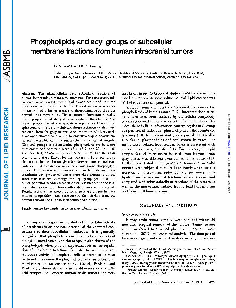
Phospholipids and acyl groups of subcellular membrane fractions from human intracranial tumors
G. Y. Sun1 and B. S. Leung
Laboratory of Neurochemistry, Ohio Mental Health and Mental Retardation Research Center, Cleveland, Ohio 44109, and Department of Surgery, University of Oregon Medical School, Portland, Oregon 97201
Abstract The phospholipids from subcellular fractions of human intracranial tumors were examined. For comparison, mi- crosomes were isolated from a fetal human brain and from the gray matter of adult human brains. The subcellular membranes of tumors had a higher protein-to-phospholipid ratio than the normal brain membranes. The microsomes from tumors had a lower proportion of diacylglycerophosphorylethanolamine and higher proportions of alkenylacylglycerophosphorylcholine and sphingomyelin (plus diacylglycerophosphorylinositol) than mi- crosomes from the gray matter. Also, the ratios of alkenylacyl- glycerophosphorylethanolamine to diacylglycerophosphoryletha- nolamine were higher in the tumors than in the normal controls. The acyl groups of ethanolamine phosphoglycerides in tumor microsomes had relatively more 18:1, 18:2, and 20:4(n - 6) and less 18:0, 22:4(n - 6), and 22:6(n - 3) than the adult brain gray matter. Except for the increase in 18:2, acyl group changes in choline phosphoglycerides between tumors and con- trols were not as extensive as in the ethanolamine phosphoglyc- erides. The characteristic features of phospholipids and their. constituent acyl groups of tumors were often present in all the subcellular fractions. Although the acyl group profiles of the tumor phosphoglycerides were in closer resemblance to the fetal brain than to the adult brain, other differences were observed. Results indicate that neoplastic brain cells are unique in their cellular composition, and consequently they deviate from the normal neurons and glials in metabolism and functions.
mal brain tissue. Subsequent studies (2-6) have also indi- cated alterations in some minor neutral lipid components of the brain tumors in general.
Although some attempts have been made to examine the phospholipids of brain tumors (7-9), interpretations of re- sults have often been hindered by the cellular complexity of unfractionated tumor tissues taken for the analysis. Be- sides, there is little information concerning the acyl group composition of individual phospholipids in the membrane fractions (10). In a recent study, we reported that the dis- tribution of phospholipids and acyl groups in subcellular membranes isolated from human brain is consistent with respect to age, sex, and diet (11). Furthermore, the lipid composition of microsomes isolated from human brain gray matter was different from that in white matter (1 1). In the present study, homogenates of human intracranial tumors were subjected to subcellular fractionation for the isolation of microsomes, mitochondria, and nuclei. The lipids from the microsomal fractions were examined and compared with other subcellular fractions of the tumors as well as the microsomes isolated from a fetal human brain and from adult human brains.
MATERIALS AND METHODS Supplementary key words microsomes ' fetal brain . gray matter
An important aspect in the study of the cellular activity of neoplasms is an accurate account of the chemical con- stituents of their subcellular membranes. It is generally recognized that phospholipids are essential components of biological membranes, and the nonpolar side chains of the phospholipids often play an important role in the regula- tion of membrane functions. In order to understand the metabolic activity of neoplastic cells, it seems to be most pertinent to examine the phospholipids of their subcellular membranes. In an earlier study, Grossi, Paoletti, and Paoletti (1) demonstrated a gross difference in the fatty acid composition between human brain tumors and nor-
Source of materials Biopsy brain tumor samples were obtained within 30
min after surgical removal of the tumors. Tumor tissues were transferred to a sealed plastic container and were stored at -20°C until chemical analysis. The time period' between surgery and chemical analysis usually did not ex-
Presented in part at the Third Meeting of the American Society for Neurochemistry, Seattle, Wash., 1972.
Abbreviations: TLC, thin-layer chromatography; GLC, gas-liquid chromatography; diacyl-GPE, diacylglycerophosphorylethanolamine; diacyl-GPC, diacylglycerophosphorylcholine; diacyl-GPI, diacylglycero- phosphorylinositol; diacyl-GPS, diacylglycerophosphorylserine.
Present address: Department of Chemistry, University of Missouri- Kansas City, Kansas City, Mo. 64110.
Journal of Lipid Research Volume 15, 1974 423
by guest, on June 25, 2018w
ww
.jlr.orgD
ownloaded from
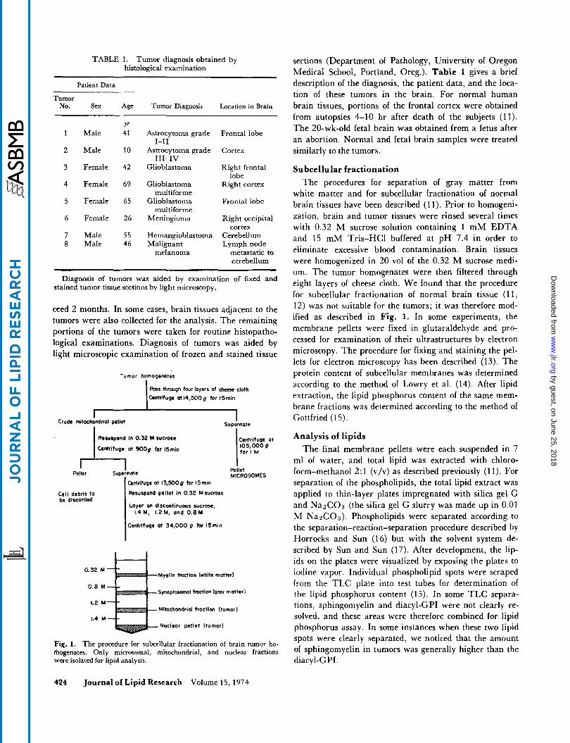
TABLE 1. Tumor diagnosis obtained by histoIogica1 examination
-
Patient Data
Tumor No. Sex Age Tumor Diagnosis Location in Brain
Y' 1 Male 41 Astrocytoma grade Frontal lobe
1-11 2 Male 10 Astrocytoma grade Cortex
111-IV 3 Female 42 Glioblastoma Right frontal
4 Female 69 Glioblastoma Right cortex
5 Female 65 Glioblastoma Frontal Iobe
6 Female 26 Meningioma Right occipital
7 Male 55 Hemangioblastoma Cerebellum 8 Male 46 Malignant Lymph node
lobe
mul ti forme
multiforme
cortex
melanoma metastatic to cerebellum
Diagnosis of tumors was aided by examination of fixed and stained tumor tissue sections by light microscopy.
ceed 2 months. In some cases, brain tissues adjacent to the tumors were also collected for the analysis. The remaining portions of the tumors were taken for routine histopatho- logical examinations. Diagnosis of tumors was aided by light microscopic examination of frozen and stained tissue
Tumor homogenotes
Poss through four layers at deest cloth Cantrifuge at14,500g far l5min
1 - I Supernote Crude mitochondrial pelld
Reaurpend In 0.32 M sucrase
Clntrituga at 9OOg far I5min
m Supernote Pellet
Cell debris lo be discarded
Centrifuge at 13,500g for I5 min
Resuspend pellet in 0.32 M sucrase
Layer on discontinuous sucrase, 1.4M, L2M, and 0 . 8 M
Centrifuge at 34.000 g far ldmin
Centrifuqa at 105,000 P tor I hr
Pellet MICROSOMES
0.32 M Myelin fmction (white mutter1
Synoptosamal fraction (gmy motterl
Mitochondrial fractlcm (tumor)
Nuclear pellet (tumor)
0.8 M
1.2 M
1.4 M
Fig. 1. The procedure for subcellular fractionation of brain tumor ho- fhogenates. Only microsomal, mitochondrial, and nuclear fractions were isolated for lipid analysis.
sections (Department of Pathology, University of Oregon Medical School, Portland, Oreg.). Table 1 gives a brief description of the diagnosis, the patient data, and the loca- tion of these tumors in the brain. For normal human brain tissues, portions of the frontal cortex were obtained from autopsies 4-10 hr after death of the subjects (11). The 20-wk-old fetal brain was obtained from a fetus after an abortion. Normal and fetal brain samples were treated similarly to the tumors.
Subcellular fractionation The procedures for separation of gray matter from
white matter and for subcellular fractionation of normal brain tissues have been described (1 1). Prior to homogeni- zation, brain and tumor tissues were rinsed several times with 0.32 M sucrose solution containing 1 m M EDTA and 15 m M Tris-HC1 buffered at p H 7.4 in order to eliminate excessive blood contamination. Brain tissues were homogenized in 20 vol of the 0.32 M sucrose medi- um. The tumor homogenates were then filtered through eight layers of cheese cloth. We found that the procedure for subcellular fractionation of normal brain tissue (1 1, 12) was not suitable For the tumors; it was therefore mod- ified as described in Fig. 1. In some experiments, the membrane pellets were fixed in glutaraldehyde and pro- cessed for examination of their ultrastructures by electron microscopy. The procedure for fixing and staining the pel- lets for electron microscopy has been described (13). The protein content of subcellular membranes was determined according to the method of Lowry et al. (14). After lipid extraction, the lipid phosphorus content of the same mem- brane fractions was determined according to the method of Gottfried (1 5).
Analysis of lipids The final membrane pellets were each suspended in 7
ml of water, and total lipid was extracted with chloro- form-methanol 2:l (v/v) as described previously (1 1). For separation of the phospholipids, the total lipid extract was applied to thin-layer plates impregnated with silica gel G and NaZC03 (the silica gel G slurry was made up in 0.01 M Na2COs). Phospholipids were separated according to the separation-reaction-separation procedure described by Horrocks and Sun (16) but with the solvent system de- scribed by Sun and Sun (17). After development, the lip- ids on the plates were visualized by exposing the plates to iodine vapor. Individual phospholipid spots were scraped from the T L C plate into test tubes for determination of the lipid phosphorus content (15). In some TLC separa- tions, sphingomyelin and diacyl-GPI were not clearly re- solved, and these areas were therefore combined for lipid phosphorus assay. In some instances when these two lipid spots were clearly separated, we noticed that the amount of sphingomyelin in tumors was generally higher than the diacyl-GPI.
424 Journal of Lipid Research Volume 15,1974
by guest, on June 25, 2018w
ww
.jlr.orgD
ownloaded from
For analysis of acyl groups, the phospholipids were sep- arated by two-dimensional T L C without exposing the plates to HCl fumes (16). After the plates were developed, lipids were visualized by spraying the thin-layer plates with 2% 2’7’-dichlorofluorescein in ethanol. The acyl groups of individual phosphoglycerides were converted to their methyl esters by alkaline methanolysis (18). The methyl esters were then analyzed by GLC as described previously (1 2).
RESULTS
Subcellular fractionation of human brain tumors
After centrifugation of the tumor homogenates at 14,500 g for 15 min, the sedimented pellets were generally grayish with small reddish brown portions at the bottom. These pellets were resuspended in 0.32 M sucrose, and further centrifugation yielded mainly the nuclear fraction (Table 2). When normal brain homogenates were subjected to subcellular fractionation, the major fractions were myelin and synaptosomes depending on the propor- tion of gray and white matter present. The microsomal pellets isolated from the tumors were reddish. Based on
. electron microscopic examination, the microsomes isolated from brain tumors were quite homogeneous and were ap- parently not contaminated by intact red blood cell mem- branes or collagen (Fig. 2). Although a few small mito- chondria may have been present in this fraction, contami- nation by myelin and by synaptosomal particles was quite rare. The microsomal membranes from the tumors were vesicular in nature and resembled the rough endoplasmic reticulum with ribosomes still attached to the membranes. Electron microscopic examination of the mitochondrial- synaptosomal fraction from the tumor homogenates indi- cated that the membranes were quite heterogeneous. Un-
like those isolated from the normal brain homogenates, there were very few synaptosomes present, and those that were present appeared to be greatly L‘degenerated”’ with no defined synaptic components. Occasionally, some gly- cogen granules were also present. Due to the heterogeneity and the low yield, a detailed biochemical analysis of the mitochondrial fractions was not performed.
Phospholipid composition of brain tumors As shown in Table 2, the protein-to-phospholipid ratios
of cellular membranes were higher in tumors than in the brain homogenates. The phospholipids from human gray matter microsomes contained a high proportion of diacyl- GPC and a relatively low proportion of sphingomyelin (Table 3). The ratios of alkenylacyl-GPE to diacyl-GPE in the microsomes from gray matter were less than 1. The proportion of sphingomyelin (plus diacyl-GPI) in the one astrocytoma analyzed was almost three times higher than that in the gray matter. The two glioblastomas also had elevated proportions of sphingomyelin (plus diacyl-GPI), although to a different extent. In general, the tumors had lower proportions of diacyl-GPE, and the ratios of alk- enylacyl-GPE to diacyl-GPE were greater than 1. Ordi- narily, alkenylacyl-GPC was not detected in normal brain tissue, but a significant amount of this phospholipid was found in the tumors, especially in the two glioblastomas analyzed.
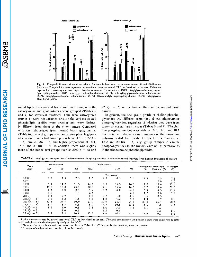
The phospholipid compositions of the mitochondrial fraction from an astrocytoma and the nuclear fraction from a glioblastoma were compared with the correspond- ing microsomal fractions (Fig. 3). Both microsomes and mitochondria of the astrocytoma had a high content of sphingomyelin in the subcellular fractions. There were high proportions of alkenylacyl-GPE found in the micro- somes of the glioblastoma as well as in the nuclear mem- branes.
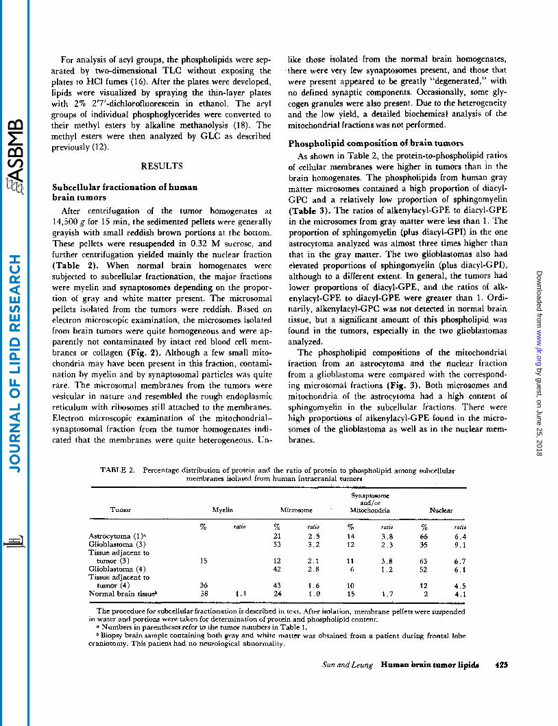
TABLE 2. Percentage distribution of protein and the ratio of protein to phospholipid among subcellular membranes isolated from human intracranial tumors
Tumor
Synaptomme and/or
Myelin Microsome Mitochondria Nuclear
% ratio % ratio % ratio % ralio
Astrocytoma (1 21 2.5 14 3.8 66 6.4 Glioblastoma (3) 53 3 .2 12 2 .3 35 9 .1 Tissue adjacent to tumor (3) 15 12 2.1 11 3.8 63 6.7
Glioblastoma (4) 42 2.8 6 1 . 2 52 6 . 1 Tissue adjacent to
Normal brain tissu@ 58 1.1 24 1 .o 15 1.7 2 4 .1 tumor (4) 36 43 1 . 6 10 12 4.5
~
The procedure for subcellular fractionation is described in text. After isolation, membrane pellets were suspended in water and portions were taken for determination of protein and phospholipid content.
Numbers in parentheses refer to the tumor numbers in Table 1. * Biopsy brain sample containing both gray and white matter was obtained from a patient during frontal lobe
craniotomy. This patient had no neurological abnormality.
Sun and Leung Human brain tumor lipids 425
by guest, on June 25, 2018w
ww
.jlr.orgD
ownloaded from
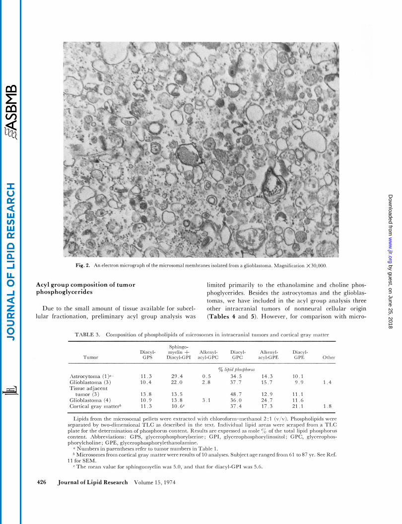
Fiq. 2. An electron micrograph of the microsomal membranes isolated from a glioblastoma. \lag-nification X30,OOO
Acyl group composition of tumor phosphogl ycerides
limited primarily to the ethanolamine and choline phos- phoglycerides. Besides the astrocytomas and the glioblas- tomas, we have included in the acyl group analysis three
Due to the small amount of tissue available for subcel- other intracranial tumors of nonneural cellular origin (Tables 4 and 5). However, for comparison with micro- lular fractionation, preliminary acyl group analysis was
TABLE 3. Composition of phospholipids of microsomes in intracranial tumors and cortical gray matter
Sphinqo- Diacyl- myelin + Alkenyl- Diacyl- Alkenyl- Diacyl-
Tumor GPS Diacyl-GPI acyl-GPC GPC acyl-GPE GPE Other
yo lipid phosphorrrr hstrocytoma (1 )n 11.3 2 9 . 4 0 . 5 34 .5 14 .3 10.1
tumor (3) 13 .8 13 .5 48 .7 12 .9 1 1 . 1 Glioblastoma (4) 10 .9 13 .8 3 . 1 36 .0 24 .7 11 .6
Glioblastoma (3) 10 .4 22 .0 2 . 8 37 .7 1 5 . 7 9 . 9 1 . 4 Tissue adjacent
Cortical gray matterh 1 1 . 3 10.be 37 .4 1 7 . 3 21 .1 1 . 8
Lipids from the microsomal pellets were extracted with chloroform-melhanol 2 : 1 (v/v). Phospholipids were separated by two-dimensional T I E as described in the text. Individual lipid areas were scraped from a T I E plate for the determination of phosphorus content. Results arc expressed as inole T: of the total lipid phosphorus content. Abbreviations: GPS, glyccrophosphorylserine; GPI, qlycerophosphorvlinositol; GPC, qlycerophos- phorylcholine; GPE, glyccrophosphorylethanolamine.
Numbers in parentheses refer to tumor nuinbms in Table 1. * Microsomes from cortical gray matter were results of 10 analyses. Subject aqe ranqcd from 61 to 87 yr. See Ref.
11 for SEM. The mean value for sphingomyelin was 5.0, and that for diacyl-GPI was 5.6.
426 Journal of Lipid Research Volume 15,1974
by guest, on June 25, 2018w
ww
.jlr.orgD
ownloaded from
Artrocytomo (1)
microsome
0 mitochondria
Glioblortomo (4 )
microsome
0 nuclei
U P
Fig. 3. Phospholipid composition of subcellular fractions isolated from astrocytoma (tumor 1) and glioblastoma (tumor 4). Phospholipids were separated by reactional two-dimensional TLC as described in the text. Values are expressed as percentages of total lipid phosphorus content. Abbreviations: dGPS, diacylglycerophosphorylserine; Sph, sphingomyelin; dGPI, diacylglycerophosphorylinositol; aGPE, alkenylacylglycerophosphorylethanolamine; dGPE, diacylglycerophosphorylethanolamine; aGPC, alkenylacylglycerophosphorylcholine; dGPC, diacylglycero- phosphorylcholine
soma1 lipids from normal brain and fetal brain, only the astrocytomas and glioblastomas were grouped (Tables 6 and 7) for statistical treatment. Data from astrocytoma (tumor 1 ) were not included because the acyl group and phospholipid profiles were peculiar and were distinct- ly different from those of the other tumors. Compared with the microsomes from normal brain gray matter (Table 6), the acyl groups of ethanolamine phosphoglycer- ides in the tumors had lower proportions of 18:0, 22:4(n - 6), and 22:6(n - 3) and higher proportions of 18:1, 18:2, and 20:4(n - 6). In addition, there was slightly more of the minor acyl groups such as 20:3(n - 6) and
22:5(n - 3) in the tumors than in the normal brain tissues.
In general, the acyl group profile of choline phospho- glycerides was different from that of the ethanolamine phosphoglycerides, regardless of whether they were from tumor or normal brain tissues (Tables 5 and 7). The cho- line phosphoglycerides were rich in 16:0, 18:0, and 18:l but contained relatively small amounts of the long-chain polyunsaturated fatty acids. Except for the increase in 18:2 and 20:4(n - 6), acyl group changes in choline phosphoglycerides in the tumors were not as extensive as in the ethanolamine phosphoglycerides.
TABLE 4. Acyl group composition of ethanolamine phosphoglycerides in the microsomal fraction from human intracranial tumors
Astrocytomas Glioblastomas Fatty Meningioma Hemangio- Melanoma Acid (1P (2) (3) (3.4) (4) (4.4) (5) ( 6 ) blastoma (7) (8)
~ ~~~
% by wcighf 16:Ob 6.6 7 .5 7 .5 8.0 4 .3 6 . 3 7.6 12.6 7 . 3 7.3 16:l 2.0 2 .0 18:O 5.1 11.7 11.3 14.6 8 . 3 1.5.3 14.0 17.0 13.6 12.4 1 8 : l 45.3 15.0 24.7 22.3 17.1 15.4 16.9 19.7 18.6 32.0 18:2 1 .4 3 . 0 2 . 1 1 . 7 3 .2 4.0 4 . 9 3 .6 6 . 5 11.8 18:3 1 . o 1 ..5 2.4 6.2 1 . 2 3 . 8 1 . 9 20: 1 1.1 0 .9 0 . 6 0 . 7 1 .o 0 . 7 0 .5 0 . 5 20:3(n - 6) 0 .6 1 . 2 1 .6 1 . 5 1 . 3 1 . 2 1 . 3 1 .4 1 . 9 0.8 20:4(n - 6) 21.3 37.7 30.9 21.7 39.9 25.4 21.8 30.0 26.1 20.4 22:4(n - 6) 3.9 13.5 6 . 9 8 .9 7.7 10.6 13.1 4.5 7.6 2 . 3
22:5(n - 3) 3 .0 0 . 9 1 . 5 0 . 6 3 . 3 1 .9 22:6(n - 3) 7 . 9 5.5 10.9 13.9 12.5 16.6 12.2 7 . 9 9 .7 6.6
22:5(n - 6 ) 1 . 2 1 . o 0 .9 0 .5 3.6 3.0 1 . 3 1 . 5 1 . 2
Lipids were separated by two-dimensional TLC as described in the text. T h e acyl groups from the phospholipids were converted to fatty
a Numbers i n parentheses refer to tumor numbers in Table 1. “A” denotes brain tissue adjacent to tumors. * Number of carbon atoms: number of double bonds.
acid methyl esters and subsequently analyzed by GLC.
Sun and Leung Human brain tumor lipids 427
by guest, on June 25, 2018w
ww
.jlr.orgD
ownloaded from
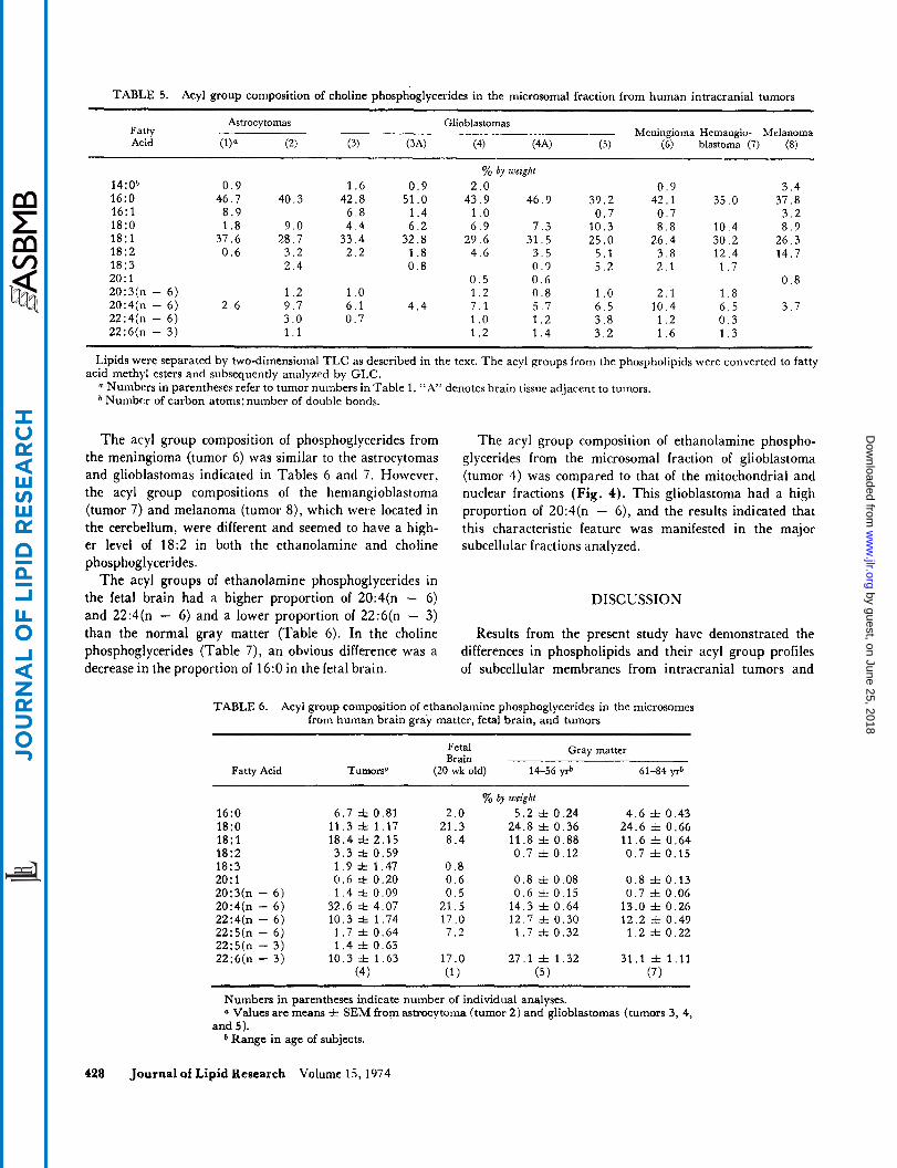
TABLE 5. Acyl group composition of choline phosphoglycerides in the microsomal fraction from human intracranial tumors
Astrocytomas Glioblastomas Fatty Meningioma Hemangio- Melanoma Acid (2) (3) (3.4) (4) (4‘4) (5) (6) blastoma (7) (8)
% by weight 14:Ob 0 . 9 1 .6 0 .9 2.0 0 . 9 3 . 4 16:O 46.7 40.3 42.8 51 .O 43.9 46.9 39.2 42.1 35.0 37.8 16: l 8 . 9 6 . 8 1 . 4 1 .o 0 . 7 0 . 7 3 . 2 18:O 1 . 8 9.0 4 .4 6 . 2 6 . 9 7 . 3 10 .3 8 .8 10 .4 8 . 9 18: 1 37.6 28.7 33.4 32.8 29.6 31.5 25.0 26.4 30.2 26.3 18:2 0 .6 3.2 2 . 2 1 .8 4 . 6 3 . 5 5 . 1 3 . 8 12 .4 14.7 18:3 2 . 4 0 . 8 0 . 9 5 .2 2 . 1 1 . 7 20: 1 0 . 5 0 . 6 0 . 8 20:3(n - 6) 1 . 2 1 .o 1 . 2 0 . 8 1 . o 2 . 1 1 . 8 20:4(n - 6) 2 . 6 9 . 7 6 . 1 4 .4 7 .1 5.7 6 . 5 10 .4 6 . 5 3 . 7 22:4(n - 6) 3 .O 0 . 7 1 .o 1 . 2 3 . 8 1 . 2 0 . 3 22:6(n - 3) 1 . 1 1 . 2 1 . 4 3 . 2 1 . 6 1 . 3
Lipids were separated by two-dimensional TLC as described in the text. The acyl groups from the phospholipids were converted to fatty acid methyl esters and subsequently analyzed by GLC.
* Numbm of carbon atoms: number of double bonds. Numbcrs in parentheses refer to tumor numbers in Table 1. “A” denotes brain tissue adjacent to tumors.
The acyl group composition of phosphoglycerides from the meningioma (tumor 6) was similar to the astrocytomas and glioblastomas indicated in Tables 6 and 7. However, the acyl group compositions of the hemangioblastoma (tumor 7) and melanoma (tumor B), which were located in the cerebellum, were different and seemed to have a high- er level of 18:2 in both the ethanolamine and choline phosphoglycerides.
The acyl groups of ethanolamine phosphoglycerides in the fetal brain had a higher proportion of 20:4(n - 6) and 22:4(n - 6) and a lower proportion of 22:6(n - 3 ) than the normal gray matter (Table 6). In the choline phosphoglycerides (Table 7) , an obvious difference was a decrease in the proportion of 16:O in the fetal brain.
The acyl group composition of ethanolamine phospho- glycerides from the microsomal fraction of glioblastoma (tumor 4) was compared to that of the mitochondrial and nuclear fractions (Fig. 4). This glioblastoma had a high proportion of 20:4(n - 6), and the results indicated that this characteristic feature was manifested in the major subcellular fractions analyzed.
DISCUSSION
Results from the present study have demonstrated the differences in phospholipids and their acyl group profiles of subcellular membranes from intracranial tumors and
TABLE 6. Acyl group composition of ethanolamine phosphoglycerides in the microsomes from human brain gray matter, fetal brain, and tumors
Fetal Gray matter Brain
Fatty Acid Tumors” (20 wk old) 14-56 yrb 61-84 yl”
7% by weight 16:O 6 . 7 f 0.81 2 .o 5 .2 f 0.24 4 .6 f 0.43 l8:O 11 .3 f 1.17 21.3 24.8 f 0.36 24.6 f 0.66 18 : l 18.4 i 2.15 8 . 4 11.8 f 0.88 11.6 f 0.64 18:2 3 . 3 f 0.59 0 . 7 f 0.12 0 . 7 f 0.15 18:3 1 . 9 f 1.47 0.8 2O:l 0 . 6 f 0.20 0 . 6 0 .8 f 0.08 0 .8 f 0.13 20:3(n - 6) 1 . 4 f 0.09 0 . 5 0 .6 f 0.15 0 .7 f 0.06 20:4(n - 6) 32.6 f 4.07 21.5 14 .3 f 0.64 13.0 f 0.26 22:4(n - 6) 10.3 f 1.74 17 .O 12.7 f 0.30 12.2 f 0.49
22:5(n - 3) 22:6(n - 3) 10.3 f 1.63 17.0 27.1 f 1.32 31.1 f 1.11
22:5(n - 6) 1 . 7 =!z 0.64 7 . 2 1 . 7 f 0.32 1 . 2 f 0.22 1 . 4 =k 0.63
(4) (1) (5 ) (7 1
Numbers in parentheses indicate number of individual analyses. a Values are means f SEM from astrocytoma (tumor 2) and glioblastomas (tumors 3, 4,
b Range in age of subjects. and 5).
428 Journal of Lipid Research Volume 15,1974
by guest, on June 25, 2018w
ww
.jlr.orgD
ownloaded from
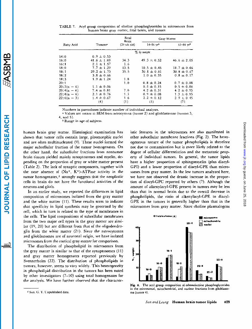
TABLE 7. Acyl group composition of choline phosphoglycerides in microsomes from human brain gray matter, fetal brain, and tumors
Fetal Gray Matter Brain
Fatty Acid Tumors" (20 wk old) 14-56 yr" 61-84 yrb
14:O 16:O 16:l 18:O 18: 1 18:2 18:3 20:l 20:3(n - 6) 20:4(n - 6) 22:4(n - 6) 22:6(n - 3)
0 . 9 f 0 .53 41.6 f 1.09
2 .1 f 1.57 7 .7 f 1.29
29.2 f 1 .73 3 .8 f 0.66 1 . 9 f 1 .24
1 . 1 f 0.06 7 . 4 f 0.81 2 . 1 rt 0.76 1 . 4 rt 0.67
(4 1
34.3 1 . 6
13.5 35.5
1 . 8 1 . o 7 .0 1 . 5 1 . 6 ( 1 )
% by weight
49 .3 f 0.52
10 .5 f 0.45 30.1 f 0.61
1 . 0 f 0.35
0 . 8 f 0.24 0 .5 f 0.15 4 .2 f 0.31 0 . 9 f 0.08 2 . 2 f 0.12
(5)
46.6 f 2.05
11.7 f 0.66 30.8 f 0.67 0 .8 f 0.17
0 . 7 f 0.08 0 . 5 f 0.06 4 . 2 f 0.15 1 . 3 f 0.15 2 . 9 f 0.45
(7)
Numbers in parentheses indicate number of individual analyses. Values are means f. SEM from astrocytoma (tumor 2) and glioblastomas (tumors 3,
4, and 5). b Range in age of subjects.
human brain gray matter. Histological examination has shown that tumor cells contain large, pleomorphic nuclei and are often multinucleated (9). These nuclei formed the major subcellular fraction of the tumor homogenates. O n the other hand, the subcellular fractions from nontumor brain tissues yielded mainly synaptosomes and myelin, de- pending on the proportion of gray or white matter present (Table 2). The lack of synaptic components, together with the near absence of (Na+, K+)-ATPase activity in the tumor homogenates,z strongly suggests that the neoplastic cells in brain do not have the functional activities of the neurons and glials.
In an earlier study, we reported the differences in lipid composition of microsomes isolated from the gray matter and the white matter (11). These results seem to indicate that specificity in lipid synthesis may be governed by the cell, which in turn is related to the type of membranes in the cells. The lipid compositions of subcellular membranes from the two major cell types in the gray matter are simi- lar (19, 20) but are different from that of the oligodendro- glia from the white matter (21). Since the astrocytomas and glioblastomas are of neuronal origin, we have isolated microsomes from the cortical gray matter for comparison.
The distribution of phospholipid in microsomes from the gray matter is similar to that of the synaptosomes (11) and gray matter homogenates reported previously by Svennerholm (22). The distribution of phospholipids in tumors, however, seems to vary widely. This heterogeneity in phospholipid distribution in the tumors has been noted by other investigators (7-10) using total homogenates for the analysis. We have further observed that the character-
2 Sun, G. Y. Unpublished data
istic features in the microsomes are also manifested in other subcellular membrane fractions (Fig. 2). The heter- ogeneous nature of the tumor phospholipids is therefore not due to contamination but is more likely related to the degree of cellular differentiation and the metastatic prop- erty of individual tumors. In general, the tumor lipids have a higher proportion of sphingomyelin (plus diacyl- GPI) and a lower proportion of diacyl-GPE than micro- somes from gray matter. In the few tumors analyzed here, we have not observed the drastic increase in the propor- tion of diacyl-GPC reported by others (7). Although the amount of alkenylacyl-GPE present in tumors may be less than that in normal brain due to the overall decrease in phospholipids, the ratio of alkenylacyl-GPE to diacyl- GPE in the tumors is generally higher than that in the microsomes from gray matter. Since choline plasmalogens
microrome 0 mitochondria
nuclei
Olioblostomo ( 4 )
Fig. 4. The acyl group composition of ethanolamine phosphoglycerides in the microsomal, mitochondrial, and nuclear fractions from glioblasto- ma (tumor 4).
Sun and Leung Human brain tumor lipids 429
by guest, on June 25, 2018w
ww
.jlr.orgD
ownloaded from
are not normally detected in brain, their presence in the tumors together with the increase in ethanolamine plas- malogens with respect to the diacyl-GPE suggest that the metabolism of plasmalogens during neoplastic transforma- tion is abnormal. The increase in plasmalogens in the phospholipids seems to be in agreement with the general increase in alkyl and alkenyl ethers of glycerol in neoplas- tic human tissues (2).
The acyl group composition of phosphoglycerides in the microsomal fraction of the brain tumors is in general sim- ilar to that reported by White (10) for the tumor homoge- nates. An obvious feature of the acyl groups of phospho- glycerides in tumors is the increase in 18:2, which is found in both ethanolamine and choline phosphoglycer- ides. Since 18:2 is abundant in the phospholipids of serum and red blood cell membranes but not in normal brain tissues, its association with the brain tumors implies that the neoplastic brain cells may have lost the specificity in fatty acid uptake. It is also possible that the transformed cells, being in a stage of rapid proliferation, are utilizing nutrients from the blood due to vascular infiltration.
The neuronal membranes in brain are known to con- tain a high proportion of 22:6(n - 3 ) (17-23), whose function is believed to be the maintenance of the mem- branes in a semifluid state in order to facilitate the trans- location of molecules. Since neoplastic brain cells are not designed to carry out normal neuronal functions, a high level of 22:6(n - 3 ) in their membranes would then be unnecessary. In the fetal brain, where cells are also in a rapid growing stage, the acyl groups of ethanolamine phosphoglycerides showed a higher proportion of 20:4(n - 6) and 22:4(n - 6) and a lower proportion of 22:6(n - 3 ) than the mature brain. Our results with respect to acyl group composition of fetal and adult brain are in good agreement with those reported previously by Sven- nerholm (22). Like the fetal brain membranes, these brain tumors also had relatively more 20:4(n - 6) and less 22:6(n - 3) in the phosphoglycerides compared with the adult human brain. Possibly, a higher proportion of 20:4(n - 6) is characteristic of rapid cell growth, whereas the increase in 22:6(n - 3) during development may be an indication of the maturity of the synaptic membranes. The acyl group changes towards that of the fetal brain composition also suggest that neoplastic cells are less dif- ferentiated than the adult neurons. In spite of the observed similarities, tumors are different from the fetal brain lipids in other ways, such as the differences in proportions of 18:l and 22:4(n - 6). In the fetal brain, the increase of 20:4(n - 6) in ethanolamine phosphoglycerides is also accompanied by the increase of 22:4(n - 6), an elongat- ed product of 20:4(n - 6) (Table 6). However, this is not seen in the tumors.
The acyl groups of ethanolamine phosphoglycerides from the tumors had a higher proportion of unsaturated
430 Journal of Lipid Research Volume 15,1974
fatty acids than the adult brain gray matter (Table 6)., However, a more interesting finding is the marked in- crease in 20:4(n - 6) and decrease in 22:6(n - 3) fatty acids in the tumors. Since the increase in 20:4(n - 6) is not accompanied by an increase in 22:4(n - 6), the re- sults suggest that the neoplastic brain cells may lack the enzymes for the elongation and desaturation processes. Results have demonstrated obvious alterations in mem- brane lipid composition of neoplastic brain cells. Conse- quently, further studies are necessary for correlating the altered membrane lipids with cellular metabolism and functions during transformation and malignancy.
Thanks are due to Ms. H. Winniczek and Mr. J. Go for their technical assistance; to Dr. P. Weitz and Dr. P. Tang for dis- section of tumors and fetal and adult brain materials; and to Ms. E. Koo, Ms. C. Rolsten, and Dr. T . Samorajski for assis- tance in electron microscopy. This investigation was supported in part by PHS research grant NS 09338 from the National In- stitute of Neurological Diseases and Stroke, by grants from the American Cancer Society (Oregon and Ohio divisions), and by project grant 473-02 from the Ohio Mental Health and Mental Retardation Research Center. Manuscript received 75 October 1973; accepted 11 April 7974.
1.
2.
3.
4.
5.
6.
7.
8.
9.
10.
REFERENCES
Grossi, E., P. Paoletti, and R. Paoletti. 1961. A gas-liquid chromatographic analysis of fatty acid compositions of human normal and tumoral nervous tissue. In Proceedings of the Fourth International Congress of Neuropathology. Vol. 1. H. Jacob, editor. Georg Thieme Verlag, Stuttgart.
Snyder, F. and R. Wood. 1969. Alkyl and alk-1-enyl ethers of glycerol in lipids from normal and neoplastic human tissues. Cancer Res. 29: 251- 257. Smith, R. R., and H. B. White. 1968. Neutral lipid pat- terns of normal and pathologic nervous tissue. A M A Arch. Neurol. 19: 54-59. White, H. B., Jr., and R. R. Smith. 1968. Cholesteryl esters of the glioblastoma. J. Neurochem. 15: 293-299. Weiss, J. F., E. D. Paoletti, P. Paoletti, D. Schiffer, and A. Fabiani. 1970. Occurrence of desmosterol in tumors of the nervous system induced in the rat by nitrosourea derivatives. Cancer Res. 30: 2107-2109. Fumagalli, R., E. Grossi, P. Paoletti, and R. Paoletti. 1964. Studies on lipids in brain tumors. I . Occurrence and signifi- cance of sterol precursors of cholesterol in human brain tu- mors. J. Neurochem. 11: 561-565. Christensen Lou, H. O., J. Clausen, and F. Bierring. 1965. Phospholipids and glycolipids of tumours in the central ner- vous system. J. Neurochem. 12: 61 9-627. Christensen Lou, H. O., and J. Clausen. 1968. Polar lipids of oligodendrogliomas. J . Neurochem. 15: 263-264. Slagel, D. E., J. C. Dittmer, and C. B. Wilson. 1967. Lipid composition of human glial tumor and adjacent brain. J . Neurochem. 14: 789-798. White, H. B. 1973. Normal and neoplastic human brain tissues: phospholipid, fatty acid and unsaturation number modifications in tumors. In Tumor Lipids, Biochemistry
29-35.
by guest, on June 25, 2018w
ww
.jlr.orgD
ownloaded from
and Metabolism. R. Wood, editor. American Oil Chemists Society Press, Champaign, 111.75-88.
11. Sun, G. Y. 1973. Phospholipids and acyl groups in subcel- lular fractions from human cerebral cortex. J. Lipid Res. 14: 656-663.
12. Sun, G. Y., and L. A. Horrocks. 1970. The acyl and alk- I-enyl groups of the major phosphoglycerides from ox brain myelin and mouse brain microsomal, mitochondrial and myelin fractions. Lipids. 5 : 1006-1012.
13. Sun, A. Y., and T. Samorajski. 1970. Effects of ethanol on the activity of adenosine triphosphatase and acetylcholines- terase in synaptosomes isolated from guinea-pig brain. J. Neurochem. 17: 1365-1 372.
14. Lowry, 0. H., N. J. Rosebrough, A. L. Farr, and R. J. Randall. 1951. Protein measurement with the Folin phenol reagent. J. Biol. Chem. 193: 265-275.
15. Gottfried, E. L. 1967. Lipids of human leukocytes: relation to cell type. J. Lipid Res. 8: 321-327.
16. Horrocks, L. A,, and G. Y. Sun. 1972. Ethanolamine plas- malogens. In Research Methods in Neurochemistry. Vol. 1. N. Marks and R. Rodnight, editors. Plenum, New York. 223-231.
17. Sun, G. Y., and A. Y. Sun. 1974. Synaptosomal plasma membranes: acyl group composition of phosphoglycerides and (Na, K) ATPase activity during fatty acid deficiency. J. Neurochem. 22: 15-18.
18. Sun, G. Y., and L. A. Horrocks. 1968. The fatty acid and aldehyde composition of the major phospholipids of mouse brain. Lipids. 3: 79-83.
19. Hamberger, A., and L. Svennerholm. 1971. Composition of gangliosides and phospholipids of neuronal and glial cell en- riched fractions. J. Neurochem. 18: 1821-1 829.
20. Norton, W. T., and S. E. Poduslo. 1971. Neuronal perikar- ya and astroglia of rat brain: chemical composition during myelination. J. Lipzd Res. 12: 84-90.
21. Fewster, M. E., and J. F. Mead. 1968. Fatty acid and fatty aldehyde composition of glial cell lipids isolated from bovine white matter. J. Neurochem. 15: 1303-1312.
22. Svennerholm, L. 1968. Distribution and fatty acid composi- tion of phosphoglycerides in normal human brain. J. Lipid Res. 9: 570-579.
23. Breckenridge, W. C., G. Gombos, and I. G. Morgan. 1971. The docosahexaenoic acid of the phospholipids of synaptic membranes, vesicles and mitochondria. Brain Res. 33: 581 -583.
Sun and Leung Human brain tumor lipids 431
by guest, on June 25, 2018w
ww
.jlr.orgD
ownloaded from