Embed Size (px)
Citation preview
1
PROTOCOL AVF 10 Feb 2017 version A 2
PHIL evaluation in the endovascular treatment of intracranial dural AVF
European multi-center, observational, prospective, single arm and open label study.

AVFRegistryPHILprotocol10Feb2017versionA Page2
Tableofcontents31
SUMMARY...............................................................................................................................................332
1. INTRODUCTION...................................................................................................................1033
2. MEDICAL DEVICE INFORMATION................................................................................2134
3.0. Study Design........................................................................................................................2235
3.1. Study Patients......................................................................................................................2236
3.2. Study Schedule Table..........................................................................................................2237
4. STUDY OBJECTIVES...........................................................................................................2338
4.0. Primary endpoint................................................................................................................2339
4.1. Secondary endpoints...........................................................................................................2340
5. ASSESSMENT CRITERIAS.................................................................................................2341
5.0. Main Criteria Assessment..................................................................................................2342
5.1. Secondary Criteria Assessments........................................................................................2443
6. DATA TO BE COLLECTED PER SCHEDULED VISITS................................................2444
6.0. Selection...............................................................................................................................2445
6.1. Inclusion...............................................................................................................................2546
6.2. Procedure.............................................................................................................................2647
6.3. Discharge..............................................................................................................................2848
6.4. 1 Month Follow up..............................................................................................................2849
6.5. 3-6 Months Follow up.........................................................................................................2950
7. CLINICAL SITE.....................................................................................................................3051
8. STUDY DURATION..............................................................................................................3052
9. PATIENT SELECTION.........................................................................................................3053
9.0. Inclusion criteria.................................................................................................................3054
10. STATISTICAL ANALYSIS PLAN.......................................................................................3055
10.0. Statistical Analysis..........................................................................................................3156
10.1. Population size.................................................................................................................3157
10.2. Invalid, Unused and Missing data..................................................................................3158
10.3. Minimizing bias strategy................................................................................................3259
11. END OF STUDY PARTICIPATION....................................................................................3260
12. ADVERSE EVENTS...............................................................................................................3261
13. PROTOCOL DEVIATIONS..................................................................................................3362
14. QUALITY CONTROL...........................................................................................................3363
15. ETHICS AND REGULATIONS............................................................................................3464

AVFRegistryPHILprotocol10Feb2017versionA Page3
16. STUDY DOCUMENTATION AND DATA RETENTION.................................................3465
17. DATA ANALYSIS and REPORTS.......................................................................................3466
17.0. Access to the data and source documentation...............................................................3467
17.1. Study Report....................................................................................................................3568
17.2. Presentation and publication rules................................................................................3569
18. RISKS AND BENEFITS........................................................................................................3570
18.0. Risks.................................................................................................................................3571
18.1. Benefits.............................................................................................................................3572
19. APPENDIX..............................................................................................................................3573
19.0. Patient Informed Consent Form....................................................................................3574
19.1. Clinical sites list...............................................................................................................3575
19.2. Cognard & al. Classification..........................................................................................3576
19.3. Bibliography....................................................................................................................3677
78
79
80
81
82
83
84
85
86
87
88
89
90
91

AVFRegistryPHILprotocol10Feb2017versionA Page4
SUMMARY 92
ProtocolSummary93
Titleandstudydesign: PHIL evaluation in the endovascular treatment of intracranial dural
AVF
European multi-center Study, observational, prospective, single arm
and open label.
Device: PHIL® Liquid Embolic System
(PHIL-25, PHIL-30, PHIL-35)
PHIL device, is CE marked for the evaluated indication
Studyobjective: Safety evaluation at 1 month after each embolization, 3-6 months
after last embolization and efficacy 3-6 months after the last
embolization treatment of dAVF with PHIL used with or without
other embolization products (cyanoacrylate glue, coils, balloon, etc.).
Primaryendpoint: • Safety: Number of adverse events (resulting in death or not)
at 1 month after each embolization and neurological
assessment (mRS) 1 monthsafter each embolization
compared to baseline
• Angiographic Efficacy: The cure rate by Angiographic
assessment at 3-6 months after last embolization
• Clinical efficacy: clinical course of the patient assessed as
(Stable / improved / deteriorated) at 3-6 months after last
embolization compared to baseline
Secondaryendpoints: • Safety: Number of adverse events (resulting in death or not)
3-6 months after last embolization and neurological
assessment (mRS) performed by a Independent neurologist
3-6 months after last embolization compared to baseline
• Improvement in the quality of life of the patient (QOL EQ
5D assessment) and changes in patients symptoms (Stable /
improved / deteriorated) present at baseline versus at 3-6
months after last embolization

AVFRegistryPHILprotocol10Feb2017versionA Page5
Studypopulation: Selection Phase: All patients with an intracranial dAVF requiring
treatment that has not been previously treated (regardless of the
intended mode of treatment and who provide appropriate consent).
Patient population: All patients with an intracranial dAVF that has
not been previously treated and will be treated by embolization with
PHIL® used with or without other embolization products and who
provide appropriate consent. At the end of the screening visit these
patients will be documented into a screening log.
Patientenrollment: Up to 16 institutions will enroll up to 60 subjects.
Each center will include a minimum of 4 patients.
Samplesize: 60 patients
With a sample size of 55 patients, we will be able to estimate the
success rate with a 95% confidence interval size of ±10.0%
considering the observed success rates in a previous study (83%)
(Cognard and al.). Thus, the lower bound of the 95% confidence
interval will be higher than the lowest estimation found in the
literature (Lv and al. 61%).
In addition, even with 10% loss to follow-up patients, this sample
size (N=60) ensures an accuracy of at least ±10% for observed
parameters.
Studyduration: The duration of the study will be determined by the time required to
recruit 60 consecutive patients within 16 investigational centers. As
the centers may not be activated simultaneously, the total period of
inclusion is estimated at 12 months.
The majority of dAVFs treated by embolization are treated in one or
two sessions. As this is an observational study in which all patients
will be followed 3 to 6 months after the last embolization by PHIL®
as part of a therapeutic path in which the number of embolizations
and the time between sessions can be variable, the expected
approximate study duration is 24 months.

AVFRegistryPHILprotocol10Feb2017versionA Page6
Inclusioncriteria: • Patient or patient’s legally authorized representative has received
information about data collection and has signed and dated an
Informed Consent Form
• Patient has an intracranial dAVF that can be treated by
embolization with PHIL® used with or without other embolization
products except other non-adhesive liquid embolization products
(i.e. Onyx or Squid)
• Patient is at least 18 years of age.
Exclusioncriteria: • - Patient has multiple dAVFs to be treated
• - Patient participates in a study evaluating another medical device,
procedure, or medication during the course of dAVF treatment and
follow up per this study protocol
• - Patient does not give consent to the collection and processing of
data required for centralized monitoring
• (For patients who refused collection of their personal data or
meeting an exclusion criteria only, their age, sex, the reason for
non-inclusion and the date of screening will be documented in a
screening log.)
-Any condition that could prevent patient follow up
Followup: Per each study center’s standard of care follow up paradigm
Independentcentral
review:
Centralized review of laboratory tests (Core Lab): An independent
committee will evaluate the angiographic / MRA assessments at the
following times:
• Before the first embolization session, in order to assess the
dAVF anatomy.
• At the end of the treatment, after the last embolization
session.
• At 3-6 months post last embolization (imaging control).

AVFRegistryPHILprotocol10Feb2017versionA Page7
Clinical events Committee (CEC):
This independent committee will evaluate any adverse events (serious
or not) per/post-procedure throughout the study duration. Any AEs
will be evaluated as serious or not and their relationship to the device
and / or procedure will be evaluated throughout the study.
Statisticalmethod: All patients included and treated with PHIL® during the study will
be analyzed (ITT).
Categorical variables will be described by their frequency
distribution and ranges bilateral 95% confidence. Continuous
variables will be described by their average, minimum standard
deviation, maximum, median and quartiles.
Variables (3-6 months cured (yes / no) or improvement of mRS at 1
month (yes / no)) will be described by their distribution frequencies
and intervals bilateral associated 95% confidence.
Relevant group comparisons will be performed using an adequate test
for the variable type:
• Person's chi-square (or Fisher's exact test if the expected
frequency in a group is smaller than five) for qualitative
variables
• Variance analyses for quantitative variables
• Variance analyses based on ranks for ordinal variables
Statistical test will be performed with a type I error risk of 5%.
The rate of events for which a date of onset has been collected will be
described survival curve according to Kaplan-Meier method and the
associated Kaplan Meier estimators will be calculated.
Regulatory: Prior to inclusion of patients, this non-interventional evaluation will
be submitted in accordance with the appropriate regulations in each
participating country, in order to obtain opinions and authorizations.
Patient information:
The patient (or his/her legal representative) must sign and date an
Informed Consent Form, prior to inclusion in the study, and be
informed of his/her rights to be able to object, if desired, to the

AVFRegistryPHILprotocol10Feb2017versionA Page8
collection and transmission of data. In accordance with appropriate
regulation, a note containing all required information will be given to
the patient or his legal representative.
Datacollectionandstudy
management:
Data will be collected by the participating investigators in an
electronic CRF developed specifically for this evaluation. Access is
secure and limited to authorized persons.
Microvention Europe is responsible for monitoring the data
collection. To ensure data quality, a plan for monitoring visits will be
implemented.
Data from the study will be stored in a European or international
public database.
94
Acronyms 95
Abbreviations Definitions
AE/SAE Adverse Event/Serious Adverse Event
AICA Anterior Inferior Cerebral Artery
CA Competent Authority
cAVM Cerebral Arterio Venous Malformation
CEC Clinical Event Committee
CE Conformité Européenne (European Conformity)
CRA Clinical Research Associate
CT/CTA Computed Tomography/Computed Tomography Angiography
CVD Cortical Venous Drainage
dAVF dural Arterio Venous Fistula

AVFRegistryPHILprotocol10Feb2017versionA Page9
DSA Digital Subtraction Angiography
DMSO DiMethyl SulfOxide
EoS End of Study
EVOH Ethyl vinyl alcohol
ICA Internal Carotid Artery
ICF Inform Consent Form
ICH Intra Cerebral Hemorrhage
IVH Intra Ventricular Hemorrhage
LOCF Last Observation Carried Forward
MRA/MRI Magnetic Resonance Angiography/ Imaging
mRS modified Rankin Scale
NBCA N-Butyl CyanoAcrylate
NSR NeuroSurgery
PICA Posterior Inferior Cerebral Artery
RT Radio Therapy
SAS Statistical Analysis System
TIA Transient Ischemic Attack
WFNS World Federation of Neurological Surgeons
96
97
98

AVFRegistryPHILprotocol10Feb2017versionA Page10
99
1. INTRODUCTION 100
101
Intracranial dural arteriovenous fistulas(dAVFs)are acquired transdural arteriolo-venous shunts.102
They are most often encountered in adults more than 50 years old but may also be seen in103
newborns. They present with a very wide spectrum of symptoms (pulsatile tinnitus, ocular104
symptoms, intracranial hypertension, dementia, intracranial hemorrhages, myelopathy, etc.).105
Dependingontheirvenousdrainagetheycanbeeitherbenignwithoutanyneurologicalriskforthe106
patient or on the contrary aggressive carrying a very high risk of intracranial hemorrhage. Perfect107
understandingoftheangio-architectureandvenousdrainagepatternsismandatorytoevaluatethe108
individualneurologicalriskofeachpatient.Treatmentstrategybasicallydependsonvenousdrainage109
andconsequentneurologicalrisk.110
Physiopathologyandclassification:111
dAVFs have long been regarded as a benign disease when compared to brain arteriovenous112
malformations1.However,thefirstdescriptionsofintracranialhemorrhagefromdAVFsmodifiedthis113
ideaandledtothebeliefthatalldAVFswerepotentiallyatrisk.dAVFswerefirstclassifiedaccording114
to their venous drainage in 1978 by Djindjian, Merland and Theron2and in 1995Cognardandal.3115
reviewedaseriesof205consecutivepatientsinordertocorrelateaggressiveneurologicalbehavior116
ofdAVFs to angiographic patterns. The initial classification of Djindjian, Merland, and Theron was117
consequentlymodifiedtofiveTypes(Table1and2).118
ThisseriesconfirmedstrongcorrelationbetweenTypeofvenousdrainageandneurologicalrisk.All119
patientswithTypeIdAVFshadbenignsymptoms.PatientswithTypeIIadAVFshadbenignsymptoms120
in 63% of the casesand intracranial hypertension symptoms in 37% but no bleeding. Focal121
neurologicalsymptoms,venousinfarctionorhemorrhageoccurredonlyincaseswithcorticalvenous122
drainage(TypeIIbtoV).Riskofhemorrhagewashigherincaseswithdirectcorticaldrainageandin123
caseswith ectasia on the draining vein. Type VdAVFs (Type III or IVdAVFs with associated124

AVFRegistryPHILprotocol10Feb2017versionA Page11
perimedullarydrainage)presentedinhalfofthecaseswithprogressivemyelopathyasaspinaldAVF.125
ThisclassificationcanbeappliedtoallintracranialdAVFswhatevertheirlocation.126
Bordenetal.4proposedasimplifiedclassificationforbothspinalandcranialdAVFs:-TypeIdAVFs127
drainintoduralsinusormeningealveins,-TypeIIdrainintoduralsinusormeningealveinsbutalso128
with retrograde drainage into subarachnoid veins,-Type III drain directly into subarachnoid veins.129
Thisclassificationwasnotbasedonaclinicalseriesandnocorrelationwithaggressiveneurological130
coursewasmade.Daviesetal.5evaluatedthevalidityofthosetwoclassificationsinaseriesof102131
patientswithdAVFs.InCognard’sclassification,aggressiveclinicalpresentationwasseenin:-0%of132
TypeI,-7%ofTypeIIa,-38%ofTypeIIb,-40%ofTypeIIa+b,-69%ofTypeIII,-83%ofTypeIVand133
100%ofTypeVdAVFs.InBorden’sclassification,aggressiveclinicalpresentationwasseenin2%of134
Type I, 28% of Type II and 31% of type IIIdAVFs.We personally prefer using the Cognard135
Classification,whichmorepreciselyallowsevaluationofpatients’risksandtherapeuticdecision.136
LocationofdAVFs:137
The most frequent locationsarethe transversesinus (50%),cavernous sinus (16%),tentorium138
cerebelli(12%),andsuperiorsagittalsinus(8%).Otherfrequentlocationsincludetheanteriorcranial139
fossa, Torcular, vein of Galen and straight sinus, superior or inferior petrosal sinus, foramen140
magnum,andcondylarvein.SomecommentsmustbemaderegardingdAVFlocations:141
-dAVFscanbeobservedeverywhereonthemeningesofthecraniumandspine.142
-ArteriesfeedingthedAVFsaremeningealfeedersnormallysupplyingtheareawheretheshuntis143
located.This may influence the therapeutic strategy and make endovascular treatment difficult or144
impossible in some locationswhere feeding arteries are distal branches of internal carotid or145
vertebralarteries(anteriorcranialfossa,tentoriumcerebella,etc.).146
-TypeofvenousdrainagedependsondAVFlocation.dAVFslocatedonthewallofvenoussinuses147
(transverse/signmoid, superior sagittal, cavernous sinuses) more often drain directly into the148
affected sinus (Type I or II). On the contrarydAVFs distantfromdural sinuses always drain into149
cortical veins and are consequently at higher risk of aggressive clinical course.Therefore, the150

AVFRegistryPHILprotocol10Feb2017versionA Page12
presenceorabsenceofaggressivesymptomsvariedwiththelocationofthedAVFbecauseanatomy151
dictatedthetypeofvenousdrainage most frequently encountered in each location. However, it is152
likewiseimportanttonotethatwhencomparingthesametypesofvenousdrainage,thelocationdid153
notinfluencetheriskofhemorrhage.154
Symptoms,clinicalpresentationandrisks:155
AlmostallthesymptomsofdAVFsarerelatedtothearterializationsofthesinus/veinsdrainingthe156
fistulas(Table3).157
Pulsatiletinnitusandotherbenignsymptoms:arethemostcommonsymptomsofdAVFs.Patients158
may also present with pain at the level of mastoid region or headaches. Vertigoisfrequently159
encountered. Most Type I or IIdAVFs of the transverse/sigmoid, Torcular and superior sagittal160
sinusespresentwithsuchsymptoms.Theymaybeencounteredinthecavernoussinusbutrarelyin161
otherlocationsandrarelyindirectcorticalvenousdrainagedAVFs(TypeIIItoV).162
Ocular symptoms:are encountered indAVFs draining into the superior/inferior ophthalmic veins163
mainlybutnotonlyinthecaseofcavernoussinusdAVFs.Reversedbloodflowintotheophthalmic164
veins producesvarious ophthalmic symptoms(chemosis, exophtalmos, cranial nerve palsy,165
intracranial pressure, diplopia, and impaired vision). Nevertheless, one should keep in mind that166
thosefistulasmaypresentwithcorticalvenousdrainageandassociatedneurologicalsymptoms.167
Intracranialhypertension(ICH):ICHhasinfactbeenfrequentlydescribedasasymptomofdAVFs.168
ThemostlikelyhypothesistoexplainintracranialhypertensioninTypeIIdAVFsisthattheincreased169
pressureinthesuperiorsagittalsinusproducesareductionoftheCSFresorption.Herebyitfollows170
that an increase in SSS pressure lowers the CSF resorption which in turn leads to increased171
intracranial pressure.Therefore,it must be kept in mind that high flow Type IIdAVFs can induce172
potentiallydevastatingintracranialhypertension.173
Dementia:SomedAVF cases showing only progressive dementia or Parkinsonism as their initial or174
main symptoms have been reported. It should be kept in mind that venous hypertensive175

AVFRegistryPHILprotocol10Feb2017versionA Page13
encephalopathy resulting fromdAVFs should be considered as a cause of reversible dementia in176
patientswithprogressivecognitivedecline.177
Seizures,venousinfarction,hemorrhages:These“aggressive”neurologicalsymptomsonlyoccurin178
casesofdAVFswithretrogradecorticalvenousdrainage(TypeIIbtoV).VanDijketal.reportedthat179
thepersistenceofcorticalvenousdrainageyieldsanannualmortalityrateof10.4%.Excludingevents180
at presentation,the annual risk for hemorrhage or non-hemorrhagic neurological deficit during181
follow-up was 8.1% and 6.9% respectively, resulting in an annual event rate of 15%6. Duffau et al.182
reported a 35% rebleeding rate in the two weeks following the initial hemorrhage7. Davies et al.183
reporteda20%annualmortalityandmorbidityrate8.Inconclusion,dAVFswithcorticalvenousreflux184
carry a high risk of morbidity-mortality due to hemorrhagic or non hemorrhagic events both at185
presentation andin the disease course. Furthermore the risk of rebleeding is high in patients186
presenting with a hemorrhage. This suggests the need for an urgent and complete cure of those187
dAVFstoavoidbleedingorrebleeding.188
Myelopathy:Some intracranialdAVFs may present as a spinal dural fistula withthesame clinical189
symptoms and MR pattern. Thus it shouldbekeptin mind that inthecase of patients with190
progressive myelopathy and no spinal dural AVF visible on spinal angiography,a cerebral191
angiographyshouldbeperformedlookingforintracranialshunt.192
193
Indicationfortreatment194
ThetherapeuticimplicationsresultingfromtheclassificationissummarizedinTable4.195
-TypeIdAVFs:aredrainingintoasinus,withanormalantegradeflowdirection.Thesefistulashave196
alwaysabenignbehaviorandpresentonlywithfunctionalsymptomssuchastinnitus,retro-auricular197
painorocularsymptoms.Nevertheless,thepermeabilityoftheothermainduralsinuseshastobe198
carefullystudiedaswellasthecerebralvenousdrainagepatterns.ThetreatmentofTypeIDAVFsis199
performed when tinnitus is disturbing. Because of the total absence of neurological risk in the200
evolutionofthesefistulasthetreatmentitselfhastobewithoutrisk.201

AVFRegistryPHILprotocol10Feb2017versionA Page14
-TypeIIadAVFs:aredrainingintoasinuswithinsufficientorabsentanterogradevenousdrainage202
and reflux into other sinuses. The reflux is most often due to stenosis or thrombosis downstream203
fromthe fistula but may be due to high flow fistulas with normal patent sinus. These DAVFs may204
produce:-Intracranial hypertension symptoms with headaches, transient visual obscurations,205
decreasedvisualacuity,diplopiaduetoVIthcranialnervepalsywitheitherbilateralpapilledemaor206
opticdiskatrophy,-progressivedementiaor–Parkinsonism.Thegoalofthetreatmentistocurethe207
fistulaortoreducetheflowenoughinsidethefistulatoallowanormalcerebralvenousdrainageand208
intracranialpressure.209
-TypeIIbora+b:aredrainingintoasinuswithinsufficientorabsentanterogradevenousdrainage210
andrefluxintocorticalveinsand/orintocorticalveinsandsinuses.Refluxintovenoussinusesmay211
produceintracranialhypertensionduetovenouscongestion.Refluxintocorticalveinsmayproduce212
seizures, venousinfarction or subarachnoid hemorrhage and intracranial hematomas. The213
neurological risk (hemorrhagic or not) requires in these cases a cure of the fistula that must be214
completeanddurable.Inmostofthecasesthebesttreatmentissinusocclusion.215
-Type III and IV:are draining directly into cortical veins without (type III) or with (type IV) venous216
ectasia(s)andareassociatedwithahighriskofhemorrhage.Completeanddurablecureofthefistula217
isrequiredandsubtotalocclusionmayproducerebleeding.218
-TypeV:aredrainingintothespinalveins.Whatevertheclinicalpresentation(myelopathyornot)a219
completecureisrequiredduetotheneurologicalrisk.220
Ineverycase,treatmentindicationandstrategyhavetobediscussedinamultidisciplinaryapproach221
todecidethebeststrategybasedonpatientsymptomsandrisksandtypeofvenousdrainage.222
EndovascularTreatment:223
TypeIdAVFs:224
Abstaining:TypeIfistulamaynotneedtobetreatediffunctionalsymptomsarenotdisturbingand225
cerebralvenousdrainageisnormal.Nevertheless,thepatienthastobeawarethatanymodification226
ofsymptomssuchasincreasedordecreasedtinnitus,headachesorretro-auricularpain,vertigo,or227

AVFRegistryPHILprotocol10Feb2017versionA Page15
visualobscurations, require a new medical consultation. Angio-MR orangiography has to be228
performedtodepictanychangesinthevenousdrainage.229
Onyx/PHILarterialinjectionwithsinusballoonprotection:Themostcommontreatmentperformed230
in TypeIfistula consistsofan arterial injection through meningeal feeders (usually the middle231
meningealartery)togetherwithavenousapproach(femoralorjugularvein)inordertoperforma232
venousballooninflationtoavoidOnyxmigrationwithinthesinus.Thisway,thearterialnetworkmay233
beoccludedandthepatencyofthesinusmaybeobtainedduetoballoonprotection.234
TypeII:235
These fistulas drain initially into a sinus with a reflux into other sinuses and /or cortical veins. The 236
target of embolization is the junction between arterial feeders and the sinus wall. They can be treated 237
by venous embolization with sinus occlusion using coils or by arterial embolization using glue or 238
Onyx/PHIL. Sinus occlusion with coils: The aim of venous embolization is to occlude the draining 239
sinus with coils. It almost always allows a complete cure of the fistula9, 10, 11, 12, 13 . The best indication 240
for sinus occlusion is Type IIb fistulas. These fistulas are risky and require a complete treatment. Sinus 241
and cortical veins are arterialized and not used for cerebral venous drainage. Sinus occlusion is safe. In 242
Type IIa (and even in some rare high flow Type I) fistulas, sinus occlusion can be performed. It 243
obtains a complete occlusion of the shunt when arterial embolizations often fail because of multiple 244
feeders. However, because in Type IIa and I, the sinus may be functional and used to drain the 245
temporal lobe and cerebellum, there is a higher risk of venous infarction. Venous catheterization can 246
be extremely easy when the sinus downstream from the fistula is patent. In some cases it is very 247
difficult and hazardous or impossible because the affected sinus is stenosed, compartmentalized, or 248
even occluded downstream and/or upstream from the fistula. 249
Transarterial sinus occlusion with Onyx/PHIL of Type IIa or IIb dAVFs is a new approach recently 250
published14 Cognard et al. reported this technique in cases in which venous access to the fistulous 251
sinus is difficult or impossible and transarterial sinus coil occlusion is also impossible to avoid 252
surgical puncture of the sinus. In such cases, arterial Onyx embolization may be an efficient and safe 253
alternative. Due to the ease of sinus filling from the arterial approach we can consider that type II 254

AVFRegistryPHILprotocol10Feb2017versionA Page16
dAVFs should be treated with Onyx/PHIL arterial embolization without a previous attempt at venous 255
catheterization if it is expected to be difficult. 256
Arterial embolization with glue or Onyx: These fistulas are often fed by numerous dilated arteries 257
from both external/internal carotid or vertebral arteries’ meningeal feeders. Complete occlusion is 258
difficult to achieve from arterial embolization with glue. The Onyx / PHIL/ Squid Liquid Embolic 259
System are non adhesive agents containing a mixture of a copolymer and DMSO. The advantages of a 260
non adhesive liquid are a decreased risk of gluing the catheter and the ability to inject a larger volume 261
of agent per administration. There have been several recent reports describing the use of Onyx for 262
dAVFs15, 16. The main advantage of Onyx is the ability to administer a large volume via a long-263
duration injection through one pedicle. This results in progressive filling of the arteriovenous network 264
and veins with arterio-arterial retrograde migration of the agent, which avoids the need for multiple 265
catheterizations and embolizations. 266
Sinus recanalization, angioplasty or stent placement: Sinus thrombosis is probably the inciting 267
event of dAVF formation. In type II dAVFs, insufficient antegrade venous drainage due to sinus 268
stenosis or thrombosis produce a retrograde drainage into other sinuses or cortical veins. The idea that 269
sinus thrombosis is the origin of dAVFs and the cause of retrograde drainage led some authors to 270
propose sinus recanalization and angioplasty or stenting as a treatment of Type II dAVFs. 271
Type III to V: 272
Arterial embolization with glue or Onyx: For those direct CVR dAVFs, the old technique is arterial 273
embolization with an injection of diluted NBCA after arterial feeder distal catheterization in a wedge 274
position. The goal of embolization is to glue the origin of the draining vein. However, NBCA injection 275
can be hazardous, and the use of glue generally requires a highly experienced operator having the 276
ability to determine the appropriate glue dilution (depending on the catheter tip positioning and 277
evaluation of the flow). An injection that is too proximal and does not occlude the vein can result in 278
arterial recruitment and a persistent fistula, whereas an injection too distal may lead to migration 279
within the draining veins resulting in an increased risk of infarction or hemorrhage. Cognard et al.17 280
reported the use of Onyx in 30 dAVFs with cortical venous drainage (10 Type Iib and 20 Type III and 281
IV dAVFs). Onyx embolization resulted in complete shunt closure in 24 cases. Complete cure was 282

AVFRegistryPHILprotocol10Feb2017versionA Page17
achieved in 23 of 25 patients who were not embolized previously and in only 1 of 5 patients who were 283
embolized previously with NBCA or coils. These findings indicate that Onyx should be used as the 284
primary treatment and that the rate of success is far lower after a previously failed embolization that 285
has closed the main feeders. The main advantage of Onyx is the ability to administer a large volume 286
via a long-duration injection through 1 pedicle. This results in progressive filling of the arteriovenous 287
network and veins with arterio-arterial retrograde migration of the agent, which avoids the need for 288
multiple catheterizations and embolizations. The more efficient access is through the middle 289
meningeal artery (even if of very small diameter), than through other more dilated feeders (occipital 290
and posterior auricular arteries). Onyx injection duration may last for more than 1 hour compared to a 291
maximum of one or two minutes with glue. Venous Onyx migration can be controlled by temporarily 292
halting its administration. There is less risk of inadvertent uncontrolled venous migration with Onyx 293
than with cyanoacrylates that can fly into the draining vein. Indeed, a few minutes after administration, 294
the initially liquid Onyx becomes pasty, and the Onyx column can be pushed progressively from the 295
catheter tip to the vein and from the vein backwards to the distal aspect of the other feeders. 296
Nevertheless, in some cases the Onyx does not reach the venous side and associated treatment by 297
Radiosurgery or surgery may be required. Regardless of the agent used, it is important to consider that 298
a complete fistula cure may produce devastating complications due to extensive thrombosis of the 299
draining veins and consequent venous infarction or hemorrhage. 300
Venous embolization with glue or Onyx: Retrograde catheterization has been done in few cases 18, 301
19It consists of a catheterization of the draining sinus followed by a microcatheterization of the vein 302
from its origin in the sinus retrograde to the fistula site. The aim is the occlusion of the origin of the 303
draining vein which produces a complete cure of the fistula. 304
1. Surgery: 305
Surgery is obviously indicated in some dAVFS with direct cortical venous drainage when the 306
endovascular approach seems too difficult and risky. Superior petrosal sinus or petrous ridge, anterior 307
fossa and tentorial dAVFs are today the best candidates for such approach. They are all type III to V 308
and they often drain in a single vein. The arterial approach is dangerous because feeders are coming 309
AVFRegistryPHILprotocol10Feb2017versionA Page18
from meningeal arteries of the ICA or ophthalmic arteries. In very difficult cases combined 310
embolization and surgical approaches have been described. 311
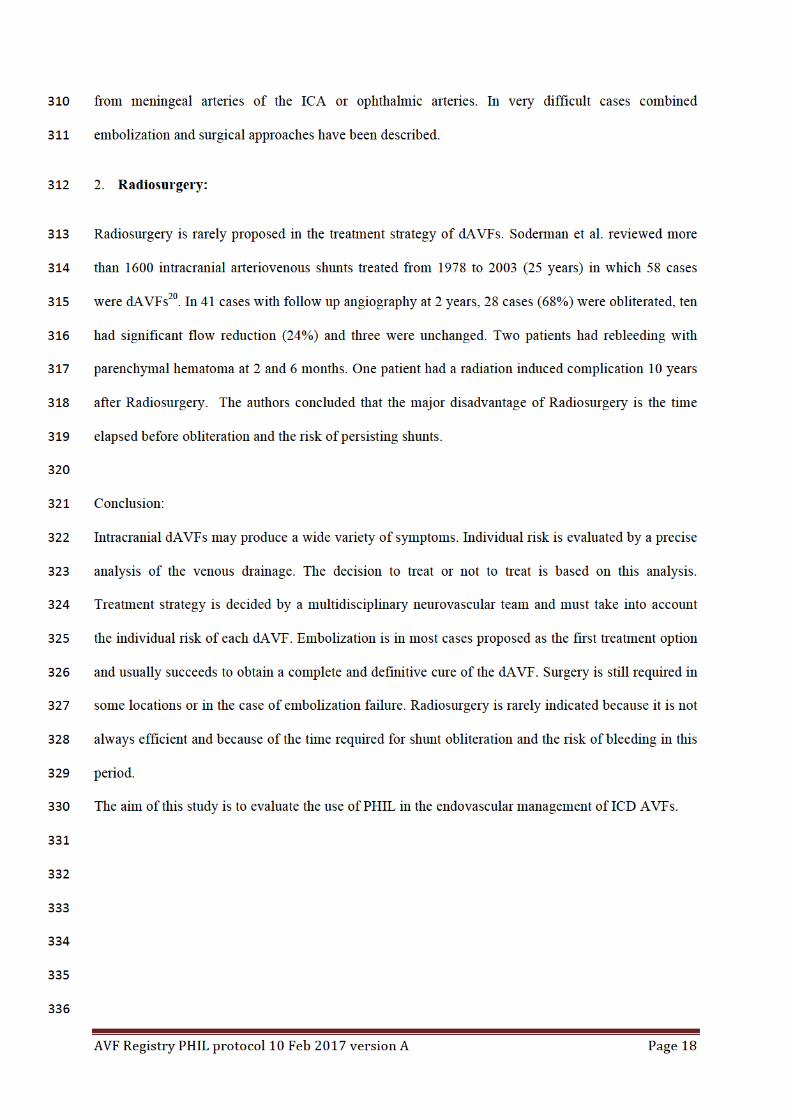
2. Radiosurgery: 312
Radiosurgery is rarely proposed in the treatment strategy of dAVFs. Soderman et al. reviewed more 313
than 1600 intracranial arteriovenous shunts treated from 1978 to 2003 (25 years) in which 58 cases 314
were dAVFs20. In 41 cases with follow up angiography at 2 years, 28 cases (68%) were obliterated, ten 315
had significant flow reduction (24%) and three were unchanged. Two patients had rebleeding with 316
parenchymal hematoma at 2 and 6 months. One patient had a radiation induced complication 10 years 317
after Radiosurgery. The authors concluded that the major disadvantage of Radiosurgery is the time 318
elapsed before obliteration and the risk of persisting shunts. 319
320
Conclusion: 321
Intracranial dAVFs may produce a wide variety of symptoms. Individual risk is evaluated by a precise 322
analysis of the venous drainage. The decision to treat or not to treat is based on this analysis. 323
Treatment strategy is decided by a multidisciplinary neurovascular team and must take into account 324
the individual risk of each dAVF. Embolization is in most cases proposed as the first treatment option 325
and usually succeeds to obtain a complete and definitive cure of the dAVF. Surgery is still required in 326
some locations or in the case of embolization failure. Radiosurgery is rarely indicated because it is not 327
always efficient and because of the time required for shunt obliteration and the risk of bleeding in this 328
period. 329
The aim of this study is to evaluate the use of PHIL in the endovascular management of ICD AVFs. 330
331
332
333
334
335
336

AVFRegistryPHILprotocol10Feb2017versionA Page19
337
338
Types Patterns of venous drainage
Type I Drainage into a sinus with a normal antegrade flow direction.
Type II Drainage into a sinus with insufficient antegrade venous drainage and
reflux:
IIa: into sinus(es) only.
IIb: into cortical vein(s) only.
IIa+b: into sinus(es) and cortical vein(s).
Type III Direct drainage into a cortical vein.
Type IV Drainage into a cortical vein with a venous ectasia.
Type V Drainage into spinal perimedullary veins.
Table 1: Classification of Djindjian and Merland according to the venous drainage. 339
340

AVFRegistryPHILprotocol10Feb2017versionA Page20
Table 2: Drawings of the types of venous drainage according to the revised Classification of 341
Djindjian and Merland. 342
343

AVFRegistryPHILprotocol10Feb2017versionA Page21
Venousdrainage Symptomsandrisks
Transverse/sigmoidsinus Pulsatiletinnitus
Ophthalmicveins Ocularsymptoms
Refluxintosuperiorsagittalor
straightsinus
Intracranialhypertension,dementia
Corticalveins Seizures,focaldeficits,venousinfarction,hemorrhages
Perimedullaryveins Myelopathy
Table3:Symptomsandrisksdependingonthedrainingvein/sinus.344
345
346
2. MEDICAL DEVICE INFORMATION 347
348
The PHIL® device has been CE marked since July 2014. It is intended for use in the embolization of 349
lesions in the peripheral and neurovasculature, including arteriovenous malformations and 350
hypervascular tumors. 351
The PHIL® device is a non-adhesive liquid embolic agent comprised of a co-polymer dissolved in 352
DMSO (dimethyl sulfoxide). An iodine component is chemically bonded to the co-polymer to provide 353
a radio pacifier element during fluoroscopic visualization. The PHIL® Liquid Embolic System 354
consists of a sterile, pre-filled, 1.0 mL syringe of PHIL® liquid embolic, a sterile, pre-filled 1.0 mL 355
syringe of DMSO, and microcatheter hub adaptors. A DMSO compatible delivery microcatheter that 356
is indicated for use in the neurovascular or peripheral vasculature is used to access the embolization 357
target site. The PHIL® Liquid Embolic System is available in several product formulations: PHIL
® 358
25%, PHIL® 30%, and PHIL
® 35%. PHIL
® 25% liquid embolic will travel more distally and 359
penetrate deeper into the nidus due to its lower viscosity compared to PHIL® 30% or 35% liquid 360
embolic. Final solidification occurs within five minutes for all product formulations. 361
The PHIL® device is delivered by slow, controlled injection through a microcatheter into the vascular 362
malformation under fluoroscopic control. The DMSO solvent dissipates into the blood, causing the 363
copolymer to precipitate in situ into a coherent embolus. The PHIL® device immediately forms a skin 364
as the polymeric embolus solidifies from the outside to the inside, while traveling more distally in the 365
vascular lesion. 366
367
368

AVFRegistryPHILprotocol10Feb2017versionA Page22
3. STUDY DESIGN AND SCHEDULE 369
370
3.0. Study Design 371
372
This study is a European multi-center Study, observational, prospective, single arm and open label. 373
Each patient will be treated per hospital standard of care. 374
375
3.1. Study Patients 376
377
Any patients having an untreated dural arteriovenous fistula requiring treatment will be screened by 378
the investigational sites. Those eligible to be treated with PHIL® will be enrolled after having dated 379
and signed an informed consent form as per appropriate regulation in the participating country. 380
381
3.2. Study Schedule Table 382
383
384
Selection1/Inclusion Procedure 3 Discharge
3 1 month FU
3,6 3-6 months FU
Patient demographic
x
dAVF information x Patient informed consent
x
Inc/excl criteria x Clinical status (mRS7, WFNS2)
x7 x x7
Symptoms x xQoL x5 x
dAVF & embolized vessels info
x
Procedure x Irradiation info x Procedure complications
x
Procedure results x AE(s) x x x x xAngio control4 x
385
1 Any patient to be treated for a dAVF will be listed in a consecutive screening log (for those not enrolled in the study only 386date of screening, age, sex and reason for non-inclusion will be collected) 387
2 Only in the case of hemorrhage 388
3 To be repeated for each single procedure. (In the case of a second procedure, 1 month FU can be done just prior) 389
4 DSA preferred but MRA can performed depending on patient health status 390
5 To be performed before the patient enters the catheterization room. 391
6Optional by Phone 392
7Performed by an independent Neurologist 393

AVFRegistryPHILprotocol10Feb2017versionA Page23
4. STUDY OBJECTIVES 394
395
4.0. Primary endpoint 396
397
Safety evaluation at 1 month after each embolization, 3-6 months after last embolization and efficacy 398
at 3-6 months after the last embolization treatment of dAVF with PHIL used with or without other 399
embolization products (cyanoacrylate glue, coils, balloon, etc.) except other non-adhesive liquid 400
embolic agents (i.e. Squid, Onyx). 401
402
4.1. Secondary endpoints 403
404
• Number of adverse events (resulting in death or not) 3-6 months after embolization alone 405
• Improvement in the quality of life of the patient as assessed by changes in symptoms present 406
at pre-treatment 3-6 months after the first and last sessions of embolization treatment (Stable / 407
improved / deteriorated) clinical course of the patient 408
409
5. ASSESSMENT CRITERIA 410
411
5.0. Main Assessment Criteria 412
413
• Clinical course of the patient assessed as (Stable / improved / deteriorated) at 3-6 months (during 414
angiography) after each embolization compared to its state at 1 months and baseline. 415
416
• Safety 1 month after the embolization procedure: 417
418
- The number of adverse events (resulting in death or not) at 1 month after each embolization. 419
Any adverse events reported in this study will be reviewed by an Independent Review 420
Committee (CEC) and neurological assessment (mRS) performed by an independent 421
neurologist 1 months after each embolization and compared to baseline. 422
423
• The cure rate at 3-6 months after the last embolization. Cure rate definition depends on the type of 424
fistula: 425
426
- Cured dAVF> IIa is defined by no venous drainage 3-6 months after the last embolization. In 427
an elderly patient, assessment can be done by MR angiography. 428
- Cured dAVF I or IIa is based on clinical improvement assessed by the patient’s self-reported 429
QOL questionnaire, progression of symptoms present pre-treatment (improved / stable / 430
deteriorated gradient) For elderly patients or if the fistula is assessed as benign, assessment 431
will be made by MR angiography. 432
433
• The last angiographic embolization evaluation after 3-6 months: The degree of occlusion of the 434
dAVF will be assessed by an Independent Imaging Core Lab versus baseline dAVF angiography and 435
classified into 3 categories: 436
437
For dAVF I or IIa: 438

AVFRegistryPHILprotocol10Feb2017versionA Page24
- Cured 439
- Residual Shunt Type I or IIa 440
- Residual Shunt Type> IIa 441
- Failed 442
443
For dAVF> IIa: 444
- Cured 445
- Residual Shunt Type I or IIa 446
- Residual Shunt >IIa 447
- Failed 448
449
5.1. Secondary Assessment Criteria 450
451
• The number of adverse events (resulting in death or not) 3-6 months after embolization. Adverse 452
events reported in this study will be reviewed by an Independent Review Committee which will 453
evaluate the relationship between AEs and the device and/or procedure. 454
455
• Improvement in the quality of life of the patient as assessed by changes in symptoms present at pre-456
treatment 3-6 months after the last sessions of embolization treatment and as assessed by the EuroQoL 457
EQ-5D scale. 458
459
460
6. ASSESSMENT AND DATA TO BE COLLECTED PER SCHEDULED VISITS 461
462
6.0. Selection 463
464
Patient demography: 465
- Age 466
- Sex 467
468
Patient informed consent form: 469
470
- Date of signature 471
472
Inclusion/exclusion criteria review 473
474
All patients with an intracranial dAVF requiring treatment that has not been previously treated will be 475
screened by the investigational sites. (If the patient is not enrolled the reason will be documented.) At 476
the end of the screening visit enrolled patients will be documented into a screening log. 477
478
(For patients who refused collection of their personal data or meeting an exclusion criteria only, their 479
age, sex, the reason for non-inclusion and the date of screening will be documented in a screening 480
log.) 481

AVFRegistryPHILprotocol10Feb2017versionA Page25
482
483
6.1. Inclusion 484
485
dAVF Information: 486
487
- Type: I, IIa, IIb, IIa + b, III, IV, V (according to the classification of Cognard et al – see 488
Appendix 19.2). 489
490
- Location of the fistula (multiple choice): 491
492
• Transverse and sigmoid Sinus 493
• Cavernous Sinus and paracavernous 494
• Petrosal sup. Or inf. Sinus 495
• Superior longitudinal Sinus 496
• Tentorial 497
• Ethmoid 498
• Vein of Galen, Straight sinus 499
• Foramen magnum 500
• Condylar Canal 501
502
503
Clinical status: 504
- mRS 505
- WFNS (in the case of hemorrhage) 506
507
Symptoms: 508
509
- Minor symptoms (tinnitus, dizziness, headache) 510
- Ocular symptoms (visual or oculomotor) 511
- Seizures 512
- Intracranial hypertension 513
- Cognitive disorders 514
- Venous infarction 515
- Intracerebral hematoma 516
- Subarachnoid hemorrhage 517
- Subdural hemorrhage 518
- Cranial nerves 519
- Myelopathy 520
521
522
523
524
If any new AE(s) occurs it will be documented. 525

AVFRegistryPHILprotocol10Feb2017versionA Page26
526
6.2. Procedure(s) 527
528
QoL EQ 5D(to be completed before the procedure.) 529
530
No. of arteries/veins catheterized 531
532
Type of catheter: 533
534
- Scepter 535
- Ultraflow 536
- Apollo 537
- Marathon 538
- Echelon 539
- Duo 540
- Sonic 541
- other 542
543
Arteries: 544
545
- Middle Meningeal artery. 546
- Occipital artery 547
- Posterior auricular artery 548
- Accessory Meningeal artery 549
- Ascending pharyngeal artery 550
- Ophthalmic artery 551
- Post meningeal. (Vertebral) artery 552
- Meningeal branches of the internal carotid 553
- Cortical artery 554
- Superficial temporal artery 555
556
Veins: 557
558
- Inferior petrosal Sinus (IPS) 559
- Superior petrosal Sinus (SPS) 560
- Cavernous sinus 561
- Facial Veins 562
- Superior longitudinal Sinus 563
- Straight sinus 564
- Transverse/Sigmoid sinus 565
- Cortical veins 566
567
568
569
Duration of the PHIL® injection by artery / vein 570
AVFRegistryPHILprotocol10Feb2017versionA Page27
571
Volume of PHIL® injected by artery / vein 572
573
Backflow (in cm) by artery / vein 574
575
Batch number 576
Other material used: 577
578
- Intravenous coils 579
- Arterial coils 580
- Intravenous glue 581
- Arterial glue 582
- Balloon for protection (intravenous) 583
- Injection through the balloon 584
- Balloon for reducing the flow (arterial) 585
- Other 586
587
Total dose of radiation: mGy / cm2 588
589
Procedural Complications: 590
591
• Type of complications: catheterization/catheter / injection PHIL® / the dAVF / other: 592
593
- Dissection, arterial rupture 594
- Catheter stuck, broken, occluded 595
- Migration of PHIL® in the vein (cortical veins, sinus, lung) 596
- Arterio-arterial migration 597
- Bleeding (Subarachnoid hemorrhage, parenchymal hematoma) 598
- Venous infarction 599
- Cerebral edema 600
601
14 Other 602
603
604
• with or without clinical sequelae 605
- Epileptic seizures 606
- Intracranial hypertension 607
- Cognitive disorders 608
- Venous infarction 609
- Hematoma 610
- Focal neurological deficit 611
- Cranial nerve deficit 612
- Blindness or other visual impairment 613
- Other 614
615
Results of the angiographic procedure: 616

AVFRegistryPHILprotocol10Feb2017versionA Page28
617
For dAVF I or IIa: 618
- Cured 619
- Residual Shunt Type I or IIa 620
- Residual Shunt Type> IIa 621
- Failed 622
- Decision not to treat 623
624
For dAVF > IIa: 625
- Cured 626
- Residual Shunt Type I or IIa 627
- Residual Shunt >IIa 628
- Failed 629
- Decision not to treat 630
631
632
Further treatment: 633
634
- Radiosurgery 635
- Surgery 636
- second embolization 637
638
6.3. Discharge 639
640
If any new AE(s) occurs it will be documented. At this stage the expected AEs are: 641
642
- Epileptic seizures 643
- Intracranial hypertension 644
- Cognitive disorders 645
- Venous infarction 646
- Hematoma 647
- Subarachnoid hemorrhage 648
- Focal neurological deficit 649
- Cranial nerve 650
- Blindness or other visual impairment 651
- Other 652
653
654
655
6.4. 1 Month Follow up (Optional by Phone) 656
657
Clinical status: 658
- mRS 659
- WFNS (in the case of hemorrhage) 660
661
If any new AE(s) occurs it will be documented. 662
663

AVFRegistryPHILprotocol10Feb2017versionA Page29
This visit can be performed over the phone if a face to face visit cannot be performed 664
665
666
6.5. 3-6 Month Follow up 667
668
Angiography (DSA strongly recommended, but MR / MRA is acceptable based on patient age, 669
specific health status, etc.) 670
671
Procedure Result: 672
673
- Cured 674
- Residual Shunt Type I or IIa 675
- Residual Shunt Type> IIa 676
- Failure 677
- Decision not to treat 678
679
For dAVF> IIa: 680
- Cured 681
- Residual Shunt Type I or IIa 682
- Residual Shunt >IIa 683
- Failure 684
- Decision not to treat 685
Additional Proposed Treatment: 686
687
- Radiosurgery 688
- Surgery 689
- Second embolization (in this case procedure, discharge, 1 month FU and 3-6 month FU visits and 690
assessments should be repeated) 691
692
Clinical status: 693
- mRS 694
Changes in the symptoms compared to baseline (Stable / improved / deteriorated) 695
If any new AE(s) occurs it will be documented. At this stage the expected AEs are: 696
697
- Epileptic seizures 698
- Intracranial hypertension 699
- Cognitive disorders 700
- Venous infarction 701
- Hematoma 702
- Subarachnoid hemorrhage 703
- Focal neurological deficit 704
- Cranial nerve 705
- Blindness or other visual impairment 706
- Other 707
708

AVFRegistryPHILprotocol10Feb2017versionA Page30
QoL EQ 5D (assessment will be completed before the patient leaves the hospital) 709
710
711
7. CLINICAL SITES 712
713
Up to 16 sites will participate in the study representing 4 to 5 European countries. Each site will 714
demonstrate its ability to recruit at least 5 patients within one year. All sites will have demonstrated 715
significant experience with liquid embolic procedures and will have received specific training in the 716
use of PHIL®. 717
718
8. STUDY DURATION 719
720
It is expected to recruit 60 consecutive patients in approximatively one year in 16 investigational sites. 721
Follow-up for each patient is expected to take up to 12 months after the first embolization session, 722
since each patient may require up to 2 embolizations maximum and the final follow-up visit is to take 723
place 3-6 months after the last session. 724
Therefore, the overall study duration will be approximatively 2 years. 725
726
9. PATIENT SELECTION 727
728
9.0. Inclusion criteria 729
1. Patient or patient’s legally authorized representative has received information about data 730
collection and has signed and dated an Informed Consent Form 731
2. Patient has an intracranial dAVF that can be treated by embolization with PHIL® used with or 732
without other embolization products except other non-adhesive liquid embolic agents (i.e. Squid, 733
Onyx). 734
3. Patient is at least 18 years of age. 735
736
9.1. Exclusion criteria 737
1. Patient has multiple dAVFs to be treated. 738
2. Patient participates in a study evaluating another medical device, procedure, or medication during 739
the course of dAVF treatment and follow-up per the study protocol. 740
(For patients who refused collection of their personal data or meeting an exclusion criteria only, 741
their age, sex, the reason for non-inclusion and the date of screening will be documented in a 742
screening log.) 743
3. Any condition that could prevent patient follow up. 744
745
10. STATISTICAL ANALYSIS PLAN 746

AVFRegistryPHILprotocol10Feb2017versionA Page31
747
10.0. Statistical Analysis 748
All patients included in the study will be included in the analyses (ITT). 749
Qualitative parameters are described by their frequency distribution and ranges bilateral 95% 750
confidence associated quantitative parameters by their average, minimum standard deviation, 751
maximum, median and quartiles, number of missing values. 752
Quality parameters (i.e. 3-6 months cured (yes / no) or improvement of mRS 1 month (yes / no)) 753
will be described by their distribution frequencies and intervals bilateral associated 95% 754
confidence. 755
Relevant group comparisons will be performed using an adequate test for the variable type: 756
• Person’s chi-square (or Fisher’s exact test if the expected frequency in a group is smaller than 757
five) for qualitative variables 758
• Variance analyses for quantitative variables 759
• Variance analyses based on ranks for ordinal variables 760
Statistical test will be performed with a type I error risk of 5%. 761
The rate of events for which a date of onset has been collected will be described survival curve 762
according to Kaplan-Meier method and the associated Kaplan Meier estimators will be calculated. 763
10.1. Population size 764
11. Sample size: 60 patients 765
12. With a sample size of 55 patients, we will be able to estimate the success rate with a 95% 766
confidence interval size of ±10.0% considering the observed success rates in a previous study 767
(83%) (Cognard and al.). Thus, the lower bound of the 95% confidence interval will be higher than 768
the lowest estimation found in the literature (Lv and al. 61%)21. 769
13. In addition, even with 10% loss to follow-up patients, this sample size (N=60) ensures an accuracy 770
of at least ±10% for observed parameters. 771
772
13.0. Invalid, Unused and Missing data 773
Missing data will be treated in the three following ways: 774
• The LOCF method will be used, with the last observation seen used as a reference value 775
for a missing value at the end of follow-up 776
• The basic method to exclude patients with missing data will be used 777
• The missing data will be charged through multiple imputation procedure 778

AVFRegistryPHILprotocol10Feb2017versionA Page32
779
13.1. Minimizing bias strategy 780
781
In order to minimize bias the two following independent review committees will be established: 782
Centralized review of laboratory tests (Core Lab): An independent committee will evaluate the 783
angiographic / MRA examinations at the following times: 784
Angiography before/during the first embolization session, in order to assess the dAVF anatomy. 785
At the end of the treatment after each embolization. 786
At 3-6 months after the last embolization (imaging control). 787
Clinical events Committee (CEC): 788
This independent committee will evaluate any adverse events per-procedure and for the overall 789
study duration. It will assess the existence of a relationship with the device and/or procedure and 790
identify any Serious Adverse Events. 791
792
793
14. END OF STUDY PARTICIPATION 794
795
End of patient study participation will occur in the following cases: 796
797
• Patient consent withdrawal for any reason (if possible, the patient will be asked to 798
attend the EoS visit in order to assess his/her final safety and clinical outcomes before 799
he/she stops participation in the study) 800
• Investigator decision to stop patient participation 801
• Lost to follow up 802
• Patient death 803
• Scheduled end of the study 804
805
It will be requested of the investigator to take any action in order to avoid patients lost to 806
follow up (including calling the patient, contacting the patient’s family doctor, etc.). 807
808
15. ADVERSE EVENTS 809
Any adverse event, whether serious or not, whether symptomatic or not, per- or post-operative, 810
related or unrelated to the use of the device will be identified. Problems associated with the use of 811
the device will be communicated to the appropriate country’s authority following the procedures in 812
force. 813
Any device that is believed to have malfunctioned or have a defect must be returned to the 814
manufacturer for analysis. 815
AVFRegistryPHILprotocol10Feb2017versionA Page33
Serious Adverse Events(SAEs) are adverse events that meet the following criteria: 816
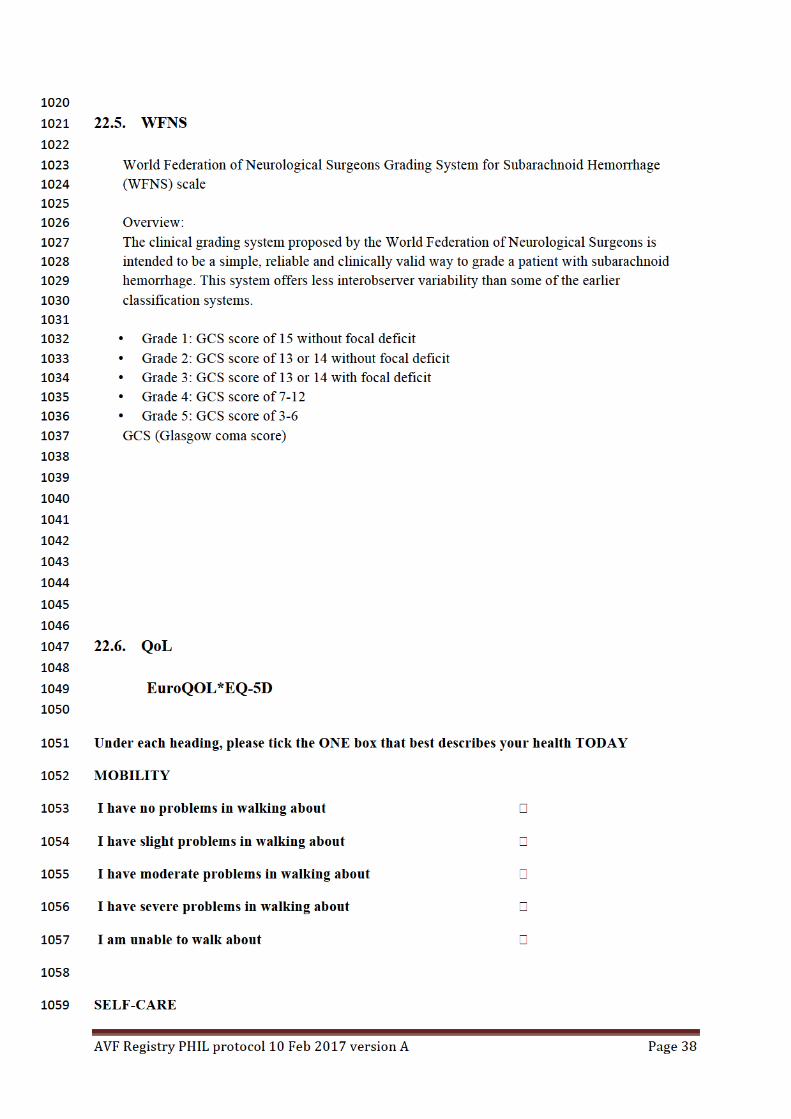
a) led to a death, 817
b) led to a serious deterioration in health that either: 818
1) resulted in a life-threatening illness or injury, or 819
2) resulted in a permanent impairment of a body structure or a body function, or 820
3) required in-patient hospitalization or prolongation of existing hospitalization, or 821
4) resulted in medical or surgical intervention to prevent life threatening illness or injury or 822
permanent impairment to a body structure or a body function. 823
c) led to fetal distress, fetal death or a congenital abnormality or birth defect. 824
825
826
In this study, all serious adverse events must be reported to the safety officer. SAEs will also result 827
in a declaration to the appropriate countries’ authorities in accordance with procedures in force. 828
Adverse events are unanticipated when they are not included in the instructions for use of the 829
medical device. 830
If the case of an unexpected serious adverse event, the safety officer will immediately contact the 831
investigator to prepare an initial report to be forwarded to the competent authorities in the prescribed 832
procedures material-vigilance and Oversight Committee. 833
834
835
836
837
16. PROTOCOL DEVIATIONS 838
839
A study/protocol deviation occurs when the investigator or other study personnel did not conduct the 840
study according to the Clinical Investigation Plan or according to regulations. 841
The investigator will document and inform the sponsor or his mandated representative of any 842
deviations that occur at his site. If needed, he will take any reasonable corrective action in order to 843
avoid deviation repetition. 844
845
846
847
17. QUALITY CONTROL 848
Progress of research in the study centers and support issues will be performed in accordance with the 849
ethics and medical recommendations. 850
Case report form completion 851
Data from the study will be recorded by authorized study site staff on an eCRF. The information 852

AVFRegistryPHILprotocol10Feb2017versionA Page34
reported in the medical questionnaire should be a reflection of that in the medical records of the 853
subject. 854
Data quality entered into the eCRF will be assessed during monitoring visits performed by a CRA. 855
Monitoring visits will be conducted as per standard operating procedure and the monitoring plan of the 856
company as mandated by the sponsor. 857
Data management quality check will be implemented in order to enhance data quality and consistency 858
as per standard operating procedure and the monitoring plan of the company as mandated by the 859
sponsor. 860
Quality assurance at any step of the study will be performed on a regular basis as per standard 861
operating procedure and the monitoring plan of the company as mandated by the sponsor. 862
18. ETHICS AND REGULATION 863
864
The final version of the Investigational Plan with the Patient Information document and Consent Form 865
will be submitted to an appropriately constituted EC or relevant country Competent Authority (CA) by 866
the Investigator/sponsor prior to commencement of the Study. A copy of the EC or CA 867
opinion/approval letter along with, for the EC document, the list of EC voting members will be 868
provided to the Sponsor prior to initiating the Study at any center. 869
The Investigator/sponsor will submit the appropriate documentation if any extension, renewal or 870
amendment of the EC or CA approval must be obtained. In particular Investigational Plan 871
amendments, Informed Consent Form changes or other written information provided to the patient 872
and/or Study procedures directly affecting the patient must be approved by the EC or CA in writing. 873
Each Investigator must sign the Investigational Plan Amendment before implementing the change at 874
his/her Site. 875
The Investigator/sponsor will report to the EC any new information that may affect patient safety or 876
the conduct of the Study. Written summaries of the Study status shall be sent by the Investigator to 877
the EC according to local requirements, and at least once annually. 878
Upon completion of the Study, the Investigator shall provide the EC with a brief report of the outcome 879
of the Study as required by the local EC. 880
881
19. STUDY DOCUMENTATION AND DATA RETENTION 882
Research-related documentation must be filed by each site for the appropriate period of time based 883
on its country’s regulation. The sponsor will file these documents based on the longest period of 884
time required by the regulation of any participating country. 885
The database used for statistical analysis will be archived by the entity performing the analysis. 886
20. DATA ANALYSIS and REPORTS 887
888
20.0. Access to the data and source documentation 889
890
Confidentiality of data shall be observed by all parties involved at all times throughout the clinical 891
investigation. All data shall be secured against unauthorized access. 892

AVFRegistryPHILprotocol10Feb2017versionA Page35
The privacy of each subject and confidentiality of his/her information shall be preserved in reports and 893
when publishing any data. 894
The principal investigator or institution shall provide direct access to source data during and after the 895
clinical investigation for monitoring, audits, EC review and regulatory authority inspections. 896
897
20.1. Study Report 898
899
A final study report will be written under the sponsor’s supervision. This report will be sent to any 900
investigators and EC/CA as per regulation in force in the participating countries. 901
902
20.2. Presentation and publication rules 903
904
Microvention Europe Company owns all the data, analysis and results for the PHIL® resulting from 905
this study and no use or transmission to a third party can be made without prior consent. However, 906
each individual center is free to use and develop independently the data from the medical records of its 907
own patients. 908
909
21. RISKS AND BENEFITS 910
911
21.0. Risks 912
913
This is an observational study with no change required to standard of care. As such, there will be no 914
additional risks to patients participating in the study. 915
916
21.1. Benefits 917
918
All patients will be managed in the same way as they would have been managed if they had refused to 919
participate in the study. There is no individual benefit to participating in this study. 920
921
922
923
22. APPENDIX 924
925
22.0. Patient Informed Consent Form 926
927
Provided separately 928
929
22.1. Clinical sites list 930
931
List attached to the protocol. 932
933
22.2. Cognard & al. Classification 934

AVFRegistryPHILprotocol10Feb2017versionA Page36
The Cognard classification is based on the direction of dural sinus drainage, the presence or absence of 935
CVD, and venous outflow architecture (nonectatic cortical veins, ectasia cortical veins, or spinal 936
perimedullary veins). 937
Type I lesions drain into the dural sinus, have an antegrade flow direction, and lack of CVD. 938
Type II lesions are subdivided into 3 subcategories: 939
Type IIa lesions drain retrogradely into a dural sinus with- out CVD, 940
Type IIb lesions drain antegradely into a dural sinus with CVD, 941
Type IIa+b lesions drain retrogradely into a dural sinus with CVD. 942
Types III (Direct drainage into a cortical vein without venous ectasia), IV (Direct drainage into a 943
cortical vein with ectasia >5mm and 3x larger than the diameter of the draining vein), and V (Direct 944
drainage into spinal perimedullary veins) lesions all have CVD, absent dural venous drainage, and 945
varying cortical venous outflow architecture Lack of CVD 946
947
22.3. Bibliography 948
9491[NewtonTH,GreitzT.Arteriovenouscommunicationbetweentheoccipitalarteryandtransverse950
sinus.Radiology1966;87:824-828].9512[DjindjianRMJ,TheronJ.Superselectivearteriographyoftheexternalcarotidartery.952
SpringerVerlagBerlin,New-York;1977:606-628.]9533[CognardC,GobinYP,PierotL,etal.Cerebralduralarteriovenousfistulas:clinicalandangiographic954
correlationwitharevisedclassificationofvenousdrainage.Radiology1995;194:671-680]9554[BordenJA,WuJK,ShucartWA.Aproposedclassificationforspinalandcranialduralarteriovenous956
fistulousmalformationsandimplicationsfortreatment.Journalofneurosurgery1995;82:166-179]9575[DaviesMA,TerBruggeK,WillinskyR,CoyneT,SalehJ,WallaceMC.Thevalidityofclassificationfor958
theclinicalpresentationofintracranialduralarteriovenousfistulas.Journalofneurosurgery959
1996;85:830-837]9606[vanDijkJM,terBruggeKG,WillinskyRA,WallaceMC.Clinicalcourseofcranialduralarteriovenous961
fistulaswithlong-termpersistentcorticalvenousreflux.Stroke2002;33:1233-1236]9627[DuffauH,LopesM,JanosevicV,etal.Earlyrebleedingfromintracranialduralarteriovenous963
fistulas:reportof20casesandreviewoftheliterature.Journalofneurosurgery1999;90:78-84]9648[DaviesMA,TerBruggeK,WillinskyR,CoyneT,SalehJ,WallaceMC.Thevalidityofclassificationfor965
theclinicalpresentationofintracranialduralarteriovenousfistulas.Journalofneurosurgery966
1996;85:830-837]9679[HalbachVV,HigashidaRT,HieshimaGB,MehringerCM,HardinCW.Transvenousembolizationof968
duralfistulasinvolvingthetransverseandsigmoidsinuses.Ajnr1989;10:385-392]96910[MironovA.Selectivetransvenousembolizationofduralfistulaswithoutocclusionofthedural970
sinus.Ajnr1998;19:389-391]97111[PiskeRL,CamposCM,ChavesJB,etal.Duralsinuscompartmentinduralarteriovenousshunts:a972
newangioarchitecturalfeatureallowingsuperselectivetransvenousduralsinusocclusiontreatment.973
Ajnr2005;26:1715-1722]974

AVFRegistryPHILprotocol10Feb2017versionA Page37
12[RoyD,RaymondJ.Theroleoftransvenousembolizationinthetreatmentofintracranialdural975
arteriovenousfistulas.Neurosurgery1997;40:1133-1141;discussion1141-1134]976
13[UrtasunF,BiondiA,CasacoA,etal.Cerebralduralarteriovenousfistulas:percutaneous977
transvenousembolization.Radiology1996;199:209-217]97814[CognardC,JanuelAC,SilvaNA,Jr.,TallP.EndovascularTreatmentofIntracranialDural979
ArteriovenousFistulaswithCorticalVenousDrainage:NewManagementUsingOnyx.Ajnr2007].98015[AratA,InciS.TreatmentofasuperiorsagittalsinusduralarteriovenousfistulawithOnyx:981
technicalcasereport.Neurosurgery2006;59,169-170]98216[RezendeMT,PiotinM,MounayerC,SpelleL,AbudDG,MoretJ.Duralarteriovenousfistulaofthe983
lessersphenoidwingregiontreatedwithOnyx:technicalnote.Neuroradiology2006;48:130-134].98417[CognardC,JanuelAC,SilvaNA,Jr.,TallP.EndovascularTreatmentofIntracranialDural985
ArteriovenousFistulaswithCorticalVenousDrainage:NewManagementUsingOnyx.Ajnr2007]98618[DeasyNP,GholkarAR,CoxTC,JeffreeMA.Tentorialduralarteriovenousfistulae:endovascular987
treatmentwithtransvenouscoilembolisation.Neuroradiology1999;41:308-312]988
19[DefreyneL,VanlangenhoveP,VandekerckhoveT,etal.Transvenousembolizationofadural989
arteriovenousfistulaoftheanteriorcranialfossa:preliminaryresults.Ajnr2000;21:761-765].99020[SodermanM,EdnerG,EricsonK,etal.Gammaknifesurgeryforduralarteriovenousshunts:25991
yearsofexperience.Journalofneurosurgery2006;104:867-875].992
21[NogueiraRG,DabusG,RabinovJD,EskeyCJ,OgilvyCS,HirschJAetal.Preliminaryexperiencewith993
Onyxembolizationforthetreatmentofintracranialduralarteriovenousfistulas.AJNR.2008994
Jan;29(1):91-7.Epub2007Nov1].995
996
22.4. mRS 997
998
999
Score Description 1000
1001
0 No symptoms at all 1002
1003
1 No significant disability despite symptoms; able to carry out all usual duties and activities 1004
1005
2 Slight disability; unable to carry out all previous activities, but able to look after own affairs 1006
without assistance 1007
1008
3 Moderate disability; requiring some help, but able to walk without assistance 1009
1010
4 Moderately severe disability; unable to walk without assistance and unable to attend to own 1011
bodily needs without assistance 1012
1013
5 Severe disability; bedridden, incontinent and requiring constant nursing care and attention 1014
1015
6 Dead 1016
1017
TOTAL (0–6): _______ 1018
1019
AVFRegistryPHILprotocol10Feb2017versionA Page38
1020
22.5. WFNS 1021
1022
World Federation of Neurological Surgeons Grading System for Subarachnoid Hemorrhage 1023
(WFNS) scale 1024
1025
Overview: 1026
The clinical grading system proposed by the World Federation of Neurological Surgeons is 1027
intended to be a simple, reliable and clinically valid way to grade a patient with subarachnoid 1028
hemorrhage. This system offers less interobserver variability than some of the earlier 1029
classification systems. 1030
1031
• Grade 1: GCS score of 15 without focal deficit 1032
• Grade 2: GCS score of 13 or 14 without focal deficit 1033
• Grade 3: GCS score of 13 or 14 with focal deficit 1034
• Grade 4: GCS score of 7-12 1035
• Grade 5: GCS score of 3-6 1036
GCS (Glasgow coma score) 1037
1038
1039
1040
1041
1042
1043
1044
1045
1046
22.6. QoL 1047
1048
EuroQOL*EQ-5D 1049
1050
Under each heading, please tick the ONE box that best describes your health TODAY 1051
MOBILITY 1052
I have no problems in walking about 1053
I have slight problems in walking about 1054
I have moderate problems in walking about 1055
I have severe problems in walking about 1056
I am unable to walk about 1057
1058
SELF-CARE 1059

AVFRegistryPHILprotocol10Feb2017versionA Page39
I have no problems washing or dressing myself 1060
I have slight problems washing or dressing myself 1061
I have moderate problems washing or dressing myself 1062
I have severe problems washing or dressing myself 1063
I am unable to wash or dress myself 1064
1065
USUAL ACTIVITIES (e.g. work, study, housework, family or leisure activities) 1066
I have no problems doing my usual activities 1067
I have slight problems doing my usual activities 1068
I have moderate problems doing my usual activities 1069
I have severe problems doing my usual activities 1070
I am unable to do my usual activities 1071
1072
PAIN / DISCOMFORT 1073
I have no pain or discomfort 1074
I have slight pain or discomfort 1075
I have moderate pain or discomfort 1076
I have severe pain or discomfort 1077
I have extreme pain or discomfort 1078
1079
ANXIETY / DEPRESSION 1080
I am not anxious or depressed 1081
I am slightly anxious or depressed 1082
I am moderately anxious or depressed 1083
I am severely anxious or depressed 1084
I am extremely anxious or depressed 1085
1086
1087

AVFRegistryPHILprotocol10Feb2017versionA Page40
1088
1089
• We would like to know how good or bad your health is TODAY. 1090
• This scale is numbered from 0 to 100. 1091
• 100 means the best health you can imagine. 1092
• 0 means the worst health you can imagine. 1093
• Mark an X on the scale to indicate how your health is TODAY. 1094
• Now, please write the number you marked on the scale in the box below. 1095
1096
YOUR HEALTH TODAY = 1097
1098
1099
1100
1101
1102
1103
90
80
70
60
50
40
30
20
10
100
Worstimaginable
healthstate
Bestimaginable
healthstate
0
95
85
75
65
55
45
35
25
15
5