Embed Size (px)
Citation preview
Complications of endovascular treatment of May-Thurner syndrome
George GeroulakosProfessor of Vascular Surgery, National and Kapodistrian
University of Athens Director, Department of Vascular Surgery, Attikon University Hospital

• Ι have no conflict of interests
• Endovenous treatment of May-Thurnersyndrome is effective, safe and durable.
• The purpose of this review is to highlight the small number of complications that may occur with this procedure and suggest treatment options.
Definitions
• It is not known at what degree, venous stenosis is haemodynamically significant
• Morphological obstruction causing >50% area stenosis as measured by IVUS has arbitrarily been chosen for stenting.
Effect of stent sizing
• Stent sizing and apposition has been shown to be an important determinant of clinical outcome.
• Since sizing is typically approximated, large oversizing (20%) can significantly increase the vessel wall stress and this contributes to neointimal hyperplasia.
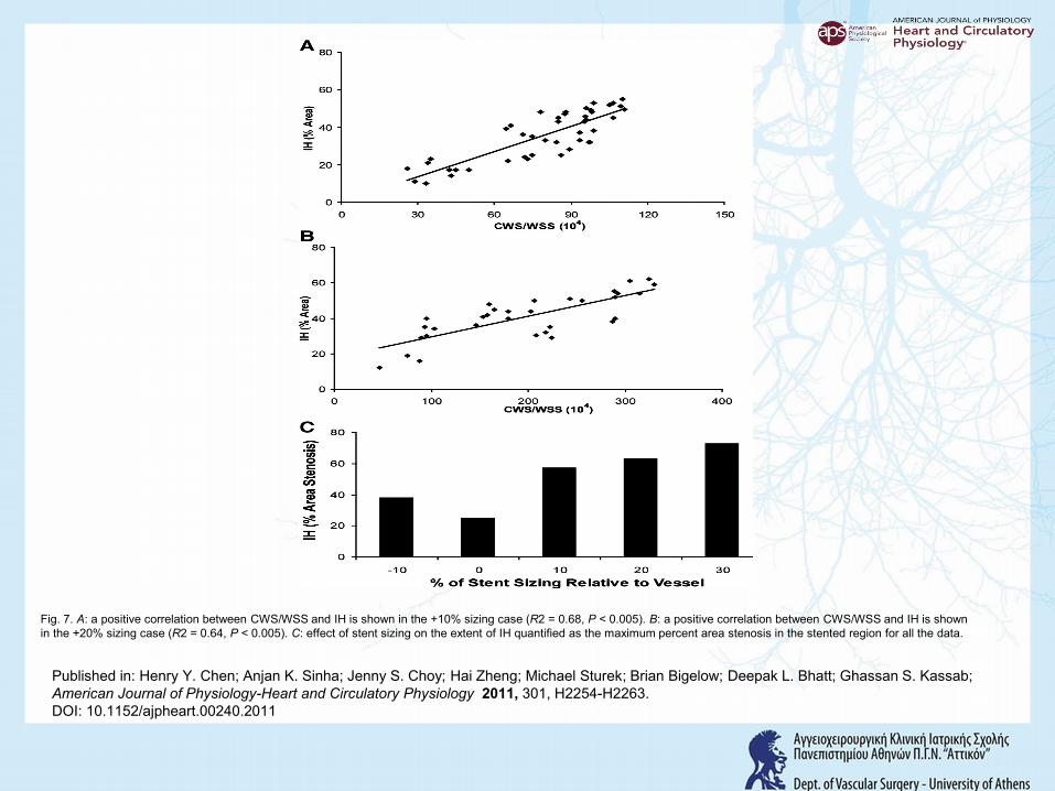
Fig. 7. A: a positive correlation between CWS/WSS and IH is shown in the +10% sizing case (R2 = 0.68, P < 0.005). B: a positive correlation between CWS/WSS and IH is shown in the +20% sizing case (R2 = 0.64, P < 0.005). C: effect of stent sizing on the extent of IH quantified as the maximum percent area stenosis in the stented region for all the data.
Published in: Henry Y. Chen; Anjan K. Sinha; Jenny S. Choy; Hai Zheng; Michael Sturek; Brian Bigelow; Deepak L. Bhatt; Ghassan S. Kassab; American Journal of Physiology-Heart and Circulatory Physiology 2011, 301, H2254-H2263.DOI: 10.1152/ajpheart.00240.2011
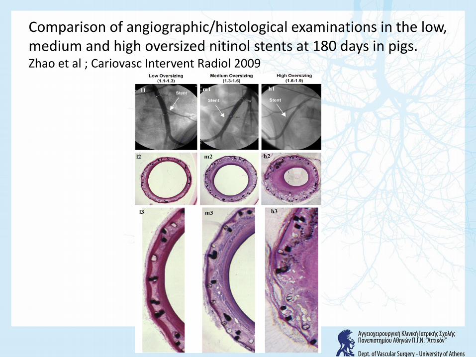
Comparison of angiographic/histological examinations in the low, medium and high oversized nitinol stents at 180 days in pigs.Zhao et al ; Cariovasc Intervent Radiol 2009
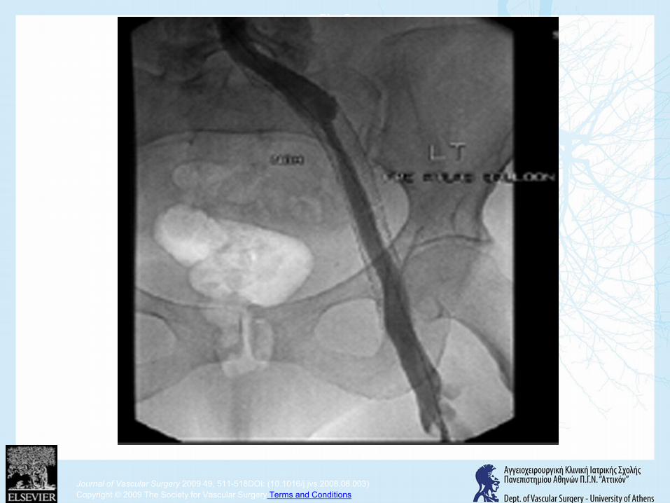
Fig 6
Journal of Vascular Surgery 2009 49, 511-518DOI: (10.1016/j.jvs.2008.08.003) Copyright © 2009 The Society for Vascular Surgery Terms and Conditions
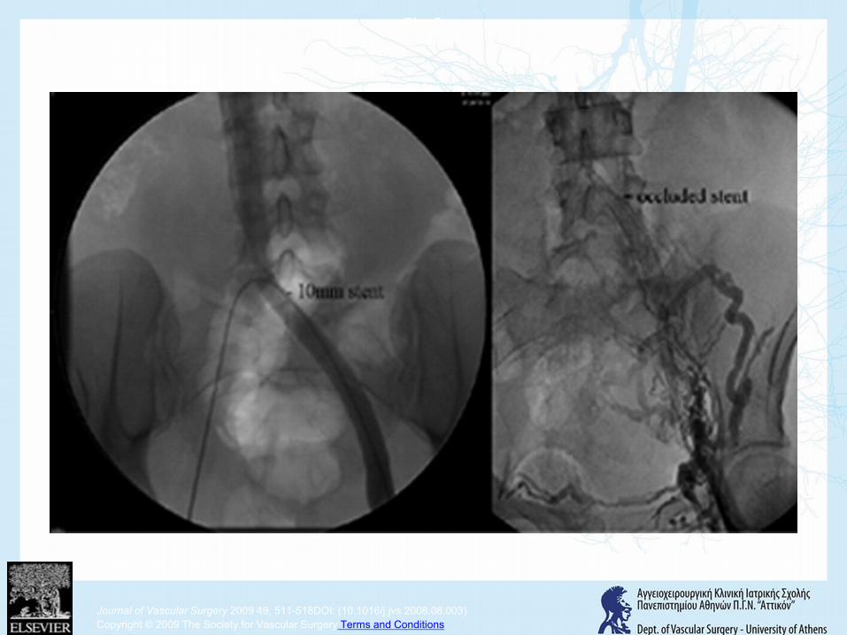
Fig 9
Journal of Vascular Surgery 2009 49, 511-518DOI: (10.1016/j.jvs.2008.08.003) Copyright © 2009 The Society for Vascular Surgery Terms and Conditions
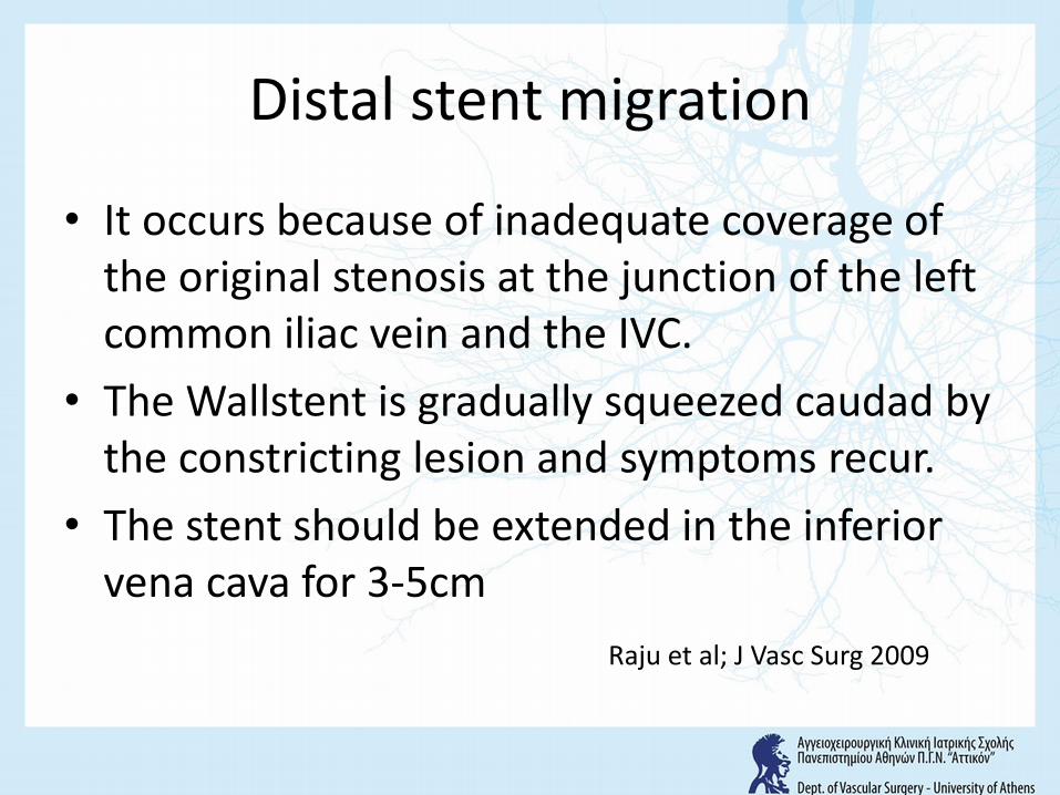
Distal stent migration
• It occurs because of inadequate coverage of the original stenosis at the junction of the left common iliac vein and the IVC.
• The Wallstent is gradually squeezed caudad by the constricting lesion and symptoms recur.
• The stent should be extended in the inferior vena cava for 3-5cm
Raju et al; J Vasc Surg 2009
Collapse and coning
• Collapse and coning of the proximal end of the Wallstent occurs when the end of it is deployed right across the stenosis with no extension in the IVC.
• This complication is specific of the Wallstentand is the result of lack of radial force at the stent ends.
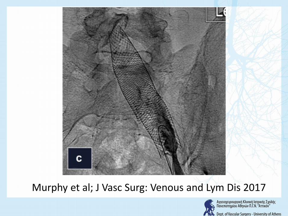
Murphy et al; J Vasc Surg: Venous and Lym Dis 2017
Proximal stent migration
• Causes of proximal migration:1. Miscalculation of the recipient vein
size2. Balloon failure3. Poor insertion technique
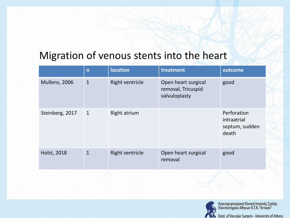
Migration of venous stents into the heartn location treatment outcome
Mullens, 2006 1 Right ventricle Open heart surgical removal, Tricuspid valvuloplasty
good
Steinberg, 2017 1 Right atrium Perforation intraatrialseptum, sudden death
Holst, 2018 1 Right ventricle Open heart surgical removal
good
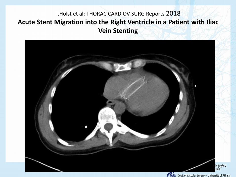
T.Holst et al; THORAC CARDIOV SURG Reports 2018Acute Stent Migration into the Right Ventricle in a Patient with Iliac
Vein Stenting
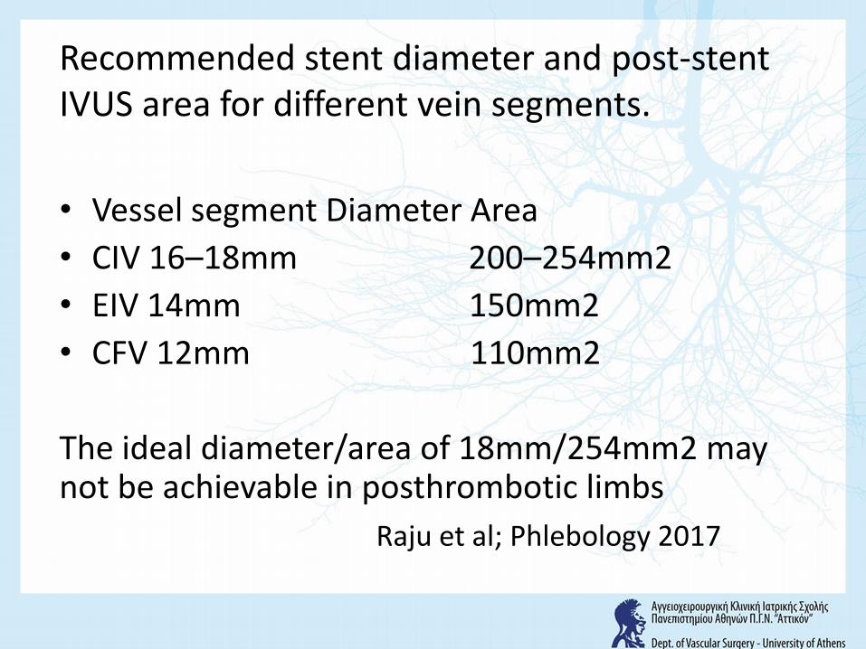
Recommended stent diameter and post-stent IVUS area for different vein segments.
• Vessel segment Diameter Area• CIV 16–18mm 200–254mm2• EIV 14mm 150mm2 • CFV 12mm 110mm2
The ideal diameter/area of 18mm/254mm2 may not be achievable in posthrombotic limbs
Raju et al; Phlebology 2017
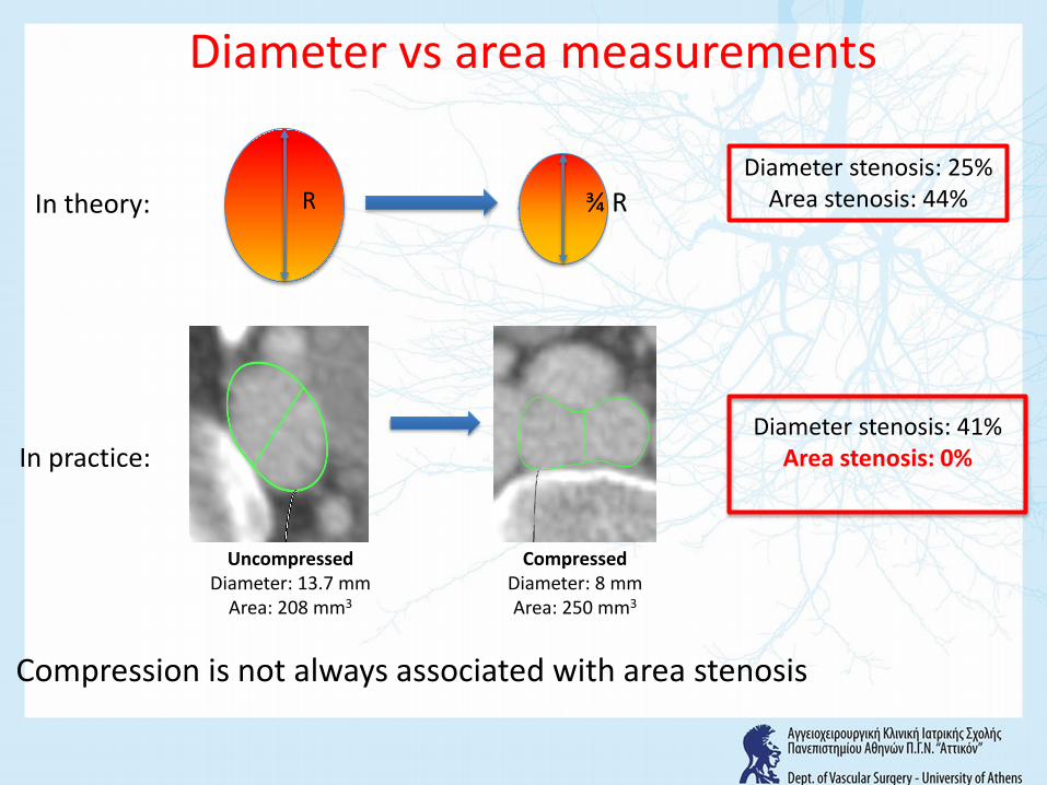
Compression is not always associated with area stenosis
Diameter stenosis: 41%Area stenosis: 0%
In theory:
In practice:
Diameter stenosis: 25%Area stenosis: 44%R ¾ R
Diameter vs area measurements
CompressedDiameter: 8 mmArea: 250 mm3
UncompressedDiameter: 13.7 mm
Area: 208 mm3
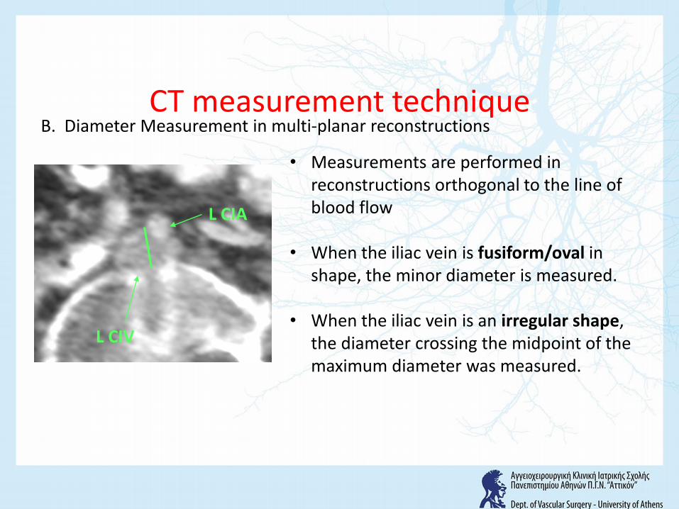
CT measurement techniqueB. Diameter Measurement in multi-planar reconstructions
• Measurements are performed in reconstructions orthogonal to the line of blood flow
• When the iliac vein is fusiform/oval in shape, the minor diameter is measured.
• When the iliac vein is an irregular shape, the diameter crossing the midpoint of the maximum diameter was measured.
L CIA
L CIV
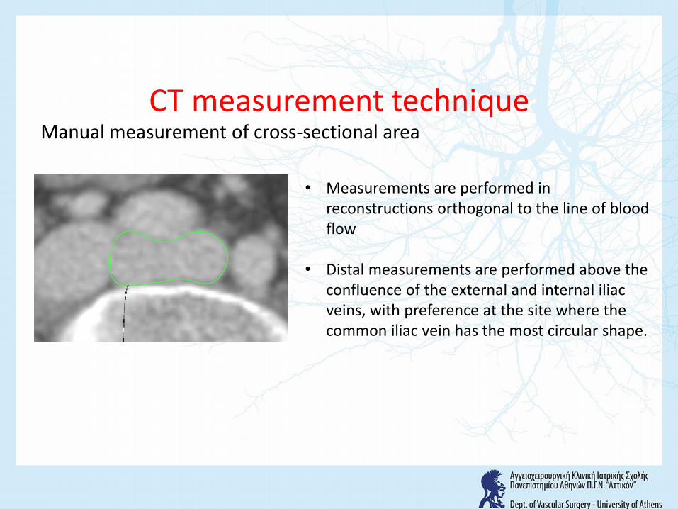
CT measurement techniqueManual measurement of cross-sectional area
• Measurements are performed in reconstructions orthogonal to the line of blood flow
• Distal measurements are performed above the confluence of the external and internal iliac veins, with preference at the site where the common iliac vein has the most circular shape.
• Extension of the stent to the vena cava to avoid migration/compression contributes to partial jailing of the contralateral flow and is associated with 2,4% DVT (Caliste et al, Ann Vasc Surg 2014).
• We present a bail out for this complication.
Contralateral DVT
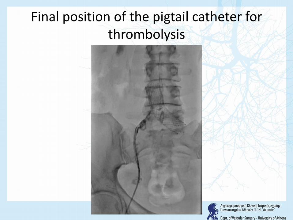
Final position of the pigtail catheter for thrombolysis
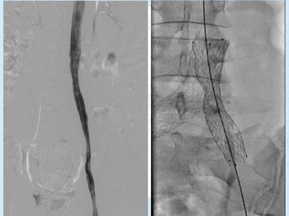
Raju’s Ζ stent modification for the reduction of contralateral DVT
• Overlapping Wallstents end at the iliac confluence and a 2cm caval extension is performed with Gianturco Z-stents placed inside and on the top of the Wallstents.
• There is a 10-20% oversizing compared to the diameter of the Wall stents to prevent migration.
Stent Patency
• 70 patients with May-Thurner syndrome underwent 77 lower extremity interventions.
• Post thrombotic (group 1) n=56• De novo presentation of swelling/pain
ulceration (group 2) n=21
Hager et al; J Vasc Surg: Venous and Lym Dis 2013
• In group 1, five patients (8.9%) developed in stent thrombosis 3 within 48 hours all with subtherapeutic IV anticoagulation, one at 1 week and one at 6 months
• In group 2, two had stent thrombosis at 12 months (9.4%)
Hager et al; J Vasc Surg: Venous and Lym Dis 2013
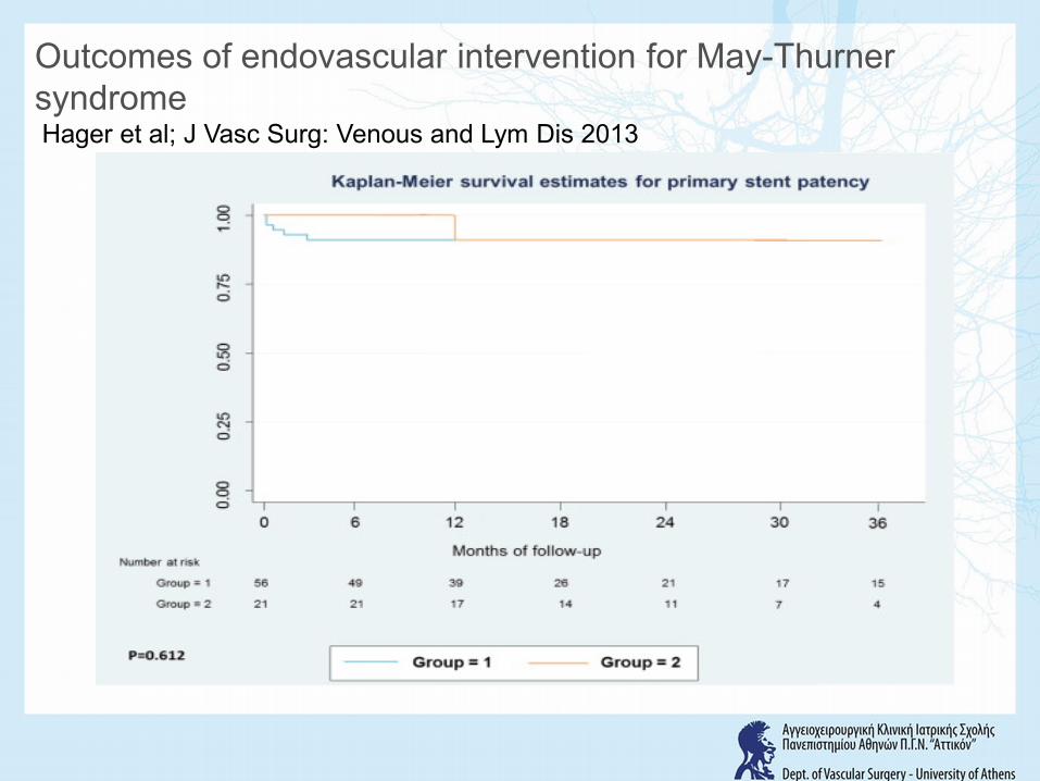
Outcomes of endovascular intervention for May-ThurnersyndromeHager et al; J Vasc Surg: Venous and Lym Dis 2013
Conclusions
• Complications in endovenous management of May Thurner syndrome are rare and reinterventions are infrequent.
• Attention to detail and in particular insertion technique, deployment of the stent to cover the full length of the stenosis and appropriate stent sizing may contribute to a reduction of complications.
ΑΤΤΙΚΟΝ UNIVERSITY HOSPITAL