Embed Size (px)
Citation preview
1
PhD Thesis
NEUROTOXICITY OF A MODELLED COMPLEX
ENVIRONMENTAL HEAVY METAL EXPOSURE IN RATS
Dr. Edina Horváth
2
Supervisor: András Papp PhD
Department of Public Health
University of Szeged
Szeged
2011
3
The Applicant’s Relevant Publications
I. Papp, A., Sárközi, L., Horváth, E., Horváth, E., Sápi, A., Kozma, G., Kónya, Z.,: Effects in the
central nervous system of rats after six weeks exposure to metal oxide nanoparticles
through the airways. In: Szilágyi M, Szentmihályi K (Eds) Trace Elements in the Food Chain
Vol. 3, Deficiency or Excess of trace Elements In Environment as a Risk of Health.
Hungarian Academy of Sciences, Budapest, 442-446. (2009)
II. Horváth, E., Oszlánczi, G., Máté, Zs., Szabó, A., Kozma, G., Sápi, A., Kónya, Z., Paulik, E.,
Nagymajtényi, L., Papp, A.:Nervous system effects of dissolved and nanoparticulate
cadmium in rats in subacute exposure. Journal of Applied Toxicology 31, 471-476 (2011)
Imp. f: 2.322
III. Horváth, E., Máté, Zs., Oszlánczi, G.,Papp, A., Sárközi, L., Kozma, G., Sápi, A., Paulik, E., Szabó,
A.,: Central nervous system effevts of combined nanoparticulate lead exposure. Arad
Medical Journal 21/1 (69), 13-16 (2011)
Abstracts:
Horváth Edina: Funkcionális idegrendszeri elváltozások ólom különböző formáival kezelt
patkányokban. MHT Ifjúsági Tagozatának VI. Fóruma; Debrecen, 2010. Összefoglalók. 30. old.
Egészségtudomány, LIV(2), p. 105 (2010)
Horváth E., Oszlánczi G., Máté Zs., Takács Sz., Szabó A., Vezér T.: Magatartási és
elektrofiziológiai hatások oldott és nanopartikuláris állapotú kadmiummal szubakutan kezelt
patkányokban. MÉT LXXI Vándorgyűlése; Szeged, 2010. Programfüzet 130. old. Acta Physiologica Hungarica, 97(4), p. 445 (2010)
Horváth, E.: Nervous system effects of experimental oral CD exposure in rats alone and in
combination with Cd nanoparticles applied into airways. Népegészségügyi Képző- és
Kutatóhelyek Országos Egyesületének IV. Konferenciája; Szombathely, 2010.
Népegészségügy, 88(3). p. 203. (2010)

4
Horváth E., Oszlánczi G., Máté Zs., Szabó A., ., Papp, A., Kozma, G., Sápi, A., Kónya, Z.,
Nagymajtényi L.: The effect of cadmium on behavioral and electrophysiological parameters
of rats efter subacute exposure in two different forms. 9th Göttingen Metting of the German
Neuroscience Societ;, Göttingen, 2011. Neuroforum, XVII(1)Suppl (2011) ISSN 0947-0875
Horváth E., Máté Zs.,Nagy V.,Takács Sz., Szabó A., Papp, A., Pusztai P., Sápi, A., Kónya, Z.,Paulik
E., Nagymajtényi L.: Functional alterations in the nervous system of rats treated with nano-
sized and other forms of lead. EuroNanoForum in partnership with Nanotech Europe;
Budapest, 2011 (p. 219.)
5
SUMMARY
Metals are major environmental pollutants due to the long periods of use (in case of lead, from ca.
3500 BC on) and the immense amounts produced. Many metals are xenobiotics because they used to
have minimal presence (and, hence, bioavailability) before man-made emission into the environment
had begun, and because they either are completely useless and toxic for the human organism (e.g.
mercury, lead or cadmium) or are essential micronutrients but toxic when overdosed (manganese,
chromium, copper, etc.).
Metal-containing dusts and fumes are generated along the complete life cycle of metal articles and
are found in the workplace atmosphere. Airborne metals cause primarily inhalational exposure, the
extent of which is influenced by the chemical form of the metals and the particle size. Environmental
nanoparticles (NPs) have important health effects on their own. Their submicroscopic size and large
specific surface area, together with the high numbers of NPs entering the organism in a typical
exposure situation, result in great biological activity. Metal ions dissolved from the surface of NPs
also must be considered among the mechanisms of action.
The second most important route of exposure to heavy metals is probably ingestion. Dust can
contaminate food or drink. Further, incorporation and accumulation of toxic metals from the soil is
characteristic for numerous plants, including ones used for staple food production, the case cadmium
content in rice being an example.
Regarding the multitude of applications of metals, the broad spectrum of heavy metal toxicity and its
consequences, as well as the modes of their possible entrance into the organism, it is of paramount
importance study this problem further on, among others by animal experiments based on more and
more adequate models. Based on practical importance and on previous experiences of the
Department, lead, cadmium and manganese were chosen for the work subserving this thesis.
Lead (Pb) has been a ubiquitous environmental pollutant, and is toxic even in low doses.
Primary production and reprocessing of Pb is based on smelting, with substantial emission of
metal fumes. Airborne Pb causes significant internal exposure both in humans and in
experimental animals, and the harms of Pb ingested with contaminated food is well-known.
Its human nervous systems effects include encephalopathy, diminished learning ability and
behavioral problems in children. In adults occupationally exposed to Pb, alterations of various
forms of central and peripheral evoked activity were described.
Cadmium, used for industrial purposes (electroplating, batteries, pigments, alloys etc.) is one
of the most toxic environmental pollutants; damaging the lungs, liver, kidney, testis, brain etc.
6
Significant inhalation of Cd can occur from tobacco smoke and in occupational settings.
Cereals, especially rice, also tend to accumulate Cd from the soil, resulting in foodborne
exposure. Amyotrophic lateral sclerosis, optic nerve damage, striatal damage and peripheral
polyneuropathy were observed as long term neurotoxic consequences. In children, a straight
relationship between hair Cd and altered visual or auditory evoked potential parameters was
found.
Manganese (Mn) is, in contrast to lead and cadmium, an essential micronutrient, e.g. as cofactor in
metallo-enzymes. It is used in many important alloys, so welding fumes and similar industrial
emissions are a source of Mn-containing NPs. Chronic inhalation of manganese compounds causes
severe neurologic disorders; starting with apathy, asthenia and headache, and ending in a Parkinson-
like syndrome. Oral or parenteral overdosing of Mn can induce the same symptoms. Disorders with
electrophysiological signs after Mn exposure include e.g. myoclonus in welders and epileptic activity
in an accidentally exposed child.
In previous experiments it was found that recording and analysis of central and peripheral
electrophysiological signals, and of certain behavioral phenomena, is sufficiently sensitive to detect
the effects of lead, cadmium and manganese on the nervous system of rats, applied subacutely in
dissolved form orally, or in the form of nanoparticles by intratracheal instillation. In order to obtain a
more adequate model, these two forms of metal application were combined in the present work to
imitate exposure coming both from environmental (food/waterborne) and occupational
(inhalational) sources.
The aims of the work were specified in the following questions:
• Are the treatment schemes and doses used previously for application of manganese, lead and
cadmium in dissolved form orally, and in nanoparticulate form intratracheally, usable also in
combination exposure?
• What quantitative and qualitative differences can be observed in the general toxicological,
electrophysiological and behavioral changes obtained by applying the mentioned metals only
in dissolved oral form, only in nanoparticulate intratracheal form, , and in combining these
forms of application?
• Is the dose-response relationship in oral-only, intratracheal-only, and combined application
different?
7
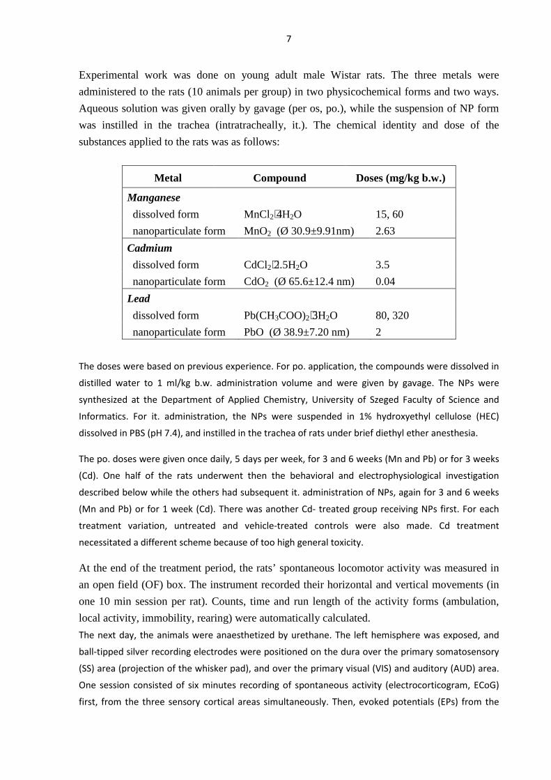
Experimental work was done on young adult male Wistar rats. The three metals were
administered to the rats (10 animals per group) in two physicochemical forms and two ways.
Aqueous solution was given orally by gavage (per os, po.), while the suspension of NP form
was instilled in the trachea (intratracheally, it.). The chemical identity and dose of the
substances applied to the rats was as follows:
Metal Compound Doses (mg/kg b.w.)
Manganese
dissolved form MnCl2⋅4H2O 15, 60
nanoparticulate form MnO2 (Ø 30.9±9.91nm) 2.63
Cadmium
dissolved form CdCl2⋅2.5H2O 3.5
nanoparticulate form CdO2 (Ø 65.6±12.4 nm) 0.04
Lead
dissolved form Pb(CH3COO)2⋅3H2O 80, 320
nanoparticulate form PbO (Ø 38.9±7.20 nm) 2
The doses were based on previous experience. For po. application, the compounds were dissolved in
distilled water to 1 ml/kg b.w. administration volume and were given by gavage. The NPs were
synthesized at the Department of Applied Chemistry, University of Szeged Faculty of Science and
Informatics. For it. administration, the NPs were suspended in 1% hydroxyethyl cellulose (HEC)
dissolved in PBS (pH 7.4), and instilled in the trachea of rats under brief diethyl ether anesthesia.
The po. doses were given once daily, 5 days per week, for 3 and 6 weeks (Mn and Pb) or for 3 weeks
(Cd). One half of the rats underwent then the behavioral and electrophysiological investigation
described below while the others had subsequent it. administration of NPs, again for 3 and 6 weeks
(Mn and Pb) or for 1 week (Cd). There was another Cd- treated group receiving NPs first. For each
treatment variation, untreated and vehicle-treated controls were also made. Cd treatment
necessitated a different scheme because of too high general toxicity.
At the end of the treatment period, the rats’ spontaneous locomotor activity was measured in
an open field (OF) box. The instrument recorded their horizontal and vertical movements (in
one 10 min session per rat). Counts, time and run length of the activity forms (ambulation,
local activity, immobility, rearing) were automatically calculated.
The next day, the animals were anaesthetized by urethane. The left hemisphere was exposed, and
ball-tipped silver recording electrodes were positioned on the dura over the primary somatosensory
(SS) area (projection of the whisker pad), and over the primary visual (VIS) and auditory (AUD) area.
One session consisted of six minutes recording of spontaneous activity (electrocorticogram, ECoG)
first, from the three sensory cortical areas simultaneously. Then, evoked potentials (EPs) from the
8
same cortical areas were recorded, and finally the compound action potential of the tail nerve. SS
stimulation was done by a pair of needles inserted into the whisker pad, delivering square electric
pulses. VIS stimulation was performed by flashes, and AUD stimulation by sound clicks. Compound
action potential of the tail nerve was evoked by means of a pair of stimulating needle electrodes
inserted at the base of tail, and recorded by another pair of needles 50 mm distally.
Body weight gain was reduced by all three metals. With Mn and Cd, the effect of the NP form
on body weight gain was disproportionately strong, regarding the dose applied, much stronger
than the effect of the dissolved form and also stronger than that of the NP form given alone in
previous experiments. The relative weight of the liver (on the basis of brain weight) was
decreased by the NP form of Mn and by both forms of Cd. Lung weight was massively
increased by it. application of each metal. In the OF test, ambulation and rearing was
decreased by Mn, and the changes were stronger after 6+6 (po., then it.) than after 6 weeks
treatment. The Pb-treated rats showed more overall time and longer periods of ambulation,
but less rearing. Also Cd caused reduced OF motility, most efficiently in the po.+it. scheme.
In the ECoG, 6 weeks of po. Mm application caused a shift to higher frequencies. This change
was not mad more intense by subsequent it. application. Orally applied Pb increased the slow
and decreased the fast waves in the ECoG but in rats with po.+it. application this change was
no more observed. Cd had no significant effect on the spontaneous cortical activity.
The cortical EPs generally showed latency lengthening on the effect of the metals. Pb also
caused increased extra lengthening of the latency on higher frequency stimulation (10 vs. 1
Hz). It was conspicuous that the effect on the SS and VIS EP latency of Mn and Pb was about
as strong after 3 weeks po. plus 3 weeks it. as after 6 weeks po. administration, although the
summed dose was much lower in the former case.
The conduction velocity of the tail nerve was decreased by Mn, Pb and Cd, and the relative
refractory period increased by Mn and Cd. The anomalous dose dependence seen on the EP
latency was present also on the nerve conduction velocity in case of Mn.
Changes observed during the experiments described in this thesis suggested on several
instances that there can be a more-than-additive interaction between the amounts of heavy
metals given by po. and it. application. This can be due to the blood-brain barrier weakened
by the op. given metal, being less able to exclude NPs, but the extreme mobility of NPs itself
can result in higher metal levels in the CNS. The role of oxidative stress induced by the
metals ond/or the NPs containing them must also be considered, and is one of the likely
common mechanisms explaining the similar character of alteration induced by the three
metals studied. Further such mechanisms are interference with Ca-dependent phenomena and
with mitochondrial energy production.
9
Based on the results described and evaluated above, it can be stated that the attempt to model
the complex exposure, coming both from environmental (food/waterborne) and occupational
(inhalational) sources, was successful. And, investigation of general and nervous system
effects of toxic environmental heavy metals in a combined exposure (dissolved form po. plus
NP form it.) by means of a set of neuro-functional tests is apparently a model to which no
direct parallel was found in the literature.
Having in mind the presence of xenobiotic heavy metals in the (occupational and residential)
environment and in commodities of environmental origin (drinking water and food) the health
effects in general, and in particular the effects on sensitive systems like the nervous system,
are of primary concern. The occurrence of metals in nanoparticulate form, adds a new feature
to the old problem.
The questions listed above can finally be answered as follows:
• The treatment schemes and doses used previously for oral and intratracheal application could
be adapted without significant change in case of Mn and Pb treatment. In case of Cd, strong
general toxicity required the development of a new scheme with shorter it. exposures. The
doses, although caused comparable alterations in the electrophysiological (and partly in the
behavioral) parameters, were at the systemic level not “equitoxic”.
• In quantitative aspect it became clear that the NP form of the metals, applied after weeks of
oral exposure to the dissolved form, had disproportionately strong effect on the body weight
gain (Mn, Cd) and on some of the open field behavioral parameters and on parameters of
evoked electrophysiological responses (all three metals). Qualitative difference was seen
mainly in the electrocorticograms, which possibly reflected the interference of NP-specific
(oxidative stress) and metal-specific (altered synaptic transmission etc.) actions. Comparison
with earlier intratracheal-only results also indicated that lower NP dose was enough to evoke
the same effects when applied after oral treatment.
• The differences in dose-response relationship could be examined only in terms of the external
dose. Measurements of internal dose (tissue metal levels) constitute the necessary next step
of the studies. In case of or al application of the metals in aqueous solution, thee effects
were of similar kind and magnitude as seen earlier. It was known from the preceding
intratracheal NP exposures that in this form much lower per kg doses are sufficient in terms
of internal dose and functional effects. Comparison of the results from the present study to
those mentioned above indicated that a lower NP dose was enough to evoke the same
effects when applied after oral treatment.
10
TABLE OF CONTENTS
1. INTRODUCTION ................................................................................................................................. 1
1.1. General properties of metals. Heavy metals in history and human culture.................................. 1
1.2. Toxicity of heavy metals and nanoparticles: general aspects........................................................ 3
1.3. Properties and toxicity of manganese ........................................................................................... 7
1.4. Properties and toxicity of lead ....................................................................................................... 9
1.5. Properties and toxicity of cadmium ............................................................................................... 10
1.6. Aims................................................................................................................................................ 13
2. MATERIALS AND METHODS .............................................................................................................. 14
2.1. Experimental animals and chemicals ............................................................................................. 14
2.2. Modes and time schemes of treatment ..............................................................................17
2.3. Behavioral investigation by the open field method...........................................................20
2.4. Electrophysiological investigation ....................................................................................21
2.5. General toxicological investigations .................................................................................23
2.6. Statistical analysis of the data........................................................................................................ 23
3. RESULTS............................................................................................................................................. 24
3.1. Effects of manganese ..................................................................................................................... 24
3.2. Effects of lead................................................................................................................................. 32
3.3. Effects of cadmium......................................................................................................................... 40
4. DISCUSSION....................................................................................................................................... 45
5. REFERENCES ...................................................................................................................................... 51
6. ACKNOWLEDGEMENT ....................................................................................................................... 58
7. APPENDIX .......................................................................................................................................... 59
11
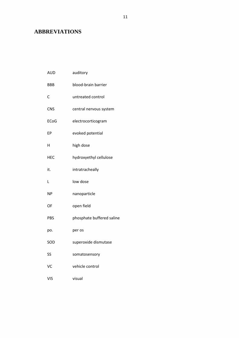
ABBREVIATIONS
AUD auditory
BBB blood-brain barrier
C untreated control
CNS central nervous system
ECoG electrocorticogram
EP evoked potential
H high dose
HEC hydroxyethyl cellulose
it. intratracheally
L low dose
NP nanoparticle
OF open field
PBS phosphate buffered saline
po. per os
SOD superoxide dismutase
SS somatosensory
VC vehicle control
VIS visual
12
1. INTRODUCTION
1.1. General properties of metals. Heavy metals in history and human culture
The majority of the chemical elements belong to the metals. Metals have good heat and
electric conductivity, metallic shine, and more or less plastic deformability. Most metals are
solid at room temperature. Electric conductivity and shine are due to the loosely bound
electrons on the outer shell of metal atoms which are easily delocalized. These electrons are
also lost in chemical reactions so that metals themselves are typically oxidized while reducing
the other reacting substance. Metal atoms with more than one oxidation state, e.g. iron and
manganese, tend to be involved in redox interactions which, under protoplasmic conditions,
may result in the generation of oxidative free radicals. To achieve octet state, numerous metal
atoms tend to form coordinate bonds with partners having a lone electron pair. Of the
complexes and chelates formed this way, many – e.g. metallothioneins – fulfil biological
functions.
It is customary to group metals into light and heavy ones. This was originally based on
density, and those with a density over 5 g/cm3 were regarded as heavy metals, but this
categorization has been oft times superseded by other aspects including toxicity, resulting in
confusion (Duffus, 2002).
When prehistoric peoples started to use metals, it meant an unprecedented impact not only on
technological development and life circumstances but also on the environment and their own
health. Stone Age lasted until 4000 BC, when the use of copper (the first metal to be utilized
by mankind) appeared and the cooper era began. Copper can be found in the nature, although
rarely, in “native form” so using copper did not absolutely require the use of fire. All other
metals are, however, found only in their ores, in form of compounds formed with oxygen,
sulfur or other elements. Obtaining these metals required smelting them from the ores in fire,
which was on one hand a substantial technical achievement but on the other hand a source of
human exposure and environmental pollution. The exploitation of lead ore steadily increased
in the antiquity, and at the Roman Empire’s heyday, lead production reached 80,000
tons/year. The symptoms – loss of appetite, fatigue, lead colic, irritability, nervous spasm –
seen in lead-mine workers were documented by Hippocrates and are described the same way
even today. The environmental effects were not recognized in those times but the emissions
13
from ancient mines and smelters can today be identified (Breitenlechner et al., 2010;
Thevenon et al., 2011).
Around 3500 BC, lead was discovered, and around 3000 BC, tin. Tin and copper were melted
together to bronze so that the period between 3000 BC and 800 BC is called the Bronze Age.
Finally, in ca. 800 BC, iron was discovered and the list of metals with major practical
applications was complete for centuries. Lead and its compounds were widely used in
cosmetics, for preserving foodstuffs, or to prevent fermentation of the wine. Lead-containing
alloys were made to “tin” cups, plates, pots and coins. This would be inconceivable today, for
the known toxicity, but the human health effect was not obvious at that time and the “food-
preserving” effect of metal kitchenware, due to the antimicrobial action of the dissolved
metals, was appreciated. Romans e.g. liked to keep wine in lead pots because it became
sweetened by the lead acetate (called for long lead sugar) generated in the interaction of the
wine (that must have been rather sour, with so much acetic acid present) with the wall of the
vessel. Lead was also used in building, in shipbuilding, and in the famous Roman water
supply system. There are studies stating that the decline of the Roman Empire was promoted
by their aristocracy’s chronic lead intoxication via wine and aqueduct water (Eaton and
Robertson, 1994).
Mercury (for us, another notoriously toxic metal) was also popular in the antique and
medieval times and was amply used in beauty products and medicines of those ages, but these
often proved to be rather harmful. The first Chinese emperor, Qin Shi Huang-Di, died in
multi-organ failure after drinking a liquid mercury preparation promising „eternal life”.
Cinnabar (HgO, the main ore of this metal) is still in use in traditional Chinese medicine.
Today’s rapidly growing industry and agriculture is almost forced to use ever newer
materials, including metal-based ones. The resulting chemical risk, manifested in
environmental and human health damage, is present even today, in spite of the more and more
strict regulations which have been evolving from the numerous negative lessons of past
centuries. Xenobiotics – substances which are “alien” for the metabolism, which can be
neither utilized nor neutralized by the organism in question – are found in soil, groundwater,
drinking water, air and, consequently, in plants and animals. In this aspect, a lot of metals are
xenobiotics because they used to have minimal presence (and, hence, bioavailability) before
man-made emission into the environment had begun, and because they either are completely
useless and toxic for the human organism (e.g. mercury, lead or cadmium) or are essential
micronutrients but toxic when overdosed (manganese, chromium, copper, etc.). Regarding the
multitude of applications of metals in our days, the broad spectrum of toxic effects of heavy
14
metals (see below) and their consequences; as well as the varieties of their possible entrance
into the organism, it is of utmost importance to learn more about this problem, among others
by animal experiments based on more and more adequate models.
1.2. Toxicity of heavy metals and nanoparticles: general aspects
1.2.1. Typical forms and ways of heavy metal exposure. Importance of nanoparticles
Exposure to heavy metals is mostly occupational. Metal-containing dusts and fumes are
generated along the complete life cycle of metal articles, from ore mining through smelting
and final product manufacturing to waste management and recycling, and are found in the
workplace atmosphere, sometimes at hazardous concentrations. Airborne metals cause
primarily inhalational exposure, the extent of which is influenced by numerous factors
including the type (nasal/oral), volume and intensity of breathing on the exposed organism’s
side, and on the other side by the chemical form in which the metal is present in the particles
(solubility, etc.) and the particle size. According to particle diameter, one can speak of
sedimenting dust (>10 µm), suspended or fine dust (100 nm-10 µm; often called PM10) and
ultrafine dust or nanoparticles (NPs, <100 nm). Traditionally, PM10 received especial
attention because this “thoracic” fraction can reach the alveoli by inspiration and cause toxic
alveolitis, and because they can cause systemic exposure due to particle-laden alveolar
phagocytes being transported by lymph, then blood, circulation to other body parts. Larger
grains are, on the contrary, trapped in the upper airways.
However, as soon as it became technically possible to investigate the origin and
environmental presence of NPs and their interaction with living organisms, they turned out to
have important health effects on their own. All high-temperature industrial processes –
smelting, casting, welding, etc. of metals (Antonini et al., 2003) or plastics (e.g. Teflon:
Seidel et al., 1991) – can generate NPs, and various combustion processes are not less
important NP sources. NPs are emitted as primary (pre-formed) particles or are generated
from gaseous precursors in an atmospheric chemical process called nucleation. Their
composition is determined by the composition of the materials worked with or burnt. So, in
the heavy metal exposure of the general population the metal content of petrol – lead (largely
phased out, in Hungary since 1999: Kertész et al., 2001) or manganese (still in use as lead
supplement, to increase octane rating, in a few countries: Davis, 1998) – played a major role
15
because most of the metal left the engine with the exhaust gases in form of microscopic and
submicroscopic oxide particles.
Intentionally manufactured nanomaterials (that is, not the nano-sized byproducts from other
industrial processes) contain at least one component that has at least one dimension in the 1-
100 nm range. Nanomaterials may add to the load of primary NPs in the workplace
atmosphere. Application of NPs and nanofibres in consumers’ commodities (personal care
products, household chemicals, electronics, tires, etc.: Oberdörster et al., 2005) means that
routes of uptake such as ingestion and dermal absorption must be considered in addition to
inhalation.
On inhalation, particles are deposited at different section of the airways, determined first of all
by their size. NPs are either deposited in the nasopharynx or get down to the alveoli (ICRP,
1994). A fundamental difference against microscopic and larger particles is that NPs are not
held back by the usual biological barriers like the alveolar and capillary wall, and reach other
target organs, beside being phagocyted like the PM10 fraction, by different transfer routes and
mechanisms. One such mechanism is transcytosis across epithelia of the respiratory tract into
the interstitium (Oberdörster et al., 2005) by means of caveolae (50-100 nm sized
invaginations of the cell membrane, serving endo- and transcytosis of a number of molecules
and microstructures: Razani and Lisanti, 2001) formed around NPs. Having crossed the
alveolar and capillary membrane, NPs are distributed throughout the body by the circulation
(Oberdörster et al., 2005).
The NPs’ small size and large specific surface area, together with the high numbers of NPs
entering the organism in a typical exposure situation, result in great biological activity. In
vivo and in vitro toxicological studies confirmed that even relatively inert materials become
more toxic and inflammatogenic in NP form. It was found, e.g., that nanosized TiO2 (ca. 20
nm diameter) caused more severe inflammation than the same compound in pigment grade
(ca. 250 nm) grains (Oberdörster, 2000). “Titanium white” as pigment is normally harmless
enough to be applied in the coating of tablets. The mentioned problem is general, concerning
both nano-sized environmental pollutants and nanotechnological products.
NPs can contribute to adverse health effects in the respiratory tract as well as in
extrapulmonary organs (Oberdörster et al., 2005). A healthy blood-brain barrier is supposed to
prevent foreign particles from entering the brain; NPs of various compositions were, however,
detected in the brain of rats after application through the airways (Kreyling et al., 2006). Such
extrapulmonary effects of NPs depend on several factors including particle solubility, particle
or aggregate size, the site of deposition, and the integrity of the alveolar epithelial lining
16
(Elder et al., 2006). Metal ions dissolved from the surface of metal-containing NPs in the
acidic microenvironment of phagosomes – after the NPs had been phagocyted by alveolar
macrophages (Lundborg et al., 1985) – also must be considered among the mechanisms of
action. The applicant’s results with Cd exposure supported the interpretation that the blood-
brain barrier may be weakened by the toxic metal ions and so will be more permeable for NPs
(Horváth et al., 2011)
Apart from inhalation, the second most important route of exposure to heavy metals is
probably ingestion. Airborne particles can be ingested if expectorated airways secretions are
swallowed instead of being spat. Dust can contaminate food or drink, e.g. under unhygienic
eating conditions at the workplace or if dust particles are captured on the surface of raw-eaten
vegetables and fruits that have not been washed adequately. The settled particles’ metal
content can also be directly incorporated into the edible parts of the plants, or soil pollution by
the particles can result in toxic metals being absorbed from the soil to the plants.
Both mechanisms were evidenced by the samples taken near to the defunct metal reprocessing
plant “Metallokémia” in Budapest. This plant was reprocessing copper, zinc, lead and other
metals from 1908 on. Lead smelting was stopped in 1977 but other metals were processed
until the end of activities in 1990 and the byproducts (slag containing heavy metals) were
stored on the plant’s premises further on (Pápay and Horváth, 1992). Windblown dust was
settling on the soil and on plants grown in the local residents’ gardens. Downwind, near the
factory, more than 1000 mg/kg Pb was in the upper soil, 1000 m farther away it was 100
mg/kg, at 2000 m, 50 mg/kg. Cd level near the plant was 3-6 mg/kg. Pb content of the
groundwater was three times, and Cd content eight times, the allowable limit (Szabó, 1991).
Leafy vegetables contained Pb over the limit in 40-50%, root vegetables in 8-50%, and
various fruits in 24-40%. Pb in the soil and in the produces was loosely correlated, but for Cd
levels the correlation was robust. The local children’s blood Pb level was higher if they lived
nearer to the factory; and among those living within the 500 m radius, eating or not eating
home-grown fruits or vegetables had a considerable influence on blood Pb (Groszmann et al.,
1992).
Incorporation and accumulation of toxic metals from the soil is characteristic for numerous
plants, including ones used for staple food production, the case cadmium content in rice being
repeatedly cited (Shigematsu, 1984).
From the intestines, most heavy metals are absorbed to around 10%. Common metal transport
mechanisms, responsible for the uptake of essential metals, are involved, which explains why
individuals with calcium- or iron-deficiency absorb more of the toxic metals. Children,
17
requiring much calcium for bone growth, can absorb 50% of the ingested lead while adults
absorb not more than 15% (Järup, 2003). Toxic metals are transported as “dead-heads” not
only from the intestine to the blood but also from the blood through the blood-brain barrier to
the CNS. Trivalent metal ions, like Mn3+ or Al3+, use transferrin to pass the blood-brain
barrier; aluminium being the culprit for “dialysis dementia”, a kind of brain damage seen in
dialysed patients.
The dermal absorption route is less well described, but presence of soil particles in inguinal
lymph nodes of persons who usually walk barefoot on the soil indicate that this is possible
(Oberdörster et al., 2005). Also some viruses, being NPs by their size, can cause transdermal
infection.
1.2.2. General mechanisms of heavy metal toxicity
Heavy metals exert a profound action on living matter, affecting the growth, metabolism,
morphology of cells.
Denaturation of proteins is one of the general damaging effects of heavy metals. The metal
ions interact with ionized moieties of amino acids in the polypeptide chain and disrupt the
non-covalent polar and ionic interactions which stabilize the secondary (or higher order)
structure of the protein. The covalent link provided by disulfide bridges is also attacked by
numerous heavy metal ions. Lead and cadmium have an especial affinity to sulfur, even their
most abundant ores are sulfides. This chemically-based destructive effect of heavy metals on
the most essential constituent of all living organisms – that is, a type of toxicity – can also be
exploited as a means of antimicrobial protection (e.g., in Credé’s solution and in
organomercurial antiseptics), but for the purposes of this thesis the metals in question are
regarded as human (neuro)toxicants.
Denaturing an enzyme protein means elimination its biocatalytic activity. Glutathion-S-
transferase was, e.g., reported to be inactivated by lead (Daggett et al., 1997) or lead and
cadmium (Planas-Bohne and Montserrat, 1992). This also exemplifies that disturbed redox
balance and oxidative stress is another frequent consequence of heavy metal exposure. Some
of the toxic heavy metals (iron, chromium, manganese) are capable of redox cycling while
others (e.g. cadmium) affect cellular antioxidant defence (Valko et al., 2005). Apart from that,
all NPs, with or without toxic metal content, tend to be absorbed to organic macromolecules
(possibly resulting in altered functionality) and to induce oxidative stress (Li et al., 2003).
NPs generate reactive oxygen species more intensely than larger particles, leading to
18
increased synthesis of pro-inflammatory mediators via intracellular signalling pathways (Long
et al., 2007; Stone et al., 2007).
It has long been known that many cellular functions (among others, muscle contraction,
exocrine and endocrine secretion) are regulated by local changes in Ca2+ concentration.
Increased intracellular Ca concentration is essential for intracellular enzyme activation and for
neurotransmission in chemical synapses. Ca2+ ion is also a charge carrier, carrying inward
current via selective voltage- and ligand-gated Ca channels. The latter are parts of the
postsynaptic (e.g. glutamate or nicotine-type acetylcholine) receptors so that neither side of a
chemical synapse can function properly in case another metal interferes with Ca (Malenka and
Nicoll, 1999). Voltage-gated Ca channels are blocked by a variety of divalent and trivalent
metal cations including manganese (Pumain et., 1987); as well as lead, mercury and zinc
(Büsselberg, 1995). Disturbed neurotransmission also belongs to the toxic spectrum of certain
neurotoxic heavy metals (Braga et al., 1999; Takeda et al., 2003).
In the trafficking of metals between parts of the cells, and between different cells or tissues,
metallothioneins play an important role. These are relatively small (500-14000 Da) proteins
unusually rich in cysteine units. By means of the sulfhydryl groups, metallothioneins bind a
range of metal ions. Physiological metals (like zinc and copper) are bound, stored and
transported to the appropriate sites, while toxic ones (mercury, cadmium, etc.) are neutralized
by the binding. The presence of –SH groups provides metallothioneins with antioxidant
properties. Both metal exposure and oxidative stress (which can also be linked, see above) can
induce the synthesis of metallothioneins.
1.3. Properties and toxicity of manganese
In pure state, manganese (Mn) is a silvery-grey, hard and brittle metal. It finds practical
application in a variety of alloys and compounds. Mn can have a series of oxidation states
between 2 and 7. For all living organisms, Mn is an essential trace element, but toxic when
overdosed. The human body contains about 10 mg Mn, stored mainly in the liver and kidneys.
The daily demand is 2-3 mg (ATSDR, 2008b). Mn is a component in many enzymes, e.g. in
one of the superoxide dismutases (Mn-SOD; Law et al., 1998) playing important role in
counteracting oxidative stress. Glutamine synthetase, alkaline phosphatase, arginase, and
pyruvate carboxylase also contain Mn (Quintanar, 2007). Glutamine synthetase (a glia-
specific Mn metalloprotein) catalyzes in the CNS the conversion of glutamic acid to
glutamine, thereby inactivating the transmitter. This enzyme requires Mn, but is inhibited by
19
its excess which is of importance in the neurotoxic mechanisms of Mn (Normandin and
Hazell, 2001).
Mn can be found, to various amounts, in all soils; originating from weathering of rocks and
volcanic activity. The latter emits also atmospheric Mn, but today the main Mn load comes
from man-made pollution (mining and metal industry, the above-mentioned fuel additive,
etc.). Mn is found in steel and other alloys, and also in the coating of welding rods (for
protection of the glowing hot welded metal parts from oxidation). MnO2 is used in the
production of dry cells. Mn is also used in paints and for glass and ceramics.
Methylcyclopentadienyl manganese tricarbonyl (MMT) was an anti-knock petrol additive the
use of which is still allowed in certain countries. Semiconductor nanocrystals (Yang et al.,
2005) and ZnS:Mn2+ nanoflowers (three-dimensional synthetic nanostructures; Chen et al.,
2005) represent the use of Mn in nanotechnology. Mn is used in certain agricultural
fungicides (Maneb, Mancozeb; Ferraz et al., 1988). A new application with direct human
exposure is in magnetic resonance imaging for diagnostic use, in form of the contrast agent
trisodium mangafodipir (Mn-DPDP, Teslascan).
Chronic inhalation of manganese compounds is the typical form of occupational Mn
exposure. The resulting “manganese madness” among ore miners was first reported in the 19th
century (Couper, 1837). The Mn-related chronic neurological disorder is today called
manganism, and it usually progresses in three stages (Saric et al., 1977; Calne et al., 1994).
The symptoms are nonspecific like apathy, anorexia, asthenia, headache, hypersomnia,
spasms, arthralgia, weakness of the legs, and irritability. In the second stage, psychomotor and
psychic disturbances dominate, such as dysarthria, excess salivation, and difficulty in
walking. The third stage represents a Parkinson-like syndrome with its associated symptoms.
But, in spite of the similar symptoms, the site of damage in manganism and in Parkinson’s
disease is different. The site of action for Mn are the striatal, and not the mesencephalic,
dopaminergic neurons (Erikson and Aschner, 2003).
Inhalation is, however, not the only way of Mn exposure leading to CNS damage. Parkinson-
like disorder was also observed in patients undergoing maintenance hemodialysis (Ohtake et
al., 2005) or in inadvertent overdosing due to long-term ingestion of a health supplement
containing high levels of Mn. This indicated that other physicochemical forms of Mn and
other routes of exposure are also relevant to the health of the CNS (and deserve being
included in neurotoxicological studies). For geological reasons (e.g. in Greece: Kondakis et
al., 1989) or due to man-made pollution (such as improper disposal of used dry cells in Japan:
Kawamura et al., 1941) abnormally high Mn levels in the drinking water were observed,
20
causing CNS symptoms in the affected population. In regions of the USA with high-Mn
drinking water, loss of visual and verbal memory, typical for Mn-induced brain damage, was
described (Woolf et al., 2002). Foodborne overexposure by Mn in babies fed on cow milk- or
soybean-based formulas was reported (Marlowe and Bliss, 1993) as was
hypermanganesaemia following long term parenteral nutrition (Crook, 2001).
The neurotoxic spectrum of Mn is variable and goes beyond the classical manganism.
Epileptic activity was observed in children following inhalational (Hernandez et al., 2003) or
parenteral (Komaki et al., 1999) exposure; and myoclonus in welders (Ono et al., 2002). In
young shipyard workers, EEG and visual evoked potential alterations were observed and
blood Mn levels up to 14 µg/L were measured (Halatek et al., 2005), while in reference
groups blood Mn is 5–7 µg/L (Bader et al., 1999). EEG and evoked potential disturbances
following occupational Mn exposure were also reported by Sinczuk-Walczak et al. (2001) and
Sjögren et al. (1996).
1.4. Properties and toxicity of lead
Lead (Pb) is a soft, malleable metal with low melting point. It is one of the oldest metals
mined and used. Egyptians and Romans widely used this metal in everyday life (see above).
In antique Rome, 4 kg lead was used per year and person, compared to 6 kg in the United
States in 1965. From Gutenberg’s times to the advent of photo typesetting, printing
technology was based on types cast from Pb-based alloys. In spite of the known toxic effects,
Pb remains to be used in several large-scale applications, an example being lead-acid batteries
found in motor vehicles, in emergency power supplies, etc. Also, leaded car fuel disappeared
from the market only in the last 15 years, so that in the mid-90’s, approximately 90% of all Pb
emission into the atmosphere was due to leaded petrol (Lovei, 1996). Lead-based pigments,
like lead white (PbCO3) and minium (Pb3O4) are no more used but demolition or renovation
of old buildings or iron structures (bridges, ships) painted originally with such paints can
expose the workers involved. On one criminal occasion, minium was used in Hungary to
improve the color of ground red paprika (Kákosy et al., 1995, 1996). Today, half of the newly
produced lead articles are made of recycled lead, which is good for the environment but the
occupational hazard is the same. Hot molten lead emits metal fumes consisting of microscopic
and submicroscopic aerosol particles. In absence of proper safety measures, working with
lead results in serious intoxication, as shown, among others, by the case of the illegal battery
recyclers in Heves (Epinfo, 1995).
21
Pb has no biological functions, it is purely toxic. It is also one of the most common
environmental xenobiotics (present in soil, groundwater, air, and foodstuffs) for which the
large-scale and careless use during previous centuries is to blame. Occupational health
damage due to Pb used to be so grave that a separate “Lead Examination Station”
(Ólomvizsgáló Állomás, an early predecessor of the present National Institute of
Occupational Health) was set up in Hungary in 1934 (Galgóczy, 1999)
The general population is exposed mainly by contaminated drinking water or food: roots,
leafy vegetables, meats, dairy products or fishes (ATSDR, 1999). High Pb level in home-
grown vegetables was a major source of risk also in the above-mentioned case of the
Metallokémia plant. Tobacco smoke is another notable source of Pb exposure. By the oral
route, 5-50% of Pb is absorbed; but by inhalation, almost the complete amount. Before the
ban of leaded petrol, the population had, mainly in urban areas and along main roads,
significant airborne Pb exposure (Rudnai et al., 1998). Pb is accumulated in the central
nervous system, first of all in the cortex and hippocampus (Gerhardsson et al., 1995), and
produces encephalopathy at blood Pb levels of 1000-1200 µg/L in adults and 800-1000 µg/L
in children (Chisolm, 1965). Exposure to low levels of Pb has been associated with behavioral
abnormalities, learning impairment, decreased hearing, and impaired cognitive functions in
humans and in experimental animals (Shannon and Graef, 1992; Ruff et al., 1996). Some
estimates suggest that, in exposed children, every 100 µg/L increase in blood Pb level is
associated with a 1-5 point decrease in the IQ (Goyer, 1996). IQ differences in Hungarian
schoolchildren, attributable to airborne Pb, were published by Füzesi (1997). Pb can also pass
the placenta and cause serious damage to the nervous system before birth. In workers with
chronic Pb exposure, less severe neurologic effects were documented with headache, lethargy,
dizziness, diminished reaction time, worsened cognitive and visuomotor performance, and
slowed nerve conduction (Lille et al., 1988; Araki et al., 2000).
1.5. Properties and toxicity of cadmium
Cadmium (Cd) is a soft, malleable bluish-white metal with low melting point. It has no
biological function in higher-order organisms and is toxic even in small amounts. It is
chemically related to zinc which explains several of its toxic effects.
Cd has been used for industrial purposes since the 19th century. The most significant
application of Cd used to be in corrosion resistant electroplating layers on steel. Metallic Cd
and its compounds were also used as pigments, UV-resistance stabilizers in plastics, coatings,
22
special alloys, Cd-based semiconductors, and on large scale in rechargeable Ni-Cd batteries.
Such applications resulted in significant workplace exposure, mainly by inhalation. However,
due to the toxic hazard involved, the use of Cd has been largely reduced in the last 20 years –
but novel technologies and materials using Cd appeared in the meantime, like quantum dots
(Rzigalinski and Strobl, 2009).
The presence of Cd in the elements of environment (air, soil, groundwater, etc.) is to a large
part due to human activity, and the general population is exposed mainly by contaminated
drinking water or food. Cd is emitted into the environment with solid and liquid industrial
(and to a lesser extent, residential) waste. The fertilizer superphosphate can also contain
significant amounts of Cd (tens of mg/kg) originating from the mineral raw material, leading
to build-up of Cd in the treated fields (Syers et al., 1986).
Cd is one of the most toxic environmental and industrial pollutants because it can damage, all
important organs (ATSDR, 2008a) and also the CNS (Méndez-Armenta, 2007). It is a Group
1 human carcinogen (IARC, 1993).
Inhalation of Cd containing aerosol is typically an industrial hazard. Depending on the size of
the particles, airborne Cd is absorbed from the respiratory tract to 2-50% (Chaney et al.,
2004). In a published acute case of intoxication due to Cd inhalation, the victim had first
respiratory signs which transformed over 3-6 months to a Parkinson-like state (stiffness of the
limbs, bradykinesia, muscle stiffness) that did not improve on antiparkinsonian medication
(Okuda et al., 1997). Amyotrophic lateral sclerosis, optic nerve damage, striatal damage and
peripheral polyneuropathy were also observed as long term neurotoxic consequences of Cd
(Bar-Sela et al., 2001; Fern et al., 1996; O’Callaghan and Miller, 1986; Viaene et al., 1999).
At population level, Cd exposure is mostly foodborne, an important exception being tobacco
use. Tobacco plants accumulate the metal in the leaves so that one cigarette may contain 1–2
µg Cd. Of that, ca. 10% is inhaled, and approximately 50% of that is absorbed in the lungs
(Elinder et al., 1983). By smoking 20 cigarettes day, a person’s internal daily Cd dose is ca. 1
µg.
Several other cultivated plants, most notably cereals, also tend to take up Cd from the soil
(Järup, 1998). Cd accumulates easily via the food chain: its level can reach 10 µg/kg in crops
(grain, fruits, vegetables) but goes up to 100-1000 µg/kg in offal (kidney and liver). Shellfish
can contain 200-2000 µg/kg Cd (Galal-Gorchev, 1991) but are themselves not poisoned,
surprisingly.
Gastrointestinal absorption of Cd in humans is 5-20% (Chaney et al., 2004). Absorbed Cd is
transported via the blood to the main target organs such as lung, kidney, liver, bones, brain,
23
testis, even to the placenta (Casalino et al., 2002; Méndez-Armenta et al., 2003; Morselt et al.,
1991; Wier et al., 1990).
In children, a straight relationship between hair Cd and altered visual or auditory evoked
potential parameters was found (Thatcher et al., 1982), and school behavioral problems were
reported (Marlowe et al., 1985).
As to mechanism of neurotoxicity, Cd2+ can block the influx of Ca2+ in the presynaptic
terminal, which may result in altered transmitter release (Antonio et al., 1998). Excitatory
neurotransmitters (glutamate and aspartate) were found decreased, while the inhibitory
neurotransmitters (glycine and GABA) were increased in the amygdala of Cd-exposed
animals, suggesting that Cd affects the balance of excitation/inhibition in synaptic
transmission (Minami et al., 2001). Contents of dopamine, serotonin and norepinephrine in
adult male rats were found decreased in all brain regions after a 24 h exposure to Cd
(Lafuente et al., 2003). Cd is also potent inhibitor of the brain (Na+/K+)-ATPase (Antonio et
al., 2002), and inhibits choline transport in synaptosomes (Chandra et al., 1994).
Beside the nervous system, Cd damages its typical sites of deposition, liver and kidneys, and
the bones. The latter is due to a Zn/Cd competition at the active centre of bone alcalic
phosphatase (E + Zn2+, Cd2+ → CdE + Zn2+) which results in bone material loss and
pathological fractures. Vertebral fracture and the subsequent pain (“itai-itai” is a painful cry in
Japanese) were described from Japan where a rice field was flooded with metal-laden
industrial wastewater (Shigematsu, 1984).
24
1.6. Aims
As described in the previous sections, human exposure to certain environmental heavy metals,
where the workplace environment is a leading source of exposure, has been a major issue of
hygiene at population level. Any effect of these metals on the nervous system deserves
especial attention, because of the importance of the affected functions in the quality of life
and productivity at the level of individuals, and because mental power (depending on healthy
brains) is a most precious human resource.
In previous experiments at the Department it was found that recording and analysis of central
and peripheral electrophysiological signals, and of certain behavioral phenomena, is
sufficiently sensitive to detect the effects of lead, cadmium and manganese on the nervous
system of rats, applied subacutely in dissolved form orally (Nagymajtényi et al., 1997; Papp
et al., 2003; Vezér et al., 2005) or in the form of nanoparticles by intratracheal instillation
(Oszlánczi et al., 2010a). In order to obtain a more adequate model, these two forms of metal
application were combined in the present work to imitate exposure coming both from
environmental (food/waterborne) and occupational (inhalational) sources.
The aims of the work were specified in the following questions:
• Are the treatment schemes and doses used previously for application of manganese, lead
and cadmium in dissolved form orally, and in nanoparticulate form intratracheally,
usable also in combination exposure?
• What quantitative and qualitative differences can be observed in the general
toxicological, electrophysiological and behavioral changes obtained by applying the
mentioned metals only in dissolved oral form, only in nanoparticulate intratracheal
form, and in combining these forms of application?
• Is the dose-response relationship in oral-only, intratracheal-only, and combined
application different?
25
2. MATERIALS AND METHODS
2.1. Experimental animals and chemicals
Young adult male Wistar rats (280–350 g body weight at start) obtained from the Breeding
Centre of the University, were used for the experiments. The animals were housed under
standard conditions (22–24 °C, 12 h light/dark cycle with light on at 6:00 a.m., up to four rats
in one cage) with free access to conventional pellet food and drinking water.
The three metals, Mn, Cd and Pb, were administered to the rats in two physicochemical forms
and two ways. Aqueous solution was given orally by gavage (per os, po.), while the
suspension of NP form was instilled in the trachea (intratracheally, it.). The chemical identity
and dose of the substances applied to the rats is given in Table 1.
Table 1 The substances used for treatment
Metal Compound Doses (mg/kg b.w.)
Manganese
dissolved form MnCl2⋅4H2O 15, 60
nanoparticulate form MnO2 2.63
Cadmium
dissolved form CdCl2⋅2.5H2O 3.5
nanoparticulate form CdO2 0.04
Lead
dissolved form Pb(CH3COO)2⋅3H2O 80, 320
nanoparticulate form PbO 2
For oral application, the compounds given in Table 1 were dissolved in distilled water to 1
ml/kg b.w. administration volume. Metal salts with high water solubility and non-toxic anion
were chosen for po. administration to imitate the presence of metals in food and drink. The
NPs used for it. application were oxides, because methods to produce them were found in the
literature and because metal oxide NPs are found in real-world emissions e.g. in welding
fumes. The final form, exerting toxic effects, is most probably a metal ion, absorbed from the
intestine or dissolved from the surface of NPs in the phagosomes of alveolar macrophages. In
26
case of NP application, their own surface effects, resulting e.g. in oxidative stress, are also
present.
The NPs were synthesized at the Department of Applied Chemistry, University of Szeged
Faculty of Science and Informatics. MnO2 NPs were made by a technique combining
sonication and hydrothermal treatment. An appropriate amount of aqueous KMnO4 solution
was mixed with ethylene glycol and sonicated with a Hielscher UIP1000 ultrasound device.
The resulting dark suspension was loaded to a Teflon-lined stainless steel autoclave. The
autoclave was heated at 200°C for 16 h in an oven and then allowed to cool to room
temperature naturally. The brownish precipitate formed was filtered and washed with 80°C
preheated distilled water to remove any unreacted starting material and the soluble byproducts
formed during the reaction. The precipitate was dried at 100°C for 1 hour.
CdO2 NPs were synthesized by a dry process. Stochiometric amount of CdCl2 and Na2CO3
were put, using NaCl as matrix, in the drum of a planetary ball mill (Fritsch Pulverisette 6)
and rotated with 20 stainless steel mill balls at 400 rpm for 4 hours (reaction 1: CdCl2 +
Na2CO3 → CdCO3 + 2 NaCl). The mixture milled this way was then calcined at 480 °C for 4
hours in air (reaction 2: CdCO3 + ½ O2 → CdO2 + CO2). After calcination, the synthesis
mixture was filtered (0.45 µm PTFE membrane filter) and washed with 80°C preheated water
to remove any unreacted starting material and the soluble NaCl matrix. The precipitate was
dried at 100 °C for 1 hour.
PbO NPs were produced in a similar way, milling a mixture of Pb(CH3COO)2 and NaOH
(reaction 1: Pb(CH3COO)2 + 2 NaOH → 2 Na(CH3COO) + Pb(OH)2 ). After calcination
(reaction 2: (Pb(OH)2 → PbO + H2O), the mixture was washed and filtered as above.
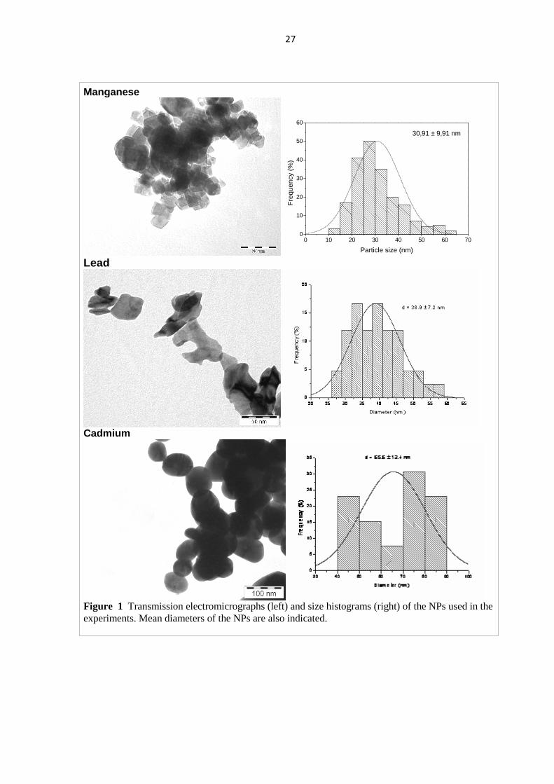
The chemical purity of the nanoparticles was checked by X-ray diffraction, and their particle
size, by X-ray diffraction and transmission electron microscopy. The size histograms and
images of the NPs are shown in Fig. 1.
For intratracheral administration, the NPs were suspended in 1% hydroxyethyl cellulose
(HEC) dissolved in PBS (pH 7.4). This vehicle was physiologically neutral and slowed the
aggregation of the NPs. The suspension was intensively sonicated as it was made, and was
sonicated again before administration.
Diethyl ether (used for brief anesthesia) and HEC was obtained from the Central Pharmacy of
the University of Szeged. Materials for synthesis of the NPs and for po. metal treatment, and
urethane for terminal anesthesia (see below), were purchased from Reanal, Budapest.
27
Manganese
0 10 20 30 40 50 60 700
10
20
30
40
50
60
Fre
quen
cy (
%)
Particle size (nm)
30,91 ± 9,91 nm
Lead
Cadmium
Figure 1 Transmission electromicrographs (left) and size histograms (right) of the NPs used in the experiments. Mean diameters of the NPs are also indicated.
28
2.2. Modes and time schemes of treatment
Gavage (for po. application) was performed using an appropriately bent and fire-polished thin
glass tube attached to a 1 ml syringe. Gavage could be performed on awake rats.
For it. instillation, the animals had a brief diethyl ether anesthesia. The rat was put in a glass
jar with air-tight lid, saturated with ether vapor. The completely anesthetized rat was
suspended, on a board tilted to 60° from horizontal, by hanging its upper incisors in a wire
loop. Keeping this way the rat in place and its mouth open, the trachea was illuminated
transdermally by means of a fibre optic light guide brought into direct contact with the
animal’s neck. The tongue was pulled forward with a pair of non-traumatic forceps, and a
custom-made laryngoscope was used to gain access to the glottis. The NP suspension (or the
vehicle, 1% HEC in the controls) was instilled into the trachea by means of a 1 ml syringe and
1.2 mm diameter plastic tubing, inserted between the vocal chords. Before taking up the
materials, an equal quantity of air was drawn into the syringe, and was pushed out after the
suspension to assure that the whole amount was emptied from the syringe and tube and
delivered into the trachea. Treatment was performed under an exhaust hood to remove ether
vapors.
The time scheme of treatment was, based on previous experience, determined so that
detectable effects could be obtained in a reasonable time span. The basic idea, outlined in
Aims, was to treat the rat first orally to imitate food/waterborne background exposure, then to
apply intratracheal treatment, as if it were by workplace metal fumes. This was to be realized
in a 3 and 6 weeks treatment scheme presented in Table 1. The treatment started with 3 or 6
weeks po. administration of the doses given in Table 1, to 2 x 10 rats. Then half of the group
was finished (underwent behavioral and electrophysiological investigation, and was dissected)
while the other half was further treated by it. administration for an equal length of time (3 or 6
weeks). This scheme worked well with Mn and Pb but produced too high general toxicity and
excessive loss of animals in case of po.+it. Cd treatment. So, a different scheme with shorter
it. application period was used instead, with only one po. dose, and it. application was done
before, and also after, po. treatment (in two different groups, see Table 2C). All the same, the
controls and the Cd3 group of thee first (abandoned) experiments with Cd exposure were
usable and are included in the evaluation.
29
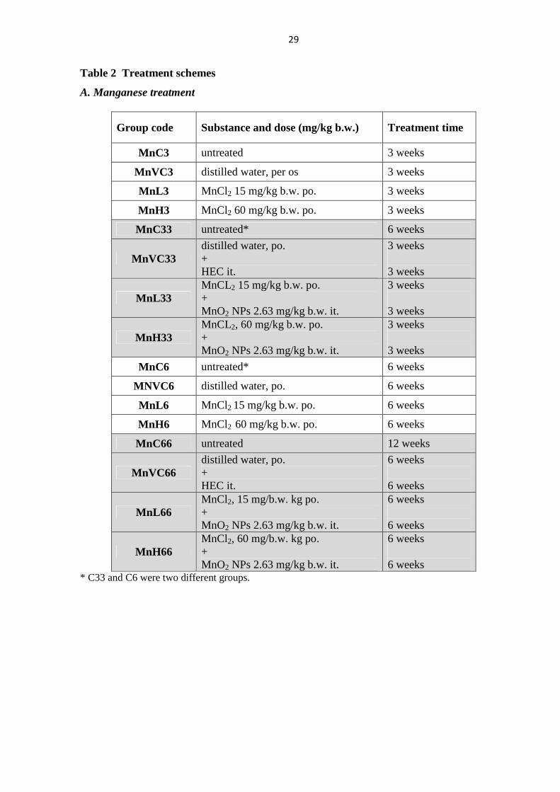
Table 2 Treatment schemes
A. Manganese treatment
Group code Substance and dose (mg/kg b.w.) Treatment time
MnC3 untreated 3 weeks
MnVC3 distilled water, per os 3 weeks
MnL3 MnCl2 15 mg/kg b.w. po. 3 weeks
MnH3 MnCl2 60 mg/kg b.w. po. 3 weeks
MnC33 untreated* 6 weeks
MnVC33 distilled water, po. + HEC it.
3 weeks 3 weeks
MnL33 MnCL2 15 mg/kg b.w. po. + MnO2 NPs 2.63 mg/kg b.w. it.
3 weeks 3 weeks
MnH33 MnCL2, 60 mg/kg b.w. po. + MnO2 NPs 2.63 mg/kg b.w. it.
3 weeks 3 weeks
MnC6 untreated* 6 weeks
MNVC6 distilled water, po. 6 weeks
MnL6 MnCl2 15 mg/kg b.w. po. 6 weeks
MnH6 MnCl2 60 mg/kg b.w. po. 6 weeks
MnC66 untreated 12 weeks
MnVC66 distilled water, po. + HEC it.
6 weeks 6 weeks
MnL66 MnCl2, 15 mg/b.w. kg po. + MnO2 NPs 2.63 mg/kg b.w. it.
6 weeks 6 weeks
MnH66 MnCl2, 60 mg/b.w. kg po. + MnO2 NPs 2.63 mg/kg b.w. it.
6 weeks 6 weeks
* C33 and C6 were two different groups.
30
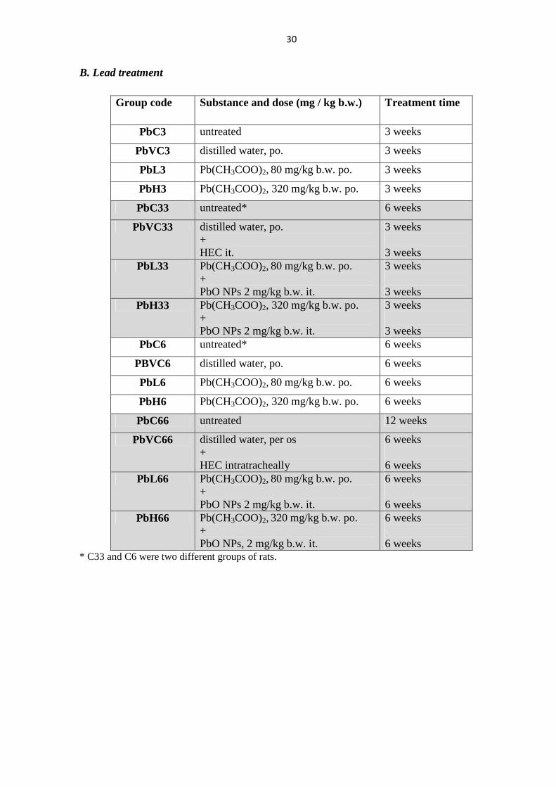
B. Lead treatment
Group code Substance and dose (mg / kg b.w.) Treatment time
PbC3 untreated 3 weeks
PbVC3 distilled water, po. 3 weeks
PbL3 Pb(CH3COO)2, 80 mg/kg b.w. po. 3 weeks
PbH3 Pb(CH3COO)2, 320 mg/kg b.w. po. 3 weeks
PbC33 untreated* 6 weeks
PbVC33 distilled water, po. + HEC it.
3 weeks 3 weeks
PbL33 Pb(CH3COO)2, 80 mg/kg b.w. po.
+ PbO NPs 2 mg/kg b.w. it.
3 weeks 3 weeks
PbH33 Pb(CH3COO)2, 320 mg/kg b.w. po.
+ PbO NPs 2 mg/kg b.w. it.
3 weeks 3 weeks
PbC6 untreated* 6 weeks
PBVC6 distilled water, po. 6 weeks
PbL6 Pb(CH3COO)2, 80 mg/kg b.w. po. 6 weeks
PbH6 Pb(CH3COO)2, 320 mg/kg b.w. po. 6 weeks
PbC66 untreated 12 weeks
PbVC66 distilled water, per os + HEC intratracheally
6 weeks 6 weeks
PbL66 Pb(CH3COO)2, 80 mg/kg b.w. po. + PbO NPs 2 mg/kg b.w. it.
6 weeks 6 weeks
PbH66 Pb(CH3COO)2, 320 mg/kg b.w. po. + PbO NPs, 2 mg/kg b.w. it.
6 weeks 6 weeks
* C33 and C6 were two different groups of rats.
31
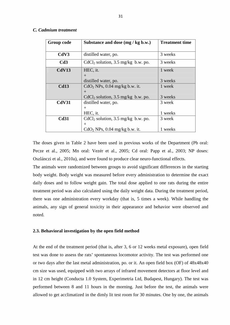
C. Cadmium treatment
Group code Substance and dose (mg / kg b.w.) Treatment time
CdV3 distilled water, po. 3 weeks
Cd3 CdCl2 solution, 3.5 mg/kg b.w. po. 3 weeks
CdV13 HEC, it. + distilled water, po.
1 week 3 weeks
Cd13 CdO2 NPs, 0.04 mg/kg b.w. it. + CdCl2 solution, 3.5 mg/kg b.w. po.
1 week 3 weeks
CdV31 distilled water, po. + HEC, it.
3 week 1 weeks
Cd31 CdCl2 solution, 3.5 mg/kg b.w. po. + CdO2 NPs, 0.04 mg/kg b.w. it.
3 week 1 weeks
The doses given in Table 2 have been used in previous works of the Department (Pb oral:
Pecze et al., 2005; Mn oral: Vezér et al., 2005; Cd oral: Papp et al., 2003; NP doses:
Oszlánczi et al., 2010a), and were found to produce clear neuro-functional effects.
The animals were randomized between groups to avoid significant differences in the starting
body weight. Body weight was measured before every administration to determine the exact
daily doses and to follow weight gain. The total dose applied to one rats during the entire
treatment period was also calculated using the daily weight data. During the treatment period,
there was one administration every workday (that is, 5 times a week). While handling the
animals, any sign of general toxicity in their appearance and behavior were observed and
noted.
2.3. Behavioral investigation by the open field method
At the end of the treatment period (that is, after 3, 6 or 12 weeks metal exposure), open field
test was done to assess the rats’ spontaneous locomotor activity. The test was performed one
or two days after the last metal administration, po. or it. An open field box (OF) of 48x48x40
cm size was used, equipped with two arrays of infrared movement detectors at floor level and
in 12 cm height (Conducta 1.0 System, Experimetria Ltd, Budapest, Hungary). The test was
performed between 8 and 11 hours in the morning. Just before the test, the animals were
allowed to get acclimatized in the dimly lit test room for 30 minutes. One by one, the animals
32
were placed into the centre of the box, for one 10-min session. The instrument recorded the
animal’s horizontal and vertical motor activity based on the interruptions of the infrared
beams. From these data, counts, time and run length of the activity forms (ambulation, local
activity, immobility, rearing) were automatically calculated. More than 40 mm shift in the
location of interrupted beams at the floor level during a time unit of 1 s was interpreted as
ambulation (i.e. walking, horizontal activity), less shift, as local activity (motions without
changing the place), and no shift at all, as immobility. Rearing was recorded if beams at floor
level and at the higher level were interrupted simultaneously. It was known form previous
experience (e.g., Vezér et al., 2005) that the OF test was suitable for investigating the
impairment of higher nervous functions caused by heavy metals.
2.4. Electrophysiological investigation
The electrophysiological recording was done on the same day after the OF test or on the
following day. The animals were anaesthetized by intraperitoneal injection of 1000 mg/kg
b.w. urethane (Mook, 2006). The head of the rats was fixed in a head holder, the skin was
opened by a mid-sagittal cut and the muscles and connective tissues (galea aponeurotica)
attached to the skull were removed. Finally the left temporal bone was cut along its inner
circumference by a dental drill bit attached to a mini drill, and the left hemisphere was thus
exposed. Lidocaine spray (10%) was applied to the wounds and the exposed cortex was
protected with a thin layer of petroleum jelly. The animals were then wrapped in a warm cloth
to maintain body temperature and were put aside for at least 30 min for recovery. For
recording, the rat was placed into the stereotaxic frame of the electrophysiological setup. To
stabilize body temperature, a thermostated (+36.5°C) base plate was used to support the rat’s
underside during the recording procedure.
To record spontaneous and evoked cortical activity, ball-tipped silver recording electrodes
were positioned on the dura over the primary somatosensory (SS) area (projection of the
whisker pad, barrel field), and over the primary visual (VIS) and auditory (AUD) area. These
regions were determined on the basis of a somatotopic map (Zilles, 1984). A stainless steel
clamp was attached to the cut skin edge as indifferent electrode. SS stimulation was done by a
pair of needles inserted into the whiskery part of the nasal skin, delivering square electric
pulses (for stimulation parameters, see below). VIS stimulation was performed by flashes
delivered by a flash generator via an optical fibre conductor directed into the contralateral eye
of the rat. For acoustic stimulation, sound clicks were applied into the ear of the rat.
33
The recorded biological signals were amplified (104x), fed into the digitizer interface of the
recording setup, and stored on PC. The complete recording and evaluation was executed by
the software Neurosys 1.11 (Experimetria Ltd, Budapest, Hungary).
The session started with six minutes recording of spontaneous activity (electrocorticogram,
ECoG) first, from the three sensory cortical areas simultaneously. From the EcoG records, the
relative spectral power of the frequency bands: delta, 0.5-4 Hz; theta, 4-7 Hz; alpha, 8-13 Hz;
beta1, 13-20 Hz; beta2, 20-30 Hz; gamma, 30-50 Hz (Kandel and Schwartz, 1985) was
determined by the software automatically.
Then EPs from the same cortical areas were recorded via the same surface electrodes. Sensory
stimuli were delivered by a digital time base and stimulator unit (Experimetria Ltd, Budapest,
Hungary). All stimuli were set and applied as of just supramaximal strength (meaning that,
e.g., the stimulus voltage was increased until the evoked response reached maximal amplitude
and ca. 5% was added) and well above background. Electrical stimulation of the whiskers and
the base of tail was done by delivering rectangular electric stimuli (3-4 V, 0.05 ms). The
intensity of the visual stimulation was ca. 60 lux, and that of the auditory stimuli, 40 dB.
Trains of 50 stimuli were applied and the evoked potentials (EPs) recorded. The standard
frequency of the stimulation was 1 Hz. Previous studies in our laboratory (Papp et al., 2001,
2004) demonstrated that varying the frequency of stimulation can sensitively detect the
dynamic interaction of successive excitation processes in the sensory system which in turn
reflects the actual state of the CNS. Accordingly, the frequency dependence in the parameters
of the cortical evoked activity was determined by delivering stimuli to the somatosensory
system (i.e. to the whisker pad) beyond the standard 1 Hz, also with 2 and 10 Hz frequency.
Finally, compound action potentials of the tail nerve were recorded. These were evoked by
means of a pair of stimulating needle electrodes inserted at the base of tail (delivering similar
electric stimuli as used to stimulate the whiskers), and were recorded distally by another pair
of needles at a distance of 50 mm.
Evoked activity (cortical responses and tail nerve action potential) were automatically
averaged off-line, and their parameters were measured manually by means of screen cursors
of the software. Exemplified on the SS EP, onset latency was measured between the stimulus
artefact (designated 0 in Fig. 2) and onset of the first wave (A in Fig. 2). Duration of the EP
was calculated as the difference of the 0-D and 0-A times. In case of the visual and auditory
EPs, onset latency and duration was measured, in the same way. The tail nerve action
potential had also a biphasic shape. There, onset latency was defined analogously with the 0-A
distance. Tail nerve conduction velocity was calculated from the onset latency and the
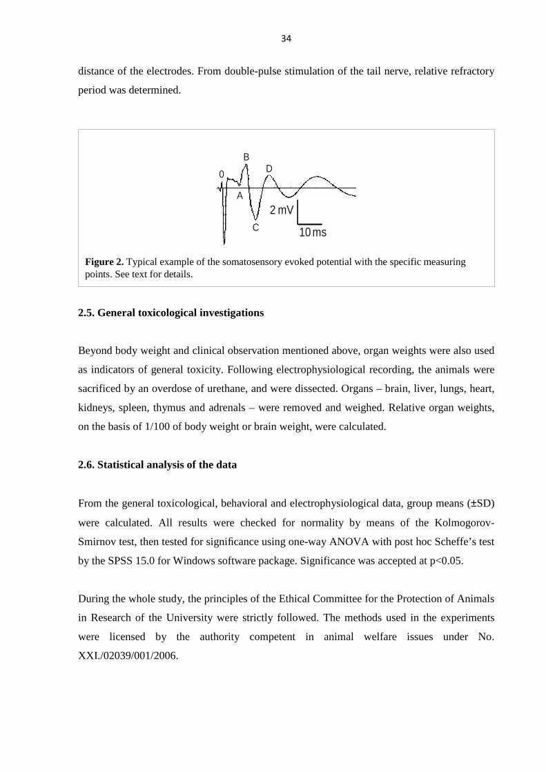
34
distance of the electrodes. From double-pulse stimulation of the tail nerve, relative refractory
period was determined.
2.5. General toxicological investigations
Beyond body weight and clinical observation mentioned above, organ weights were also used
as indicators of general toxicity. Following electrophysiological recording, the animals were
sacrificed by an overdose of urethane, and were dissected. Organs – brain, liver, lungs, heart,
kidneys, spleen, thymus and adrenals – were removed and weighed. Relative organ weights,
on the basis of 1/100 of body weight or brain weight, were calculated.
2.6. Statistical analysis of the data
From the general toxicological, behavioral and electrophysiological data, group means (±SD)
were calculated. All results were checked for normality by means of the Kolmogorov-
Smirnov test, then tested for significance using one-way ANOVA with post hoc Scheffe’s test
by the SPSS 15.0 for Windows software package. Significance was accepted at p<0.05.
During the whole study, the principles of the Ethical Committee for the Protection of Animals
in Research of the University were strictly followed. The methods used in the experiments
were licensed by the authority competent in animal welfare issues under No.
XXI./02039/001/2006.
0
A
B
C
D
10 ms
2 mV
Figure 2. Typical example of the somatosensory evoked potential with the specific measuring points. See text for details.
35
3. RESULTS
3.1. Effects of manganese
3.1.1. Body and organ weights
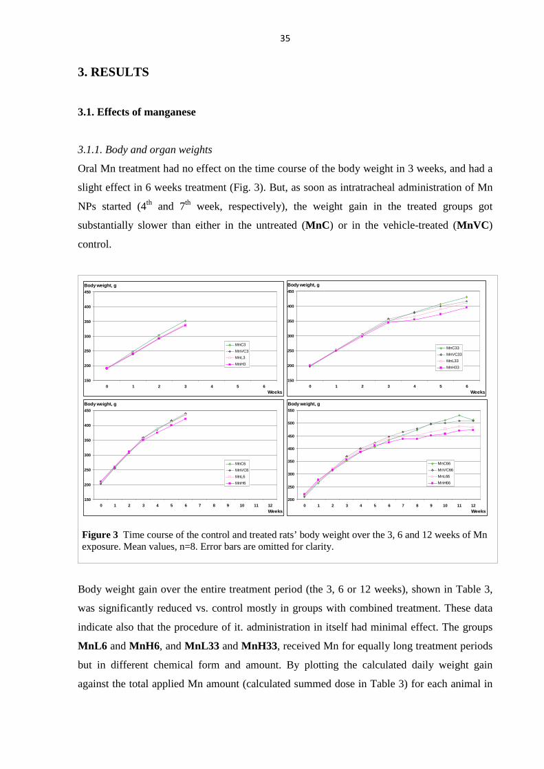
Oral Mn treatment had no effect on the time course of the body weight in 3 weeks, and had a
slight effect in 6 weeks treatment (Fig. 3). But, as soon as intratracheal administration of Mn
NPs started (4th and 7th week, respectively), the weight gain in the treated groups got
substantially slower than either in the untreated (MnC) or in the vehicle-treated (MnVC)
control.
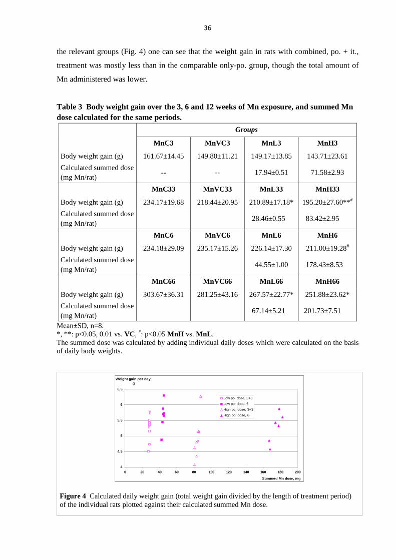
Body weight gain over the entire treatment period (the 3, 6 or 12 weeks), shown in Table 3,
was significantly reduced vs. control mostly in groups with combined treatment. These data
indicate also that the procedure of it. administration in itself had minimal effect. The groups
MnL6 and MnH6, and MnL33 and MnH33, received Mn for equally long treatment periods
but in different chemical form and amount. By plotting the calculated daily weight gain
against the total applied Mn amount (calculated summed dose in Table 3) for each animal in
150
200
250
300
350
400
450
0 1 2 3 4 5 6Weeks
Body weight, g
MnC3
MnVC3
MnL3
MnH3
150
200
250
300
350
400
450
0 1 2 3 4 5 6
Weeks
Body weight, g
MnC33
MnVC33
MnL33
MnH33
150
200
250
300
350
400
450
0 1 2 3 4 5 6 7 8 9 10 11 12
Weeks
Body weight, g
MnC6
MnVC6
MnL6
MnH6
200
250
300
350
400
450
500
550
0 1 2 3 4 5 6 7 8 9 10 11 12Weeks
Body weight, g
MnC66
MnVC66
MnL66
MnH66
Figure 3 Time course of the control and treated rats’ body weight over the 3, 6 and 12 weeks of Mn exposure. Mean values, n=8. Error bars are omitted for clarity.
36
the relevant groups (Fig. 4) one can see that the weight gain in rats with combined, po. + it.,
treatment was mostly less than in the comparable only-po. group, though the total amount of
Mn administered was lower.
Table 3 Body weight gain over the 3, 6 and 12 weeks of Mn exposure, and summed Mn dose calculated for the same periods.
Groups
MnC3 MnVC3 MnL3 MnH3
Body weight gain (g) 161.67±14.45 149.80±11.21 149.17±13.85 143.71±23.61
Calculated summed dose (mg Mn/rat)
-- -- 17.94±0.51 71.58±2.93
MnC33 MnVC33 MnL33 MnH33
Body weight gain (g) 234.17±19.68 218.44±20.95 210.89±17.18* 195.20±27.60**#
Calculated summed dose (mg Mn/rat)
28.46±0.55 83.42±2.95
MnC6 MnVC6 MnL6 MnH6
Body weight gain (g) 234.18±29.09 235.17±15.26 226.14±17.30 211.00±19.28#
Calculated summed dose (mg Mn/rat)
44.55±1.00 178.43±8.53
MnC66 MnVC66 MnL66 MnH66
Body weight gain (g) 303.67±36.31 281.25±43.16 267.57±22.77* 251.88±23.62*
Calculated summed dose (mg Mn/rat)
67.14±5.21 201.73±7.51
Mean±SD, n=8. *, **: p<0.05, 0.01 vs. VC, #: p<0.05 MnH vs. MnL. The summed dose was calculated by adding individual daily doses which were calculated on the basis of daily body weights.
4
4,5
5
5,5
6
6,5
0 20 40 60 80 100 120 140 160 180 200
Summed Mn dose, mg
Weight gain per day, g
Low po. dose, 3+3
Low po. dose, 6
High po. dose, 3+3
High po. dose, 6
Figure 4 Calculated daily weight gain (total weight gain divided by the length of treatment period) of the individual rats plotted against their calculated summed Mn dose.
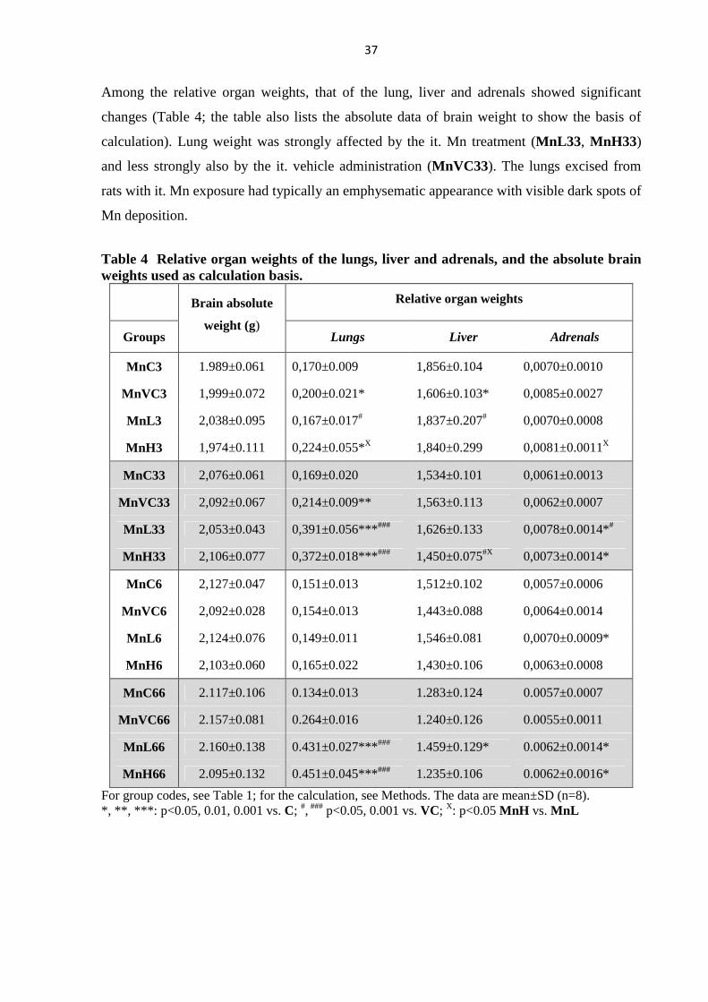
37
Among the relative organ weights, that of the lung, liver and adrenals showed significant
changes (Table 4; the table also lists the absolute data of brain weight to show the basis of
calculation). Lung weight was strongly affected by the it. Mn treatment (MnL33, MnH33)
and less strongly also by the it. vehicle administration (MnVC33). The lungs excised from
rats with it. Mn exposure had typically an emphysematic appearance with visible dark spots of
Mn deposition.
Table 4 Relative organ weights of the lungs, liver and adrenals, and the absolute brain weights used as calculation basis.
Relative organ weights
Groups
Brain absolute
weight (g) Lungs Liver Adrenals
MnC3 1.989±0.061 0,170±0.009 1,856±0.104 0,0070±0.0010
MnVC3 1,999±0.072 0,200±0.021* 1,606±0.103* 0,0085±0.0027
MnL3 2,038±0.095 0,167±0.017# 1,837±0.207# 0,0070±0.0008
MnH3 1,974±0.111 0,224±0.055*X 1,840±0.299 0,0081±0.0011X
MnC33 2,076±0.061 0,169±0.020 1,534±0.101 0,0061±0.0013
MnVC33 2,092±0.067 0,214±0.009** 1,563±0.113 0,0062±0.0007
MnL33 2,053±0.043 0,391±0.056***### 1,626±0.133 0,0078±0.0014*#
MnH33 2,106±0.077 0,372±0.018***### 1,450±0.075#X 0,0073±0.0014*
MnC6 2,127±0.047 0,151±0.013 1,512±0.102 0,0057±0.0006
MnVC6 2,092±0.028 0,154±0.013 1,443±0.088 0,0064±0.0014
MnL6 2,124±0.076 0,149±0.011 1,546±0.081 0,0070±0.0009*
MnH6 2,103±0.060 0,165±0.022 1,430±0.106 0,0063±0.0008
MnC66 2.117±0.106 0.134±0.013 1.283±0.124 0.0057±0.0007
MnVC66 2.157±0.081 0.264±0.016 1.240±0.126 0.0055±0.0011
MnL66 2.160±0.138 0.431±0.027***### 1.459±0.129* 0.0062±0.0014*
MnH66 2.095±0.132 0.451±0.045***### 1.235±0.106 0.0062±0.0016*
For group codes, see Table 1; for the calculation, see Methods. The data are mean±SD (n=8). *, **, ***: p<0.05, 0.01, 0.001 vs. C; #, ### p<0.05, 0.001 vs. VC; X: p<0.05 MnH vs. MnL
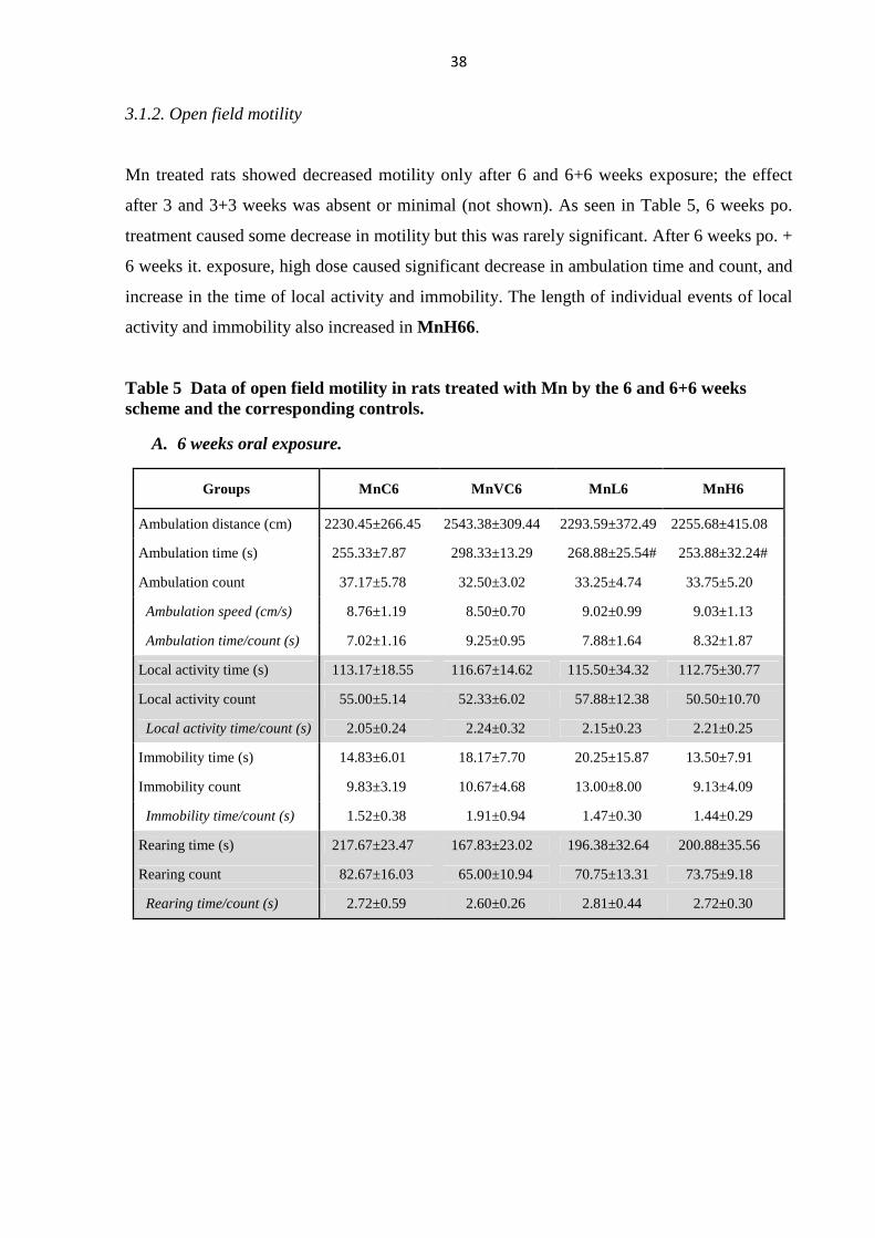
38
3.1.2. Open field motility
Mn treated rats showed decreased motility only after 6 and 6+6 weeks exposure; the effect
after 3 and 3+3 weeks was absent or minimal (not shown). As seen in Table 5, 6 weeks po.
treatment caused some decrease in motility but this was rarely significant. After 6 weeks po. +
6 weeks it. exposure, high dose caused significant decrease in ambulation time and count, and
increase in the time of local activity and immobility. The length of individual events of local
activity and immobility also increased in MnH66.
Table 5 Data of open field motility in rats treated with Mn by the 6 and 6+6 weeks scheme and the corresponding controls.
A. 6 weeks oral exposure.
Groups MnC6 MnVC6 MnL6 MnH6
Ambulation distance (cm) 2230.45±266.45 2543.38±309.44 2293.59±372.49 2255.68±415.08
Ambulation time (s) 255.33±7.87 298.33±13.29 268.88±25.54# 253.88±32.24#
Ambulation count 37.17±5.78 32.50±3.02 33.25±4.74 33.75±5.20
Ambulation speed (cm/s) 8.76±1.19 8.50±0.70 9.02±0.99 9.03±1.13
Ambulation time/count (s) 7.02±1.16 9.25±0.95 7.88±1.64 8.32±1.87
Local activity time (s) 113.17±18.55 116.67±14.62 115.50±34.32 112.75±30.77
Local activity count 55.00±5.14 52.33±6.02 57.88±12.38 50.50±10.70
Local activity time/count (s) 2.05±0.24 2.24±0.32 2.15±0.23 2.21±0.25
Immobility time (s) 14.83±6.01 18.17±7.70 20.25±15.87 13.50±7.91
Immobility count 9.83±3.19 10.67±4.68 13.00±8.00 9.13±4.09
Immobility time/count (s) 1.52±0.38 1.91±0.94 1.47±0.30 1.44±0.29
Rearing time (s) 217.67±23.47 167.83±23.02 196.38±32.64 200.88±35.56
Rearing count 82.67±16.03 65.00±10.94 70.75±13.31 73.75±9.18
Rearing time/count (s) 2.72±0.59 2.60±0.26 2.81±0.44 2.72±0.30
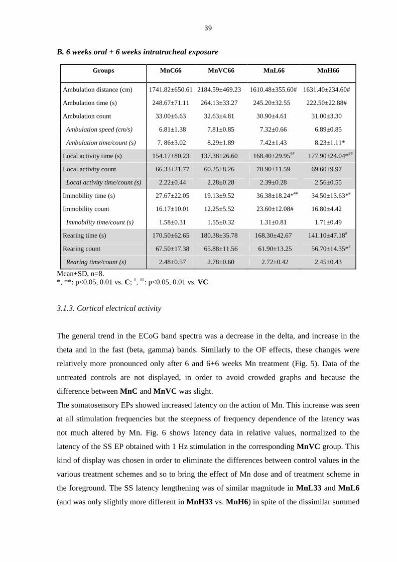
39
B. 6 weeks oral + 6 weeks intratracheal exposure
Groups MnC66 MnVC66 MnL66 MnH66
Ambulation distance (cm) 1741.82±650.61 2184.59±469.23 1610.48±355.60# 1631.40±234.60#
Ambulation time (s) 248.67±71.11 264.13±33.27 245.20±32.55 222.50±22.88#
Ambulation count 33.00±6.63 32.63±4.81 30.90±4.61 31.00±3.30
Ambulation speed (cm/s) 6.81±1.38 7.81±0.85 7.32±0.66 6.89±0.85
Ambulation time/count (s) 7. 86±3.02 8.29±1.89 7.42±1.43 8.23±1.11*
Local activity time (s) 154.17±80.23 137.38±26.60 168.40±29.95## 177.90±24.04*##
Local activity count 66.33±21.77 60.25±8.26 70.90±11.59 69.60±9.97
Local activity time/count (s) 2.22±0.44 2.28±0.28 2.39±0.28 2.56±0.55
Immobility time (s) 27.67±22.05 19.13±9.52 36.38±18.24*## 34.50±13.63*#
Immobility count 16.17±10.01 12.25±5.52 23.60±12.08# 16.80±4.42
Immobility time/count (s) 1.58±0.31 1.55±0.32 1.31±0.81 1.71±0.49
Rearing time (s) 170.50±62.65 180.38±35.78 168.30±42.67 141.10±47.18#
Rearing count 67.50±17.38 65.88±11.56 61.90±13.25 56.70±14.35*#
Rearing time/count (s) 2.48±0.57 2.78±0.60 2.72±0.42 2.45±0.43
Mean+SD, n=8. *, **: p<0.05, 0.01 vs. C; #, ##: p<0.05, 0.01 vs. VC.
3.1.3. Cortical electrical activity
The general trend in the ECoG band spectra was a decrease in the delta, and increase in the
theta and in the fast (beta, gamma) bands. Similarly to the OF effects, these changes were
relatively more pronounced only after 6 and 6+6 weeks Mn treatment (Fig. 5). Data of the
untreated controls are not displayed, in order to avoid crowded graphs and because the
difference between MnC and MnVC was slight.
The somatosensory EPs showed increased latency on the action of Mn. This increase was seen
at all stimulation frequencies but the steepness of frequency dependence of the latency was
not much altered by Mn. Fig. 6 shows latency data in relative values, normalized to the
latency of the SS EP obtained with 1 Hz stimulation in the corresponding MnVC group. This
kind of display was chosen in order to eliminate the differences between control values in the
various treatment schemes and so to bring the effect of Mn dose and of treatment scheme in
the foreground. The SS latency lengthening was of similar magnitude in MnL33 and MnL6
(and was only slightly more different in MnH33 vs. MnH6) in spite of the dissimilar summed

40
dose (see Table 3) which was approximately in accordance with the relationship of summed
Mn dose and body weight. VIS latency in the groups MnH33 and MnH6 was in similar
relationship (Fig. 7). AUD latencies changed less consequently.
SS
0%
20%
40%
60%
80%
100%
MnVC6 MnL6 MnH6 MnVC66 MnL66 MnH66Groups
Rel. power
gamma
beta2
beta1
alpha
theta
delta
** *
*#
VIS
0%
20%
40%
60%
80%
100%
MnVC6 MnL6 MnH6 MnVC66 MnL66 MnH66Groups
Rel. power
gamma
beta2
beta1
alpha
theta
delta* *#
*#
#
AUD
0%
20%
40%
60%
80%
100%
MnVC6 MnL6 MnH6 MnVC66 MnL66 MnH66Groups
Rel. power
gamma
beta2
beta1
alpha
theta
delta
*
*
#
Figure 5 Band spectrum of the ECoG in rats treated with Mn by the 6 and 6+6 weeks scheme. *: p<0.05 vs. MnVC; #: p<0.05 high vs. low dose.
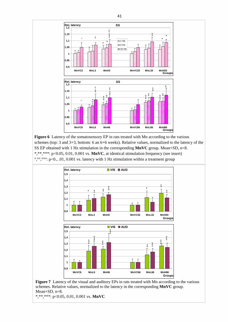
41
SS
0,9
0,95
1
1,05
1,1
1,15
1,2
MnVC3 MnL3 MnH3 MnVC33 MnL33 MnH33Groups
Rel. latency
1 Hz
2 Hz
10 Hz
° °°
**
**
* °
°°
**
*
*
SS
0,9
0,95
1
1,05
1,1
1,15
1,2
MnVC6 MnL6 MnH6 MnVC66 MnL66 MnH66Groups
Rel. latency°
**
°°
* **
* **
**
°°°
**
*
**
***
**
**
° *°
Figure 6 Latency of the somatosensory EP in rats treated with Mn according to the various schemes (top: 3 and 3+3, bottom: 6 an 6+6 weeks). Relative values, normalized to the latency of the SS EP obtained with 1 Hz stimulation in the corresponding MnVC group. Mean+SD, n=8. *,**,***: p<0.05, 0.01, 0.001 vs. MnVC, at identical stimulation frequency (see insert) °,°°,°°°: p<0., .01, 0.001 vs. latency with 1 Hz stimulation within a treatment group
0,9
1
1,1
1,2
1,3
1,4
1,5
MnVC3 MnL3 MnH3 MnVC33 MnL33 MnH33Groups
Rel. latency VIS AUD
* ** * *
*
** *
**
**
0,9
1
1,1
1,2
1,3
1,4
1,5
MnVC6 MnL6 MnH6 MnVC66 MnL66 MnH66Groups
Rel. latency VIS AUD
**
*
**
*
**
**
*
**
*
**
*
**
*
**
*
Figure 7 Latency of the visual and auditory EPs in rats treated with Mn according to the various schemes. Relative values, normalized to the latency in the corresponding MnVC group. Mean+SD, n=8. *,**,***: p<0.05, 0.01, 0.001 vs. MnVC
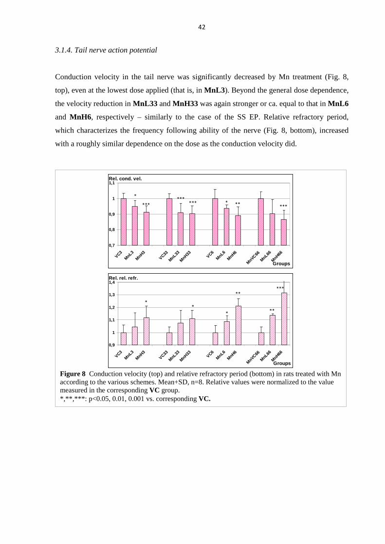
42
3.1.4. Tail nerve action potential
Conduction velocity in the tail nerve was significantly decreased by Mn treatment (Fig. 8,
top), even at the lowest dose applied (that is, in MnL3). Beyond the general dose dependence,
the velocity reduction in MnL33 and MnH33 was again stronger or ca. equal to that in MnL6
and MnH6, respectively – similarly to the case of the SS EP. Relative refractory period,
which characterizes the frequency following ability of the nerve (Fig. 8, bottom), increased
with a roughly similar dependence on the dose as the conduction velocity did.
0,7
0,8
0,9
1
1,1
VC3MnL3
MnH3
VC33
MnL33
MnH33
VC6MnL6
MnH6
MnVC66
MnL66
MnH66
Groups
Rel. cond. vel.
*
******
*** * ** ***
0,9
1
1,1
1,2
1,3
1,4
VC3M
nL3
MnH3
VC33
MnL33
MnH33VC6
MnL6
MnH6
MnVC66
MnL66
MnH66
Groups
Rel. rel. refr.
**
*
*****
**
Figure 8 Conduction velocity (top) and relative refractory period (bottom) in rats treated with Mn according to the various schemes. Mean+SD, n=8. Relative values were normalized to the value measured in the corresponding VC group. *,**,***: p<0.05, 0.01, 0.001 vs. corresponding VC.
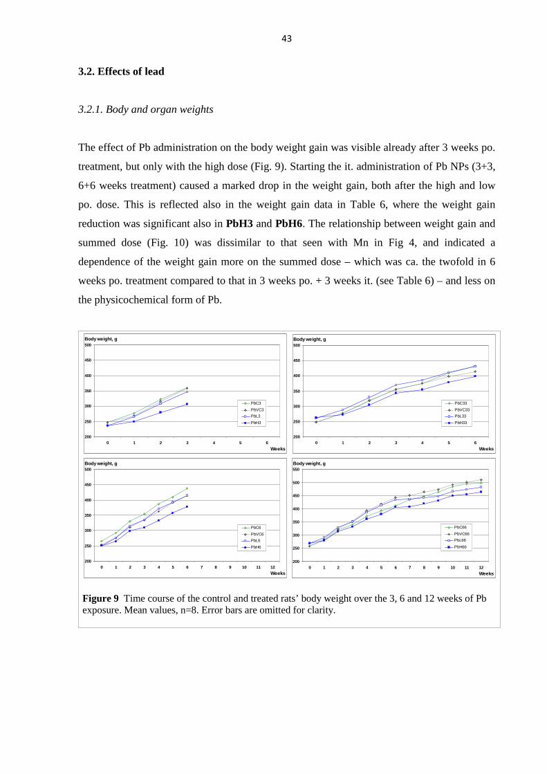
43
3.2. Effects of lead
3.2.1. Body and organ weights
The effect of Pb administration on the body weight gain was visible already after 3 weeks po.
treatment, but only with the high dose (Fig. 9). Starting the it. administration of Pb NPs (3+3,
6+6 weeks treatment) caused a marked drop in the weight gain, both after the high and low
po. dose. This is reflected also in the weight gain data in Table 6, where the weight gain
reduction was significant also in PbH3 and PbH6. The relationship between weight gain and
summed dose (Fig. 10) was dissimilar to that seen with Mn in Fig 4, and indicated a
dependence of the weight gain more on the summed dose – which was ca. the twofold in 6
weeks po. treatment compared to that in 3 weeks po. + 3 weeks it. (see Table 6) – and less on
the physicochemical form of Pb.
200
250
300
350
400
450
500
0 1 2 3 4 5 6
Weeks
Body weight, g
PbC33
PbVC33
PbL33
PbH33
200
250
300
350
400
450
500
0 1 2 3 4 5 6
Weeks
Body weight, g
PbC3
PbVC3
PbL3
PbH3
200
250
300
350
400
450
500
0 1 2 3 4 5 6 7 8 9 10 11 12
Weeks
Body weight, g
PbC6
PbVC6
PbL6
PbH6
200
250
300
350
400
450
500
550
0 1 2 3 4 5 6 7 8 9 10 11 12
Weeks
Body weight, g
PbC66
PbVC66
PbL66
PbH66
Figure 9 Time course of the control and treated rats’ body weight over the 3, 6 and 12 weeks of Pb exposure. Mean values, n=8. Error bars are omitted for clarity.
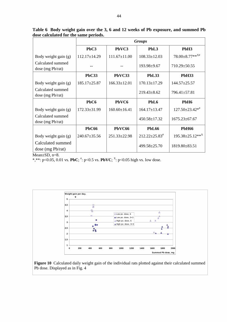
44
Table 6 Body weight gain over the 3, 6 and 12 weeks of Pb exposure, and summed Pb dose calculated for the same periods.
Groups
PbC3 PbVC3 PbL3 PbH3
Body weight gain (g) 112.17±14.29 111.67±11.00 108.33±12.03 78.00±8.77**X#
Calculated summed dose (mg Pb/rat)
-- -- 193.98±9.67 710.29±50.55
PbC33 PbVC33 PbL33 PbH33
Body weight gain (g) 185.17±25.87 166.33±12.01 170.13±17.29 144.57±25.57
Calculated summed dose (mg Pb/rat)
219.43±8.62 796.41±57.81
PbC6 PbVC6 PbL6 PbH6
Body weight gain (g) 172.33±31.99 160.60±16.41 164.17±13.47 127.50±23.42*#
Calculated summed dose (mg Pb/rat)
450.58±17.32 1675.23±67.67
PbC66 PbVC66 PbL66 PbH66
Body weight gain (g) 240.67±35.56 251.33±22.98 212.22±25.83# 195.38±25.12**X
Calculated summed dose (mg Pb/rat)
499.58±25.70 1819.80±83.51
Mean±SD, n=8. *,**: p<0.05, 0.01 vs. PbC; #: p<0.5 vs. PbVC; X: p<0.05 high vs. low dose.
1
1,5
2
2,5
3
3,5
4
4,5
5
0 200 400 600 800 1000 1200 1400 1600 1800 2000
Summed Pb dose, mg
Weight gain per day, g
Low po. dose, 6
Low po. dose, 3+3
High po. dose, 6
High po. dose, 3+3
Figure 10 Calculated daily weight gain of the individual rats plotted against their calculated summed Pb dose. Displayed as in Fig. 4
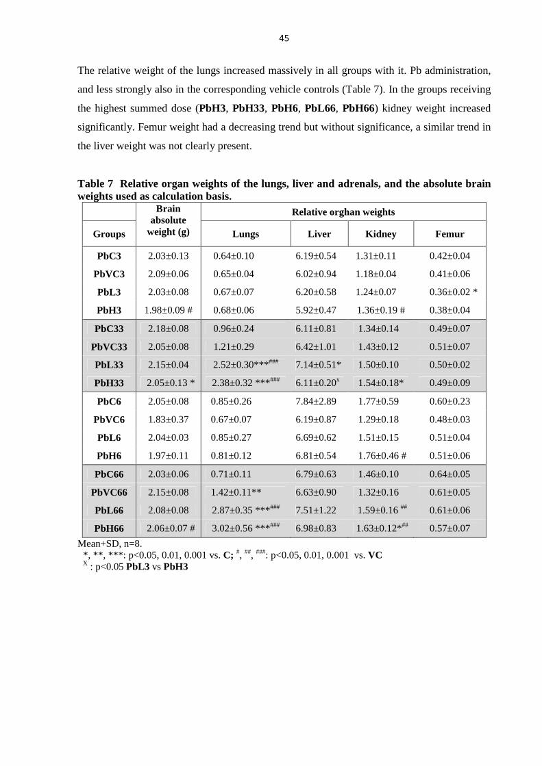
45
The relative weight of the lungs increased massively in all groups with it. Pb administration,
and less strongly also in the corresponding vehicle controls (Table 7). In the groups receiving
the highest summed dose (PbH3, PbH33, PbH6, PbL66, PbH66) kidney weight increased
significantly. Femur weight had a decreasing trend but without significance, a similar trend in
the liver weight was not clearly present.
Table 7 Relative organ weights of the lungs, liver and adrenals, and the absolute brain weights used as calculation basis.
Relative orghan weights
Groups
Brain absolute
weight (g) Lungs Liver Kidney Femur
PbC3 2.03±0.13 0.64±0.10 6.19±0.54 1.31±0.11 0.42±0.04
PbVC3 2.09±0.06 0.65±0.04 6.02±0.94 1.18±0.04 0.41±0.06
PbL3 2.03±0.08 0.67±0.07 6.20±0.58 1.24±0.07 0.36±0.02 *
PbH3 1.98±0.09 # 0.68±0.06 5.92±0.47 1.36±0.19 # 0.38±0.04
PbC33 2.18±0.08 0.96±0.24 6.11±0.81 1.34±0.14 0.49±0.07
PbVC33 2.05±0.08 1.21±0.29 6.42±1.01 1.43±0.12 0.51±0.07
PbL33 2.15±0.04 2.52±0.30***### 7.14±0.51* 1.50±0.10 0.50±0.02
PbH33 2.05±0.13 * 2.38±0.32 ***### 6.11±0.20x 1.54±0.18* 0.49±0.09
PbC6 2.05±0.08 0.85±0.26 7.84±2.89 1.77±0.59 0.60±0.23
PbVC6 1.83±0.37 0.67±0.07 6.19±0.87 1.29±0.18 0.48±0.03
PbL6 2.04±0.03 0.85±0.27 6.69±0.62 1.51±0.15 0.51±0.04
PbH6 1.97±0.11 0.81±0.12 6.81±0.54 1.76±0.46 # 0.51±0.06
PbC66 2.03±0.06 0.71±0.11 6.79±0.63 1.46±0.10 0.64±0.05
PbVC66 2.15±0.08 1.42±0.11** 6.63±0.90 1.32±0.16 0.61±0.05
PbL66 2.08±0.08 2.87±0.35 ***### 7.51±1.22 1.59±0.16 ## 0.61±0.06
PbH66 2.06±0.07 # 3.02±0.56 ***### 6.98±0.83 1.63±0.12*## 0.57±0.07
Mean+SD, n=8. *, **, ***: p<0.05, 0.01, 0.001 vs. C; #, ##, ###: p<0.05, 0.01, 0.001 vs. VC X : p<0.05 PbL3 vs PbH3
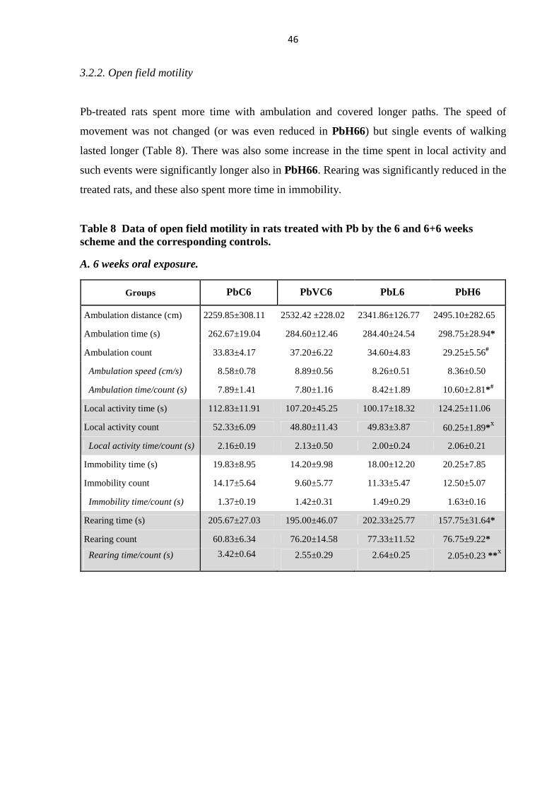
46
3.2.2. Open field motility
Pb-treated rats spent more time with ambulation and covered longer paths. The speed of
movement was not changed (or was even reduced in PbH66) but single events of walking
lasted longer (Table 8). There was also some increase in the time spent in local activity and
such events were significantly longer also in PbH66. Rearing was significantly reduced in the
treated rats, and these also spent more time in immobility.
Table 8 Data of open field motility in rats treated with Pb by the 6 and 6+6 weeks scheme and the corresponding controls.
A. 6 weeks oral exposure.
Groups PbC6 PbVC6 PbL6 PbH6
Ambulation distance (cm) 2259.85±308.11 2532.42 ±228.02 2341.86±126.77 2495.10±282.65
Ambulation time (s) 262.67±19.04 284.60±12.46 284.40±24.54 298.75±28.94*
Ambulation count 33.83±4.17 37.20±6.22 34.60±4.83 29.25±5.56#
Ambulation speed (cm/s) 8.58±0.78 8.89±0.56 8.26±0.51 8.36±0.50
Ambulation time/count (s) 7.89±1.41 7.80±1.16 8.42±1.89 10.60±2.81*#
Local activity time (s) 112.83±11.91 107.20±45.25 100.17±18.32 124.25±11.06
Local activity count 52.33±6.09 48.80±11.43 49.83±3.87 60.25±1.89*x
Local activity time/count (s) 2.16±0.19 2.13±0.50 2.00±0.24 2.06±0.21
Immobility time (s) 19.83±8.95 14.20±9.98 18.00±12.20 20.25±7.85
Immobility count 14.17±5.64 9.60±5.77 11.33±5.47 12.50±5.07
Immobility time/count (s) 1.37±0.19 1.42±0.31 1.49±0.29 1.63±0.16
Rearing time (s) 205.67±27.03 195.00±46.07 202.33±25.77 157.75±31.64*
Rearing count 60.83±6.34 76.20±14.58 77.33±11.52 76.75±9.22*
Rearing time/count (s) 3.42±0.64 2.55±0.29 2.64±0.25 2.05±0.23 **x
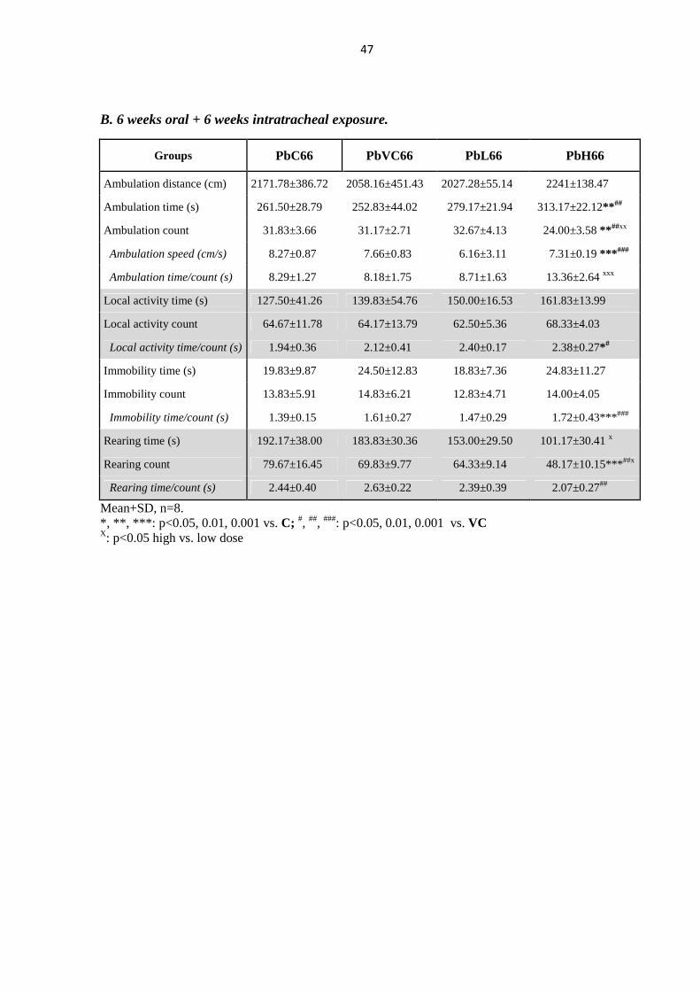
47
B. 6 weeks oral + 6 weeks intratracheal exposure.
Groups PbC66 PbVC66 PbL66 PbH66
Ambulation distance (cm) 2171.78±386.72 2058.16±451.43 2027.28±55.14 2241±138.47
Ambulation time (s) 261.50±28.79 252.83±44.02 279.17±21.94 313.17±22.12**##
Ambulation count 31.83±3.66 31.17±2.71 32.67±4.13 24.00±3.58 **##xx
Ambulation speed (cm/s) 8.27±0.87 7.66±0.83 6.16±3.11 7.31±0.19 ***###
Ambulation time/count (s) 8.29±1.27 8.18±1.75 8.71±1.63 13.36±2.64 xxx
Local activity time (s) 127.50±41.26 139.83±54.76 150.00±16.53 161.83±13.99
Local activity count 64.67±11.78 64.17±13.79 62.50±5.36 68.33±4.03
Local activity time/count (s) 1.94±0.36 2.12±0.41 2.40±0.17 2.38±0.27*#
Immobility time (s) 19.83±9.87 24.50±12.83 18.83±7.36 24.83±11.27
Immobility count 13.83±5.91 14.83±6.21 12.83±4.71 14.00±4.05
Immobility time/count (s) 1.39±0.15 1.61±0.27 1.47±0.29 1.72±0.43***###
Rearing time (s) 192.17±38.00 183.83±30.36 153.00±29.50 101.17±30.41 x
Rearing count 79.67±16.45 69.83±9.77 64.33±9.14 48.17±10.15***##x
Rearing time/count (s) 2.44±0.40 2.63±0.22 2.39±0.39 2.07±0.27##
Mean+SD, n=8. *, **, ***: p<0.05, 0.01, 0.001 vs. C; #, ##, ###: p<0.05, 0.01, 0.001 vs. VC X: p<0.05 high vs. low dose
48
3.2.3. Cortical electrical activity
After 3 and 3+3 weeks, there were no noteworthy changes in the band spectrum of the ECoG
(not shown). In the groups PbL6 and PbH6, increased slow and decreased fast activity was
seen, compared to their control PbVC6. However, in the rats receiving 6 weeks it. Pb
exposure after 6 weeks po. treatment, this frequency shift disappeared (Fig. 11).
Latency of the SS EP was increased in the Pb-treated rats (Fig. 12). The change was
significant vs. vehicle control in rats with 3+3, 6 and 6+6 weeks treatment. The change in
PbH33 and PbH6 was nearly equal, and in PbL33 was somewhat bigger than in PbL6.
SS
0%
20%
40%
60%
80%
100%
PVC6 PL6 PH6 PVC66 PL66 PH66Groups
Rel. power
gamma
beta2
beta1
alpha
theta
delta
*
*
VIS
0%
20%
40%
60%
80%
100%
PVC6 PL6 PH6 PVC66 PL66 PH66Groups
Rel. power
gamma
beta2
beta1
alpha
theta
delta
*
**
AUD
0%
20%
40%
60%
80%
100%
PVC6 PL6 PH6 PVC66 PL66 PH66Groups
Rel. power
gamma
beta2
beta1
alpha
theta
delta
***
***
Figure 11 Band spectrum of the ECoG in rats treated with Pb by the 6 and 6+6 weeks scheme. *,**: p<0.05, 0.0 vs. PbVC.
49
SS
0,9
0,95
1
1,05
1,1
1,15
1,2
PVC3 PL3 PH3 PVC33 PL33 PH33Groups
Rel. latency
1 Hz
2 Hz
10 Hz
°
°° * °
° **
°°
*°
SS
0,9
0,95
1
1,05
1,1
1,15
1,2
PVC6 PL6 PH6 PVC66 PL66 PH66Groups
Rel. latency
1 Hz
2 Hz
10 Hz
° **
°°
* °
°
°
***
°°
**
°
*
Figure 12 Latency of the somatosensory EP in rats treated with Pb according to the various schemes. Relative values, normalized to the latency of the SS EP obtained with 1 Hz stimulation in the corresponding PbVC group. Mean+SD, n=8. *,**,***: p<0.05, 0.01, 0.001 vs. PbVC, at identical stimulation frequency (see insert). °,°°: p<0.05, 0.01 vs. latency with 1 Hz stimulation within a treatment group.
0,8
0,9
1
1,1
1,2
1,3
1,4
1,5
PVC3 PL3 PH3 PVC33 PL33 PH33Groups
Rel. latency VIS AUD
***
* ***
** **
** ***
0,8
0,9
1
1,1
1,2
1,3
1,4
1,5
PVC6 PL6 PH6 PVC66 PL66 PH66Groups
Rel. latency VIS AUD
***
* **
*** ***
***
***
*
Figure 13 Latency of the visual and auditory EPs in rats treated with Pb according to the various schemes. Relative values, normalized to the latency in the corresponding PbVC group. Mean+SD, n=8. *,**,***: p<0.05, 0.01, 0.001 vs. PbVC
50
In both combination groups, the frequency dependent lengthening of the latency became also
more intense. The relationship of latencies in PbH33 and PbH6 was dissimilar to that of body
weight gain in these groups (Fig. 12). Also in the VIS EP, the latency in PbH33 and PbH6
was nearly equal. The latency of AUD EP was also lengthened but in a rather irregular way
(Fig. 13).
3.2.4. Tail nerve action potential
Conduction velocity in the tail nerve was generally decreased in the Pb-treated rats (Fig. 14).
Again it seemed that, as in the case of weight gain, the change reflected more the dose and
less the chemical form of Pb. The length of relative refractory period showed some changes
but no clear tread (not shown).
0,7
0,8
0,9
1
1,1
PVC3PL3
PH3
PVC33PL33
PH33PVC6
PL6PH6
PVC66PL66
PH66
Groups
Rel. cond. vel.
* ***
**
** * ***
Figure 14 Conduction velocity in rats treated with Pb according to the various schemes. Mean+SD, n=8. Relative values were normalized to the value measured in the corresponding VC group. *,**,***: p<0.05, 0.01, 0.001 vs. corresponding VC
51
3.3. Effects of cadmium
3.3.1. Body and organ weights
The time course of the body weight was markedly different in the groups Cd13 and Cd31,
and suggested that the NP form of Cd had, in spite of the much lower dose, more pronounced
effect on this toxicological parameter (Fig. 15; Cd3 and CdV3 are not included because these
are practically identical to the first 3 weeks of Cd31 and CdV31). The effect is also seen in
the weight gain and Cd dose data given separately for the po. and it. administration weeks in
Table 9.
Table 9 Body weight gain and summed Cd doses in the oral and intratracheal treatment periods.
Groups Body weight gain (g)
Calculated summed dose (mg Cd/rat)
Body weight gain (g)
Calculated summed dose (mg Cd/rat)
Week 1 - 3 Week 4
CdV31 120.50±15.66 -- 21.33±4.89 --
Cd31 80.38±35.58* 8.603±0.269 -27.14±24.07*** 0.042±0.003
Week 1 Week 2 - 4
CdV13 11.67±6.65 97.67±26.09
Cd13 -37.86±13.52*** 0.032±0.004 138.51±19.57* 6.425±0.589
Mean+SD, n=8. *,***: p<0.05, 0.001 vs. corresponding control.
150
200
250
300
350
400
450
0 1 2 3 4Weeks
Body weight, g
CdV31
Cd31
CdV13
Cd13
Figure 15 Time course of the control and treated rats’ body weight treated by the 1+3 and 3+1 schemes (see Table 8). Mean values, n=8. Error bars are omitted for clarity.
52
The relative weight of the lungs was massively influenced by the it. application (Table 9).
Weight decrease of the liver and kidneys was also significant only in the combined treatment
groups, and was strongest in Cd31, suggesting a disproportionately high toxicity of
nanoparticulate Cd.
Table 10 Relative organ weights of the lungs, liver and adrenals, and the absolute brain weights used as calculation basis
Relative organ weights
Groups
Brain absolute
weight (g) Lungs Liver Kidneys
CdV3 2.093±0.059 0.650±0.036 6.025±0.940 1.181±0.042
Cd3 2.022±0.074 0.655±0.047 5.697±0.345 1.199±0.084
CdV31 2.064±0.061 0.908±0.051 7.238±0.569 1.387±0.074
Cd31 1.997±0.086 1.615±0.220*** 5.910±1.233* 1.153±0.133**
CdV13 1.918±0.186 0.940±0.095 6.521±0.222 1.353±0.089
Cd13 1.976±0.118 1.338±0.162** 6.254±0.647 1.191±0.077*
Mean+SD, n=8. *,**,***: p<0.05, 0.01, 0.001 vs. corresponding control.
3.3.2. Open field motility
Decreased motility was observed in all Cd-treated rats vs. the corresponding controls. In Cd3,
only the run length was reduced; but in Cd31, nearly all parameters were altered including
slower ambulation speed and longer events of local activity and immobility. In Cd13 the
changes were similar but less strong and mostly not significant. The data are shown in Table
11.
53
Table 11 Data of open field motility in rats treated with Cd by the various treatment schemes. A. 3 weeks oral exposure.
Groups CdC3 CdV3 Cd3
Ambulation distance (cm) 2848.64±175.82 2631.98±585.19 2420.33±201.35 *xxx
Ambulation time (s) 324.00±45.40 289.33±61.72 292.67±18.38 xxx
Ambulation count 26.00±6.36 32.00±10.00 27.33±4.23
Ambulation speed (cm/s) 8.46±0.63 9.11±1.10 8.28±0.64 xx
Ambulation time/count (s) 13.53±5.31 10.60±6.83 11.00±2.37
Local activity time (s) 129.33±30.47 115.33±43.03 144.67±25.52 xxx
Local activity count 58.50±11.48 50.17±12.66 61.83±6.68 #xx
Local activity time/count (s) 2.22±0.33 2.26±0.47 2.33±0.23 xx
Immobility time (s) 26.33±11.84 16.00±13.97 25.83±6.62 xx
Immobility count 16.00±5.69 10.33±6.47 14.50±3.27 xxx
Immobility time/count (s) 1.67±0.39 1.41±0.29 1.82±0.50 x
Rearing time (s) 121.33±35.59 180.33±69.88 137.83±36.27 xx
Rearing count 57.00±15.26 65.17±19.21 57.67±17.96 xx
Rearing time/count (s) 2.13±0.39 2.72±0.62 2.42±0.35
B. Combined exposures
Groups CdV31 Cd31 CdV13 Cd13
Ambulation distance (cm) 2493.07±289.12 392.38±358.48 ### 2705.97±108.61 2226.76±363.10 #ooo
Ambulation time (s) 290.67±26.27 210.17±44.83 ## 297.17±21.84 265.86±19.39 o
Ambulation count 29.33±2.07 26.67±6.35 30.67±1.63 31.43±4.43
Ambulation speed (cm/s) 8.59±0.91 6.58±0.43 ### 9.15±0.77 8.34±0.81 ooo
Ambulation time/count (s) 9.95±1.14 8.24±2.71 9.73±1.10 8.59±1.21
Local activity time (s) 146.50±33.64 241.00±34.30 ### 129.83±33.62 156.14±37.65 ooo
Local activity count 61.17±9.99 81.50±8.87 ## 53.00±8.20 65.86±9.60 #oo
Local activity time/count (s) 2.39±0.40 2.96±0.29 # 2.44±0.50 2.37±0.38 oo
Immobility time (s) 17.50±5.24 87.00±52.22 ### 15.33±6.92 18.71±8.71 ooo
Immobility count 12.00±2.28 34.17±13.44 ### 11.67±3.83 13.29±4.89 ooo
Immobility time/count (s) 1.44±0.21 2.41±0.67 ### 1.29±0.26 1.40±0.21 ooo
Rearing time (s) 146.33±23.64 62.83±42.96 ## 158.67±41.35 160.29±41.01 ooo
Rearing count 69.17±11.62 26.67±12.04 ### 70.67±12.97 61.71±17.16 ooo
Rearing time/count (s) 2.13±0.27 2.20±0.71 2.23±0.22 2.65±0.58
*: p<0,05 CdC3 vs. Cd3 #, ##, ###: p<0.05, 0.01, 0.001 CdV31 vs. Cd31 or CdV13 vs. Cd13 x, xx, xxx: p<0.05, 0.01, 0.001 Cd31 vs. Cd3 o, oo, ooo: p<0.05, 0.01, 0.001 Cd31 vs. Cd13
54
3.3.3. Cortical electrical activity
In the band spectrum of the ECoG, there was practically no change in any of the Cd-treated
groups.
In the somatosensory EP, increase of the latency was seen in all treated groups. In Cd31, the
increase was significant at all stimulation frequencies (1, 2 and 10 Hz) vs. CdV31; whereas in
the other treatment schemes (Cd3, Cd13) only at the highest stimulation frequency (Fig.16,
top). The body weight gain difference was also the highest in Cd31 vs. CdV31 around the
time of electrophysiological recording. The frequency-dependent extra lengthening of latency
was, however, more pronounced also in Cd3 and in Cd31 than in the corresponding controls.
The VIS and AUD EPs were also significantly lengthened by Cd application in the groups
Cd3 and Cd31, but not in Cd13 (Fig. 16, bottom).
SS
0,8
0,9
1
1,1
1,2
1,3
CdV3 Cd3 CdV31 Cd31 CdV13 Cd13Groups
Rel. latency1 Hz 2 Hz 10 Hz
°°° °°
** °°
***
°°°
°°°
* °°
0,6
0,7
0,8
0,9
1
1,1
1,2
1,3
1,4
CdV3 Cd3 CdV31 Cd31 CdV13 Cd13Groups
Rel. latency VIS AUD
****** *** **
Figure 16 Latency of the somatosensory EP (top) and visual and auditory EP (bottom) in rats treated with Cd according to the various schemes. Relative values, normalized to the latency of the EP in the corresponding control group (for SS: with 1 Hz stimulation). Mean+SD, n=8. *,**: p<0.05, 0.01 vs. control (V) at identical stimulation frequency (see insert). °,°°,°°°: p<0.05, 0.01, 0.001 vs. latency with 1 Hz stimulation within a treatment group.
55
3.3.4. Tail nerve action potential
Decreased conduction velocity of the tail nerve was found in all treated groups (Fig. 17) but
here there was no difference between various treatments, unlike what was seen in the cortical
evoked responses. The lengthening of the relative refractory period, which indicated increased
fatigability of the nerve in the treated rats, was significant in Cd3 and Cd13.
0,9
0,95
1
1,05
CdV3 Cd3 CdV31 Cd31 CdV13 Cd13Groups
Rel. cond. vel.
****
***
0,9
1
1,1
1,2
1,3
1,4
CdV3 Cd3 CdV31 Cd31 CdV13 Cd13Groups
Rel. rel. refr.
*
**
Figure 17 Conduction velocity (top) and relative refractory period (bottom) in rats treated with Mn according to the various schemes. Mean+SD, n=8. Relative values were normalized to value measured in the corresponding VC group. *,**,***: p<0.05, 0.01, 0.001 vs. corresponding VC.
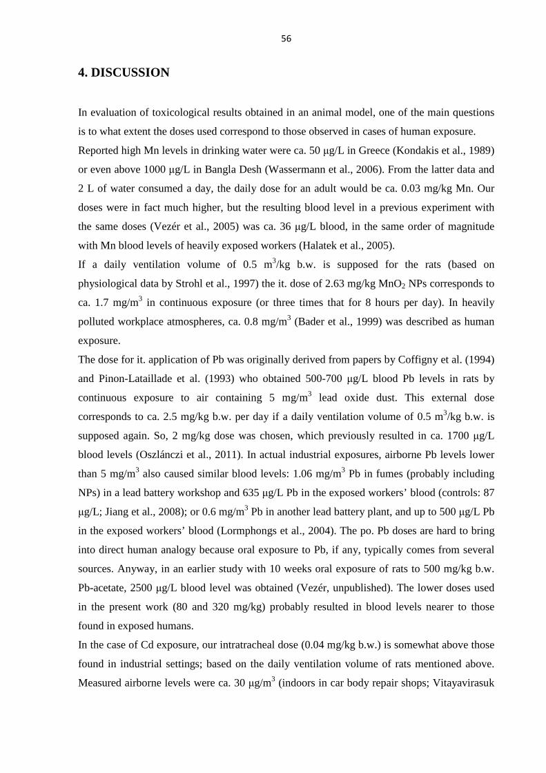
56
4. DISCUSSION
In evaluation of toxicological results obtained in an animal model, one of the main questions
is to what extent the doses used correspond to those observed in cases of human exposure.
Reported high Mn levels in drinking water were ca. 50 µg/L in Greece (Kondakis et al., 1989)
or even above 1000 µg/L in Bangla Desh (Wassermann et al., 2006). From the latter data and
2 L of water consumed a day, the daily dose for an adult would be ca. 0.03 mg/kg Mn. Our
doses were in fact much higher, but the resulting blood level in a previous experiment with
the same doses (Vezér et al., 2005) was ca. 36 µg/L blood, in the same order of magnitude
with Mn blood levels of heavily exposed workers (Halatek et al., 2005).
If a daily ventilation volume of 0.5 m3/kg b.w. is supposed for the rats (based on
physiological data by Strohl et al., 1997) the it. dose of 2.63 mg/kg MnO2 NPs corresponds to
ca. 1.7 mg/m3 in continuous exposure (or three times that for 8 hours per day). In heavily
polluted workplace atmospheres, ca. 0.8 mg/m3 (Bader et al., 1999) was described as human
exposure.
The dose for it. application of Pb was originally derived from papers by Coffigny et al. (1994)
and Pinon-Lataillade et al. (1993) who obtained 500-700 µg/L blood Pb levels in rats by
continuous exposure to air containing 5 mg/m3 lead oxide dust. This external dose
corresponds to ca. 2.5 mg/kg b.w. per day if a daily ventilation volume of 0.5 m3/kg b.w. is
supposed again. So, 2 mg/kg dose was chosen, which previously resulted in ca. 1700 µg/L
blood levels (Oszlánczi et al., 2011). In actual industrial exposures, airborne Pb levels lower
than 5 mg/m3 also caused similar blood levels: 1.06 mg/m3 Pb in fumes (probably including
NPs) in a lead battery workshop and 635 µg/L Pb in the exposed workers’ blood (controls: 87
µg/L; Jiang et al., 2008); or 0.6 mg/m3 Pb in another lead battery plant, and up to 500 µg/L Pb
in the exposed workers’ blood (Lormphongs et al., 2004). The po. Pb doses are hard to bring
into direct human analogy because oral exposure to Pb, if any, typically comes from several
sources. Anyway, in an earlier study with 10 weeks oral exposure of rats to 500 mg/kg b.w.
Pb-acetate, 2500 µg/L blood level was obtained (Vezér, unpublished). The lower doses used
in the present work (80 and 320 mg/kg) probably resulted in blood levels nearer to those
found in exposed humans.
In the case of Cd exposure, our intratracheal dose (0.04 mg/kg b.w.) is somewhat above those
found in industrial settings; based on the daily ventilation volume of rats mentioned above.
Measured airborne levels were ca. 30 µg/m3 (indoors in car body repair shops; Vitayavirasuk
57
et al., 2005) or 1-19 µg/m3 (outdoors, in bridge maintenance; Conroy et al., 1995). However,
in a rat inhalation experiment 70 and 550 µg/m3 were used (Takenaka et al., 2004).
Both the general and functional neurotoxicological parameters indicated several times during
the experiments described in this thesis that there can be a non-additive interaction between
the amounts of heavy metals given by po. and it. application.
The toxic effects of Cd were mostly stronger in the combination groups (Cd13 and Cd31)
than in the group treated with oral Cd only (Cd3); exemplified both by body weight gain and
SS EP latency (Fig. 15,16). In Cd31, where the rats received 3 weeks oral then 1 week
intratracheal exposure, the amount of Cd given it. was much less than that given po.
previously, yet the final effect suggested potentiating interaction. During the 3 weeks of oral
Cd treatment, the blood-brain barrier (BBB) may have been weakened (as described by
Shukla et al., 1996), so the Cd-containing NPs applied afterwards had higher chance to enter
the brain. A potentiation-like effect was also seen in the time course of the body weight, and
in the relative liver weight. In an experiment with 6 weeks intratracheal application of CdO2
NPs, a tenfold dose (0.4 mg/kg) was needed to achieve significant CNS effects (Papp et al.,
2009) which is another argument for the potentiating interaction.
Pb is another metal known to affect BBB (Goldstein et al., 1974), which may explain that,
among the Pb-treated rats, lengthening of SS EP latency was nearly equal in PbH33 and
PbH6, despite the ca. twice higher summed dose in the 6 weeks po. Pb administration.
In the experiment with Mn and with Cd (but not with Pb), the disproportionately strong effect
of NP exposure was seen also on the body weight gain, a general parameter entirely
independent of the state of the BBB. Metal level data from previous experiments with the
same doses and routes of application as used here (Mn: Oszlánczi et al., 2010b; Vezér et al.,
2005) favor the reasoning that inhaled NPs cause internal exposure more efficiently than
ingested, dissolved metals. In the works referred to above, 9 weeks it. application of 5.26
mg/kg b.w. of MnO2 NPs resulted in ca. 7000 µg/kg Mn in the treated rats’ brain while 60
mg/kg MnCl2 applied orally for 10 weeks gave only ca. 290 µg/kg. It is an open question, to
be settled in further experiments (by electron microscopy), whether NPs themselves appear in
the brain and in other organs – due to their extreme mobility within the organism – or their
metal content is dissolved and absorbed, inducing the known general and organ-specific toxic
effects.
When discussing toxicity of NPs, oxidative stress must not be forgotten. Lungs are affected
the most directly, and welding fumes containing Mn and other metals were reported to cause
oxidative stress and inflammation in the airways, with depletion of glutathione (McNeilly et
58
al., 2004). CdTe NPs (quantum dots) are cytotoxic (Zhang et al., 2007) and Cd increases
oxidative stress by enzyme inhibition (Casalino et al., 2002). Metabolites indicating oxidative
damage and induction of antioxidant enzymes upon Pb exposure were detected both in
animals (Adonaylo and Oteiza, 1999) and in humans (Ahamed and Siddiqui, 2007). Even the
reduced body weight gain observed in the treated animals might be due to oxidative stress,
more exactly to the metabolic disturbance caused by the presence of free radicals (Merry,
2002). Generation of reactive oxygen species was detected in the brain in case of all three
metals, (Mn: Zhang et al., 2009; Cd: Kumar et al., 1996; Pb: Patra et al., 2001) resulting in
membrane lipid peroxidation (Mn: Avila et al., 2008; Cd and Pb: Zanchi et al., 2010; Pb:
Ahamed and Siddiqui, 2007). Damage to these important functional units of the cell
membrane results in changes of fluidity and other membrane properties which, in turn,
disturbs all membrane- and receptor-bound phenomena, such as synaptic transmission, and
these are likely to be reflected e.g. in various forms of cortical activity.
Cortical evoked potentials reflect alterations in synaptic transmission most directly. The
increased latency observed in the rats treated with each of the metals might be, at least partly,
due to decreased synaptic efficiency. Pb2+ ions block the voltage-gated Ca-channels in
presynaptic endings, impeding this way intracellular Ca2+ rise and depolarization on arrival of
an axonal discharge, and preventing synchronous emptying of vesicles. Inside, however, Pb2+
ions can activate a number of Ca-dependent processes including those involved in exocytosis,
so the spontaneous release of the transmitter will be more likely; and the same has been
reported also for Cd2+ ions (Suszkiw et al., 1984). For Mn2+, a similar mechanism (blocked
membrane Ca-channels and increased intracellular Ca release at the same time) was reported
by Kita et al. (1981). In rats exposed to traffic-generated dust containing Pb and Cd, short-
term memory was impaired (Zanchi et al., 2010), and memory formation and consolidation is
known to depend on Ca-regulated neuronal events.
The main excitatory transmitter in the CNS is glutamate. After being released into the
synaptic cleft and having activated the receptors, glutamate is taken up and converted to
glutamine by the astrocytes. Mn2+ decreases both the uptake (Erikson and Aschner, 2003) and
the transformation to glutamine (Normandin and Hazell, 2001). The transporter (astrocytic
GLT-1) is inhibited also by Pb2+ (Struzynska et al., 2005), and Cd2+ (Liu et al., 2008). Excess
glutamate may desensitize the postsynaptic receptors leading to weaker and/or slower
postsynaptic excitation, resulting in smaller/slower cortical evoked responses, and may also
exert excitotoxicity (Coyle and Puttfarcken, 1993).
59
The decrease of conduction velocity in the tail nerve was in line with the lengthened latency
of the cortical EPs and was probably caused by the metal ions’ effect on the channels
involved in action potential propagation and/or by energetic shortage due to mitochondrial
inhibition. First of all the increased sensitivity to frequent stimulation, seen primarily on the
somatosensory cortical EPs (increased difference between identical parameter of the response
measured at 1 Hz vs. at much faster stimulation), suggested the latter. Mn is a well-
documented mitochondrial toxin, acting on complex II (Malecki, 2001) and complex III
(Zhang et al., 2003). The mitochondrial damage by Pb was described by Lidsky and
Schneider (2010).
Energetic shortage could have a slowing effect on the basal cortical activity (ECoG) –
similarly to cases of mitochondrial encephalopathy (Smith and Harding, 1993). Glutamatergic
overactivity could have the opposite effect: glutamatergic collaterals from the specific
afferents to the ascending reticular formation could increase cortical activity by cholinergic
modulation (Metherate et al., 1992). The resulting net change may be (and in fact was)
different depending on the relative strength of the two opposite influences.
The changes in open field motor behavior may partly result from the general CNS effects of
the metals/NPs but the dopaminergic system plays here the crucial role. Motivation,
determining spontaneous open field locomotor activity, is regulated by
mesolimbic/mesocortical dopaminergic structures (Alexander et al., 1990). Dopaminergic
neurons are especially vulnerable to oxidative stress due to the auto-oxidizing tendency of
dopamine and to the presence of monoamine oxidase producing hydrogen peroxide (Alexi et
al., 2000). The tendency of Mn, Pb and Cd to induce oxidative stress is well known and was
mentioned above as a common point in their effects on the nervous system. Of the activity
forms measured, vertical motility is an specially sensitive indicator of striatal dopaminergic
activity (Sedelis et al., 2001), and diminished rearing was observed in the rats exposed to Mn
(Table 5) Pb (Table 8) and Cd (Table 11). Diminished horizontal movements after high dose
Mn (MnH66) fits well in the picture of general hypomotility, a phenomenon possibly
analogous to what has been described in welders suffering from the Mn-dependent Parkinson-
like syndrome (Bowler et al., 2006). The increased locomotion of the Pb-treated rats may be
analogous to what was reported by Ma et al. (1999) in rats following Pb exposure during
intra-and extrauterine development, and was explained by decreased cortical D2 receptor
level. A possible human analog is “attention deficit hyperactivity disease” found more
frequently among children with elevated blood Pb (Needleman and Gatsonis, 1990). Another
60
symptom reflecting impaired central motor control, postural sway, was also seen in Pb-
exposed workers (Yokoyama et al., 2002).
Based on the results described and evaluated above, it can be stated that the attempt to model
the complex exposure, coming both from environmental (food/waterborne) and occupational
(inhalational) sources, was successful. In a world where the large-scale use of various metals
is daily reality, where occupational exposure still endangers the workers’ health, and where
emissions from present and past times can be found in the environment and in commodities of
environmental origin – notably, drinking water and food – the health effects in general, and in
particular the effects on sensitive systems like the nervous system, are of primary concern.
The occurrence of metals in nanoparticulate form, let it be nanotechnological materials or
unwanted pollutants, adds a new feature to the old problem.
Especially when speaking of neurotoxicity, the study of functional alterations is important
because by classical biomarkers – by detecting the levels of toxic metals in available human
biological samples (blood or urine) – the damage to central or peripheral parts of the nervous
system cannot be characterized adequately. This problem has been repeatedly raised in the
literature (e.g. Manzo et al., 1996; Myers et al., 2003). Animal model experiments can
contribute to the development of neuro-functional biomarkers which may be better suited for
this purpose.
The questions specified in Aims (1.6.) can finally be answered as follows:
• The treatment schemes and doses used previously for oral and intratracheal application
could be adapted without significant change in case of Mn and Pb treatment. In case of
Cd, strong general toxicity required the development of a new scheme with shorter it.
exposures. The doses, although caused comparable alterations in the
electrophysiological (and partly in the behavioral) parameters, were at the systemic
level not “equitoxic”.
• In quantitative aspect it became clear that the NP form of the metals, applied after weeks
of oral exposure to the dissolved form, had disproportionately strong effect on the
body weight gain (Mn, Cd), as well as on some of the open field behavioral
parameters and on parameters of evoked electrophysiological responses (all three
metals). Qualitative difference was seen mainly in the electrocorticograms, which
possibly reflected the interference of NP-specific (oxidative stress) and metal-specific
(altered synaptic transmission etc.) actions. Comparison with earlier intratracheal-only
61
results also indicated that lower NP dose was enough to evoke the same effects when
applied after oral treatment.
• The differences in dose-response relationship could be examined only in terms of the
external dose. Measurements of internal dose (tissue metal levels) constitute the
necessary next step of the studies. In case of oral application of the metals in aqueous
solution, the effects were of similar kind and magnitude as seen earlier. It was known
from the preceding intratracheal NP exposures that in this form much lower per kg
doses are sufficient in terms of internal dose and functional effects. Comparison of the
results from the present study to those mentioned above indicated that a lower NP
dose was enough to evoke the same effects when applied after oral treatment.
62
5. REFERENCES
1 Adonaylo VN, Oteiza I (1999) Lead intoxication: antioxidant defences and oxidative damage in rat brain. Toxicology 135, 77-85.
2 Ahamed M, Siddiqui MKJ (2007) Low level lead exposure and oxidative stress: Current opinions. Clin Chim Acta 383, 57–64.
3 Alexander GE, Crutcher MD, DeLong MR (1990) Basal ganglia-thalamocortical circuits: parallel substrates for motor, oculomotor, “prefrontal” and “limbic” functions. Prog Brain Res 85, 119-146.
4 Alexi T, Borlongan CV, Faull RLM, Williams CE, Clark RG, Gluckmann PD, Hughes PE (2000) Neuroprotective strategies for basal ganglia degeneration: Parkinson’s and Huntington’s diseases. Prog Neurobiol 60, 409-470.
5 Antonini JM, Lewis AB, Roberts JR, Whaley DA (2003) Pulmonary effects of welding fumes: review of worker and experimental animal studies. Am J Ind Med 43, 350-360.
6 Antonio MT, Benito MJ, Leret ML, Corpas I (1998) Gestational administration of cadmium alters the neurotransmitter levels in newborn rat brains. J Appl Toxicol18, 83-88.
7 Antonio MT, López N, Leret ML (2002) Pb and Cd poisoning during development alters cerebellar and striatal function in rats. Toxicology176, 59-66.
8 Araki S, Sato H, Yokoyama K, Murata K (2000) Subclinical neurophysiological effects of lead: A review on peripheral, central, and autonomic nervous system effects in lead workers. Am J Ind Med 37, 193-204.
9 ATSDR (1999) Toxicological profile for lead. Agency for Toxic Substances and Disease Registry, U.S. Department of Health and Human Services. Atlanta, USA
10 ATSDR (2008a) Draft toxicological profile for cadmium. Agency for Toxic Substances and Disease Registry, U.S. Department of Health and Human Services. Atlanta, USA
11 ATSDR (2008b) Draft toxicological profile for manganese. Agency for Toxic Substances and Disease Registry, U.S. Department of Health and Human Services. Atlanta, USA
12 Avila DS, Gubert P, Fachinetto R, Wagner C, Aschner M, Rocha JB, Soares FA (2008) Involvement of striatal lipid peroxidation and inhibition of calcium influx into brain slices in neurobehavioral alterations in a rat model of short-term oral exposure to manganese. NeuroToxicol 29, 1062–1068.
13 Bader M, Dietz MC, Ihring A, Triebig G (1999) Biomonitoring of manganese in blood, urine and axillary hair following low-dose exposure during the manufacture of dry cell batteries. Int Arch Occup Environ Health 72, 521-527.
14 Bar-Sela S, Reingold S, Richter ED (2001) Amyotrophic lateral sclerosis in a battery-factory worker exposed to cadmium. Int J Occup Environ Health 7, 109–112.
15 Bowler R, Koller W, Schultz PE (2006) Parkinsonism due to manganism in a welder: Neurological and neuropsychological sequelae. NeuroToxicol 27, 327-332.
16 Braga MFM, Pereira EFR, Albuquerque EX (1999) Nanomolar concentrations of lead inhibit glutamatergic and GABAergic transmission in hippocampal neurons. Brain Res 826, 22-34.
17 Breitenlechner E, Hilber M, Lutz J, Kathrein Y, Unterkircher A, Oeggl K (2010) The impact of mining activities on the environment reflected by pollen, charcoal and geochemical analyses. J Archaeol Sci 37, 1458–1467.
18 Büsselberg D (1995) Calcium channels as target sites of heavy metals. Toxicol Lett 82/83, 255-261.
19 Calne DB, Chu NS, Huang CC, Lu CS, Olanow W (1994) Manganism and idiopathic Parkinsonism: Similarities and differences. Neurology 44, 1583-1586.
20 Casalino E, Calzaretti G, Sblano C, Landriscina C (2002) Molecular inhibitory mechanisms of antioxidant enzymes in rat liver and kidney by cadmium. Toxicology 30, 37-50.
63
21 Chandra SV, Musthy RC, Husain T, Bansal SK (1994) Effect of interaction of heavy metals on (Na+K+)ATPase and uptake of H-DA and H-NA in rat brain synaptosomes. Acta Pharmacol Toxicol 54, 210–213.
22 Chaney RL, Reeves PG, Ryan JA, Simmons RW, Welch RM, Angle JS (2004) An improved understanding of soil Cd risk to humans and low cost methods to phytoextract Cd from contaminated soils to prevent soil Cd risks. Biometals 17, 549-53.
23 Chen W, Bovin JO, Wang S, Joly AG, Wang Y, Sherwood PMA (2005) Fabrication and luminescence of ZnS:Mn2+ nanoflowers. J Nanosci Nanotechnol 5, 1309-1322.
24 Chisolm JJ (1965) Chronic lead intoxication in children. Develop Med Child Neurol 7, 529-536.
25 Coffigny H, Thoreux-Manlay A, Pinon-Lataillade G, Monchaux G, Masse R, Soufir JC (1994) Effects of lead-poisoning of rats during pregnancy on the reproductive system and fertility of their offsprings. Hum Exp Toxicol 13, 241-246.
26 Conroy LM, Lindsay RM, Sullivan PM (1995) Lead, chromium and cadmium emission factors during abrasive blasting operations by bridge painters. Am Ind Hyg Assoc J 56, 266-271.
27 Couper J (1837) On the effects of black oxide of manganese when inhaled into the lungs. Br Ann Med Pharm Vit Stat Gen Sci 1, 41–42.
28 Coyle JT, Puttfarcken P (1993) Oxidative stress, glutamate and neurodegenerative disorders. Science 262, 689-695.
29 Crook MA (2001) Organic-metal toxicity and total parenteral nutrition. Nutrition 17, 683.
30 Daggett D, Nuwaysir EF, Nelson SA, Wright LS, Kornguth SE, Siegel FL (1997) Effects of triethyl lead administration on the expression of glutathione S-transferase isoenzymes and quinone reductase in rat kidney and liver. Toxicology 117, 61-71.
31 Davis JM (1998) Methylcyclopentadienyl manganese tricarbonyl: Health risk uncertainties and research directions. Environ Health Persp 106, 191-201.
32 Duffus JH (2002) "Heavy metals" a meaningless term? (IUPAC Technical Report) Pure Appl Chem 74, 793–807.
33 Eaton DL, Robertson WO (1994) Toxicology In: Rosenstick L, Cullen MR (Eds.) Textbook of Clinical Occupational and Environmental Medicine. Saunders, Philadelphia, pp. 116-117.
34 Elder A, Gelein R, Silva V, Feikert T, Opanashuk L, Carter J, Potter R, Maynard A, Ito Y, Finkelstein J, Oberdörster G (2006) Translocation of ultrafine manganese oxide particles to the central nervous system. Environ Health Persp 8, 1172-1178.
35 Elinder CG, Kjellström T, Lind B, Linnman L, Piscator M, Sundstedt K. (1983) Cadmium exposure from smoking cigarettes: variations with time and country where purchased. Environ Res 32, 220-227.
36 Epinfo (1995) Hazai információ – Heves községben elıfordult ólommérgezés. EPINFO 2, 409-414.
37 Erikson KM, Aschner M (2003) Manganese neurotoxicity and glutamate-GABA interaction. Neurochem Int 43, 475–480.
38 Fern R, Black JA, Ransom BR, Waxman SG (1996) Cd(2+)-induced injury in CNS white matter. J Neurophysiol 76, 3264–3273.
39 Ferraz HB, Bertolucci PH, Pereira JS, Lima JG, Andrade LA. 1988. Chronic exposure to the fungicide maneb may produce symptoms and signs of CNS manganese intoxication. Neurology 38, 550-553.
40 Füzesi Zs, Levy BS, Levenstein C (1997) A tudománytól a cselekvésig: ólomveszély Magyarországon - jelentés a tényekrıl. From science to action: the lead hazard in Hungary - a fact report. Fact Alapítvány, Pécs.
41 Galal-Gorchev H (1991) Dietary intake of pesticide residues, cadmium, mercury and lead. Food Add Contam 8, 793–806.
42 Galgóczy G (1999) A foglalkozás-egészségügy központi intézménye. Hippocrates 1, 26-28.
43 Gerhardsson L, Englyst V, Lundstrom NG, Nordberg G, Sandberg S, Steinvall F (1995) Lead in tissues of deseased lead smelter workers. J Trace Elem Med Biol 9, 136-143.
64
44 Goldstein GW, Asbury AK, Diamond I (1974) Pathogenesis of lead encephalopathy. Uptake of lead and reaction of brain capillaries. Arch Neurol 1, 382-389.
45 Goyer RA (1996) Results of Lead Research: Prenatal Exposure and Neurological Consequences. Environ Health Persp 104(10), 1050.
46 Groszmann M, Náray M, Szépvölgyi E, Hudák A (1992) A vérólomszint alakulása a Metallochemia környékéln élı lakosok és az üzem dolgozói körében. Budapesti Közegészségügy 24, 156-166.
47 Halatek T, Sinczuk-Walczak H, Szymcsak M, and Rydzynski K (2005) Neurological and respiratory symptoms in shipyard welders exposed to manganase. Int J Occup Med Environ Health 18, 265-274.
48 Hernandez EH, Discalzi G, Dassi P, Jarre L, Pira E (2003) Manganese intoxication: The cause of an inexplicable epileptic syndrome in a 3 year old child. NeuroToxicol 24, 633–639.
49 Horváth E, Oszlánczi G, Máté Zs, Szabó A, Kozma G, Sápi A, Kónya Z, Paulik E, Nagymajtényi L, Papp A (2011) Nervous system effects of dissolved and nanoparticulate cadmium in rats in subacute exposure. J Appl Toxicol 31, 471-476.
50 IARC (1993) Cadmium and Cadmium Compounds. IARC Summary & Evaluation Volume 58. http://www.inchem.org/documents/iarc/vol58/mono58-2.html
51 ICRP (1994) Human respiratory tract model for radiological protection. A report of a task group of the ICRP. Annals of the International Commission on Radiation Protection, ICRP Publication 66, Pergamon Press, Oxford
52 Järup L (1998) Health effects of cadmium exposure—a review of the literature and a risk estimate. Scand J Work Environ Health 24, 11–51.
53 Järup L (2003) Hazards of heavy metal contamination. Brit Med Bull 68, 167–182.
54 Jiang Y-M, Long L-L, ZhuaX-Y, Zheng H, Fu X, Ou S-Y, Wei D-L, Zhou H-L, Zheng W. (2008). Evidence for altered hippocampal volume and brain metabolites in workers occupationally exposed to lead: A study by magnetic resonance imaging and 1H magnetic resonance spectroscopy. Toxicol Lett 181, 118-125.
55 Kákosy T, Hudák A, Náray MJ (1996) Lead intoxication epidemic caused by ingestion of contaminated ground paprika. J Toxicol Clin Toxicol 34, 507-511.
56 Kákosy T, Soós G, Lászlóffy M (1995). Klinikai tapasztalatok ólommal szennyezett főszerpaprika okozta mérgezés során. Orvosi Hetilap 136, 2903-2909.
57 Kandel ER, Schwartz JH (1985) Principles of Neural Science. Elsevier, New York, pp. 643-644.
58 Kawamura R, Ikuta H, Fukuzumi S, Yamada R, and Tsubaki S (1941) Intoxication by manganese in well water. Kitasato Arch Exper Med 18, 145-171.
59 Kertész M, Vaskövi B, László B, Merétei T (2001) A hazai ólom-imisszió alakulása összefüggésben a forgalmazott üzemanyagok ólomtartalmának csökkentésével. Egészségtudomány 45, 328-343.
60 Kita H, Narita K, Van der Kloot W (1981) Tetanic stimulation increase the frequency of miniature end-plate potentials at the frog neuromuscular junction in Mn2+-, Co2+-, and Ni2+-saline solutions. Brain Res 205, 111–121.
61 Komaki H, Maisawa S, Sugai K, Kobayashi Y, Hashimoto T (1999) Tremor and seizures associated with chronic manganese intoxication. Brain Devel 21, 122-124.
62 Kondakis XG, Makris N, Leotsinidis M, Prinou M, Papapetropoulos T (1989) Possible health effects of high manganese concentrations in drinking water. Arch Environ Health 44, 175-178.
63 Kreyling WG, Semmler-Behnke M, Möller W (2006) Ultrafine particle–lung interactions: Does size matter? J Aerosol Med 19, 74-83.
64 Kumar R, Agarwal AK, Seth PK (1996) Oxidative stress-mediated neurotoxicity of cadmium. Toxicology Letters 89, 65–69.
65 Lafuente A, González-Carracedo A, Romero A, Esquifino AI (2003) Effect of cadmium on 24-h variations in hypothalamic dopamine and serotonin metabolism in adult male rats. Exp Brain Res 149, 200-206.
65
66 Law NA, Caudle MT, Pecoraro VL (1998) Manganese redox enzymes and model systems: Properties, structures, and reactivity. Adv Inorg Chem 46, 305-440.
67 Li N, Sioutas C, Cho A, Schmitz D, Misra C, Sempf J, Wang M, Oberley T, Froines J, Nel A (2003) Ultrafine particulate pollutants induce oxidative stress and mitochondrial damage. Environ Health Persp 4, 455-460.
68 Lidsky TI, Schneider JS (2010) Lead neurotoxicity in children: basic mechanisms and clinical correlates. Brain 126, 5-19.
69 Lille F, Hazemann P, Garnier R, Dally S (1988) Effects of lead and mercury intoxications on evoked potentials. J Toxicol Clin Toxicol 26, 103-116.
70 Liu YP, Yang CS, Tzeng SF (2008) Inhibitory regulation of glutamate aspartate transporter (GLAST) expression in astrocytes by cadmium-induced calcium influx. J Neurochem 105, 137-150.
71 Long TC, Tajuba J, Sama P, Saleh N, Swartz C, Parker J, Hester S, Lowry GV, Veronesi B (2007) Nanosize titanium dioxide stimulates reactive oxygen species in brain microglia and damages neurons in vitro. Environ Health Persp 115, 1631-1637.
72 Lormphongs S, Morioka I, Miyai N, Yamamoto H, Chaikittiporn C, Thiramanu T, Miyashita K. (2004). Occupational health education and collaboration for reducing the risk of lead poisoning of workers in a battery manufacturing plant in Thailand. Ind Health 42, 440-445.
73 Lovei M (1996) Phasing out lead from gasoline: World-wide experience and policy implications. Technical paper 397. The World Bank, Washington (DC).
74 Lundborg M, Eklund A, Lind DB, Camner P (1985). Dissolution of metals by human and rabbit alveolar macrophages. Br J Ind Med 42, 642-645.
75 Ma T, Chen HH, Ho IK. (1999). Effects of chronic lead (Pb) exposure on neurobehavioral function and dopaminergic neurotransmitter receptors in rats. Toxicol Lett 105, 111–121.
76 Malecki E.A. (2001) Manganese toxicity is associated with mitochondrial dysfunction and DNA fragmentation in rat primary striatal neurons. Brain Res Bull 55, 225-228.
77 Malenka RC, Nicoll RA (1999) Long-term potentiation - A decade of progress? Science 285, 1870-1874.
78 Manzo L, Artigas F, Martinez E, Mutti A, Bergamaschi E, Nicotera P, Tonini M, Candura SM, Ray DE, Costa LG (1996) Biochemical markers of neurotoxicity. A review of mechanistic studies and applications. Hum Exp Toxicol 15/Suppl 1, S20-S35.
79 Marlowe M, Bliss L (1993) Hair element concentrations and young children's behavior at school and home. J Orthomol Med 9, 1-12.
80 Marlowe M, Cossairt A, Moon C, Errera J, MacNeal A, Peak R, Ray J, Schroeder C (1985) Main and interaction effects of metallic toxins on classroom behavior. J Abnorm Child Psychol 13, 185-198.
81 McNeilly JD, Heal MR, Beverland IJ, Howe A, Gibson MD, Hibbs LR, MacNee W (2004) Soluble transition metals cause the pro-inflammatory effects of weiding fumes ion vitro. Toxicol Appl Pharmacol 196, 95-107.
82 Méndez-Armenta M, Rios C (2007) Cadmium neurotoxicity. Environ Toxicol Pharmacol 23, 350–358.
83 Méndez-Armenta M, Villeda-Hernández J, Barroso-Moguel R, Nava-Ruíz C, Jiménez-Capdeville ME, Ríos C (2003) Brain regional lipid peroxidation and metallothionein levels of developing rats exposed to cadmium and dexamethasone. Toxicology Letters 144, 151-157.
84 Merry BJ (2002) Molecular mechanisms linking calorie restriction and longevity. Intern J Biochem Cell Biol 34:1340-1354.
85 Metherate R, Cox CL, Ashe JH (1992) Cellular bases of neocortical activation: modulation of neural oscillations by the nucleus basalis and endogenous acetylcholine. J Neurosci 12, 4701-4711.
86 Minami A, Takeda A, Nishibaba D, Takefuta S, Oku N (2001) Cadmium toxicity in synaptic neurotransmission in the brain. Brain Res 894, 336-339.
87 Mook D (2006) Anesthetic Management of Rodents and Rabbits. http://www.dar.emory.edu/vet_drug_anesthetic.htm#inj
66
88 Morselt AF (1991) Environmental pollutants and diseases. A cell biological approach using chronic cadmium exposure in the animal model as a paradigm case. Toxicology 70, 1-132.
89 Myers JE, Thompson ML, Ramushu S, Young T, Jeebhay MF, London L, Esswein E, Renton K, Spies A, Boulle A, Naik I, Iregren A, Rees DJ (2003) The nervous system effects of occupational exposure on workers in a South African manganese smelter. NeuroToxicol 28, 257-262.
90 Nagymajtényi l., Schulz H., Papp A., Dési I. (1997) Behavioural and electrophysiological changes cause by subchronic lead exposure in rats. Centr Eur J Occup Environ Med 3,195-209.
91 Needleman HL, Gatsonis CA (1990) Low-level lead exposure and the IQ of children. JAMA 263, 673-678
92 Normandin L, Hazell AS (2001) Manganese neurotoxicity: an update of pathophysiologic mechanisms. Metab Brain Dis 17, 375-387.
93 O’Callaghan JP, Miller D (1986) Diethyldithiocarbamate increases distribution of cadmium to brain but prevents cadmium-induced neurotoxicity. Brain Res 370, 354–358.
94 Oberdörster G (2000) Toxicology of ultrafine particles: in vivo studies. Phil Trans R Soc Lond A 358, 2719-2740.
95 Oberdörster G, Oberdörster E, Oberdörster J (2005) Nanotoxicology: An emerging discipline evolving from studies of ultrafine particles. Environ Health Persp 7, 823-839.
96 Ohtake T, Negishi K, Okamoto K, Oka M, Maesato K, Moriya H, Kobayashi S (2005) Manganese-induced Parkinsonism in a patient undergoing maintenance hemodialysis. Am J Kidney Dis 46, 749-753.
97 Okuda B, Iwamoto Y, Tachibana H, Sugita M (1997) Parkinsonism after acute cadmium poisoning. Clin Neurol Neurosurg 99, 263–265.
98 Ono K, Komai K, Yamada M (2002) Myoclonic involuntary movement associated with chronic manganese poisoning. J Neurol Sci 199, 93-96.
99 Oszlánczi G, Horváth E, Szabó A, Horváth E, Sápi A, Kozma G, Kónya Z, Paulik E, Nagymajtényi L, Papp A (2010a) Subacute exposure of rats by metal oxide nanoparticles through the airways: general toxicity and neuro-functional effects. Acta Biol Szeged 54, 165-170.
100 Oszlánczi G, Papp A, Szabó A, Nagymajtényi L, Sápi A, Kónya Z, Paulik E, Vezér T (2011) Nervous system effects in rats on subacute exposure by lead-containing nanoparticles via the airways. Inhal Toxicol 23, 173-181.
101 Oszlánczi G, Vezér T, Sárközi L, Horváth E, Kónya Z, Papp A (2010b) Functional neurotoxicity of Mn-containing nanoparticles in rats. Ecotoxicol Environ Saf 73, 2004-2009.
102 Pápay D, Horváth A (1992) A Metallochemia üzem környezetegészségügyi vizsgálataa és értékelése. Budapesti Közegészségügy 24, 81-86.
103 Papp A, Nagymajtényi L, Dési I (2003) A study on electrophysiological effects of subchronic cadmium treatment in rats. Environ Toxicol Pharmacol 13, 181-186.
104 Papp A, Pecze L, Vezér T (2004) Dynamics of central and peripheral evoked electrical activity in the nervous system of rats exposed to xenobiotics. Centr Eur J Occup Envir Med 10, 52-59.
105 Papp A, Sárközi L, Horváth E, Horváth E, Sápi A, Kozma G, Kónya Z (2009) Effects in the central nervous system of rats after six weeks exposure to metal oxide nanoparticles through the airways. In: Szilágyi M, Szentmihályi K (Eds.) Trace Elements In the Food Chain Vol. 3, Deficiency or Excess of Trace Elements In the Environment as a Risk of Health. Hungarian Academy of Sciences, Budapest, pp. 442-446.
106 Papp A, Vezér T, Institóris L (2001) An attempt to interpret the fatigue of the somatosensory cortical evoked potential during a stimulus train as a possible biomarker of neurotoxic exposure. Centr Eur J Occup Envir Med 7, 176-281.
107 Patra RC, Swarup D, Dwivedi SK (2001) Antioxidant effects of alpha tocopherol, ascorbic acid, and L-methionine on lead-induced oxidative stress to the liver, kidney and brain in rats. Toxicology. 162, 81-88.
67
108 Pecze L, Papp A, Institoris L, Szabó A, Nagymajtényi L (2005) Acute and subchronic effects of lead on the central and peripheral nervous system in rats in combination with alcohol. Ecotoxicol Environ Saf 61, 139-144.
109 Pinon-Lataillade G, Thoreux-Manlay A, Coffigny H, Monchaux G, Masse R, Soufir JC (1993) Effect of ingestion and inhalation of lead on the reproductive system and fertility of adult male rats and their progeny. Hum Exp Toxicol 12, 165-172.
110 Planas-Bohne F, Montserrat E (1992) Activity of glutathione-S-transferase in rat liver and kidneys after administration of lead or cadmium. Arch Toxicol 66, 365-367.
111 Pumain R, Kurcewicz I, Louvel J (1987) Ionic changes induced by excitatory amino acids in the rat cerebral cortex. Can J Physiol Pharmacol 65, 1065-1077.
112 Quintanar L (2007) Manganese neurotoxicity: A bioinorganic chemist’s perspective. Inorg Chim Acta 361, 875-884.
113 Razani B, Lisanti MP (2001) Caveolins and caveolae: molecular and functional relationships. Exp Cell Res 271, 36-44.
114 Rudnai P, Vámos A, Virágh Z, Bittó A, Vanczel Gy, Farkas I, Sárkány E, Horváth A, Czinner A, Szabó T, Szaszkovszki M (1998) Vér-ólom szőrıvizsgálat Budapest V. kerületi óvodás gyermekek körében. Egészségtudomány 42, 51-60.
115 Ruff HA, Markowitz ME, Bijur PE, Rosen JF (1996) Relationship among blood lead levels iron deficiency, and cognitive development in two year old children. Environ Health Perspect 104, 180-185.
116 Rzigalinski BA, Strobl JS (2009) Cadmium-containing nanoparticles: perspectives on pharmacology and toxicology of quantum dots. Toxicol Appl Pharmacol 238, 280-288.
117 Saric M, Markicevic A, Hrustic O (1977) Occupational exposure to manganese. Br J Ind Med 34, 114-118.
118 Sedelis M, Schwarting RK, Huston JP (2001) Behavioral phenotyping of the MPTP mouse model of Parkinson’s disease. Behav Brain Res 125, 109-125.
119 Seidel WC, Scherer KV Jr, Cline D Jr, Olson AH, Bonesteel JK, Church DF, Nuggehali S, Pryor WA (1991) Chemical, physical, and toxicological characterization of fumes produced by heating tetrafluoroethene homopolymer and its copolymers with hexafluoropropene and perfluoro(propyl vinyl ether). Chem Res Toxicol 4, 229-236.
120 Shannon MV, Graef JW (1992) Lead intoxication in infancy. Pediatrics 89, 87-90.
121 Shigematsu I (1984) The epidemiological approach to cadmium pollution in Japan. Ann Acad Med Singapore 13, 231-236.
122 Shukla A, Shukla GS, Srimal RC (1996) Cadmium-induced alterations in blood- brain barrier permeability and its possible correlation with decreased microvessel antioxidant potential in rat. Hum Exp Toxicol 15, 400-405.
123 Sinczuk-Walczak H, Jakubowski M, Matczak W (2001) Neurological and neurophysiological examinations of workers occupationally exposed to manganese. Int J Occup Med Environ Health 14, 329-337.
124 Sjögren B, Iregren A, Frech W, Hagman M, Johansson L, Tesarz M, Wennberg A (1996) Effects of the nervous system among welders exposed to aluminium and manganese. Occup Environ Med 53, 32-40.
125 Smith SJ, Harding AE (1993) EEG and evoked potential findings in mitochondrial myopathies. J Neurol 240, 367-372.
126 Stone V, Johnston H, Clift MJ (2007) Air pollution, ultrafine and nanoparticle toxicology: cellular and molecular interactions. IEEE Trans Nanobioscience 6, 331-340.
127 Strohl KP, Thomas AJ, St.Jean P, Schlanker EH, Koletsky RJ, Schork NJ. (1997). Ventilation and metabolism among rat strains. J Appl Physiol 82, 317-323.
128 Struzynska L, Chalimoniuk M, Sulkowski G (2005) The role of astroglia in Pb-exposed adult rat brain with respect to glutamate toxicity. Toxicology 212, 185–194.
68
129 Suszkiw J., Toth G., Murawsky M., Cooper G.P. (1984) Effects of Pb2+ and Cd2+ on acetylcholine release and Ca2+ movements in synaptosomes and subcellular fractions from rat brain and torpedo electric organ. Brain Res 323, 31-46.
130 Syers JK, Mackay AD, Brown MW, Currie CD (1986). Chemical and physical characteristics of phosphate rock materials of varying reactivity. J Sci Food Agric 37(11), 1057–1064.
131 Szabó P (1991) A talajok ólomszennyezettsége Nagytétény környékén. Agrokémia és Talajtan. 40, 297-302.
132 Takeda A, Sotogaku N, Oku N (2003) Influence of manganese on the release of neurotransmitters in rat striatum. Brain Res 965, 279-282.
133 Takenaka S, Karg E, Kreyling WG, Lentner B, Schulz H, Ziesenis A, Schramel P, Heyder J. (2004) Fate and toxic effects of inhaled ultrafine cadmium oxide particles in the rat lung. Inhal Toxicol 16 Suppl 1, 83-92.
134 Thatcher RW, Lester ML, McAlaster R, Horst R (1982) Effects of low levels of cadmium in lead on cognitive functioning in children. Arch Environ Health 37, 159-166.
135 Thevenon F, Guédron S, Chiaradia M, Loizeau J-L, Poté J (2011) (Pre-) historic changes in natural and anthropogenic heavy metals deposition inferred from two contrasting Swiss Alpine lakes. Quaternary Sci Rev 30, 224-233.
136 Valko M, Morris H, Cronin MTD (2005) Metals, Toxicity and Oxidative Stress. Curr Med Chem 12, 1161-1208.
137 Vezér T, Papp A, Hoyk Z, Varga C, Náray M, Nagymajtényi L (2005) Behavioral and neurotoxicological effects of subchronic manganese exposure in rats. Env Toxicol Pharmacol 19, 797-810.
138 Viaene MK, Roels HA, Leenders J, De Groof M, Swerts LJVC, Lioson D, Masschelein R (1999) Cadmium. A possible etiological Factor in peripheral polyneuropathy. NeuroToxicol 20, 7-16.
139 Vitayavirasuk B, Junhom S, Tantisaeranee P (2005) Exposure to lead, cadmium and chromium among spray painters in automobile body repair shops. J Occup Health 47, 516-522.
140 Wasserman GA, Liu X, Parvez F, Ahsan H, Levy D, Factor-Litvak P, Kline J, van Geen A, Slavkovich V, Lolacono NJ, Cheng Z, Zheng Y, Graziano JH (2006) Water manganese exposure and children’s intellectual function in Araihazar, Bangladesh. Environ Health Perspect 114, 124–129.
141 Wier PJ, Miller RK, Maulik D, DiSant'Agnese PA (1990) Toxicity of cadmium in the perfused human placenta. Toxicol Appl Pharmacol 105, 156-171.
142 Woolf A, Wright R, Amarasiriwardena C, Bellinger D (2002) A child with chronic manganese exposure from drinking water. Environ Health Persp 110, 613-616.
143 Yang H, Santra S, Holloway PH (2005) Syntheses and applications of Mn-doped II-VI semiconductor nanocrystals. J Nanosci Nanotechnol 5, 1364-1375.
144 Yokoyama K, Araki S, Yamashita K, Murata K, Nomiyama K, Nomiyama H, Tao YX, Liu SJ (2002) Subclinical cerebellar anterior lobe, vestibulocerebellar and spinocerebellar afferent effects in young female lead workers in China: computerized posturography with sway frequency analysis and brainstem auditory evoked potentials. Ind Health 40, 245-253.
145 Zanchi ACT, Fagundes LS, Barbosa F Jr, Bernardi R, Ramos Rhoden C, Saldiva PNH, do Valle AC (2010) Pre and post-natal exposure to ambient level of air pollution impairs memory of rats: the role of oxidative stress. Inhal Toxicol 22, 910–918.
146 Zhang J, Fitsanakis VA, Gu G, Jing D, Ao M, Amarnath V, Montine TJ (2003) Manganese ethylene-bis-dithiocarbamate and selective dopaminergic neurodegeneration in rat: a link through mitochondrial dysfunction. J Neurochem 84, 336-346.
147 Zhang P, Wong TA, Lokuta KM, Turner DE, Vujisic K, Liu B (2009) Microglia enhance manganese chloride-induced dopaminergic neurodegeneration: Role of free radical generation. Exp Neurol 217, 219–230.
148 Zhang Y, Chen W, Zhang J, Liu J, Chen G, .Pope C (2007) In vitro and in vivo toxicity of CdTe nanoparticles. J Nanosci Nanotechnol 7, 497-503.
149 Zilles K (1984) The cortex of the rat. A stereotaxic atlas. Springer, Berlin
69
6. ACKNOWLEDGEMENT
I would like to thank to Prof. Dr. László Nagymajtényi, Head of the Department of Public
Health, for the opportunity to carry out this PhD work, and that he always provided the
appropriate conditions.
I am especially grateful to my supervisor Dr. András Papp who helped to acquire the
methodological background, to whom I could turn at any circumstances if I had any
questions. Without his help and advice this study could not have been realized.
Many thanks to my colleagues, Dr. Andrea Szabó, Dr. Gábor Oszlánczi, Zsuzsanna Máté,
Szabolcs Takács, and Viktória Nagy for supporting and helping me in the daily work.
Many thanks to Dr. Zoltán Kónya Head of the Department of Applied Chemistry, University
of Szeged Faculty of Science and Informatics, Dr. András Sápi, Péter Pusztai for providing
me the necessary nanomaterials.
And last but not the least, I would like to thank Anita Balázs, Gyuláné Kiss, János Kis and
Mihályné Németh for their assistance in working with animals, and finally to Imre Gera and
Lászlóné Szalai for their technical help.
70
7. APPENDIX
IV. Papp, A., Sárközi, L., Horváth, E., Horváth, E., Sápi, A., Kozma, G., Kónya, Z. Effects in the central nervous system of rats after six weeks exposure to metal oxide nanoparticles through the airways. In: Szilágyi M, Szentmihályi K (Eds) Trace Elements in the Food Chain Vol. 3, Deficiency or Excess of trace Elements In Environment as a Risk of Health. Hungarian Academy of Sciences, Budapest, 442-446. (2009)
V. Horváth, E., Oszlánczi, G., Máté, Zs., Szabó, A., Kozma, G., Sápi, A., Kónya, Z., Paulik, E., Nagymajtényi, L., Papp, A. Nervous system effects of dissolved and nanoparticulate cadmium in rats in subacute exposure. Journal of Applied Toxicology 31, 471-476 (2011)
VI. Horváth, E., Máté, Zs., Oszlánczi, G.,Papp, A., Sárközi, L., Kozma, G., Sápi, A.,
Paulik, E., Szabó, A. Central nervous system effevts of combined nanoparticulate lead exposure. Arad Medical Journal 21/1 (69), 13-16 (2011)