Embed Size (px)
Citation preview

NEW ASPECTS AND THE INTEGRATION OF THE
FIRST AND EARLY SECOND TRIMESTER
SCREENING TO THE FETO-MATERNAL MEDICINE
DR. KÁROLY SZILI MD.
Ph.D thesis
Supervisor:
Prof. Dr. Habil. János Szabó MD PhD DSC
Department of Medical Genetics
University of Szeged
SZEGED, HUNGARY
2015.

1
Table of contents
NEW ASPECTS AND THE INTEGRATION OF THE FIRST AND EARLY SECOND TRIMESTER
SCREENING TO THE FETO-MATERNAL MEDICINE ................................................................. 0
Table of contents ............................................................................................................... 1
List of publications related to the dissertation ................................................................... 3
Objectives ......................................................................................................................... 4
1. Introduction ............................................................................................................... 5
1.1. Health ........................................................................................................................... 5 Definition of health ......................................................................................................................................... 5
1.2. Prevention ..................................................................................................................... 5 Definitions of the prevention:......................................................................................................................... 5 Prevention in Fetal Medicine .......................................................................................................................... 6
1.3. Medical Genetics ........................................................................................................... 6
1.4. Fetal Medicine ............................................................................................................... 6 Definition of fetal medicine ............................................................................................................................ 6 Scope of the fetal medicine ............................................................................................................................ 7 Aim of the fetal medicine ............................................................................................................................... 7
1.4.1. Teratogen agents ....................................................................................................... 8
1.4.2. Prevention of feto-maternal diseases ......................................................................... 9
1.4.3. Preventive agents ...................................................................................................... 9
1.4.4. Methods in fetal medicine and medical genetics ....................................................... 11 1.4.5. Family planning ........................................................................................................................... 11 1.4.6. Genetic counselling services ....................................................................................................... 11 1.4.7. The current status of preimplantation genetic diagnosis. .......................................................... 11 1.4.8. Fetopatology and post-mortem radiology .................................................................................. 12 1.4.9. Risk estimation and assessment .................................................................................................. 12 1.4.10. Prenatal screening ....................................................................................................................... 13 1.4.11. Ultrasound screening .................................................................................................................. 13 1.4.12. Biochemical screening or maternal serum screening ................................................................. 13 1.4.14. Non-invasive prenatal testing ..................................................................................................... 14 1.4.15. Prenatal diagnostics .................................................................................................................... 14 1.4.16. Invasive procedure ...................................................................................................................... 14
2. Fetal loss and aneuploidies ....................................................................................... 16
2.1. Human karyotype ............................................................................................................................ 16 2.2. Aneuploidies .................................................................................................................................... 16 2.2.1. The Down syndrome ................................................................................................................... 17 2.2.2. Edwards syndrome ...................................................................................................................... 18 2.2.3. Patau syndrome .......................................................................................................................... 18 2.2.4. Aneuploidy of the Sex Chromosomes ......................................................................................... 18

2
3. Efficacy of prenatal screening and diagnostics for trisomies ...................................... 21
4. Basics of the ultrasound screening ............................................................................ 21
5. Material and Methods .............................................................................................. 22
5.1. Materials: ......................................................................................................................................... 22 5.2. Measurements ................................................................................................................................. 23
Facial profile (NBL and PT) ....................................................................................................................... 23 5.3. Inclusion and exclusion criteria: ...................................................................................................... 24 5.4. Statistical analysis and data collection ............................................................................................ 24
6. Results ..................................................................................................................... 26
Descriptive data analysis of the results ........................................................................................................ 26 Biparietal diameter (BPD) ............................................................................................................................. 27 Nuchal translucency (NT) .............................................................................................................................. 27 Embryonic or fetal heart rate (EHR /FHR) ..................................................................................................... 28 Femur length ................................................................................................................................................. 28 Prenasal Thickness (PT) ................................................................................................................................. 29 Nasal bone length (NBL) ............................................................................................................................... 29 Nasal bone length-to-prenasal thickness ratio (NBL:PT) .............................................................................. 30 Prenasal thickness-to-nasal bone length ratio (PT:NBL) ............................................................................... 30 Ductus venosus flow pulsatility index (DVPI) ................................................................................................ 31 Nuchal translucency versus ductus venosus flow pulsatility index (NT/DVPI) ............................................. 31 DVPI/NT-to-PT/NBL ratio .............................................................................................................................. 32 Statistical Performance of the markers ........................................................................................................ 33 IMPORTANT NOTES: ..................................................................................................................................... 33 Second trimester results on combined first and second trimester normograms ......................................... 33
7. Discussion ................................................................................................................ 35
7.1. The explanation of the results ......................................................................................................... 35 7.2. The limitations of the study ............................................................................................................. 37 7.3. Combined normograms ................................................................................................................... 37 7.4. Practical and clinical aspects............................................................................................................ 38 Place of the noninvasive prenatal tests in screening for trisomy ................................................................. 39 Conclusions ................................................................................................................................................... 40
8. Clinical conclusions of the study ................................................................................ 41
9. New findings and newly developed methods in the thesis ......................................... 42
10. New observations during my fellowship ................................................................ 43
11. Acknowledgement ................................................................................................ 44
References ...................................................................................................................... 45
Appendix ......................................................................................................................... 51
Publications of the thesis ................................................................................................. 52

3
List of publications related to the dissertation
I. Response to “Comment to “Nasal bone length: prenasal thickness ratio: a strong 2D ultrasound
marker for Down syndrome””
Károly Szili, Andrea Szabó, János Szabó
Prenatal Diagnosis 2015, (in press) Impact Factor: 2.514
II. Új módszerek a Down-szindróma második trimeszterbeli ultrahangszűrésére: az
orrcsonthosszúság és a praenasalis lágyrész-vastagság mérésének statisztikai elemzése
Szili Károly, Szabó Andrea, Vanya Melinda, Bártfai György, Szabó János
Orvosi Hetilap Orv.Hetil., 2014, 155(47), 1876–1881 (precalc. Impact Factor. :0.390)
III. Is it Possible to Improve 2nd Trimester Screening Efficacy with a Combined 1st and 2nd
Trimester Nasal Bone Length Normogram?
Szili K., Szabó A.Sz., Vanya M., Szabó J.
The Journal of Reproductive Medicine (In press, Accepted: Oct. 2014) Impact Factor: 0.688
IV. Nasal bone length:prenasal thickness ratio: a strong 2D ultrasound marker for Down syndrome
Szabó Andrea, Szili Károly, Szabó János Tamás, Sikovanyecz János, Isaszegi Dóra, Horváth Emese,
Szabó János
PRENATAL DIAGNOSIS Volume 34, Issue 12 (pages 1139–1145) Impact Factor: 2.514
V. Az egészséges élettér—az otthoni mikrokörnyezet vizsgálati modellje
Lipták-Váradi Julianna, Szili Károly, Vanya Melinda, Széll Márta, Szabó János, Szabó Andrea, Kató
Lilla
ÉPÍTÉS ÉPÍTÉSZETTUDOMÁNY 41:(3) pp. 271-282. (2013) Impact Factor: 0
VI. A prenazális lágyrész vastagodás a 21-es triszómia ultrahang jele a második
trimeszterben
Szabó Andrea, Szili Károly, Szabó János Tamás, Isaszegi Dóra, Horváth Emese, Sikovanyecz János,
Szabó János
MAGYAR NŐORVOSOK LAPJA 76: pp. 24-27. (2013) Impact Factor: 0
VII. Early embryonic heart rate and pregnancy outcome (citable abstract)
Szili K, Ferencz E, Szabó A, Szabó J, Sikovanyecz J
ULTRASOUND IN OBSTETRICS & GYNECOLOGY 40:(S1) pp. 234-235. (2012) IF:3.14
VIII. Diagnosis and counseling of women with single umbilical artery should be confined to
first‐trimester (citable abstract)
Szabó J, Horváth E, Szili K, Sikovanyecz J
ULTRASOUND IN OBSTETRICS & GYNECOLOGY 36:(S1) p. 119. 1 p. (2010) IF:3.14
IX. Effects of maternal epilepsy and antiepileptic therapy in women during pregnancy
Melinda Vanya, Nóra Árva-Nagy, Károly Szili, Délia Szok,György Bártfai
Ideggyógyászati Szemle/Clinical Neuroscience Impact Factor:0.382

4
Aims and objectives of the dissertation
1. To provide a comprehensive literate review in the main scopes of fetal
medicine including prevention; ethology of prenatal conditions and
diseases; ultrasonography, biochemical and non-invasive prenatal
screening and of course diagnostics.
2. To summarize the clinically important and published findings in the field
from the prevention up to the diagnostics.
3. To find and collect the most sensitive screening methods and markers of
the trisomies.
4. To create and publish the population specific normograms of
ultrasonography markers of the Hungarian population
5. To find and describe the best ultrasonography screening methodology for
autosomal trisomies.
6. To develop more sensitive screening methods or techniques with the
combination of new and old screening markers.
7. To confirm or deny the first and only publication in our knowledge about
combination of nuchal translucency and ductus venosus pulsatility index
in the first trimester.
8. To observer the possible efficacy of the second trimester facial markers in
the first trimester screening.
9. To observe the combination of the first and second trimester nasal bone
length normogram.

5
1. Introduction 1.1. Health
Definition of health
Health is a state of complete physical, mental and social well-being and not merely the
absence of disease or infirmity.
1.2. Prevention
Definitions of the prevention:
To define the prevention and find the correct word on it we need to check several definitions
of prevention.
(1) “Prenvention in nursing care: actions directed to preventing illness and promoting health
to reduce the need for secondary or tertiary health care. Prevention includes such nursing
actions as assessment, including disease risk; application of prescribed measures, such as
immunization; health teaching; early diagnosis and treatment; and recognition of disability
limitations and rehabilitation potential. In acute care nursing many interventions are
simultaneously therapeutic and preventive”.(1)
(2) “The management of those factors that could lead to disease so as to prevent the
occurrence of the disease.” (2)
(3) “The prevention in disease control terms includes measures designed to prevent the
introduction of a disease into areas where it does not already exist, and improve the resistance
of the population and reduce the chances of the infection spreading, when the disease already
exists in the population.” (3)
(4) “Action so as to avoid, forestall, or circumvent a happening, conclusion, or phenomenon
(for example, disease prevention).(4) (5) mid-15c., "action of stopping an event or practice,"
from Middle French prévention and directly from Late Latin praeventionem (nominative
praeventio) "action of anticipating," noun of action from past participle stem of praevenire
(see prevent). (5)
In contrast to the „promoting health” was found alone in a definite, and the words of „disease”
and „factors” were used in a several times by other definitions.

6
Prevention in Fetal Medicine
During literature review the definition of prevention in fetal medicine was not found. Thither
are many reasons, why it is necessary and why not recommended to create the definition
preventive fetal medicine. Nevertheless, to keep the ethical aspects he definition of prevention
in fetal medicine must be legalized.
To use definitions of prevention in the fetal medicine, we bear to adjust and mix their aspects.
„The prevention in fetal medicine is actions directed to promoting their health before, during,
and after the pregnancy, to decrease and assess risk of risk factors and to preventing the feto-
maternal diseases, abnormalities and adverse outcomes of pregnancy.”
This complex definition is not fully satisfactory but could be useful to see the grandness of
this thesis, and might be used by some other publication, besides.
1.3. Medical Genetics
Medical Genetics is a medical specialty made up of clinical geneticists who are physicians
certified in multiple or different clinical specialties.
The aim of the medical genetics is to find, to prevent, to observe and to cure factor or the
background of the single gene, polygenetic, epigenetic and complex disease in humans.
Before a genetic test, there is pre-test consultation where a genetic counselling with detailed
information have been given to the subject(s) (and their relatives if necessary). The result and
another detailed counselling will be given after the post-test genetic counselling.
1.4. Fetal Medicine
Definition of fetal medicine
A multidisciplinary branch of medicine that trades with the growth, development, prevent,
care, and treatment of the fetus and with environmental components that may harm the fetus.
The major area of fetal medicine is the major physical anomalies. Which could be observed
cc. They are seen in approximately 3-6% of newborns. (6) The "major physical anomaly"
means a physical anomaly that has cosmetic or functional significance, another 1-3% will
have malformations (including internal, genetic - Biochemic, structural, mental, or perceptive
condition) detected later in childhood or life. These congenital malformations account for
about 20% of deaths in the perinatal period.
7
Scope of the fetal medicine
Fetal medicine has many interrelated studies such as maternal medicine, obstetrics, public
health, midwifery, gynecology, birth rate, medical genetics and genomics, epigenetics,
Neonatology, Perinatology, pediatrics, radiology...etc.
Aim of the fetal medicine
The aim of the fetal medicine to observe, define, prevent, evaluate factors of fetal
development and find solution or prevention strategy to fetal diseases.
Many countries has been ’separated’ from another branch of medicine and using as a unique
field with or without maternal medicine (fetal medicine or feto-maternal medicine).
Thither was a substantial demographic change in the maternal age in the Hungarian
population. A drastic decline can be observed in the proportion of women under 25, while a
significant increasing tendency in the proportion of women over 30 or more. The rate of 35
year old or older pregnant increased 15.6% till 2009 with more than 7% from 2001. (see
Figure 1)
Figure 1 Live births by age-group of mother per 1000 females of corresponding age between1970 and2009 Source: Hungarian Congenital Abnormality Registry (HCAR) 2011
Department of Hungarian Congenital Abnormality Registry (HCAR or VRONY) is a good
monitor of fetal defects in Hungary. Approximately 5-6% of fetuses have congenital
abnormalities at birth in Hungary (HCAR National Report of Birth Defects, 2011).
The incidence of fetal anomalies has been increased during the last decades (Figure 2),
however many studies suggesting that’s could be the issue of the modern lifestyle or the
improved health and more developed technologies.

8
Figure 2 Birth defects (cases per thousand) between 1990 and 2009 in Hungary (Source: HCAR 2011)
(Figure 2. Orange rhomboid in 2007 is without minor anomalies: hernias, haemangioma, etc.
The blue line is online notifications.)
Not only the increasing maternal age, but there are many teratogen agents which are
threatening and effecting fetal conditions.
Fortunately, there are many new promising results which could decrease the incidence of a
numerous feto-maternal abnormalities.
1.4.1. Teratogen agents
In humans, congenital disorders resulted in nearly 510,000 deaths globally in 2010.
Teratogenic agents cause approximately 7% of congenital malformations. The teratogens
causes a higher risk of birth defects and developmental abnormalities. These factors could be
biological (Rubeolla or Parvo B13 viruses), physical (such as high temperature or X-ray), or
chemical (such as thalidomide, tetracyclines or high dosage of vitamin A). Teratogen
registries have been sorted out into classes A, B, C, D, X where A and B show no evidence of
risk and C, D, and X show evidence of danger. Many times the data arrives from retrospective
and uncontrolled studies so information is usually not complete, although there are a few,
which were well documented and proven.
There is no absolute teratogen agent so the outcome of the teratogen agent should observe by
fetal medicine specialists and consulted by a medical genetics.
*
0
10
20
30
40
50
60
70
19
90
19
91
19
92
19
93
19
94
19
95
19
96
19
97
19
98
19
99
20
00
20
01
20
02
20
03
20
04
20
05
20
06
20
07
20
08
20
09
Per
tho
usan
d

9
The effect of teratogen is highly dependent from the gestational age and the dosage of
teratogen.
1.4.2. Prevention of feto-maternal diseases
The prevention of the fetal could be happened with preventive agents and with medical
procedures, too. The levels of prevention must be included in the pregnancy care protocol.
Personalized pregnancy care and preventive direction should be offered to all pregnant.
1.4.3. Preventive agents
Many intrauterine and post-partum condition could be primarily prevented by different type
of diets and chemical agents.
Czeizel et al. have been published about primarily prevention effect of by pre- and
periconceptional folate administration in neural tube defects, limb-reduction and cardiac
defects (7-9), with this findings our upcoming study preliminary results suggesting that more
than 70% of neural tube defects and 30% of cardiac defects could be successfully prevented.
Figure 3 Sensitivity of of teratogens during pregnancy (Columbia University, USA, Thomson Higher Education, 2007)
10
However, Corby reported aspirin as contraindicated agent during pregnancy in 1978. (7) One
and two decades later Wallenberg et al. (8) and Beroyz el al. (12) have been published the
good outcome of low-dose aspirin in the prevention of preeclampsia (PE) in a randomized
trial on more than 9000 patients.mized trial on more than 9000 patient. Various tasks have
been set up to follow the beneficial effect of platelet aggregation inhibitors during pregnancy,
these subject areas highlighted the effect of Low-dose aspirin started at 16 weeks or earlier
was associated with a substantial reduction in preeclampsia, preterm delivery and intrauterine
growth restriction (IUGR). (9)
This effect could be extended with decreased level of protein intake and increase level of
greens intake. Now aspirin is a commonly used agent to prevent preeclampsia. Before
pregnancy, aspirin should be extended to adult females, who affected by anti-phospholipid
syndromes (APLS).
Preliminary studied were suggested that the optimal dose of magnesium agents (such as
magnesium lactate or magnesium sulforicum) in combination with vitamin B6 (pyridoxine)
could lessen the danger of preterm delivery (tocolytic), fetal cardiac defects, ADHD in the
childhood, maternal anxiety and tension. (10-14) In combination magnesium with Vitamin B6
and B12 have a good effect on the maternal gastrointestinal system and heart. It could also
decrease the rate of negative pregnancy symptoms (such as nausea) and miscarriage. (15)
The Vitamin D prophylaxis as a prevention could decrease the deficiency of vitamin D has
been linked with a greater hazard of pregnancy complications, such as preeclampsia; decrease
the incidence of wheezing, asthma, rhinitis and allergic in childhood; decrease the risk of
maternal gestational diabetes and a lower likelihood of a mother needing a Caesarian section.
Still, the D vitamin is acting as an important agent in healthy and normal bone and immune
development in utero. FDA recommendations for the daily intake is 200UI but for a pregnant
or lactating woman it should be increased up to the daily limit (4.000-6.000UI/day for
pregnant and 4.5-6.500UI/day for lactating women,) because the toxic dose of vitamin D is
over 40.000UI/day (during the summer up to 20.000UI/day could be put out by the sun).
Another positive side effect of vitamin D intake is the preconception effect because vitamin D
is decreased the risk autoimmune abortion and increase the pace of fecundity in the overall
population. (16-22)
Women with PCOS at least 1000UI/day intake of vitamin D had two times higher fertility rate
compared to a woman with average intake (2,2UI/day in Hungary), but the clinical detail of

11
sentiment for the readiness of the pregnancy could be the same level as inter-pregnancy
recommendations (4.000UI/day independently from the seasons). (17,18,21,23,24)
Antioxidant such as vitamin C and E could decrease the adverse pregnancy outcome (such as
preterm delivery) and positive adulthood outcome (decreased incidence of age-related
macular degeneration, amyotrophic lateral sclerosis, clogged arteries, scar, atopic eczema or
dermatitis, heart diseases, cataract, cancer (colon, breast), dementia (Alzheimer’s),
Parkinson’s and liver diseases). Some new randomized or cohort studies have been published
on these findings till now so this will not remain level D evidence, anymore. (25-29)
Vitamin E is also act as important factor in male reproduction, sufficient level of vitamin D
could increase fertility. (17,18,21,23,24)
The evolution of central nervous system is very complex. Some preliminary studies proved
the beneficial effects of Omage-3 acids, DHA and EHA. These factors could decrease the risk
of abnormal development of central nervous system (such as schizophrenia or autism) and
also could increase the level of predicted intelligence up to 20%. (21,30).
1.4.4. Methods in fetal medicine and medical genetics
1.4.5. Family planning
Family planning allows people and couples to anticipate and make their desired number of
tykes and the spacing and timing of their births. It is achieved through the use of contraceptive
methods and the treatment of involuntary infertility. A woman’s ability to space and limit her
pregnancies has a direct impact on her wellness and wellbeing as well as to the gist of each
pregnancy. (WHO, 2010)
Positive family planning helps parents to hold a (healthy) baby, it is a lot more important if
the parents have a familial disease.
1.4.6. Genetic counselling services
Genetic counselling should be necessary if family history and/or screening test are positive
and/or maternal anxiety is higher. Each counselling should personalized to the patient(s).
1.4.7. The current status of preimplantation genetic diagnosis.
Pre-implantation genetic diagnosis (PGD) is mostly defined as the in vitro genetic
(cytogenetic or molecular) testing the embryo before embryo transfer and its implantation.

12
1.4.8. Fetopatology and post-mortem radiology
Fetopatology and post-mortem radiology (such as CT or X-ray) is necessary to observe the cause of
the fetal death.
1.4.9. Risk estimation and assessment
To evaluate the efficacy of a particular screening method and to evaluate the risk of a fetal
condition, a risk estimation is necessary. Family tree should be created by the monogenic and
by some polygenic disease to observe and explain the real risk. All screening tests had to have
calculated sensitivity (detection rate), specificity (true negative rate), positive and negative
predictive values, likely-hood ratios and cut off values (this could be chosen).
Detection rate or true positive rate: provides the proportion of actual positives which are
correctly identified as true positives by the screening test. True negative rate: provides the
proportion of negatives which are correctly identified as true negatives by the screening trial.
Positive predictive value (PPV): proportion of positive results that are true positives.
Negative predictive value (NPV): proportion of negative results that are true negatives.
The false negative rate is one minus the detection rate and false positive rate one minus
specificity, the booth should be as low as possible or acceptable compared to the risk of
diagnostic procedures.
Likely-hood ratios give a good chance to the practitioner to estimate the risk of the
multifactorial hazard. The positive likely-hood ratio could increase the risk of a condition by
its multiple if the marker was positive and/or higher than cut-off. Negative likely-hood ratio
could decrease the risk of a fetal condition if the marker was negative or lower than the cut-
off. The sum total of multiple markers’ likely-hood ratio is depending on their linkage or
correlation. The clinical introduction of these ratios is easy: Example: Background risk of a
disease 1:1 =1, Marker one: Positive and its positive likely-hood ratio is 6x (increase the risk
six times) Marker two: Negative and its negative likely-hood ratio is 0.5x (decrease the risk to
half)
𝐿𝑅 (𝑜𝑣𝑒𝑟𝑎𝑙𝑙) = 𝐵𝑅 𝑥 𝐿𝑅(𝑚𝑎𝑟𝑘𝑒𝑟1) 𝑥 𝐿𝑅(𝑚𝑎𝑟𝑘𝑒𝑟2)
3 = 1 × 6 × 0.5
The result is three times higher than the overall risk of the condition or compared population’s
background risk, the likelihood ratio of the disease could be much easier explained than other
methods.

13
The higher risk of a condition does not mean that the condition is exiting because there are no
absolute screening marker. Diagnostic test or analysis of other screening factor is even
necessary.
Cut-off value is a term at what level and limit of risk is considered to be screen positive. Cut-
off values could be based-on manual or personal selection, or study proven results (for
example FPR 5% limit) or in the most case 90th-95th-or-97th-or-99th percentile were used in
the literature. To this study 97th and 99th percentiles were used.
Invasive diagnostics should be offered if the cut-off risk reaching the cut-off limit or multiple
marker of a condition is over cut-off value.
1.4.10. Prenatal screening
Prenatal screening for fetal malformations means to detect embryos or fetuses with normal or
abnormal features during their intrauterine life.
1.4.11. Ultrasound screening
The ultrasound screening is the one of the oldest way to follow-up the pregnancy. In the last
few decades, the technical development and science were reached the new era prenatal
screening. These innovations help us to observe the fetus on high-definition live images or
volumes. Ultrasound screening is able to detect developmental, chromosomal and structural
abnormalities.
The ultrasound screening needs normal ranges, these called reference ranges, normograms, or
normograms in the literature. These reference intervals are necessary to calculate the real
gestational age and follow the fetal development.
To create normal ranges an advanced statistical knowledge is necessary. The complexity and
requirements of the normogram creation was published by our group in Hungarian Medical
Journal (Orvosi Hetilap).(31) Another publication of our research group proved usefulness of
combined normograms was published.
1.4.12. Biochemical screening or maternal serum screening
Biochemical screening is introduced with AFP and HCG measurement in the middle of 80es,
its overall screening performance was not higher than 40% sensitivity at 20-40% false
positive rate. In the last decade multiple markers (such as PAPP-A, eostradiol, PlGF) were
presented in the fetal medicine.

14
1.4.13. Combined Screening
The combination of the ultrasound and biochemical markers has improved the efficacy of the
screening. The ultrasound dating by using CRL is successfully solved the vulnerabilities of
the biochemical screening. The correct dating is allowed to use them as MoMs values
(Multiple of Medians) which are age, habitual and gestational age specific values. These
values are really useful in biochemical screening but not as much as during the ultrasound
screening.
1.4.14. Non-invasive prenatal testing
Non-invasive prenatal testing (NIPT) is based-on fetal cell free DNA in maternal plasma. This
method is could be easily used in the clinical practice, but actually basically big companies
earn a huge profit. Olive Kagan and et al. has been published (in press in UOG) a QUALY
based study comparing the new and old methods for screening for trisomies, this publication
was suggested that NIPT is more expensive and not wide specturumed as combined or
ultrasound screening.
This method is brand new. Till this time, there is no restrictions, no ethical observation or no
further control of the samples is exits. This will be a very big and serious deal, because two
(mother and fetus) plus a half (father) patients genetic data could be observed without a
warning or acceptance. Private life and health insurance companies could use these data in
future to find risk gene of complex disease (such as cancer, cardiovascular diseases...etc). The
parents and fetus does not known about this risk.
Another possible field of usage is the Y detection to determine the sex of the fetus, which
could be easily lead to demographic problems such as it happened before in the Far East
regions (like China) during the introduction of CVS in 1980s.
1.4.15. Prenatal diagnostics
Prenatal diagnostics means to detect genetic diseases in the fetus using invasive procedures
and genetic techniques.
1.4.16. Invasive procedure
The aim of the invasive procedure is to get a sample from a fetal cell culture for a genetic test.
Because of the needle puncture for sampling the invasive prenatal method is the other term
used. Ultrasound controlled sampling could be performed in the first trimester (Chorionic

15
Villus Sampling - CVS), or in the second trimester (Amniocentesis - AC) or later in the
pregnancy (cordocentersis - CC). The major problem of the sampling procedures is the risk
of abortion, while the advantage is the certainty of the result which is above 99.8 per cent.
Reviewing the literature, from end of 80s up today, a huge decrease of the invasive procedure
associated risk could be observed. Commonly known, AC and CVS risk were about 1-2%,
however the education, trainings, and experience proves that there is no significant difference
if the procedure technique was appropriate and the protocols were followed. The latest
publication in this field suggesting that the procedure of miscarriage
The decision has been made by the parents, which based-on the estimated risk of aneuploidies
in contrast to the risk of miscarriage associated with invasive procedures.
Recent Guideline of the Hungarian College of Clinical Geneticist and of Obstetrics and
Gynaecology (2010) recommends that all pregnant women of 37 years of age or over should
be offered invasive testing to obtain a definitive diagnosis of fetal karyotype. However, from
an ethical point of view the couples are left to have an autonomous decision if they want to
have an invasive test or not. At genetic counselling the patient is advised to the possibility that
they can skip the expensive screening and can go straight for invasive testing.
A huge series of studies were presented on the risk of post procedure miscarriage. The
significant discordance between the number should be observed and should be implicated to
the genetic counselling. (32-37)
Table 1 Risk of misscarige with and without invasive procedures (literature review)
Invasive Procedure Risk of PPM or SM Optimal GA Sens. / Rep
Chorionic Villus Sampling 0.17-2.7% 10-12wks 98% / 2.3%
Spontaneous miscarriage cc. 1% 10-12wks N/A
Amniocentesis 0.06-2.3% 14-20wks 99% / 1.3%
Spontaneous miscarriage less than 0.5% 14-20wks N/A
Cordocentersis 0.3-3% From 18-20wks 98% / 2.6%
Spontaneous miscarriage less than 0.1% From 18-20wks N/A
PPM: Post Procedure Misscarriage; SM: Spontaneous miscarriage.Optimal GA:
Optimal gestational age of procedure, Sens.: Sensitivity (incl. polluted sample or
unsuccessful procedures); Rep.: Repetition of procedure is necessary. wks: weeks
However, the invasive techniques have been become a routine and the likelihood of fetal loss
is decreased to cc. 1.1 in the last decades. During the pre-test genetic counselling their
complications and risk of iatrogenic fetal abnormalities should be present to the patients.
16
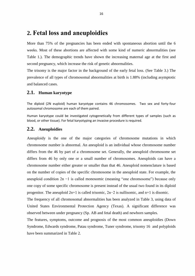
2. Fetal loss and aneuploidies
More than 75% of the pregnancies has been ended with spontaneous abortion until the 6
weeks. Most of these abortions are affected with some kind of numeric abnormalities (see
Table 1.). The demographic trends have shown the increasing maternal age at the first and
second pregnancy, which increase the risk of genetic abnormalities.
The trisomy is the major factor in the background of the early fetal loss. (See Table 3.) The
prevalence of all types of chromosomal abnormalities at birth is 1.88% (including asymptotic
and balanced cases.
2.1. Human karyotype
The diploid (2N euploid) human karyotype contains 46 chromosomes. Two sex and forty-four
autosomal chromosome are each of them paired.
Human karyotype could be investigated cytogenetically from different types of samples (such as
blood, or other tissue). For fetal karyotyping an invasive procedure is required.
2.2. Aneuploidies
Aneuploidy is the one of the major categories of chromosome mutations in which
chromosome number is abnormal. An aneuploid is an individual whose chromosome number
differs from the 46 by part of a chromosome set. Generally, the aneuploid chromosome set
differs from 46 by only one or a small number of chromosomes. Aneuploids can have a
chromosome number either greater or smaller than that 46. Aneuploid nomenclature is based
on the number of copies of the specific chromosome in the aneuploid state. For example, the
aneuploid condition 2n −1 is called monosomic (meaning “one chromosome”) because only
one copy of some specific chromosome is present instead of the usual two found in its diploid
progenitor. The aneuploid 2n+1 is called trisomic, 2n−2 is nullisomic, and n+1 is disomic.
The frequency of all chromosomal abnormalities has been analyzed in Table 3, using data of
United States Environmental Protection Agency (Texas). A significant difference was
observed between under pregnancy (Sp. AB and fetal death) and newborn samples.
The features, symptoms, outcome and prognosis of the most common aneuploidies (Down
Syndrome, Edwards syndrome, Patau syndrome, Tuner syndrome, trisomy 16 and polyploids
have been summarized in Table 2.
17
2.2.1. The Down syndrome
In 1886, Dr. John Langdon Down was described firstly Down syndrome as the part of the
“Observations on an Ethnic Classification of Idiots” (38). Nowadays, the Down syndrome is
the most frequent numerical chromosomal abnormality. Down syndrome is also known as
trisomy 21, but the old terminology, mongoloid idiotism must not be used in the clinical
practice.
The trisomy of 21 chromosomes causes several but a wide range of developmental errors,
physical and mental handicaps, most of them could be observed already in the fetal life. No
effective cure of Down syndrome has been developed until now. Fetus affect with Down
syndrome showed promising results with fetal programing, but this is only some approaches
to decrease the symptoms. Nowadays, the prevention by early diagnosis and therapeutic
abortion could be the possible choice.
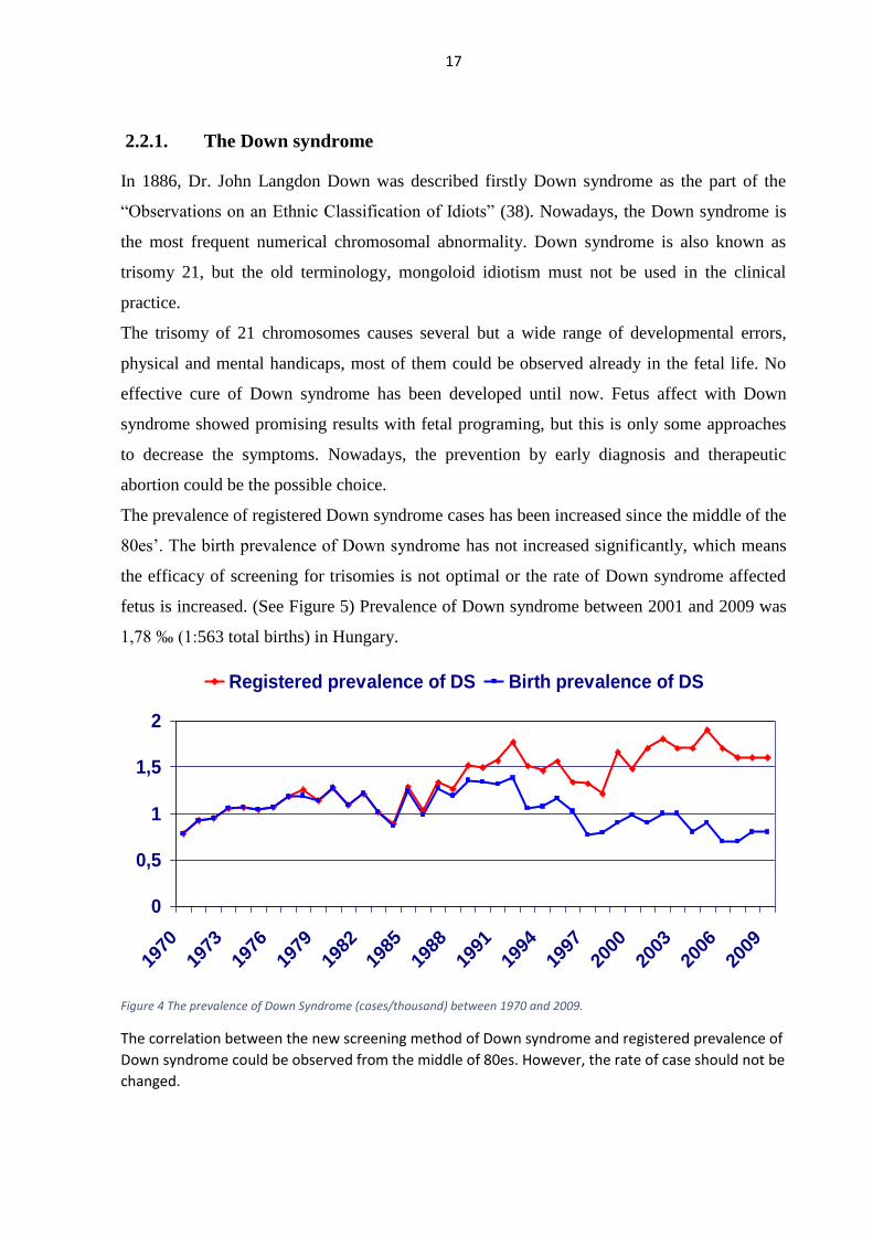
The prevalence of registered Down syndrome cases has been increased since the middle of the
80es’. The birth prevalence of Down syndrome has not increased significantly, which means
the efficacy of screening for trisomies is not optimal or the rate of Down syndrome affected
fetus is increased. (See Figure 5) Prevalence of Down syndrome between 2001 and 2009 was
1,78 ‰ (1:563 total births) in Hungary.
Figure 4 The prevalence of Down Syndrome (cases/thousand) between 1970 and 2009.
The correlation between the new screening method of Down syndrome and registered prevalence of
Down syndrome could be observed from the middle of 80es. However, the rate of case should not be
changed.
0
0,5
1
1,5
2
1970
1973
1976
1979
1982
1985
1988
1991
1994
1997
2000
2003
2006
2009
Registered prevalence of DS Birth prevalence of DS

18
2.2.2. Edwards syndrome
Trisomy 18 was independently described by Edwards (31) et al and Smith (32) et al in 1960.
Among liveborn children, trisomy 18 is the second most common autosomal trisomy after
trisomy 21. Trisomy 18 is characterized by severe psychomotor and growth retardation,
microcephaly, microphthalmia, malformed ears, micrognathia or retrognathia, microstomia,
distinctively clenched fingers, and other congenital malformations.
2.2.3. Patau syndrome
Trisomy 13 or Patau syndrome is a trisomy disorder. It is caused by the presence of a whole
extra copy (or occasionally partial extra copy) of chromosome 13. Patau syndrome, in 75%-
90% of cases, is the result of the presence of a whole extra (third) copy of chromosome 13,
hence its alternative name of trisomy 13 (or simple trisomy 13). Patau syndrome is caused by
a chromosomal translocation in 5%-10% of cases, and to mosaicism (whereby only some cells
have the extra copy of chromosome 13) in 5%. In occasional cases, it is only a part of
chromosome 13 that is extra (partial trisomy 13). Occurrence in live births is about 1 in 9,500
(in the absence of any prenatal detection program), and rises with increasing maternal age.
Trisomy 13 is associated with a high rate of spontaneous loss of pregnancy (64% loss rate
from the 2nd trimester onwards) and very poor chances of survival in neonates (median
survival is 10 days).
2.2.4. Aneuploidy of the Sex Chromosomes
However, sex chromosomes are rarely affected by aneuploidies, their screening and diagnosis
is still not solved. There are numerous syndrome known with additional X or Y chromosomes
(ei. XXX, XXY, XYY… etc.), or with the lack of one sex chromosome (X0).
Tuner-syndrome (XO) could be observed as the most common aneuploidy during fetal life.
However, the high rate of spontaneous abortion is decreasing its birth prevalence. Other sex
aneuploidy are not commonly affected by spontaneous abortion like Tuner but XXX or
Superwoman syndrome has shown a higher rate than the others, where significant difference
between lost fetuses and newborns could not be observed.

19
Table 1 Clinical feature of aneuplody, pregnancy outcome and prognosis
Clinical features of the aneuplody
Trisomy Clinical Features Outcome (without iatrogenic abortion)
21
Cardiac defects, duodenal atresia, mild-
to-moderate mental retardation,
thyroid problems, hearing loss,
increased risk of leukemia, seizures
Only 1:37 fetuses survive the intrauterine
life and 80% of children could reach 60 yr
of age or older
18
Cardiac defects, renal anomalies, severe
mental retardation, intrauterine growth
restriction, omphalocele, central
nervous system defects, breathing and
feeding difficulties, “rocker-bottom”
feet, micrognathia, low-set ears
Only 1 from 149 fetuses survives the
intrauterine life and more than 90% of
children die before or shortly after birth;
<10% reach 1 yr of age
13
Cardiac defects; renal anomalies; cleft
lip, palate, or both; holoprosencephaly;
microcephaly; omphalocele; severe
mental retardation; deafness; seizures
Only 1 from 86 fetuses survives the
intrauterine life and most children die in
the first days or weeks of life; <10% reach 1
yr of age.
16 Cardiac defects; renal anomalies;
multiplex CNS abnormalities
The most fetuses (99.9%) and remaining
children die in the first days.
X0
Smaller height, lack or less developed
primary and secondary genitals,
anovulation, infertility, recurring
miscarriage
Only 1:1800 fetuses survive the
intrauterine life and 90% of children could
reach 50 yr of age or older
XXX
Various phenotypes are known, rate of
infertility, gyn. cancer (cervix, breast,
uterus)
Only 1:5 fetuses survive the intrauterine
life and 80% of children could reach 65 yr
of age or older
XXY
Various male phenotypes are known,
rate of infertility, higher rate of breast
cc.
Only 1 from 2 fetuses survives the
intrauterine life and 80% of children could
reach 60 yr of age or older
Triploid and
tetraploid
Multiplex abnormalities and fetal
hydrops could be observed.
The most fetuses (99.9%) and remaining
children die in the first days in intensive
care.

20
Table 1 Summarize the prevalence of genetic syndromes in the intrauterine life and by birth.
Table. 2. Prevalence of aneuploidy
Affected chromosome Sp. Ab or fetal death* Newborn**
1 0.00% 0.00%
2 2.12% 0.00%
3 0.71% 0.00%
4 1.27% 0.00%
5 0.00% 0.00%
6–12 7.48% 0.00%
13 1.71% 0.02%
14 3.67% 0.00%
15 4.24% 0.00%
16 16.39% 0.00%
17 0.13% 0.00%
18 2.97% 0.02%
19–20 0.69% 0.00%
21 4.67% 0.13%
22 5.65% 0.00%
Subtotal (trisomy): 51.69% 0.17%
Trisomy or monosomy of sex chromosomes
XYY 0.05% 0.05%
XXY 0.05% 0.05%
XO 18.00% 0.01%
XXX 0.28% 0.05%
Subtotal (sex chr): 18.39% 0.17%
Translocations
Balanced 0.19% 0.19%
Unbalanced 3.00% 0.06%
Subtotal translocation 3.19% 0.25%
Polyploid
Triploid 17.00% 0.00%
Tetraploid 6.00% 0.06%
Other (such as mosacism) 3.73% 0.65%
Subtotal polyploids and other 26.73% 1.29%
Total: 100.00%*** 1.88%****
*Data based on 7500 spontaneous abortions with genetic analysis.
**Data based on 85.000 births with genetic analysis.
***Spontaneous abortion or fetal death had been associated with genetic syndromes in
cc. 50% of all abortion cases.
****Birth population had been karyotyped.
Source: Data calculated from United States Environmental Protection Agency, Texas
1998
21
3. Efficacy of prenatal screening and diagnostics for
trisomies
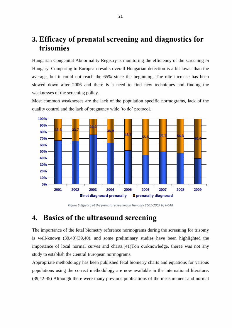
Hungarian Congenital Abnormality Registry is monitoring the efficiency of the screening in
Hungary. Comparing to European results overall Hungarian detection is a bit lower than the
average, but it could not reach the 65% since the beginning. The rate increase has been
slowed down after 2006 and there is a need to find new techniques and finding the
weaknesses of the screening policy.
Most common weaknesses are the lack of the population specific normograms, lack of the
quality control and the lack of pregnancy wide ’to do’ protocol.
Figure 5 Efficacy of the prenatal screening in Hungary 2001-2009 by HCAR
4. Basics of the ultrasound screening
The importance of the fetal biometry reference normograms during the screening for trisomy
is well-known (39,40)(39,40), and some preliminary studies have been highlighted the
importance of local normal curves and charts.(41)Ton ourknowledge, theree was not any
study to establish the Central European normograms.
Appropriate methodology has been published fetal biometry charts and equations for various
populations using the correct methodology are now available in the international literature.
(39,42-45) Although there were many previous publications of the measurement and normal
50,3 54,3
33,3 33,724,2
36,948,7
55,5 60,6
0%
10%
20%
30%
40%
50%
60%
70%
80%
90%
100%
2001 2002 2003 2004 2005 2006 2007 2008 2009
not diagnosed prenatally prenatally diagnosed
22
ranges of the human fetal biometric parameters, none of all had specific data on the Central
European Region.
The first trimester fetal biometric characteristics have been observed, analyzed and published
many times by the Fetal Medicine Foundation (FMF) London and its co-operators, but many
papers highlighted the racial growth chart differences. (41,46-50) Our study was established
based on these findings.
The new sonography era of the screening for Down Syndrome (DS) has been started by Szabó
and Gellén with their breakthrough publication about nuchal translucency thickness in 1990.
(51) Following their paper many studies have been proved its clinical importance of their
hypothesis and results. (52-56)
From the beginning of the 20th Century, facial markers introduced to the trisomy screening. In
the last decade, the importance of facial marker in the first(57-60) and second trimester (61-
68)was published several year ago and our good preliminary results also proved the high
efficacy of the fetal profile ratios in the second trimester.(69) These results were suggested
that to introduce them and to create normograms to observer these markers and ratios in the
first trimester, too.
The aim of the study was to establish the local fetal growth charts and normograms of fetal
biparietal diameter, femur and humeral length, nuchal translucency, prenasal thickness, nasal
bone length, ductus venosus PI, and PT-to-NBL and NBL-to-PT ratio from about 10weeks
and fetal heart rate and CRL from the 37 days of gestation to the midtrimester.
The secondary aim was to improve efficacy of screening for chromosomal abnormalities at
the first trimester ultrasound screening.
5. Material and Methods
5.1. Materials: This prospective observational study has been designed to measure, and describe the normal
biometric parameters. All included 4321 cases scans have been performed from January, 2008
to February, 2014 in the MEDISONO Fetal and Maternal Health Research Centre and the
Department of Medical Genetics, University of Szeged, Szeged, Hungary.
This study contains for (both low- and high) mixed-risk obstetric populations, and ethnically
over 99.8% of pregnancies were a Caucasian population of Hungary. The study protocol was
approved by the Regional Ethics Committee of the University of Szeged and all procedures
were in full accordance with the Helsinki Declarations.

23
5.2. Measurements
All measurements were performed by one experienced sonographer using transabdominal
ultrasound (GE Voluson E8 Expert, GE Healthcare Cipf, Austria). This sonographer was a
holder of The Fetal Medicine Foundation’s (FMF) Certificate of Competence for first-
trimester scanning. All measurements were repeated for 3 times and the best one were
selected.
Measurements of fetal biometry such as CRL were followed the INTERGROWTH-21st
measurement of fetal crown rump length and standardization of ultra-sonographers (2010).
CRL was measured in the mid-sagittal section, a neutral horizontal position, using the optimal
magnification with the correct calliper position. The intersection of the callipers were placed
on the outer borders of the skin over the head and rump.
NT and DVPI measurements were fully followed the FMF criteria. DVPI-to-NT and NT-to-
DVPI were established by the division NT and DVPI.
Measurements of BPD were obtained from a transverse axial plane of the fetal head showing
a central midline echo broken in the anterior third by the cavum septi pellucidi, if already
present. BPD was measured from the outer border of the skull.
The femur length (FL) was measured from the greater trochanter to the lateral condyle if it
was exiting and shown, as if on the two ossification border of the bone.
To measure the FHR, M-Mode was used in acquiring volume with automatic calculation.
Facial profile (NBL and PT) On the basis of technical descriptions of NBL (70,71) measurements and our experience, both
measurements could be obtained in the same image if the face of the transducer was
positioned parallel to the nasal bone. The insonation angle should be close to 45 degrees. The
following image settings were used: low gain, medium dynamic contrast, and maximum
magnification so that the fetal head occupied the entire screen. Images were adjusted to ensure
the correct midsagittal plane and sharp margins of the skin and the nasal bone. The
diencephalon, nasal bone, lips, maxilla, and mandible were used as reference points for the
correct measurements of NBL in the midsagittal plane (72,73). The following image settings
were used: low gain, medium dynamic contrast, and maximum magnification so that the fetal
head occupied the entire screen. Images were adjusted to ensure the correct midsagittal plane.
(58,73) Briefly, PT was measured as the shortest distance from the lower margin of the
frontal bone to the outer surface of the overlying skin. The margins of the nasal bone are the
24
proximal and the distal ends of the white ossification line. The NBL(70,71) and PT(73) were
measured using the same view. If it was possible NT, NBL and PT were measured on the
same image. PT-to-NBL and NBL-to-PT were established by the division of NBL and PT.
Additionally, between April 2008 and December 2013, 2549 women were included into
antoher study and followed-up in the first and second trimester to improve the second
trimester screening efficacy. First and second trimester measurements were combined in a
normogram and compared to second trimester Down syndrome cases.
5.3. Inclusion and exclusion criteria:
All healthy singleton pregnancies have been included were not have been confirmed any
abnormalities by the fetus and mother.
Exclusion criteria were: IUGR, any fetal (including fetal and neonatal mortality) or maternal
disease/ disorder, IVF or induced pregnancies, rejecting the participation of study.
Absent nasal bone was an exclusion criteria for the facial profile ratio group. PT and NBL
measurement were accepted if booth could be observed and measured on the same image.
The main exclusion criteria were when the difference between the three repeated
measurements were higher than 10% of the measured value. (Only the measurement of the
marker was excluded in this case, not the patient case.)
5.4. Statistical analysis and data collection
All measurement data and volumes have been sent to astraia software (astraia GMBH,
Münich, Germany) via DICOM. Data analysis performed by a single medical bioinformatics
specialist with Microsoft Office 2013 (Mircosoft, Redmond, Virginia) and SYSSTAT (SyStat
Software Inc., San Jose, CA, USA)
Woman was referred by LMP but all data were based on the CRL measurement in this study,
no correction have been made by other parameters. GA was calculated from CRL
measurement at 10th week.
Statistical analysis was performed using SigmaStat. Regression analysis was used to
determine the percentiles (mean and 5th, 95th percentile) and regression analysis was used to
estimate the relation between CRL and other parameters. Euploid and trisomy group were
compared with independent sample t-test (p ≥ 0.001).

25
No reproducibility analysis was taken, but previous studies have been confirmed a non-
significant difference. This study not concerning about newborn or child characteristics, and
no other maternal, neonatal or fetal biometrics were not observed.

26
6. Results
From the 4321 women, 3356 accepted the consent form (77.67%) and participated in our
study. Thirty seven women were excluded because of missed abortion at the first scan. One
Edward’s syndrome case and five Down syndrome cases were diagnosed during the
ultrasound measurements.
Furthermore 103 cases were excluded because of maternal (26), fetal (74) or neonatal (8)
disorders. The Edwards syndrome case had spontaneous abortion a few days after the CVS at
11th week. Down syndrome cases were cytogenetically proved by AC during the early
midtrimester.
Descriptive data analysis of the results
The mean maternal age in euploid and trisomy cases was 33.83 years (16.6–47.1 years) and
35.83 years (26.1–41.3 years), respectively. The mean gestational age was 77. 58 days (26.0–
120 days) for euploids. The population specific maternal age, CRL, FHR, FL, BPD, NT,
NBL and PT descriptive statistics of euploid fetuses (mean, standard deviation, maximum,
75%, median, 25% and minimum values) of the euploid fetuses were summarized in Table 3.
Table3. Mean Std Dev Std.
Error Max Median Min
GA by LMP (days) 77.579 14.033 0.249 120 73 26
NT (mm) 1.845 0.632 0.0323 6.9 1.8 0.6
FL (mm) 8.942 2.319 0.1 16.8 8.7 3.9
BPD (mm) 21.977 3.536 0.0786 40.9 21.5 10.08
NBL (mm) 2.381 0.415 0.0213 4.2 2.3 1
PT (mm) 1.412 0.369 0.019 3.2 1.4 0.1
FHR (bpm) 162.542 15.468 0.279 215 164 68
Ductus Venosus PI 1.099 0.165 0.00843 1.7 1.1 0.65
The following figure series represents the results of the study and the created normograms
with control cases.

27
Biparietal diameter (BPD)
All biparietal diameter measurements and distribution were summarized in Figure 6.
Normogram of Biparietal Diameter
Crown-rump length (mm)
20 40 60 80 100 120
Bip
ari
eta
l dia
me
ter
(mm
)
5
10
15
20
25
30
35
40
45
Mean
Eupolid
97% Confidence Band
3rd and 97th percentiles
Trisomy 21
Trisomy18
Where the black crosses were the measurements, black/gray line was the median, blue lines
were the confidence lower and upper lines, and red lines were the 3rd and 97th percentiles.
Red rectangles were the Down syndrome cases. The red dots were Edwards’s syndrome case.
Nuchal translucency (NT)
All nuchal translucency measurements and distribution were summarized in Figure 7.
Normogram of nucthal translucency thickness
Crown-rump length (mm)
20 40 60 80 100
Nucth
al T
ranslu
ce
ncy
Thic
kne
ss (
mm
)
0
2
4
6
8
Euploid
Mean
97% Confidence Band
3rd and 97th percentiles
Trisomy 18
Trisomy 21
Where the black crosses were the measurements, black/gray line was the median, blue lines
were the confidence lower and upper lines, and red lines were the 3rd and 97th percentiles.
Red rectangles were the Down syndrome cases. The red dots were Edwards’s syndrome case.

28
Embryonic or fetal heart rate (EHR /FHR)
All fetal heart rate measurements and distribution were summarized in Figure 8.
Normogram of Fetal Hearth Rate
CRL
0 20 40 60 80 100
Fe
tal H
ea
rth R
ate
0
50
100
150
200
Euploid
Mean
97% Confidence Band
3rd and 97th percentiles
Trisomy 21
Trisomy 18
Where the black crosses were the measurements, black/gray line was the median, blue lines
were the confidence lower and upper lines, and red lines were the 3rd and 97th percentiles.
Red dots were the Down syndrome cases. The red rectangles were Edwards’s syndrome case.
Femur length
All femoral length measurements and distribution were summarized in Figure 12.
Normogram of femoral length
Crown-rump length (mm)
40 50 60 70 80 90 100
Fe
mo
ral L
eng
th (
mm
)
0
5
10
15
20
Euploids
Mean
97% Confidence Band
3rd and 97th percentiles
Trisomy 21
Trisomy 18
Where the black crosses were the measurements, black/gray line was the median, blue lines
were the confidence lower and upper lines, and red lines were the 3rd and 97th percentiles.
Red rectangles were the Down syndrome cases. The red dots were Edwards’s syndrome case.

29
Prenasal Thickness (PT)
All prenasal thickness measurements and distribution were summarized in Figure 10.
Normogram of Prenasal Thickness
Crown-rump length (mm)
40 50 60 70 80 90 100
Pre
na
sa
l Thic
kne
ss (
mm
)
0,0
0,5
1,0
1,5
2,0
2,5
3,0
3,5
Euploid
Mean
97% Confidence Band
3rd and 97th percentiles
Trisomy 21
Trisomy 18
Where the black crosses were the measurements, black/gray line was the median, blue lines
were the confidence lower and upper lines, and red lines were the 3rd and 97th percentiles.
Red rectangles were the Down syndrome cases. The red dots were Edwards’s syndrome case.
Nasal bone length (NBL)
All nasal bone length measurements and distribution were summarized in Figure 11.
Normogram of Nasal Bone Length
Crown-rump length (mm)
40 50 60 70 80 90 100
Na
sa
l Bo
ne
Le
ng
th
0
1
2
3
4
5
6
Euploid
Mean
97% Confidence Band
3rd and 97th percentiles
Trisomy 21
Trisomy 18
Where the black crosses were the measurements, black/grey line was the median, blue lines
were the confidence lower and upper lines, and red lines were the 3rd and 97th percentiles.
Red rectangles were the Down syndrome cases. The red dots were Edwards’s syndrome case.

30
Nasal bone length-to-prenasal thickness ratio (NBL:PT)
All nasal bone length-to-prenasal thickness ratio distribution were summarized in Figure 12.
Normogram of Nasal Bone Length-to-Prenasal Thickness Ratio
Crown-rump length (mm)
40 60 80 100
Na
sa
l Bo
ne
Le
ng
th-t
o-P
rena
sa
l Thic
kne
ss R
atio
0
1
2
3
4
5
6
Euploid
Mean
97% Confidence Band
3rd and 97th percentiles
Trisomy 18
Trisomy 21
Where the black crosses were the measurements, black/grey line was the median, blue lines
were the confidence lower and upper lines, and red lines were the 3rd and 97th percentiles.
Red rectangles were the Down syndrome cases. The red dots were Edwards’s syndrome case.
Prenasal thickness-to-nasal bone length ratio (PT:NBL)
All prenasal thickness-to-nasal bone length ratio distribution were summarized in Figure 13.
Normogram of Prenasal Thickness-to-Nasal Bone Length Ratio
Crown-rump length (mm)
40 60 80 100
Pre
na
sa
l Thic
kne
ss-t
o-N
asa
l Bo
ne
Le
ng
th R
atio
0
1
2
3
4
5
6
7
Euploid
Mean
97% Confidence Band
3rd and 97th percentiles
Trisomy 18
Trisomy 21 (only 4 cases)
Where the black crosses were the measurements, black/grey line was the median, blue lines were the
confidence lower and upper lines, and red lines were the 3rd and 97th percentiles. Red rectangles were
the Down syndrome cases. The red dots were Edwards’s syndrome case. One case with absent nasal
bone was excluded because of the zero division.
31
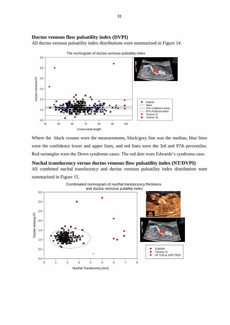
Ductus venosus flow pulsatility index (DVPI)
All ductus venosus pulsatility index distributions were summarized in Figure 14.
The normogram of ductus venosus pulsatility index
Crown-rump length
40 50 60 70 80 90 100
Ductu
s V
eno
sus P
I
0,5
1,0
1,5
2,0
2,5
3,0
3,5
Euploid
Mean
97% Confidence Band
97% Prediction Band
Trisomy 21
Trisomy 18
Where the black crosses were the measurements, black/grey line was the median, blue lines
were the confidence lower and upper lines, and red lines were the 3rd and 97th percentiles.
Red rectangles were the Down syndrome cases. The red dots were Edwards’s syndrome case.
Nuchal translucency versus ductus venosus flow pulsatility index (NT/DVPI)
All combined nuchal translucency and ductus venosus pulsatility index distribution were
summarized in Figure 15.
Combinated normogram of nucthal translucency thickness
and ductus venosus pulatility index
Nucthal Translucency (mm)
0 1 2 3 4 5 6 7 8
Ductu
s v
eno
sus P
I
0,0
0,5
1,0
1,5
2,0
2,5
3,0
3,5
Euploids
Trisomy 21
NT Tr18 vs DVPI TR18

32
Where the black crosses were the measurements, red lines were the 1st and 99th percentiles
limit circle. Red rectangles were the Down syndrome cases. The red dots were Edwards’s
syndrome case.
DVPI/NT-to-PT/NBL ratio
All combined ductus venosus pulsatility index-to-nuchal translucency ratio versus prenasal
thickness-to-nasal bone length ratio distribution were summarized in Figure 16.
Distribution of the DVPI-to-NT ratio versus PT-to-NBL ratio
DVPI/NT
0,0 0,2 0,4 0,6 0,8 1,0 1,2 1,4
PT/N
BL
0
1
2
3
4
5
6
7
Euploid
Mean
97% Confidence Band
3rd and 97th percentiles
Trisomy 21
Trisomy 18
Where the black crosses were the measurements, black/grey line was the median, blue lines
were the confidence lower and upper lines, and red lines were the 3rd and 97th percentiles.
Red rectangles were the Down syndrome cases. The red dots were Edwards’s syndrome case.

33
Statistical Performance of the markers
Table 4 summarized the statistical performance of the marker of Down syndrome.
Table 4.
Statistical performance of the markers DR Spec LR+ LR-
Fetal Heart Rate (FHR) 50.00 % 99.02 % 68.07 0.34
Femur Length (FL) 40.00 % 99.07 % 43.04 0.61
Biparietal Diameter (BPD) 40.00 % 98.79 % 33.10 0.61
Nasal Bone Length (NBL) 66.67 % 99.17 % 80.67 0.34
Prenasal Thickness (PT) 50.00 % 99.36 % 78.50 0.50
NBL/PT ratio 33.33 % 98.28 % 19.42 0.68
*PT/NBL ratio 80.00 % 98.93 % 74.56 0.20
Nucthal Translucency (NT) 83.33 % 99.63 % 225.83 0.17
Ductus Venosus PI 66.67 % 98.89 % 60.22 0.34
DVPI+NT 100 % 98.51 % 67.25 <0.00
DVPI+NT+PT/NBL ratio (3D diagram) 100 % 99.17 % 120.25 <0.00
NT/DVPI-to-PT/NBL ratios 100 % 99.20 % 125.67 <0.00
DVPI/NT-to-PT/NBL ratios 100 % 99.47 % 188.50 <0.00
DR: detection rate or sentivity; Spec: Specificity (FPR=1(00%)-Spec.); LR+:
Positive likelihood ratio; LR-: Negative likelihood ratio
Significant differences were observed between euploid and trisomy group from the aspect of
nuchal translucency, fetal heart rate, nasal bone length, prenasal thickness, ductus venosus PI,
prenasal thickness-to-nasal bone length and ductus venous-to-nuchal translucency to prenasal
thickness-to-nasal bone length ratios. (p > 0.001)
IMPORTANT NOTES:
three times repeated measurements of NBL caused almost no measurements marked
on 40-55mm of CRL region because difference of the measurements where higher
than study limit (10% difference which was higher than the callipers internal space).
Where NBL was zero, there was a zero division in the results by PT/NBL ratio. These
cases were excluded.
Second trimester results on combined first and second trimester normograms
Forty-one (1.6%) of 2549 were affected by trisomy 21. Maternal age ranged from 16 to 47
years (median 29.5) and the gestational age from 14 to 27 weeks (median 19.57 weeks). The
nasal bone length ranged from absence (0.00 mm) to 12 mm and volume capture duration
ranged from 1 to 49 minutes (median 7 minutes).

34
Where the black crosses were the euploid measurements, black/grey line was the median, blue
lines were the confidence lower and upper lines, and red lines were the 3rd and 97th
percentiles in Figure 17

35
Where the black crosses were the trisomy 21 measurements, black/grey line was the median,
blue lines were the confidence lower and upper lines, and red lines were the 3rd and 97th
percentiles in Figure 18.
Down syndrome cases were plotted on normogram (Figure 2). Cut-off value was setup to the
5th percentiles line. Forty-one cases of trisomy 21 were identified (cytogenetically) and all of
them were detected between 14th and 28thweeks. In 33 cases the measured NBL values were
lower than the 5th percentile and 8 cases of trisomy 21 fetuses were higher than 5th percentile,
respectively. These results showed 80.49% sensitivity with 98.17% specificity. (See in Table
3.). Positive and negative likelihood ratio for trisomy 21 fetuses were 43.98 and 0.2,
respectively.There was significant difference between the nasal bone length of euploid and
trisomy 21 fetuses (P =< 0.001).
7. Discussion
7.1. The explanation of the results
This study represented the high sensitivity ultrasound screening methods and reference charts
of the fetal biometric parameters of the Caucasian population.
NT was the first and the most sensitive screening marker of Down syndrome. This study
proved its strength in first trimester screening.
DVPI was found one of the most sensitive marker of trisomies during the first trimester. It has
high sensitivity and a medium-high specificity on trisomies. This finding was also confirmed
by our study.
The most sensitive marker was the combined DVPI and NT plot, but the best result was
reached when NT and DVPI were combined with the facial markers.
Our preliminary results were proved the efficacy of PT, NBL and their ratios in the second
trimester. Slightly, these markers were fitted to the first trimester scan. They could be
measured easily on the same image with NT. The common measurement, possibility could
decrease the necessary time of observation and extremely increase efficacy of screening. In
contrast with previous result PT-to-NBL is overwhelming in the first trimester.
The FL, BPD and HL should act as an important screening marker of the early IUGR and not
for the trisomies. These markers may help to identify the bi- and unilateral cranial and limb
anomalies during the first trimester.

36
The clinical aspects of these findings were the introduction of the facial profile ratio to the
first trimester screening, using DVPI-to-NT and PT-to-NBL ratios in the first trimester as new
markers of trisomies.
The ductus venosus-to-nuchal translucency to prenasal thickness-to-nasal bone length ratio
reached an impressive 100% detection rate of 0.6% false positive rate. The risk of
chromosomal defects is very high and the first line of management of such pregnancies
should be the offer of NIPT or chorionic villus sampling (CVS) for fetal karyotyping.
The latest screening strategy for first and second trimester was introduced by Nicolaides et al.
in a congress. (Advances in Fetal Medicine Dec 2013 London). Their opinion was to focus on
the neck on first and focus on the face (facial profile) in the second trimester. These was a
summary of the long development and research of the screening for trisomy, booth direction
were introduced and published from several groups in last two and the half decades.
(51,53,60,65,69,70,74-83)
CRL, BPD, FHR and FL were measured from the beginning of the obstetric ultrasound era.
These markers were easily measured with the low-resolution devices, but provided much
useful information about fetal development to the examiner. Our study had been set the
normograms of these markers and tried to use them in the screening for trisomy. Excluding
FHR, these markers have efficacy in the developmental and well-being scans, and they should
not be used for trisomy screening. FHR proved a really high sensitivity and a fair specificity
to detect fetal defects in the early pregnancy. Our previous observation also proved its
importance during the early first trimester scans to find the pregnancy outcome or the early
and late fetal loss from the 6 weeks.(84)
NT were the real first marker of autosomal trisomies (51) but several study proved its
usefulness in different conditions and diseases (See Appendix Table 1).(75) Current paper
used NT after the first trimester and proved a really good efficacy on trisomy.
Nasal bone length was proved high screening efficacy during the pregnancy. However, in the
first trimester its repeatability is very low and there is no linear increase till 60mm of CRL,
but the production lines were padded to the border of the plot and it was useful to screen out
cases in the early pregnancy.
Prenasal thickness (PT) was published a several years later by Maymon et. al. PT(65) was
improved the second trimester screening efficacy for trimsomies. Current study as a
preliminary studies(85) before used PT as a first trimester marker –successfully. Szabó et

37
al.(69) published facial profile based ratios and its inverse counterpart and proposed that they
could be utilized as well in the first trimester. This study confirms the usefulness of these
markers in the first trimester. However, during the second trimester NBL-to-PT was better
than the PT-to-NBL, although PT-to-NBL was better in the first trimester. The problem with
this ratio is the zero division so if there is no nasal bone, it is unable to use for risk estimation.
7.2. The limitations of the study
The limitations were the local population and strict inclusion policy of the healthy euploid
cases and the number of the overall cases and patient number of trisomy group.
Another limitation of the study is the nasal bone development; margins of the nasal should be
identified.
7.3. Combined normograms
Table 5. Fetal nasal bone length normogram and trisomy 21 : review of the literature
Authors Population Method Case Tr21 DR FPR LR+
Bromley et al. 2002(3) Mixed 2D 239 16 69 5 11
Cicero et al. 2003 (4) Mixed 2D 1016 34 60 1 50.50x
132x*
Bunduki et al. 2003 (5) Mixed 2D 1631 22 59.1 5.1 11.6x
Chen et al. 2004 (9) Chinese 2D 198 NI N/A N/A N/A
Benoit et al. 2005 (6) Mixed 3D 38 20 75 8 N/A
Sutthibenjakul et al. 2009 (12) Thai &
mixed 2D 295 18 77.7 0.7 N/A
Geipel et al. 2010 (17) mixed 3D 870 37 65 5.8 14x
Szabó et al. 2014 (16) Caucasian 2D 1330 33 75.8 1.88 41,32
This study in 2014 Caucasian 2D 2590 41 80.5 1.83 43.98x
Population : population of the study; Method : 2D or 3D ultrasound were performed.; cases:
number of euploid cases; Tr21 : number of trisomy 21 cases; DR : detection rate; FPR : false
positive rate; LR+ : likelihood ratio;
*by Caucasians
*N/A not applicable or not available; NI : not included
38
Our data demonstrate that hypoplastic nasal bone between 14 and 28th gestational weeks was
found in 1.83% of euploid and 80.49% of trisomy 21 fetuses, respectively. These findings are
showed a better screening performance than to previous studies with 2D ultrasound. (8, 16)
The nasal bone length was used as an isolated marker in our study. However, other studies
used nasal bone length in combination bi-parietal diameter (BPD), femur length (FL) and
moreover they used multiple of medians (MoMs) values instead of simple measurement value
in millimetres. Our analysis showed that using MoMs in a statistical evaluation is misleading.
Since the millimetre measurements day-by-day are more reliable and much easier to use in
practice than a more complex summarized and corrigated values.
Fifty-one (1.83%) out of 2549 euploid fetuses fell under 5th percentile While 8 (19.51%)
trisomy 21 fetuses had nasal bone length higher than 5th percentile. The nasal bone was
present but hypoplastic in 33 (80.49%) out of 41 trisomy 21 cases.
7.4. Practical and clinical aspects
To use these normograms some basic recommendation is still necessary.
Repeated measurements are strongly recommended to exclude the medical, technical,
measurement and visual errors. If the visualization is not enough good, then the examination,
it should be repeated in another time/day/.
Ultrasound scans and biochemical screening are still required during the trimesters. The
screening for trisomy shoud be more ultrasonographic oriented and extended with NBL, PT.
These results lead up to 90% sensitivity, if biochemical screening is preformed than it could
reach 95% of sensitivity. Higher-risk patients should be sent to the genetics department for a
counselling of the diagnostics ways of the invasive procedures which will be required for the
diagnosis and performed only after genetic counselling. This study could improved the
efficacy of screening.
If the risk was low, but fetal growth is lower the normograms than IUGR is possible, but the
measurements and dating must be rechecked to exclude the possible errors. In several cases,
using the LMP to set GA leads to the wrong dating of pregnancy, so use CRL results to GA or
changing markers when there is no automatized data transfer. In patients with a family history
or maternal (and/or parental) anxiety of genetic syndromes that can be diagnosed by DNA
analysis, the NIPT by some syndromes or the CVS sample can also be tested for these
syndromes.

39
Place of the noninvasive prenatal tests in screening for trisomy
First of all, noninvasive prenatal tests (NIPT) are more widely offered in the wealthy and
developed countries and its efficacy in screening for trisomies.
On the other hand, NIPT overall screening (not diagnostic) performance is 99,5% of 1% of
FPR which is impressive for a bit less than 1000EUR. However, ultrasound scans are still
necessary to observe the fetal biometry for screening of other fetal structural and
developmental disorder for a less than 70EUR. So it is more than dangerous, if the extended
(anomaly) scans have been spent for a NIPT only.
It must not be forget that NIPT always remains a screening test, when invasive procedure
offers
Figure 17 Show the developed screening and consultation methodology of trisomy.
This methodology provides the optimal prenatal care also in low-income countries where the
full spectrum of biochemical screening and NIPT is not widely available for average citizens.

40
Conclusions
Local normograms and the most sensitive ultrasound screening model for trisomy 21, and 18
were introduced. Using these ratios could be comparable with NIPT. These measurements and
methods should be incorporated into first trimester screening for trisomy. Further
investigation will be necessary to observe these findings on different population and also in
the second and third trimester.

41
8. Clinical conclusions of the study
The increasing prevalence of DS can be primarily attributed to the increasing ratio of
advanced age of mothers and changes of the entertainment especially the diet
Prenatal screening could show a significant improvement year by year if our
normogram and protocol could be introduced.
The prevalence of Down syndrome and the efficiency of prenatal screening was
slightly lower than the values observed in other European countries, but new
references and protocol could lead to a better result such as it happened in Denmark.
The increasing geographical inequalities in screening effectiveness demonstrated the
existence of non-exploited opportunities in certain (non-properly managed) areas of
Hungary by HCAR but 2D ultrasound is commonly used in the obstetrics and these
markers could be easily measured in 5-15 minutes.
Some clinical and practical training should be offered to involve the physicians to the
screening.
The combination of different ratio might be working in the second trimester screening,
too.
It is possible to improve screening efficacy if first and second trimester measurement
of NBL is combined on a normogram.
Combining different ratios could be beneficially by other markers, too.

42
9. New findings and newly developed methods in the
thesis
We first described the definition of “prevention in fetal medicine”
We first described the national normograms of the fetal biometry parameters of the
Hungarian population.
We first described a new practical-based, easy-to-use and cost-effective two-
dimensional measurement techniques of the NBL and PT in the first trimester.
We first described a high risk pregnancy management protocol for low income
countries
We first described that the NIPT how to be placed into the screening system in
Hungary.
We first described that the combined the first and second trimester normogram to
enhance the efficacy of the second trimester screening of nasal bone length.
We first described in the international literature who introduced NBL and PT with
their ratios in the first trimester.
We first described that the combination of different ratios could increase the screening
efficiency in the first trimester.
We first described that highest sensitivity and specificity could be reached for the
bedside in the first trimester without any biochemical or DNA test.
We first described that development of a statistical method to a practical method of the
ultrasound screening, which has comparable screening performance to NIPT but much
cheaper and could be widely used also in the low income countries.

43
10. New observations during my fellowship
1) In the major trisomy markers of euploid and trisomy fetuses a significant difference was
observed.
2) We elaborated the method how the fetal nasal bone length (NBL) and prenasal thickness
(PT) can be obtained and measured in a single volume acquisition (image) during the
first trimester anatomy scan.
3) Validated normograms have been created for the Hungarian population for the first and
second trimester.
4) We first demonstrated the combination of nasal bone length (NBL) and prenasal
thickness (PT) as a ratio could be used in the first trimester as a screening marker of
aneuploidies.
5) These data have been supported previous observations which highlighted the importance
of nuchal translucency and ductus venosus flow pulsatility index as the most effective
screening co-maker of autosomal trisomy.
6) We first demonstrated the combination of nuchal translucency and ductus venosus flow
pulsatility index as a ratio versus the combination of nasal bone length (NBL) and
prenasal thickness (PT) as a ratio, could be used in the first trimester as a more sensitive
ultrasonography screening marker of aneuploidies.
7) We first described in the international literature that the combination of first and second
trimester NBL measurement on a mixed normogram could increase the efficacy of
screening in the second trimester.
8) We first described in the international literature that the PT: NBL and NBL: PT ratio in
the first trimester.
9) We first published in the international literature that the ultrasound measurements of
these new markers can successfully be incorporated into the first and second trimester
fetal anatomy scan.
10) We first described in the Hungarian literature that how to create and validate obstetrical
normograms.

44
11. Acknowledgement
This to acknowledge with thanks to my supervisor and mentor, Professor Dr. János
Szabó, MD, DSc, allocating me this fantastic research topic and supporting me to
realize it. I am grateful for his devoted supervisor activity and for helping me to
perform the practical and theoretical part of my study.
I would like to thank Professor Dr. habil. Márta Széll, PhD, DSc, head of the
Department of Medical Genetics for her kind support in the last period of my
fellowship.
Special thanks to Dr. Emese Horváth MD, PhD for her practical recommendations.
Special thanks to my PhD-fellows, Andrea Szabó, MD, PhD and Melinda Vanya MD
for their dedicated work in preparation of the manuscripts.
Special thanks to Roland Denk Cs, MSc, CEO and team of extra software GMBH, for
their technical support and, for the invitation and sponsorship of my congress
participations.
Special thanks to my all co-authors, who helped and support my researches, especially
to Szabó Andrea MD, PhD, Vanya Melinda MD, Prof. Bártfai György MD, PhD, DSc,
Dr. János Tamás Szabó MD, Dr. János Sikovanyecz Md, PhD, Emese Horváth MD,
PhD, Dóra Isaszegi MSc, Dr. Lipták-Váradi Julianna MSc, PhD, Prof Széll Márta
MSc PhD DSc, Dr. Lilla Kató MD.,Dr. Edit Ferencz MD, Roland Denk Cs, MSc,
Prof. Dr. Hajnalka Orvos MD, PhD DSc, Prof. Dr- Attila Pál MD, PhD DSc, and of
course to Prof. Dr. habil. János Szabó MD PhD DSc.
Thanks for the members of the Department of Medical Genetics and MEDISONO
Fetal and Maternal Health Research Centre.
Thanks for the proofreading and spell checking for my friends and colleagues,
especially Julianna Lipták-Váradi and Cindita Belencita Keto and to Ginger
Proofreading Software.
I would like to thank Dr. habil. Edit Paulik, MD. PhD, head of the Department of
Public Health for her kind support.
Last but not at least, I am extremely grateful to my parents (Judit Jákó and Károly
Szili) and my sweetheart (Csilla Dézsi) and all of my family who supported me and
guaranteed me the steady and quiet background.

45
References
(1) Mosby's Medical Dictionary. 8th edition ed. USA: Elsevier; 2009.
(2) Wayne Jonas editor. Mosby's Dictionary of Complementary and Alternative Medicine. 1st edtion ed. USA: Mosby; 2005.
(3) Sameeh M. Abutarbush, editor. Saunders Comprehensive Veterinary Dictionary. 3 ed. ed.: Elsevier; 2007.
(4) Farlex editor. Farlex Partner Medical Dictionary. online: Farlex; 2012.
(5) Douglas Harper editor. Online Etymology Dictionary. 1st ed.: online; 2010.
(6) March of Dimes Birth Defects Foundation editor. Global Report On Birth Defects. 06th ed. USA: March of Dimes Birth Defects Foundation; 2006.
(7) Corby DG. Aspirin in pregnancy: maternal and fetal effects. Pediatrics 1978 Nov;62(5 Pt 2 Suppl):930-937.
(8) WALLENBURG H, MAKOVITZ J, DEKKER G, ROTMANS P. Low-Dose Aspirin Prevents Pregnancy-Induced Hypertension and Preeclampsia in Angiotensin-Sensitive Primigravidae. Lancet 1986 JAN 4;1(8471):1-3.
(9) Bujold E, Roberge S, Lacasse Y, Bureau M, Audibert F, Marcoux S, et al. Prevention of Preeclampsia and Intrauterine Growth Restriction With Aspirin Started in Early Pregnancy A Meta-Analysis. Obstet Gynecol 2010 AUG;116(2):402-414.
(10) Bolden JR. Acute and Chronic Tocolysis. Clin Obstet Gynecol 2014 SEP 2014;57(3):568-578.
(11) Berzan E, Doyle R, Brown CM. Treatment of Preeclampsia: Current Approach and Future Perspectives. Curr Hypertens Rep 2014 SEP 2014;16(9):473.
(12) Ueda A, Kondoh E, Kawasaki K, Mogami H, Konishi I. Long-Term Use of Magnesium Sulphate Improves Pregnancy Outcomes in Women with Preeclampsia. Reproductive Sciences 2014 MAR 2014;21(3):401A-401A.
(13) Xiao C, Gangal M, Abenhaim HA. Effect of magnesium sulfate and nifedipine on the risk of developing pulmonary edema in preterm births. J Perinat Med 2014 SEP 2014;42(5):585-589.
(14) Vest AR, Cho LS. Hypertension in Pregnancy. Curr Atheroscler Rep 2014 MAR 2014;16(3):395.
(15) Rumbold A, Middleton P, Pan N, Crowther CA. Vitamin supplementation for preventing miscarriage. Cochrane Database of Systematic Reviews 2011 2011(1):CD004073.
(16) Kovacs CS, Kronenberg HM. Maternal-fetal calcium and bone metabolism during pregnancy, puerperium, and lactation. Endocr Rev 1997 DEC 1997;18(6):832-872.

46
(17) Hollis BW, Wagner CL. Assessment of dietary vitamin D requirements during pregnancy and lactation. Am J Clin Nutr 2004 MAY 2004;79(5):717-726.
(18) Bodnar LM, Catov JM, Simhan HN, Holick MF, Powers RW, Roberts JM. Maternal vitamin D deficiency increases the risk of preeclampsia. Journal of Clinical Endocrinology & Metabolism 2007 SEP 2007;92(9):3517-3522.
(19) Gale CR, Robinson SM, Harvey NC, Javaid MK, Jiang B, Martyn CN, et al. Maternal vitamin D status during pregnancy and child outcomes. Eur J Clin Nutr 2008 JAN 2008;62(1):68-77.
(20) Erkkola M, Kaila M, Nwaru BI, Kronberg-Kippila C, Ahonen S, Nevalainen J, et al. Maternal vitamin D intake during pregnancy is inversely associated with asthma and allergic rhinitis in 5-year-old children. Clinical and Experimental Allergy 2009 JUN 2009;39(6):875-882.
(21) Brown AS. The environment and susceptibility to schizophrenia. Prog Neurobiol 2011 JAN 2011;93(1):23-58.
(22) Hollis BW, Johnson D, Hulsey TC, Ebeling M, Wagner CL. Vitamin D Supplementation During Pregnancy: Double-Blind, Randomized Clinical Trial of Safety and Effectiveness. Journal of Bone and Mineral Research 2011 OCT 2011;26(10):2341-2357.
(23) Hollis BW. Circulating 25-hydroxyvitamin D levels indicative of vitamin D sufficiency: Implications for establishing a new effective dietary intake recommendation for vitamin D. J Nutr 2005 FEB 2005;135(2):317-322.
(24) Wald N. Prevention of Neural-Tube Defects - Results of the Medical-Research-Council Vitamin Study. Lancet 1991 JUL 20 1991;338(8760):131-137.
(25) Krol E, Krejpcio Z, Chmurzynska A. Folic Acid and Protein Content in Maternal Diet and Postnatal High-Fat Feeding Affect the Tissue Levels of Iron, Zinc, and Copper in the Rat. Biol Trace Elem Res 2011 DEC 2011;144(1-3):885-893.
(26) Dror DK, Allen LH. Interventions with Vitamins B6, B12 and C in Pregnancy. Paediatr Perinat Epidemiol 2012 JUL 2012;26:55-74.
(27) Zhong Rong-zhen, Zhou Dao-wei. Oxidative Stress and Role of Natural Plant Derived Antioxidants in Animal Reproduction. Journal of Integrative Agriculture 2013 2013;12(10):1826-1838.
(28) Kiondo P, Wamuyu-Maina G, Wandabwa J, Bimenya GS, Tumwesigye NM, Okong P. The effects of vitamin C supplementation on pre-eclampsia in Mulago Hospital, Kampala, Uganda: a randomized placebo controlled clinical trial. Bmc Pregnancy and Childbirth 2014 AUG 21 2014;14:283.
(29) Swaney P, Thorp J, Allen I. Vitamin C Supplementation in Pregnancy-Does It Decrease Rates of Preterm Birth? A Systematic Review. Am J Perinatol 2014 FEB 2014;31(2):91-97.
(30) Helland IB, Smith L, Saarem K, Saugstad OD, Drevon CA. Maternal supplementation with very-long-chain n-3 fatty acids during pregnancy and lactation augments children's IQ at 4 years of age. Pediatrics 2003 JAN 2003;111(1):e39.

47
(31) Szili K, Szabo A, Vanya M, Bartfai G, Szabo J. News methods for second trimester screening of Down syndrome: statistical analysis of the use of nasal bone length and prenasal thickness. Orv Hetil 2014 Nov 23;155(47):1876-1881.
(32) Smidt-Jensen S, Philip J, Lundsteen C, Permin M, Zachary JM, Fowler SE. Randomised comparison of amniocentesis and transabdominal and transcervical chorionic villus sampling. The Lancet 1992 11/21;340(8830):1237-1244.
(33) Sundberg K, Bang J, Smidt-Jensen S, Brocks V, Lundsteen C, Parner J, et al. Randomised study of risk of fetal loss related to early amniocentesis versus chorionic villus sampling. The Lancet 1997 9/6;350(9079):697-703.
(34) Eiben B, Hammans W, Goebel R. Chorionic villus sampling versus early amniocentesis. The Lancet 1997 10/25;350(9086):1253-1254.
(35) Sieroszewski P, Perenc M, Bas-Budecka E, Suzin J. Ultrasound diagnostic schema for the determination of increased risk for chromosomal fetal aneuploidies in the first half of pregnancy. J Appl Genet 2006;47(2):177-185.
(36) Nizard J. Amniocentesis: Technique and education. Curr Opin Obstet Gynecol 2010;22(2):152-154.
(37) Gekas J, Durand A, Bujold E, Valle M, Forest J-, Rousseau F, et al. Cost-effectiveness and accuracy of prenatal Down syndrome screening strategies: Should the combined test continue to be widely used? Am J Obstet Gynecol 2011;204(2):175.e1-175.e8.
(38) J. Langdon H. Down, M.D. Observations on an Ethnic Classification of Idiots. London Hospital Reports 1866;3(1):259-262.
(39) ALTMAN D, CHITTY L. Charts of Fetal Size .1. Methodology. Br J Obstet Gynaecol 1994 JAN;101(1):29-34.
(40) Royston P, Wright E. How to construct 'normal ranges' for fetal variables. Ultrasound Obstet Gynecol 1998 JAN;11(1):30-38.
(41) Jacquemyn Y, Sys S, Verdonk P. Fetal biometry in different ethnic groups. Early Hum Dev 2000 JAN;57(1):1-13.
(42) CHITTY L, ALTMAN D, HENDERSON T, CAMPBELL S. Charts of Fetal Size .2. Head Measurements. Br J Obstet Gynaecol 1994 JAN;101(1):35-43.
(43) CHITTY L, ALTMAN D, HENDERSON A, CAMPBELL S. Charts of Fetal Size .3. Abdominal Measurements. Br J Obstet Gynaecol 1994 FEB;101(2):125-131.
(44) CHITTY L, ALTMAN D, HENDERSON A, CAMPBELL S. Charts of Fetal Size .4. Femur Length. Br J Obstet Gynaecol 1994 FEB;101(2):132-135.
48
(45) Papageorghiou AT, Ohuma EO, Altman DG, Todros T, Ismail LC, Lambert A, et al. International standards for fetal growth based on serial ultrasound measurements: the Fetal Growth Longitudinal Study of the INTERGROWTH-21st Project. The Lancet 2014 9/6–12;384(9946):869-879.
(46) Prefumo F, Sairam S, Bhide A, Penna L, Hollis B, Thilaganathan B. Maternal ethnic origin and fetal nasal bones at 11-14 weeks of gestation. BJOG 2004 Feb;111(2):109-112.
(47) Spencer K, Heath V, El-Sheikhah A, Ong CY, Nicolaides KH. Ethnicity and the need for correction of biochemical and ultrasound markers of chromosomal anomalies in the first trimester: a study of Oriental, Asian and Afro-Caribbean populations. Prenat Diagn 2005 May;25(5):365-369.
(48) Collado F, Bombard A, Li V, Julliard K, Aptekar L, Weiner Z. Ethnic variation of fetal nasal bone length between 11?14 weeks' gestation. Prenat Diagn 2005;25(8):690-692.
(49) Cossi PS, Bussamra LC, Araujo Junior E, Nardozza LM, Moron AF. Ethnic variation and variability of fetal nasal bone length at 11-15 weeks of gestation in a Brazilian population: preliminary results. Arch Gynecol Obstet 2008 Nov;278(5):431-435.
(50) Parikh LI, Nolan J,3rd, Tefera E, Driggers R. Fetal biometry: does patient ethnicity matter? J Matern Fetal Neonatal Med 2014 Mar;27(5):500-504.
(51) Szabó J, Gellén J. Nuchal Fluid Accumulation in Trisomy-21 Detected by Vaginosonography in 1st Trimester. Lancet 1990 NOV 3;336(8723):1133-1133.
(52) Nicolaides S K, Azar G, AZAR GS, Gosden C. Fetal nuchal edema - Associated malformations and chromosomal defects. BMJ 1992;7(2):123-131}.
(53) Nicolaides KH, Azar G, Byrne D, Mansur C, Marks K. Fetal nuchal translucency: ultrasound screening for chromosomal defects in first trimester of pregnancy. BMJ 1992 Apr 4;304(6831):867-869.
(54) SZABO J, GELLEN J, SZEMERE G. 1ST-TRIMESTER ULTRASOUND SCREENING FOR FETAL ANEUPLOIDIES IN WOMEN OVER 35 AND UNDER 35 YEARS OF AGE. ULTRASOUND IN OBSTETRICS \& GYNECOLOGY} 1995} MAR};5}(3}):161-163}.
(55) Nicolaides K, Sebire N, Snijders R. Down's syndrome screening with nuchal translucency. LANCET} 1997} FEB 8};349}(9049}):438}.
(56) Antsaklis A, Daskalakis G, Theodora M, Hiridis P, Komita O, Blanas K, et al. Assessment of nuchal translucency thickness and the fetal anatomy in the first trimester of pregnancy by two- and three-dimensional ultrasonography: a pilot study. J Perinat Med 2011 Mar;39(2):185-193.
(57) Cicero S, Curcio P, Papageorghiou A, Sonek J, Nicolaides K. Absence of nasal bone in fetuses with trisomy 21 at 11-14 weeks of gestation: an observational study. Lancet 2001 Nov 17;358(9294):1665-1667.
(58) Cicero S, Longo D, Rembouskos G, Sacchini C, Nicolaides KH. Absent nasal bone at 11-14 weeks of gestation and chromosomal defects. Ultrasound Obstet Gynecol 2003 Jul;22(1):31-35.

49
(59) Cicero S, Rembouskos G, Vandecruys H, Hogg M, Nicolaides KH. Likelihood ratio for trisomy 21 in fetuses with absent nasal bone at the 11-14-week scan. Ultrasound Obstet Gynecol 2004 Mar;23(3):218-223.
(60) Cicero S, Curcio P, Rembouskos G, Sonek J, Nicolaides KH. Maxillary length at 11-14 weeks of gestation in fetuses with trisomy 21. Ultrasound Obstet Gynecol 2004 Jul;24(1):19-22.
(61) Bromley B, Lieberman E, Shipp T, Benacerraf B. Fetal nose bone length - A marker for Down syndrome in the second trimester. J Ultrasound Med 2002 DEC;21(12):1387-1394.
(62) Benn P, Kaminsky L, Ying J, Borgida A, Egan J. Combined second-trimester biochemical and ultrasound screening for Down syndrome. Obstet Gynecol 2002 DEC;100(6):1168-1176.
(63) Bunduki V, Ruano R, Miguelez J, Yoshizaki CT, Kahhale S, Zugaib M. Fetal nasal bone length: reference range and clinical application in ultrasound screening for trisomy 21. Ultrasound Obstet Gynecol 2003 Feb;21(2):156-160.
(64) Viora E, Errante G, Sciarrone A, Bastonero S, Masturzo B, Martiny G, et al. Fetal nasal bone and trisomy 21 in the second trimester. Prenat Diagn 2005;25(6):511-515.
(65) Maymon R, Ushakov F, Waisman D, Cuckle H, Tovbin Y, Herman A. A model for second-trimester Down syndrome sonographic screening based on facial landmarks and digit length measurement. Ultrasound Obstet Gynecol 2006 Mar;27(3):290-295.
(66) Odibo A, Sehdev HM, Stamilio D, Cahill A, Dunn L, Macones G. Defining nasal bone (NB) hypoplasia in second-trimester down syndrome screening: Does the use of multiples of the median (MOM) improve screening efficiency? Obstet Gynecol 2006 DEC;195(6):S168-S168.
(67) Maymon R, Moskovitch M, Levinsohn-Tavor O, Weinraub Z, Herman A, Cuckle H. Bedside estimation of Down syndrome risk from second-trimester ultrasound prenasal thickness. Ultrasound Obstet Gynecol 2009 Dec;34(6):629-633.
(68) Mogra R, Schluter PJ, Ogle RF, O'Connell J, Fortus L, Hyett JA. A prospective cross-sectional study to define racial variation in fetal nasal bone length through ultrasound assessment at 18-20 weeks' gestation. Aust N Z J Obstet Gynaecol 2010 Dec;50(6):528-533.
(69) Szabó A, Szili K, Szabó JT, Sikovanyecz J, Isaszegi D, Horváth E, et al. Nasal bone length:prenasal thickness ratio: a strong 2D ultrasound marker for Down syndrome. Prenat Diagn 2014;In Press:n/a-n/a.
(70) Sonek J, Nicolaides K. Prenatal ultrasonographic diagnosis of nasal bone abnormalities in three fetuses with Down syndrome. Obstet Gynecol 2002 JAN;186(1):139-141.
(71) Sonek JD, Cicero S, Neiger R, Nicolaides KH. Nasal bone assessment in prenatal screening for trisomy 21. Am J Obstet Gynecol 2006 Nov;195(5):1219-1230.
(72) Cicero S, Sonek J, McKenna D, Croom C, Johnson L, Nicolaides K. Nasal bone hypoplasia in trisomy 21 at 15-22 weeks' gestation. Ultrasound Obstet Gynecol 2003 JAN;21(1):15-18.

50
(73) Persico N, Borenstein M, Molina F, Azumendi G, Nicolaides KH. Prenasal thickness in trisomy-21 fetuses at 16-24 weeks of gestation. Ultrasound Obstet Gynecol 2008 Nov;32(6):751-754.
(74) Cicero S, Curcio P, Papageorghiou A, Sonek J, Nicolaides K. Absence of nasal bone in fetuses with trisomy 21 at 11-14 weeks of gestation: an observational study. Lancet 2001 NOV 17;358(9294):1665-1667.
(75) Filkins K, Koos BJ. Ultrasound and fetal diagnosis. Curr Opin Obstet Gynecol 2005 Apr;17(2):185-195.
(76) Monni G, Zoppi MA, Ibba RM, Floris M, Manca F, Axiana C. Nuchal translucency and nasal bone for trisomy 21 screening: single center experience. Croat Med J 2005 Oct;46(5):786-791.
(77) Maymon R, Levinsohn-Tavor O, Cuckle H, Tovbin Y, Dreazen E, Wiener Y, et al. Second trimester ultrasound prenasal thickness combined with nasal bone length: a new method of Down syndrome screening. Prenat Diagn 2005 OCT;25(10):906-911.
(78) Miguelez J, Moskovitch M, Cuckle H, Zugaib M, Bunduki V, Maymon R. Model-Predicted Performance of Second-Trimester Down Syndrome Screening With Sonographic Prenasal Thickness. Journal of Ultrasound in Medicine 2010 DEC;29(12):1741-1747.
(79) Benacerraf BR. The history of the second-trimester sonographic markers for detecting fetal Down syndrome, and their current role in obstetric practice. Prenat Diagn 2010;30(7):644-652.
(80) Sehdev H, McNamara J, Cahill A, Odibo L, Macones G, Odibo A. Observed:expected ratios: assessment of nasal bone length for second-trimester Down syndrome screening in a user-friendly fashion. Obstet Gynecol 2011 JAN;204:S143-S143.
(81) De Jong-Pleij EAP, Vos FI, Ribbert LSM, Pistorius LR, Tromp E, Bilardo CM. Prenasal thickness-to-nasal bone length ratio: a strong and simple second- and third-trimester marker for trisomy 21. Ultrasound in Obstetrics & Gynecology 2012 FEB;39(2):185-190.
(82) Sonek J, Molina F, Hiett AK, Glover M, McKenna D, Nicolaides KH. Prefrontal space ratio: comparison between trisomy 21 and euploid fetuses in the second trimester. Ultrasound in Obstetrics & Gynecology 2012 SEP;40(3):293-296.
(83) Gonzalez R, Aedo S, Dezerega V, Sepulveda W. Frontonasal Fold Thickness-to-Nasal Bone Length Ratio as a Prenatal Sonographic Marker for Trisomy 21 in a Low-Risk Population. Journal of Ultrasound in Medicine 2013 MAY;32(5):795-800.
(84) Szili K, Ferencz E, Szabó A, Szabó J, Sikovanyecz J, Szabó J. P16.17: Early embryonic heart rate and pregnancy outcome. Ultrasound in Obstetrics & Gynecology 2012;40(S1):234-235.
(85) Miron JP, Cuckle H, Miron P. Prenasal thickness in first-trimester screening for Down syndrome. Prenat Diagn 2012 Jul;32(7):695-697.

51
Appendix
Appendix table 1. Increased NT thickness associated fetal disorders
Venous congestion localized to the head and neck
Cardiac defects / dysfunction
Fetal anemia
Fetal hypoproteinemia /nephrosis
Fetal infection (ei. Parvo B13)
Altered composition of extracellular matrix
Failure of lymphatic drainage
Appendix table 2. First trimester major and minor markers of Down Syndrome
Nuchal thickening
Hyperechoic bowel
Absent nasal bone
Short humerus
Short femur
EIF
Pyelectasis
Mouth/cleft disorder

52
Publications of the thesis

53
I

54
II

55
III

56
IV

57
V

58
VI

59
VII

60
VIII

61
IX