Embed Size (px)
Citation preview

1 Dr. Hisham Ah. Nematalla
Pharmacotherapy of Thrombo-embolic disorders
Intended Learning Objectives:
1. Define thromboembolic disorders
2. Identify risk factors, signs and symptoms of Venous
thromboembolism.
3. Describe the processes of thrombosis & fibrinolysis, including the
role of the vascular endothelium, platelets, coagulation cascade,
and thrombolytic proteins.
4. Determine a patient’s relative risk (low, moderate, high, or very
high) of developing venous thrombosis.
5. Potential advantages of the low-molecular-weight heparins and
fondaparinux over unfractionated heparin.
6. Role of new oral anticoagulants (NOACs) in VTE.
7. Formulate an appropriate prevention strategy for a patient at risk
for deep vein thrombosis.
8. Formulate an appropriate treatment plan for a patient who
develops a deep vein thrombosis or pulmonary embolism,
hrombo-embolic disorders is the combination of thrombus and its
main complication embolism. Thrombus is the formation of a
blood clot inside a blood vessel, obstructing the flow of blood
through the circulatory system. A clot that breaks free and begin to
travel around the body is known as an embolus. Thrombo-embolic
disorders are classified into venous thrombo-embolism (VTE) and arterial
thrombo-embolism (ATE).
T

2 Dr. Hisham Ah. Nematalla
Venous thrombo-embolism:
VTE is manifested as deep vein thrombosis (DVT) and pulmonary
embolism (PE) resulting from thrombus formation in the venous
circulation
It is often provoked by prolonged immobility and vascular injury and
is most frequently seen in patients who have been hospitalized for a
serious medical illness, trauma, or major surgery. VTE can also
occur with little or no provocation in patients who have an underlying
hypercoagulable disorder.
While VTE may initially cause few or no symptoms, the first overt
manifestation of the disease may be sudden death.
Death from PE can occur within minutes, before effective treatment can
be given.
Long-term complications of VTE include Post-Thrombotic
Syndrome (PTS) (after DVT) and Chronic Thrombo-embolic
Pulmonary Hypertension (CTPH) (after PE) and recurrent
thromboembolic events.
The treatment of VTE is fraught with substantial risks since
antithrombotic drugs require precise dosing, close monitoring as well
as ongoing patient education.
Arterial thrombo-embolism:
Arterial thrombosis is the formation of a thrombus within an artery. In
most cases, arterial thrombosis follows rupture of an atheroma, and is
therefore referred to atherothrombosis.
Arterial thrombosis can embolize and is the major cause of arterial
embolism, potentially causing infarction of almost any organ in the
body as brain (stroke), heart (myocardial infarction), limbs (peripheral
arterial occlusion), eye (central retinal artery occlusion).

3 Dr. Hisham Ah. Nematalla
Virchow’striad
The main causes of thrombosis are given by Virchow’s triad which lists
hypercoagulability, endothelial cell injury and disturbed blood flow.
Pathophysiology:

4 Dr. Hisham Ah. Nematalla
Thrombogenesis
With vascular injury, a dynamic interplay between thrombogenic
(activating) and antithrombotic (inhibiting) forces result in the local
formation of a hemostatic plug that seals the vessel wall and prevents
further blood loss.
A disruption of this delicate system of checks and balances may lead to
inappropriate clot formation within the blood vessel that can obstruct
blood flow or embolize to a distant vascular bed.
The clotting cascade is a stepwise series of enzymatic reactions that
result in the formation of a fibrin mesh. Clotting factors circulate in the
blood in inactive forms. Once a precursor is activated by specific
stimuli, it activates the next precursor in the sequence. The final steps
in the cascade are the conversion of prothrombin to thrombin and
fibrinogen to fibrin.
Thrombin plays a key role in the coagulation cascade; it is responsible
not only for the production of fibrin, but also for the conversion of
factors V and VIII, creating a positive feedback loop that greatly
accelerates the entire cascade. Thrombin also enhances platelet
aggregation.
A number of tempering mechanisms control coagulation. The intact
endothelium adjacent to the damaged tissue actively secretes several
antithrombotic substances including heparan sulfate,
thrombomodulin, protein C, and protein S.
Activated protein C inhibits factor Va and VIIIa activity. Antithrombin
and heparin cofactor II (HCII) are circulating proteins that inhibit
thrombin and factor Xa. Heparan sulfate exponentially accelerates
antithrombin and HCII activity. Tissue factor pathway inhibitor (TFPI)
inhibits the extrinsic coagulation pathway.
When these self-regulatory mechanisms are intact, the formation of the
fibrin clot is limited to the zone of tissue injury. However, disruptions
in the system often result in inappropriate clot formation.

5 Dr. Hisham Ah. Nematalla
Fibrinolysis:
The fibrinolytic protein plasmin degrades the fibrin mesh into soluble
end products collectively known as fibrin split products or fibrin
degradation products.
The fibrinolytic system is also under the control of a series of
stimulatory and inhibitory substances. Tissue plasminogen activator
(t-PA) and urokinase plasminogen activator (u-PA) convert
plasminogen to plasmin.
Plasminogen activator inhibitor-1 (PAI-1) inhibits the plasminogen
activators and α2-antiplasmin inhibits plasmin activity.
Aberrations in the fibrinolytic system have also been linked to
hypercoagulability.
1. Venous Thromboembolism:
Signs & Symptoms of VTE
The symptoms of DVT or PE are non-specific and it is extremely
difficult to differentiate VTE from other disorders.
Common symptoms of DVT:
Unilateral calf or thigh pain, leg swelling, or redness.
In up to half of the cases, this may not result in local symptoms or signs,
and the onset of PE may be the first evidence of presence of VTE.
PTS is long term complication of DVT caused by damage to the venous
valves, producing chronic lower extremity swelling, pain, tenderness,
skin discoloration and ulceration.

6 Dr. Hisham Ah. Nematalla
Common symptoms of PE:
In the majority of cases, PE is suspected due to dyspnea and pleuritic
chest pain either alone or in combination.
Patients with massive PE may experience syncope associated with
findings of hemodynamic collapse (severe hypotension & right
ventricular failure followed by death)
Acute sub-massive PE, involving only segmental or sub-segmental
pulmonary arteries may have minimal or no symptoms.

7 Dr. Hisham Ah. Nematalla
Diagnosis:
Given that VTE can be debilitating or fatal, it is important to treat it quickly
and aggressively. On the other hand, because major bleeding induced by
antithrombotic drugs can be equally harmful, it is important to avoid
treatment when the diagnosis is not reasonably certain.
1. Pretest Probability of PE & DVT (Modified Geneva & Wells score)
The clinical examination for DVT is often unreliable, therefore, clinical
decision rules (pretest probability scores) based on patient’s age, symptoms
and risk factors have been developed to stratify patient into low, moderate or
high clinical probability. This approach helps to improve the effectiveness of
diagnosing DVT and to limit the need for additional testing.
2. D-dimer: is a degradation product of a cross-linked fibrin blood clot that
is typically elevated in patients with acute VTE, but also by a variety of
non-thrombotic disorders including recent major surgery, hemorrhage,
trauma, pregnancy, or cancer.
3. Imaging tests
For DVT:
Duplex ultrasound (is the most commonly used test to diagnosis DVT.
It is a non-invasive test that can measure the rate and direction of blood
flow and visualize clot formation in proximal veins of the legs. It cannot
reliably detect small blood clots in distal veins. Coupled with a careful
clinical assessment, it can rule in or out (include or exclude) the
diagnosis in the majority of cases.
Venography is the gold standard for the diagnosis of DVT. However,
it is an invasive test that involves injection of radiopaque contrast dye
into a foot vein. It is expensive and can cause anaphylaxis and
nephrotoxicity.

8 Dr. Hisham Ah. Nematalla
For PE:
Ventilation-perfusion (V/Q) lung scans: measures the distribution of
blood and air flow in the lungs. When there is a large mismatch between
blood and air flow in one area of the lung, there is a high probability
that the patient has a PE.
Pulmonary angiography is the gold standard for the diagnosis of PE.
However, it is an invasive test that involves injection of radiopaque
contrast dye into the pulmonary artery. The test is expensive and
associated with a significant risk of mortality.
Computerized tomographic pulmonary angiography (CTPA)
currently are the most widely used and evaluated tests for the diagnosis.
Pharmacotherapy of VTE:
Drugs used in prophylaxis & treatment of VTE are classified into:
1. Parentral anticoagulants:
Unfractionated Heparin (UHF) (iv & sc)
UHF blocks coagulation by binding with antithrombin enhancing
many folds its catalytic inhibitory effect on factor IIa (thrombin)
& factor Xa.
UHF is a heterogeneous mixture of negatively charged sulfated
mucopolysaccharide with Mwt ranging from 3000Da to
30000Da.
UFH binds non-selectively to a number of plasma & cellular
protein reducing its anticoagulant effect.
UFH is cleared by zero-order enzymatic degradation (dose-
dependent) & first-order renal elimination (dose-independent).
When administrated in fixed dose, the anticoagulant response to
UFH shows inter-patient and intra-patient variability.
Protein binding and saturable elimination kinetics explain the
wide variation in anticoagulant effect of UFH.
When given sc, the bioavailability of UFH ranges from 30 to
70% depending on the dose given, delayed onset of action and
higher doses are required.
For the treatment of VTE, UFH is given by iv infusion following
an initial bolus dose.

9 Dr. Hisham Ah. Nematalla
Side effects include: bleeding, HIT (heparin-induced
thrombocytopenia), osteoporosis.
Low Molecular Weight Heparin (LMWH) (sc only)
Enoxaparin (Clexane®), Tinzaparin (Innohep®), Dalteparin
(Fragmin®).
The LMWHs are smaller heparin fragments obtained by
chemical or enzymatic depolymerization of UFH.
Due to their smaller chain length, LMWHs have relatively
greater activity against factor Xa and inhibit thrombin to a
lesser degree.
They exhibit less binding to plasma and cellular proteins,
resulting in a more predictable anticoagulant response.
Consequently, routine monitoring of anticoagulation activity
and dose adjustments are not required in the majority of
patients.
On the contrary, monitoring is important in obese (BMI > 50
kg/m2), weight < 50 kg, renal impairment CrCl < 30 ml/min
& pregnant women.

10 Dr. Hisham Ah. Nematalla
LMWHs have longer plasma half-lives, allowing once- or
twice-daily administration, improved subcutaneous
bioavailability (> 90%), and dose-independent renal
clearance.
In addition, they are also associated with a lower incidence of
bleeding, HIT and osteopenia.
Advantages of LMWHs over UFH:
Greater bioavailability
Predictability and dose-dependent plasma level
Lower risk of bleeding
Lower risk of HIT
Lower risk of heparin induced osteoporosis
No need for blood monitoring
Can be safely administrated in outpatients, cancer &
pregnancy.
Longer duration of anticoagulant effect enabling once or
twice daily administration
Fondaparinux (Arixtra®) (sc only).
Fondaparinux, the first agent in this class, is an indirect
inhibitor of factor Xa, and exerts its anticoagulant activity by
accelerating AT & has no effect on thrombin (factor IIa).
predictable and linear dose-response relationship, rapid onset
of activity, and long half-life.
Factor Xa inhibitors do not require routine coagulation
monitoring or dose adjustments.
Fondaparinux has a half-life of 17 to 21 hours, permitting
once-daily administration, but the anticoagulant effects of
fondaparinux will persist for 2 to 4 days after stopping the
drug.
Fondaparinux is as safe and effective
Fondaparinux is as safe and effective and can be used in
patients with a history of HIT who require anticoagulation
therapy.
Effective as IV UFH for the treatment of PE and SC LMWH
for DVT treatment.
Is contraindicated in patients with severe renal impairment
(CrCl less than 30 mL/minute).

11 Dr. Hisham Ah. Nematalla
As with other anticoagulants, the major side effect associated
with fondaparinux is bleeding. Fondaparinux should be used
with caution in elderly patients because their risk of bleeding
is higher.
2. Oral anticoagulants:
Vitamin K Antagonist (VKA):
Warfarin is FDA-approved for the prevention and treatment
of VTE, as well as the prevention of thromboembolic
complications in patients with myocardial infarction, atrial
fibrillation, and heart valve replacement.
While very effective, warfarin has a narrow therapeutic index,
requiring frequent dose adjustments and careful patient
monitoring.
Warfarin exerts its anticoagulant effect by inhibiting the
production of the vitamin K–dependent coagulation factors II
(prothrombin), VII, IX, and X, as well as the anticoagulant
proteins C and S and its full antithrombotic activity is delayed
for 7 to 15 days.

12 Dr. Hisham Ah. Nematalla
It is extensively bound to albumin (98-99%) & metabolized
in the liver via several isoenzymes including cytochrome P-
450 (CYP)1A2, 2C9, 2C19, 2C18, and 3A4.
Hepatic metabolism of warfarin varies greatly among
patients, leading to very large inter-patient differences in dose
requirements and genetic variations in these isoenzymes
result in a significantly lower warfarin dose requirement to
achieve a therapeutic response.
Warfarin does not follow linear kinetics. Small dose
adjustments can lead to large changes in anticoagulant
response.
The dose of warfarin is determined by each patient’s
individual response to therapy and the desired intensity of
anticoagulation.
In addition to hepatic metabolism, warfarin dose
requirements are influenced by diet, drug-drug interactions,
and health status.
Therefore, the dose of warfarin must be determined by
frequent clinical and laboratory monitoring.
The initial dose of warfarin must be overlapped with UFH or
LMWHs
Patients who are younger (less than 55 years of age) and
otherwise healthy can safely use higher warfarin initiation
doses (e.g., 7.5 or 10 mg).
A more conservative initiation dose (e.g., 5 mg or less) should
be given to elderly.
The typical maintenance dose of warfarin for most patients
will be between 25 and 55 mg per week, although some
patients require higher or lower doses.
Adjustments in the maintenance warfarin dose should be
determined on the total weekly dose and by reducing or
increasing the weekly dose by 5% to 25%.
When adjusting the maintenance dose, wait at least 7 days to
ensure that a steady state has been attained on the new dose
before checking the INR again.
Side effect include mainly GI bleeding, intra-cranial bleeding.

13 Dr. Hisham Ah. Nematalla
New/Novel/Non-vitamin K Oral Anticoagulants (NOACs)
o Direct Thrombin inhibitor (DTI) (Factor IIa inhibitor):
Dabigatran (Pradaxa®)
o Factor Xa Inhibitors: Rivaroxaban (Xarelto®), Apixaban
(Eliquis®) Edoxaban (Savaysa® or Lixiana®).
NOACs:
Pharmacodynamics:
NOACs inhibit only a single target either factor Xa in case of
rivaroxaban, apixaban & edoxaban or thrombin (factor IIa) in case of
dabigatran.
They have a rapid onset of action (peak plasma within 1-4 hours) and
half-lives of about 12 hours (once or twice daily administration), they
have also a rapid offset of action.
NOACs produce a more predictable anticoagulant response, therefore
given in fixed doses without routine monitoring.
Pharmacokinetics:
NOACs are excreted in at least in part via the kidneys. The extent of
renal clearance varies depending on the agent: 80% of dabigatran is
excreted unchanged by the kidney and 50%, 33% and 27% of edoxaban,
rivaroxaban and apixaban respectively.
Because of their renal clearance, NOACs should be used with caution
in patients with a CrCl < 30 ml/min and shouldn’t be used in patients
with CrCl < 15 ml/min.
All NOACs require some degree of hepatic metabolism, consequently
they should be avoided in patient with liver dysfunction as evidence by
increased PT/INR and reduced serum albumin at baseline.
Potent inducers of CYP3A4, as phenytoin & carbamazepine may
reduce plasma level of apixaban & rivaroxaban but will not affect the
level of dabigatran & edoxaban.
There are no dietary restrictions, except that rivaroxaban should be
administrated with meal to maximize its absorption.
Adverse effects & Limitations
There are no specific antidotes for the NOACs, but these are under
development.

14 Dr. Hisham Ah. Nematalla
Dabigatran may be not the best choice in patients with a history of
coronary artery disease because of its higher risk of MI compared with
warfarin.
Dyspepsia have reported in patients treated with dabigatran.
Except for apixaban, the rate of gastrointestinal bleeding with the
NOACs is higher than that of warfarin, particularly in the elderly.
Pharmacotherapy of VTE:
A. Prevention of VTE:
Given that VTE is often clinically silent and potentially fatal (high
morbidity & mortality), prevention strategies have the greatest potential
to improve patient outcomes.
To rely on the early diagnosis and treatment of VTE is unacceptable
because many patients will die before treatment can be initiated.
The goal of an effective VTE prophylaxis program is to identify all
patient at risk, determine each patient’s level of risk and implement
regimens that provide sufficient protection for the level of each risk.

15 Dr. Hisham Ah. Nematalla
Risk Classification and Consensus Guidelines for VTE Prevention

16 Dr. Hisham Ah. Nematalla
Low-dose UFH (5000 units every 8 or 12 hours) sc has been shown
to reduce the risk of VTE by 55% to 70% both in patients with
moderate to high level of risks.
The LMWHs and fondaparinux provide a higher degree of protection
against VTE in most high-risk & in very-high-risk populations.
Warfarin appears to be as effective as the LMWHs for the prevention
of symptomatic VTE events in the highest-risk populations. When used
to prevent VTE, the dose of warfarin must be adjusted to maintain an
International Normalized Ratio (INR) between 2 and 3.
Oral administration and low drug cost give warfarin some advantages
over the LMWHs and fondaparinux.
However, warfarin does not achieve its full antithrombotic effect for
several days, requires frequent monitoring and periodic dosage
adjustments, and carries a substantial risk of major bleeding.
Warfarin should only be used when a systematic patient monitoring
system is available. If TTR is below 65%, discontinue warfarin &
switch to LMWHs or fondaparinux.
Time in Therapeutic Range of INR of 2.0 to 3.0:
1. Percent of Visits in Range (Traditional Method)
This looks at how many visits had INR results in range, and divides by the total
number of visits. If the patient has had 8 visits, and 6 had readings within their
therapeutic range, then the patient is considered in range 75% of the time.
2. Percent of Days in Range (Rosendaal Method)
This is the most complex of the calculations, as it looks at the amount of time
between visits to determine how long the patient might have been within their
therapeutic range. If a patient has a therapeutic range of 2.0 - 3.0, and on May 1st
tested at 2.5, then tested 3.5 on May 31st, then we can determine how many days
were in range. Since there were 30 days between tests, you assume that the patient
slowly moved from 2.5 to 3.5 over those 30 days, so around May 15th, the patient
was probably over 3.0, and therefore was out of range. Therefore, we estimate that
15 days were in range, and 15 days were out of range (within the 30-day time
period), which means the patient is within range 50% of the time.
Patient who are well controlled on warfarin (TTR of 65% or more) should not
consider any priority for switching.

17 Dr. Hisham Ah. Nematalla
NOACs in orthopedic thromboprophylaxis:
Rivaroxaban (10 mg once daily), Dabigatran (220 mg once daily) &
Apixaban (2.5 mg twice daily) are currently licensed in orthopedic
thromboprophylaxis (Total Hip Arthroplasty THA & Total Knee
Arthroplasty TKA) in USA & EU.
Compared with enoxaparin, the risk of symptomatic VTE was lower
with rivaroxaban but similar with dabigatran and apixaban.
Compared with enoxaparin, clinically significant bleeding was higher
with rivaroxaban but similar with dabigatran and lower with
apixaban.
Case scenario
KK is a 69-year-old obese female who fell and fractured her right hip. She is hospitalized and will undergo
surgery to repair her fractured right hip.
PMH: Hypertension × 12 years; dyslipidemia × 10 years; obesity ×20 years; degenerative joint disease × 5
years; recurrent urinary tract infections. Smoke one-half pack per day for 25 years; occasional alcohol use.
The patient has Medicare, but due to her fixed income, has difficulty paying for medications, leading to
occasional periods of non-compliance.
Current Meds: Metoprolol 100 mg by mouth twice daily, Hydrochlorothiazide 25 mg by mouth daily,
Vytorin 10/40 (ezetimibe 10 mg/simvastatin 40 mg) by mouth daily, Salsalate 750 mg by mouth twice daily
(NSAID: salicylates), Trimethoprim-sulfamethoxazole SS tablets by mouth twice daily for 7 days (last
treatment was 1 month ago), Shark cartilage 3 tablets by mouth daily, Enteric-coated aspirin 81 mg by mouth
daily, Ginseng 2 tablets by mouth daily
Allergies: No known drug allergies
PE: VS: blood pressure 145/90 mm Hg; heart rate, 72; respiratory rate,16; temperature, 37.4؛C (99.3°F);
weight 280 lb. (127.3 kg); body mass index (BMI) 40 kg/m2
Labs: Within normal limits; estimated glomerular filtration rate =74 mL/minute
Which risk factor(s) predispose KK to VTE?
What is KK’s estimated risk for developing VTE?
Given KK’s presentation and history, create an appropriate VTE prophylaxis plan including
the pharmacologic agent, dose, route and frequency of administration, duration of therapy,
monitoring parameters, and patient education.

18 Dr. Hisham Ah. Nematalla
B. Treatment of VTE:
Clinical therapeutic outcomes:
The goal of VTE treatment is to prevent short and long-term
complications of the disease.
In the short-term (few days to 6 months), the aim of therapy is to
prevent propagation or local extension of the clot, embolization and
death.
In the long-term (more than 6 months), the aim of therapy is to
prevent complications, such as PTS, CTPH and recurrent VTE.
A. Conventional regimen for treatment of VET:
1. Initial therapy (2 overlapped steps):
Starts with a rapidly-acting parenteral anticoagulant, as UFH or
LMWHs overlapped with VKA. (why?)
The parenteral anticoagulant is given for at least 5 days and is
stopped when the anticoagulant response with VKA is
therapeutic as evidenced by INR between 2 & 3.
Once UFH commences, check aPTT at 6-hour interval and adjust
to maintained at 1.5-2.5 times the institutional therapeutic range.
LMWHs do not require aPTT monitoring.
Platelets count must be checked daily while on UFH or LMWHs
Stop UHF or LMWHs after 5 days overlap with warfarin
(minimally), when INR stable between 2-3 for 2 days.
Achieving a therapeutic aPTT (1.5-2.5 times control value) in the
first 24 hours after initiating UFH is critical because this has been
shown to lower the risk of recurrent VTE.
Weight-based dosing regimen are more likely to exceed the
therapeutic threshold in the first 24 hours.
Another approach is 5000-unit bolus dose iv followed by iv
infusion at rate of 1000 to 1200 unit/hour.

19 Dr. Hisham Ah. Nematalla
Doses of UFH, LMWHs & fondaparinux in acute VTE:
2. Long-term therapy
Once therapeutic INR is established, VKA is then continued as
monotherapy for a minimum 3 months.
At this point, the decision to stop or continue treatment depends
on the balance between the risk of recurrence if warfarin is
stopped and the risk of bleeding if it is continued.
Patients with VTE in the setting of a transient and reversible risk
factors such as surgery, long air travel, have a low risk of
recurrence if anticoagulant therapy is stopped at 3 months
provided they are fully mobile (grade 1B, AT10, 2016).
3. Extended therapy:
In contrast those with ongoing risk factors, such as active cancer,
patients with unprovoked VTE, are often prescribed extended
anticoagulant therapy as long as the bleeding risk is not excessive
(6-12 months).
If anticoagulant therapy is stopped in patients with unprovoked
VTE, the risk of recurrence is at least 10% at 1 year and 30% at
5 years.
20 Dr. Hisham Ah. Nematalla
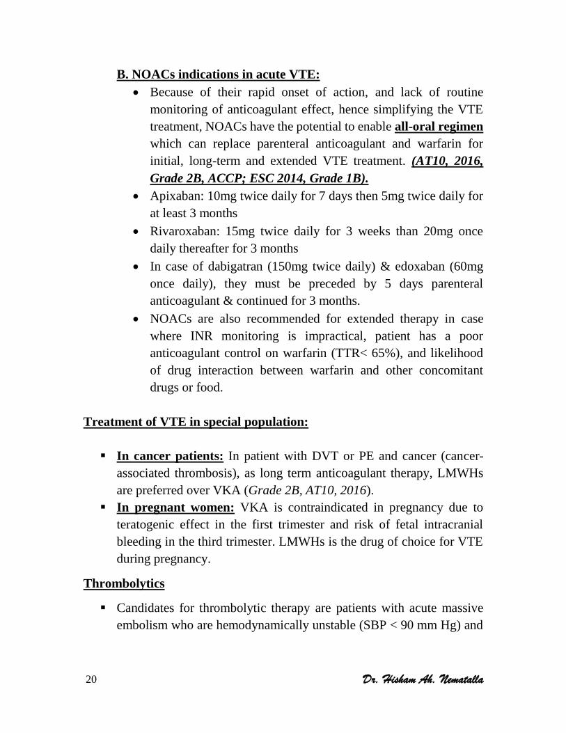
B. NOACs indications in acute VTE:
Because of their rapid onset of action, and lack of routine
monitoring of anticoagulant effect, hence simplifying the VTE
treatment, NOACs have the potential to enable all-oral regimen
which can replace parenteral anticoagulant and warfarin for
initial, long-term and extended VTE treatment. (AT10, 2016,
Grade 2B, ACCP; ESC 2014, Grade 1B).
Apixaban: 10mg twice daily for 7 days then 5mg twice daily for
at least 3 months
Rivaroxaban: 15mg twice daily for 3 weeks than 20mg once
daily thereafter for 3 months
In case of dabigatran (150mg twice daily) & edoxaban (60mg
once daily), they must be preceded by 5 days parenteral
anticoagulant & continued for 3 months.
NOACs are also recommended for extended therapy in case
where INR monitoring is impractical, patient has a poor
anticoagulant control on warfarin (TTR< 65%), and likelihood
of drug interaction between warfarin and other concomitant
drugs or food.
Treatment of VTE in special population:
In cancer patients: In patient with DVT or PE and cancer (cancer-
associated thrombosis), as long term anticoagulant therapy, LMWHs
are preferred over VKA (Grade 2B, AT10, 2016).
In pregnant women: VKA is contraindicated in pregnancy due to
teratogenic effect in the first trimester and risk of fetal intracranial
bleeding in the third trimester. LMWHs is the drug of choice for VTE
during pregnancy.
Thrombolytics
Candidates for thrombolytic therapy are patients with acute massive
embolism who are hemodynamically unstable (SBP < 90 mm Hg) and

21 Dr. Hisham Ah. Nematalla
at low risk of bleeding & in case of threatened limb loss (grade 2B,
AT10, 2016).
Thrombolytic therapy may be systemic or catheter-directed.