Embed Size (px)
Citation preview
REVIEW ARTICLE
Pharmacotherapy for Status Epilepticus
Eugen Trinka1,3,4• Julia Hofler1,3
• Markus Leitinger1,3• Francesco Brigo2
Published online: 27 August 2015
� The Author(s) 2015. This article is published with open access at Springerlink.com
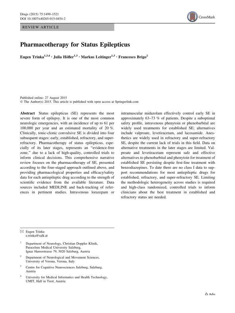
Abstract Status epilepticus (SE) represents the most
severe form of epilepsy. It is one of the most common
neurologic emergencies, with an incidence of up to 61 per
100,000 per year and an estimated mortality of 20 %.
Clinically, tonic-clonic convulsive SE is divided into four
subsequent stages: early, established, refractory, and super-
refractory. Pharmacotherapy of status epilepticus, espe-
cially of its later stages, represents an ‘‘evidence-free
zone,’’ due to a lack of high-quality, controlled trials to
inform clinical decisions. This comprehensive narrative
review focuses on the pharmacotherapy of SE, presented
according to the four-staged approach outlined above, and
providing pharmacological properties and efficacy/safety
data for each antiepileptic drug according to the strength of
scientific evidence from the available literature. Data
sources included MEDLINE and back-tracking of refer-
ences in pertinent studies. Intravenous lorazepam or
intramuscular midazolam effectively control early SE in
approximately 63–73 % of patients. Despite a suboptimal
safety profile, intravenous phenytoin or phenobarbital are
widely used treatments for established SE; alternatives
include valproate, levetiracetam, and lacosamide. Anes-
thetics are widely used in refractory and super-refractory
SE, despite the current lack of trials in this field. Data on
alternative treatments in the later stages are limited. Val-
proate and levetiracetam represent safe and effective
alternatives to phenobarbital and phenytoin for treatment of
established SE persisting despite first-line treatment with
benzodiazepines. To date there are no class I data to sup-
port recommendations for most antiepileptic drugs for
established, refractory, and super-refractory SE. Limiting
the methodologic heterogeneity across studies is required
and high-class randomized, controlled trials to inform
clinicians about the best treatment in established and
refractory status are needed.
& Eugen Trinka
1 Department of Neurology, Christian Doppler Klinik,
Paracelsus Medical University Salzburg,
Ignaz Harrerstrasse 79, 5020 Salzburg, Austria
2 Department of Neurological and Movement Sciences,
University of Verona, Verona, Italy
3 Centre for Cognitive Neurosciences Salzburg, Salzburg,
Austria
4 University for Medical Informatics and Health Technology,
UMIT, Hall in Tirol, Austria
Drugs (2015) 75:1499–1521
DOI 10.1007/s40265-015-0454-2

Key Points
Initial treatment of early status epilepticus (SE) with
intravenous lorazepam or intramuscular midazolam
is able to control seizures in 63–73 %; buccal
midazolam may be an alternative whenever
intravenous or intramuscular application of other
benzodiazepines is not possible.
In established SE, intravenous antiepileptic drugs
(phenytoin/fosphenytoin, valproate, levetiracetam,
phenobarbital) are most commonly used, but there is
no class I evidence for choosing one over the other;
valproate and levetiracetam represent safe and
effective alternatives to phenobarbital and
phenytoin; lacosamide is another potential
alternative to phenytoin and phenobarbital, but
current evidence is too sparse to give
recommendations.
Refractory and super-refractory SE is treated with
anesthetics (propofol, midazolam, thiopental/
pentobarbital) with lower success rates and a high
morbidity and mortality. Potential drugs to be
considered in super-refractory SE are ketamine,
magnesium, and immunomodulatory treatments, as
well other cause-directed and non-medical
treatments.
Other drugs which might be useful in the treatment
of SE, such as clonazepam, paraldehyde,
chlormethiazole (clomethiazole), or lidocaine, have a
long history, but there is no higher-class evidence to
support their use other than as second or third
alternatives in refractory cases.
1 Introduction
Status epilepticus (SE) can be regarded as the most severe
and extreme form of an epileptic seizure. Tonic-clonic SE
(i.e., convulsive SE, CSE) can be defined as ongoing con-
vulsive seizure activity or repeated convulsive seizures,
without regaining consciousness between seizures, for more
than 5 min [1]. Non-convulsive SE (NCSE) can be defined as
an ‘‘enduring epileptic condition with reduced or altered
consciousness, behavioral and vegetative abnormalities, or
merely subjective symptoms like auras, but without major
convulsive movements for more than 30 min’’ [2, 3]. A Task
Force of the International League Against Epilepsy (ILAE)
recently defined SE as ‘‘a condition resulting either from the
failure of the mechanisms responsible for seizure termina-
tion or from the initiation of mechanisms, which lead to
abnormally prolonged seizures (after time point t1)… which
can have long-term consequences (after time point t2),
including neuronal death, neuronal injury, and alteration of
neuronal networks, depending on the type and duration of
seizures’’. [4]. The time limits for t1 were set at 5 min for
generalized convulsive SE, and 10 min for focal SE with
impaired consciousness (formerly complex-partial SE). In
the new classification NCSE is divided into those patients
with and without coma following two broad clinical cate-
gories: while the former are ‘‘ictally comatose’’, often seen as
a progression of CSE, the ‘‘walking wounded’’ with aura
continua, absence status, or focal SE with impaired con-
sciousness have a less severe prognosis and do usually not
need the full armamentarium of emergency treatment as
described below.
SE is most prevalent in the population with structural
brain damage. In patients with epilepsy, SE can be pre-
cipitated by drug withdrawal, intercurrent illness, or
metabolic disturbance. The mortality of SE is around 20 %,
but may be as high as 40 % in the elderly with acute
symptomatic SE [5–9] and many co-morbidities [10]. The
annual incidence has been estimated to be approximately
18–28 cases per 100,000 per year, but may be as high as 61
per 100,000 per year, depending on the population studied
[11–16]. The incidence is highest in the elderly and has a
second peak in the neonatal period [17–22].
Although the first descriptions go back to Babylonian
Times (Sakikku-Board, 718–614 BC) [23] and recognition
of absence status was evident in the 16th century [24],
detailed descriptions of the clinical picture and first
pathophysiology considerations occurred in the 19th and
20th centuries. In their seminal work, Clark and Prout
recognized three phases of CSE [25–27]:
(a) In patients with epilepsy, an early phase can be
characterized, where frequency and severity of
seizures increases in a crescendo pattern. Synonyms
are premonitory status, impending status, and heraldic
status. In patients without pre-existing epilepsy, the
phase with a crescendo-like increase in seizure
frequency and severity is missing, and SE starts
abruptly. Ongoing convulsive epileptic activity for
more than 5 min is now often called early SE.
(b) Established SE designates continuous seizure activity
with convulsions or intermittent seizures without
regaining consciousness between the seizures. For
more than 10 and up to 30 min, or failure of initial
treatment (usually benzodiazepines) of early SE.
(c) With increasing duration, a decrease in motor activity
(electromechanical dissociation) occurs while the
patient remains in a coma. This phase is called
1500 E. Trinka et al.

advanced SE or refractory SE, referring to the failed
treatment (usually with antiepileptic drugs, AEDs) of
early and established SE. Other terms are subtle SE or
stuporous SE.
(d) At the third London-Innsbruck Colloquium on Status
Epilepticus [28], the fourth stage of SE, called super-
refractory SE, was characterized. At this stage
seizures continue despite maximal treatment with
intravenous (IV) anesthetics for more than 24 h in an
intensive care unit. These patients have ictal EEG
discharges when anesthesia is lessened. This stage has
also been termed malignant SE [29] (Fig. 1).
It has to be acknowledged that there is no clear defini-
tion of the stages and one might merge into the other.
While Clark and Prout described the stages of SE in mostly
untreated patients, clinical practice now defines the first
stage with a time frame (5 min of convulsive and 10 min of
focal non-convulsive), and the later stages by treatment
response: it has now been commonly accepted to designate
established SE as ‘‘benzodiazepine-resistant SE,’’ while the
term refractory SE is used, when treatment with benzodi-
azepines and one or more IV AEDs have failed. This also
implies that the timeframes given above, which are used by
most clinicians, may vary considerably with treatment. By
nature this lack of clear definitions leads to a high degree of
variability in the current literature.
In 2007, at the First London-Innsbruck Colloquium on
Status Epilepticus, a workshop was held with the purpose
of outlining the options of optimal pharmacotherapy of the
various forms of SE. A consensus was reached and a
treatment protocol published, which followed the conven-
tional pattern of tonic-clonic SE established [30, see Flow
chart in ‘‘Appendix’’]. The European Federation of Neu-
rological Societies and other groups have also published
similar recommendations [31, 32]. Recent reviews [33, 34]
covered the history of pharmacotherapy of SE outlining the
enormous range of therapies that have been advocated
since the 19th century.
Data on the pharmacotherapy of SE are most often
observational, having a high degree of heterogeneity and
high-class randomized, controlled trials are only available
for the early stages of SE. Therefore we discuss the phar-
macotherapy of SE in a narrative, rather than in a sys-
tematic review. In this article we will review the data
following the same principles of a staged approach as
outlined above.
Treatment of SE, especially of its later stages, the
pharmacological management of which represents a terra
incognita [28], is almost an ‘‘evidence-free zone,’’ due to a
lack of adequate numbers of high-quality, controlled trials
to inform clinical decisions, especially in the later stages of
the disorder. In most clinical trials performed in this area,
often burdened by severe methodologic limitations
including excessive clinical heterogeneity, investigators
use different definitions of SE (and its stages), adopt
inappropriate comparators, or use unclear methods of data
presentation [35–37], so that reaching definite evidence is
an extremely challenging task.
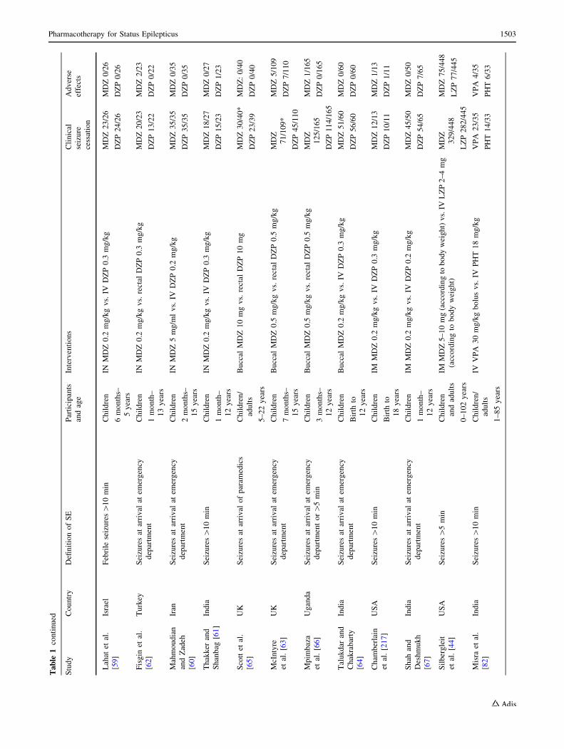
Given this serious limitation, in this narrative review we
presented the most relevant studies on this topic (Table 1)
taking into account the ‘‘evidence-pyramid’’ [38]: when-
ever available, data from controlled clinical trials (ran-
domized/not randomized) were preferred over uncontrolled
trials or case series, unless reporting relevant clinical
results in terms of efficacy or tolerability. Similarly,
Stage I
Stage II
Stage III
5 to 10 min
10 to 30 min
30 to 60 min
Early phase
Premonitory SE, impending SE
Established SE
Refractory SE: SE, that con�nues despite stage I/II treatment
subtle SE, stuporous SE
> 24 hStage IVSuper-refractory SE: SE, that con�nues despite treatment with anaesthe�cs > 24 hours
Fig. 1 Clinical course of
convulsive status epilepticus
(SE)
Pharmacotherapy for Status Epilepticus 1501

Ta
ble
1M
ain
ran
do
miz
ed,
con
tro
lled
clin
ical
tria
lsco
nd
uct
edin
dif
fere
nt
stag
eso
fst
atu
sep
ilep
ticu
s(S
E)
Stu
dy
Co
un
try
Defi
nit
ion
of
SE
Par
tici
pan
ts
and
age
Inte
rven
tio
ns
Cli
nic
al
seiz
ure
cess
atio
n
Ad
ver
se
effe
cts
Ear
lyS
E:
Sta
ge
I
Rem
yet
al.
[53
]
Fra
nce
Sei
zure
s[
20
min
or
2G
TC
Sw
ith
in
20
min
Ad
ult
s
16
–6
5y
ears
Rec
tal
DZ
P3
0m
gv
s.re
ctal
DZ
P2
0m
gD
ZP
30
mg
13
/18
DZ
P2
0m
g
6/2
1
DZ
P3
0m
g
10
/18
DZ
P2
0m
g
13
/21
Dre
ifu
sset
al.
[52
]
US
AA
cute
rep
etit
ive
seiz
ure
sA
du
lts
[1
8y
ears
Rec
tal
DZ
P0
.2m
g/k
gv
s.p
lace
bo
DZ
P3
1/4
6
Pla
ceb
o
14
/49
DZ
P1
9/4
6
Pla
ceb
o
13
/49
Cer
egh
ino
etal
.[5
0]
US
AM
ult
iple
seiz
ure
sw
ith
in1
2–
24
hA
du
lts
18
–7
6y
ears
Rec
tal
DZ
P0
.2m
g/k
gv
s.p
lace
bo
DZ
P2
2/3
1
Pla
ceb
o
11
/39
DZ
P1
0/3
1
Pla
ceb
o9
/39
Sh
aner
etal
.
[11
7]
Cal
ifo
rnia
GT
CS[
30
min
or
3G
TC
Sw
ith
in1
ho
r
GT
CS[
5m
in
Ad
ult
s
[1
5y
ears
IVD
ZP
2–
20
mg?
IVP
HT
6–
18
mg
/kg
bas
edo
nin
itia
ld
rug
lev
els
vs.
IVP
B1
0–
30
mg
/kg
IV
DZ
P?
PH
T
10
/18
PB
16
/18
DZ
P?
PH
T
9/1
8
PB
9/1
8
All
dre
dg
e
etal
.[4
0]
US
AS
eizu
res[
5m
inA
du
lts
[1
8y
ears
IVD
ZP
5m
go
rL
ZP
2m
gv
s.p
lace
bo
LZ
P3
9/6
6
DZ
P2
9/6
8
Pla
ceb
o
15
/71
LZ
P7
/66
DZ
P7
/68
Pla
ceb
o
16
/71
Ch
amb
erla
in
etal
.[4
3]
US
AS
eizu
res[
5m
inC
hil
dre
n
3m
on
ths–
18
yea
rs
IVD
ZP
0.2
mg
/kg
vs.
IVL
ZP
0.1
mg
/kg
DZ
P1
01
/14
0
LZ
P9
7/1
33
DZ
P1
57
**
LZ
P1
55
**
Lep
pik
etal
.
[41
]
US
AC
3G
TC
Sin
1h
;co
nfu
sio
nal
stat
ew
ith
on
go
ing
EE
Gab
no
rmal
itie
s
Ad
ult
s
[1
8y
ears
IVL
ZP
2m
gv
s.IV
DZ
P5
mg
LZ
P3
3/3
7
DZ
P2
5/3
2
LZ
P0
/37
DZ
P4
/32
Tre
iman
etal
.
[81
]
US
AS
eizu
res[
10
min
or
2G
TC
Sw
ith
in
10
min
or
sub
tle
gen
eral
ized
con
vu
lsiv
e
SE
(co
ma
and
icta
ld
isch
arg
eso
nE
EG
)
Ad
ult
s
[1
8y
ears
IVL
ZP
vs.
IVP
Bv
s.IV
DZ
P?
PH
Tv
s.IV
PH
TL
ZP
63
/97
PB
53
/91
DZ
P?
PH
T
53
/95
PH
T4
4/1
01
LZ
P4
2/9
7
PB
46
/91
DZ
P?
PH
T
48
/95
PH
T4
4/1
01
Ary
aet
al.
[48
]
Ind
iaS
eizu
res
atar
riv
alat
emer
gen
cy
dep
artm
ent
Ch
ild
ren
6–
14
yea
rs
INL
ZP
0.1
mg
/kg
vs.
IVL
ZP
0.1
mg
/kg
INL
ZP
59
/71
IVL
ZP
56
/70
INL
ZP
0/7
1
IVL
ZP
0/7
0
Gat
hw
ala
etal
.[5
8]
Ind
iaS
eizu
res
atar
riv
alat
emer
gen
cy
dep
artm
ent
Ch
ild
ren
6m
on
ths–
14
yea
rs
IVM
DZ
0.1
mg
/kg
vs.
IVD
ZP
0.3
mg
/kg
vs.
IVL
ZP
0.1
mg
/kg
MD
Z3
6/4
0
DZ
P2
9/4
0
LZ
P3
8/4
0
MD
Z4
/40
DZ
P2
4/4
0
LZ
P4
/40
1502 E. Trinka et al.
Ta
ble
1co
nti
nu
ed
Stu
dy
Co
un
try
Defi
nit
ion
of
SE
Par
tici
pan
ts
and
age
Inte
rven
tio
ns
Cli
nic
al
seiz
ure
cess
atio
n
Ad
ver
se
effe
cts
Lah
atet
al.
[59
]
Isra
elF
ebri
lese
izu
res[
10
min
Ch
ild
ren
6m
on
ths–
5y
ears
INM
DZ
0.2
mg
/kg
vs.
IVD
ZP
0.3
mg
/kg
MD
Z2
3/2
6
DZ
P2
4/2
6
MD
Z0
/26
DZ
P0
/26
Fis
gin
etal
.
[62
]
Tu
rkey
Sei
zure
sat
arri
val
atem
erg
ency
dep
artm
ent
Ch
ild
ren
1m
on
th–
13
yea
rs
INM
DZ
0.2
mg
/kg
vs.
rect
alD
ZP
0.3
mg
/kg
MD
Z2
0/2
3
DZ
P1
3/2
2
MD
Z2
/23
DZ
P0
/22
Mah
mo
ud
ian
and
Zad
eh
[60
]
Iran
Sei
zure
sat
arri
val
atem
erg
ency
dep
artm
ent
Ch
ild
ren
2m
on
ths–
15
yea
rs
INM
DZ
5m
g/m
lv
s.IV
DZ
P0
.2m
g/k
gM
DZ
35
/35
DZ
P3
5/3
5
MD
Z0
/35
DZ
P0
/35
Th
akk
eran
d
Sh
anb
ag[6
1]
Ind
iaS
eizu
res[
10
min
Ch
ild
ren
1m
on
th–
12
yea
rs
INM
DZ
0.2
mg
/kg
vs.
IVD
ZP
0.3
mg
/kg
MD
Z1
8/2
7
DZ
P1
5/2
3
MD
Z0
/27
DZ
P1
/23
Sco
ttet
al.
[65
]
UK
Sei
zure
sat
arri
val
of
par
amed
ics
Ch
ild
ren
/
adu
lts
5–
22
yea
rs
Bu
ccal
MD
Z1
0m
gv
s.re
ctal
DZ
P1
0m
gM
DZ
30
/40
*
DZ
P2
3/3
9
MD
Z:
0/4
0
DZ
P0
/40
McI
nty
re
etal
.[6
3]
UK
Sei
zure
sat
arri
val
atem
erg
ency
dep
artm
ent
Ch
ild
ren
7m
on
ths–
15
yea
rs
Bu
ccal
MD
Z0
.5m
g/k
gv
s.re
ctal
DZ
P0
.5m
g/k
gM
DZ
71
/10
9*
DZ
P4
5/1
10
MD
Z5
/10
9
DZ
P7
/11
0
Mp
imb
aza
etal
.[6
6]
Ug
and
aS
eizu
res
atar
riv
alat
emer
gen
cy
dep
artm
ent
or[
5m
in
Ch
ild
ren
3m
on
ths–
12
yea
rs
Bu
ccal
MD
Z0
.5m
g/k
gv
s.re
ctal
DZ
P0
.5m
g/k
gM
DZ
12
5/1
65
DZ
P1
14
/16
5
MD
Z1
/16
5
DZ
P0
/16
5
Tal
uk
dar
and
Ch
akra
bar
ty
[64
]
Ind
iaS
eizu
res
atar
riv
alat
emer
gen
cy
dep
artm
ent
Ch
ild
ren
Bir
thto
12
yea
rs
Bu
ccal
MD
Z0
.2m
g/k
gv
s.IV
DZ
P0
.3m
g/k
gM
DZ
51
/60
DZ
P5
6/6
0
MD
Z0
/60
DZ
P0
/60
Ch
amb
erla
in
etal
.[2
17]
US
AS
eizu
res[
10
min
Ch
ild
ren
Bir
thto
18
yea
rs
IMM
DZ
0.2
mg
/kg
vs.
IVD
ZP
0.3
mg
/kg
MD
Z1
2/1
3
DZ
P1
0/1
1
MD
Z1
/13
DZ
P1
/11
Sh
ahan
d
Des
hm
uk
h
[67
]
Ind
iaS
eizu
res
atar
riv
alat
emer
gen
cy
dep
artm
ent
Ch
ild
ren
1m
on
th–
12
yea
rs
IMM
DZ
0.2
mg
/kg
vs.
IVD
ZP
0.2
mg
/kg
MD
Z4
5/5
0
DZ
P5
4/6
5
MD
Z0
/50
DZ
P7
/65
Sil
ber
gle
it
etal
.[4
4]
US
AS
eizu
res[
5m
inC
hil
dre
n
and
adu
lts
0–
10
2y
ears
IMM
DZ
5–
10
mg
(acc
ord
ing
tob
od
yw
eig
ht)
vs.
IVL
ZP
2–
4m
g
(acc
ord
ing
tob
od
yw
eig
ht)
MD
Z
32
9/4
48
LZ
P2
82
/44
5
MD
Z7
5/4
48
LZ
P7
7/4
45
Mis
raet
al.
[82
]
Ind
iaS
eizu
res[
10
min
Ch
ild
ren
/
adu
lts
1–
85
yea
rs
IVV
PA
30
mg
/kg
bo
lus
vs.
IVP
HT
18
mg
/kg
VP
A2
3/3
5
PH
T1
4/3
3
VP
A4
/35
PH
T6
/33
Pharmacotherapy for Status Epilepticus 1503

Ta
ble
1co
nti
nu
ed
Stu
dy
Co
un
try
Defi
nit
ion
of
SE
Par
tici
pan
ts
and
age
Inte
rven
tio
ns
Cli
nic
al
seiz
ure
cess
atio
n
Ad
ver
se
effe
cts
Gil
adet
al.
[84]
Isra
elS
eizu
res[
30
min
Ad
ult
s
[1
8y
ears
IVV
PA
30
mg
/kg
bo
lus
vs.
IVP
HT
18
mg
/kg
bo
lus
VP
A1
3/1
8
PH
T7
/9
VP
A0
/18
PH
T2
/9
Mis
raet
al.
[11
6]
Ind
iaS
eizu
res[
5m
inC
hil
dre
n/
adu
lts
1–
75
yea
rs
IVL
EV
20
mg
/kg
ov
er1
5m
inv
s.IV
LZ
P0
.1m
g/k
go
ver
2–
4m
in
LE
V2
9/3
8
LZ
P3
1/4
1
LE
V6
2*
*
LZ
P9
4*
*
Est
abli
shed
SE
:st
age
II
Sh
aner
etal
.
[11
7]
Cal
ifo
rnia
GT
CS[
30
min
or
3G
TC
Sw
ith
in1
ho
r
GT
CS[
5m
in
Ad
ult
s
[1
5y
ears
IVD
ZP
2–
20
mg?
IVP
HT
6–
18
mg
/kg
bas
edo
nin
itia
ld
rug
lev
els
vs.
IVP
B1
0–
30
mg
/kg
DZ
P?
PH
T
10
/18
PB
16
/18
DZ
P?
PH
T
9/1
8
PB
9/1
8
Tre
iman
etal
.
[81]
US
AS
eizu
res[
10
min
or
2G
TC
Sw
ith
in
10
min
or
sub
tle
gen
eral
ized
con
vu
lsiv
e
SE
(co
ma
and
icta
ld
isch
arg
eso
nE
EG
)
Ad
ult
s
[1
8y
ears
IVL
ZP
vs.
IVP
Bv
s.IV
DZ
P?
PH
Tv
s.IV
PH
TL
ZP
63
/97
PB
53
/91
DZ
P?
PH
T
53
/95
PH
T4
4/1
01
LZ
P4
2/9
7
PB
46
/91
DZ
P?
PH
T
48
/95
PH
T4
4/1
01
Ag
arw
alet
al.
[83]
Ind
iaS
eizu
res[
5m
inre
frac
tory
toIV
DZ
PC
hil
dre
n/
adu
lts
[2
yea
rs
IVV
PA
20
mg
/kg
bo
lus
vs.
IVP
HT
20
mg
/kg
VP
A4
4/5
0
PH
T4
2/5
0
VP
A4
/50
PH
T8
/50
Ch
enet
al.
[21
8]
Ch
ina
Sei
zure
s[
5m
inre
frac
tory
toIV
DZ
PA
du
lts
15
–9
9y
ears
IVV
PA
30
mg
/kg
bo
lus
foll
ow
edb
yin
fusi
on
at1
–2
mg
/kg
vs.
IV
DZ
P0
.2m
g/k
gb
olu
sfo
llo
wed
by
infu
sio
nat
4m
g/h
for
3m
in
and
then
incr
ease
dev
ery
3m
inb
y1lg
/min
un
til
seiz
ure
cess
atio
no
rm
axim
ald
ura
tio
n(1
h)
reac
hed
VP
A1
5/3
0
DZ
P2
0/3
6
VP
A5
/30
DZ
P4
/36
Mal
amir
i
etal
.[2
19]
Iran
Sei
zure
s[
5m
in,
no
tco
ntr
oll
edb
yD
ZP
Ch
ild
ren
3–
16
yea
rs
IVV
PA
20
mg
/kg
bo
lus
vs.
IVP
B2
0m
g/k
gb
olu
sV
PA
27
/30
PB
23
/30
7/3
0
22
/30
Ref
ract
ory
SE
:st
age
III
Sin
gh
iet
al.
[12
7]
Ind
iaM
oto
rse
izu
res
un
con
tro
lled
afte
r2
do
ses
of
DZ
Pan
dP
HT
infu
sio
n
Ch
ild
ren
2–
12
yea
rs
IVM
DZ
0.2
mg
/kg
bo
lus
foll
ow
edb
y2
–1
0l
g/k
g/m
inin
fusi
on
vs.
DZ
P0
.01
–0
.1m
g/k
g/m
inin
fusi
on
MD
Z1
8/2
1
DZ
P1
7/1
9
MD
Z8
/21
DZ
P9
/19
Meh
taet
al.
[22
0]
Ind
iaS
eizu
res[
30
min
,n
ot
con
tro
lled
by
DZ
P
and
PH
T
Ch
ild
ren
5m
on
ths–
12
yea
rs
IVV
PA
30
mg
/kg
bo
lus
vs.
IVD
ZP
10lg
/Kg
/min
incr
ease
db
y
10lg
/kg
/hev
ery
5m
in
VP
A1
6/2
0
DZ
P1
7/2
0
VP
A0
**
DZ
P2
2*
*
Ro
sset
tiet
al.
[13
5]
Sw
itze
rlan
d,
US
A
Sei
zure
s[
30
min
,n
ot
con
tro
lled
by
ben
zod
iaze
pin
ean
dP
HT
or
VP
Ao
rP
B
or
LE
V
Ad
ult
s
16
–8
7y
ears
IVP
RO
2m
g/k
gb
olu
sth
enti
trat
edto
war
db
urs
t-su
pp
ress
ion
or
2m
g/k
g/h
vs.
IVP
TB
5m
g/k
gb
olu
sth
enti
trat
edto
war
db
urs
t-
sup
pre
ssio
no
r2
mg
/kg
/ho
rIV
TH
P2
mg
/kg
bo
lus
then
titr
ated
tow
ard
bu
rst-
sup
pre
ssio
no
r4
mg
/kg
/h
PR
O6
/14
PT
Bo
rT
HP
2/9
PR
O1
5*
*
PT
Bo
rT
HP
12
**
Su
per
-ref
ract
ory
SE
:st
age
IV
No
ran
do
miz
ed,
con
tro
lled
clin
ical
tria
lsav
aila
ble
toin
form
clin
ical
dec
isio
ns
1504 E. Trinka et al.

cumulative data obtained from high-quality systematic
literature reviews were reported as the best available evi-
dence on this topic.
2 Early Status Epilepticus: Stage I
All AEDs commonly used as first-line treatment in SE are
benzodiazepines. These drugs bind to the gamma-
aminobutyric acid (GABA)-A receptors, increasing chan-
nel opening frequency at the receptor, with subsequent
increased chloride conductance and neuronal hyperpolar-
ization, leading to enhanced inhibitory neurotransmission
and antiepileptic action [39].
2.1 Lorazepam (Intravenous (IV) and Intranasal
(IN))
Lorazepam can be administered either intravenously or
intranasally, although to date most evidence in the treat-
ment of SE refers to its IV use. Although it has a longer
initial duration of action than diazepam, lorazepam
administered intravenously is usually preferred as initial
treatment of early SE, because it is less lipid-soluble and
consequently does not undergo the rapid redistribution into
peripheral tissues seen with diazepam. This pharmacologic
advantage has been clinically substantiated in randomized,
controlled trials comparing IV lorazepam with placebo
[40], IV diazepam [41–43], and IM midazolam [44]. In a
meta-analysis, lorazepam was better than placebo for risk
of non-cessation of seizures (relative risk (RR) 0.52; 95 %
confidence interval (CI) 0.38–0.71), better than diazepam
for reducing risk of non-cessation of seizures (RR 0.64;
95 % CI 0.45–0.90), and had a lower risk for continuation
of SE requiring a different drug or general anesthesia (RR
0.63; 95 % CI 0.45–0.88) [36, 45]. There was no statisti-
cally significant difference between lorazepam and diaze-
pam administered intravenously in terms of respiratory
failure/depression, or hypotension [36, 45].
IM midazolam was non-inferior to IV lorazepam in a
landmark study in early SE [44] (see Sect. 2.4 for details).
Recently an intranasal (IN) administration of lorazepam
has been proposed as an alternative, non-invasive delivery
route for this drug, considering the favorable pharma-
cokinetics with rapid absorption from the IN route leading
to rapid blood concentrations required for seizure termi-
nation [46, 47]. The favorable pharmacokinetics of IN
lorazepam in relation to standard (IV) administration have
been confirmed in one randomized, open-label non-inferi-
ority trial conducted in 141 consecutive children aged
6–14 years who presented with convulsions to the emer-
gency room, showing that IN lorazepam was not inferior to
IV lorazepam in terms of clinical seizure remission withinTa
ble
1co
nti
nu
ed
Stu
dy
Co
un
try
Defi
nit
ion
of
SE
Par
tici
pan
ts
and
age
Inte
rven
tio
ns
Cli
nic
al
seiz
ure
cess
atio
n
Ad
ver
se
effe
cts
Stu
die
sn
ot
exp
lici
tly
rep
ort
ing
ad
efin
itio
no
fS
E
Ap
ple
ton
etal
.[4
2]
UK
SE
no
td
efin
edC
hil
dre
nL
ZP
0.0
.5–
0.1
mg
/kg
IVv
s.D
ZP
0.3
–0
.4m
g/k
gIV
LZ
P2
6/2
7
DZ
P2
9/3
4
LZ
P0
/27
DZ
P0
/34
McC
orm
ick
etal
.[2
21]
US
AS
En
ot
defi
ned
Ch
ild
ren
1m
on
th–
15
yea
rs
MD
Z0
.2m
g/k
gIV
vs.
LZ
P0
.1/k
gIV
MD
Z1
4/1
5
LZ
P8
/12
MD
Z1
/15
LZ
P2
/12
DZP
dia
zep
am,GTCS
gen
eral
ized
ton
ic-c
lon
icse
izu
res,IM
intr
amu
scu
lar,IN
intr
anas
al,IV
intr
aven
ou
s,LZP
lora
zep
am,mo
mo
nth
s,PB
ph
eno
bar
bit
al,PHT
ph
eny
toin
,PRO
pro
po
fol,PTB
pen
tob
arb
ital
,SE
stat
us
epil
epti
cus,THP
thio
pen
tal,VPA
val
pro
ate
*E
xp
ress
edas
nu
mb
ero
fep
iso
des
**
Ex
pre
ssed
asn
um
ber
of
epis
od
es;
som
ep
atie
nts
exp
erie
nce
dm
ore
than
on
ead
ver
seef
fect
s
Pharmacotherapy for Status Epilepticus 1505

10 min of drug administration [48]. It has to be noted that
this study included not only children in SE, but also those
who had a seizure in the emergency room, which can
explain the high rate of treatment success, potentially
leading to a bias towards non-inferiority.
2.2 Diazepam (IV, Rectal)
Diazepam is a highly lipophilic benzodiazepine, which
rapidly enters into the brain but subsequently is rapidly
redistributed into peripheral tissues [39]. This pharma-
cokinetic property is responsible for its fast anticonvulsant
effect in spite of its longer elimination half-life. Diazepam
can be administered either intravenously or rectally, with
demonstrated significantly higher efficacy over placebo in
terms of controlling acute repetitive convulsive seizures in
adults and children for both methods of administration [40,
49–52]. Diazepam 30 mg intrarectal gel was found to have
higher efficacy than 20 mg in seizure cessation without any
statistically significant increase in adverse effects [53].
A meta-analysis of the literature indicates that, com-
pared with placebo, after diazepam administration there is
a lower risk of requirement for ventilator support and
continuation of SE requiring a different drug or general
anesthesia with diazepam (304 patients included overall)
[36]. In a recent double-blind, randomized, controlled,
superiority trial IV diazepam was compared to IV lor-
azepam in pediatric SE [43]. 273 children aged 3 months to
\18 years were randomized to either 0.2 mg/kg diazepam
(n = 140) or 0.1 mg/kg lorazepam (n = 133). The rates
for cessation of SE within 10 min and without recurrence
over 30 min were 72.1 % (101/140) in the diazepam group
and 72.9 % (97/133) in the lorazepam group. There were
also no differences in all secondary outcomes (e.g.,
requirement of assisted ventilation), except that patients in
the lorazepam group were more often sedated (66.9 vs.
50 %).
2.3 Clonazepam (IV)
Clonazepam is more lipophilic than lorazepam, but less
lipophilic than diazepam, making it therefore less prone to
redistribution. Its long half-life of 17–55 hs and rapid onset
of action makes it an attractive agent for emergency
treatment of seizures and SE. To date, there is limited
evidence to support the use of IV clonazepam in the
treatment of early SE. In one uncontrolled case series (17
children) with SE treated with this drug, seizure cessation
was reported in all patients after administration of doses
between 0.25 and 0.75 mg, with no adverse effects repor-
ted [54]. In a subsequent uncontrolled, open-label trial (24
patients), the administration of an IV bolus injection of
1–2 mg clonazepam led to complete control of 100 % (7/7)
petit mal, 50 % (7/14) grand mal, and 66 % of partial
complex cases of SE (mean time to clinical seizure ces-
sation after administration was 1.75 min) [55]. Adverse
effects consisted exclusively of transient mild to moderate
drowsiness occurring in 40 % of the patients. One study
comparing IV clonazepam alone to clonazepam followed
by levetiracetam in generalized CSE was reported to recruit
in 2011, but final results have not been published yet [56].
Quite surprisingly, despite these favorable preliminary
data, no further controlled trials has been conducted to
evaluate efficacy and tolerability of this drug in the treat-
ment of SE. Despite this lack of evidence, clonazepam is
extensively used in France, The Netherlands, Belgium, and
other European countries.
2.4 Midazolam (IV, Intramuscular (IM), Intranasal,
Buccal)
Midazolam is a benzodiazepine with the advantage of
multiple routes of administration, due to its water solubil-
ity. At physiologic pH the ring structure of midazolam
closes and it becomes highly lipophilic, crossing the blood-
brain barrier rapidly [57].
Midazolam administered intravenously was found to be
similar in terms of seizure recurrence to IV diazepam or IV
lorazepam in a pediatric non-randomized, controlled trial,
with no significant differences in mean duration to clinical
seizure cessation [58].
A recent double-blind, randomized, non-inferiority trial
compared the efficacy of IM midazolam with that of IV
lorazepam for children and adults with CSE treated by
paramedics before admission to hospital [44]. Midazolam
was found to be at least as safe and effective as IV lor-
azepam: at the time of arrival in the emergency department,
seizures were absent without rescue therapy in 73.4 %
(329/448) in the IM-midazolam group and in 63.4 % (282/
445) in the IV-lorazepam group. The two treatment groups
were similar with regard to the need for endotracheal
intubation (14.1 % of subjects with midazolam and 14.4 %
with lorazepam) and recurrence of seizures (11.4 % and
10.6 %, respectively). Among subjects whose seizures
ceased before arrival in the emergency department, the
median times to active treatment were significantly lower
in the midazolam group, although the onset of action (i.e.,
seizure cessation) occurred sooner after IV lorazepam
administration, and adverse-event rates were similar in the
two groups. Overall, these findings indicate that IM
midazolam is a practical, safe, and effective alternative to
IV lorazepam for the treatment of prolonged convulsive
seizures in prehospital settings.
In general, IN and buccal routes of administration are
more convenient than IV administration for the treatment
of SE, because these formulations deliver the medication
1506 E. Trinka et al.

non-invasively and more rapidly than by the IV route, and
may be used also by paramedics. In three randomized,
controlled trials comparing IN midazolam with IV diaze-
pam, IN midazolam was equally effective as IV diazepam,
with a lower mean time to control of seizures in the
midazolam group than in the diazepam group, and no
significant side effects observed in either group [59–61]. In
addition, IN midazolam was found to be more effective
than rectal diazepam in children with prolonged convulsive
seizures, without serious complications [62].
In a prospective randomized trial, buccal midazolam
was found to be more effective than rectal diazepam in
children with convulsive febrile seizures [63]. No statisti-
cally significant differences in terms of efficacy were found
in other studies comparing buccal midazolam with IV
diazepam [64] or rectal diazepam [65, 66]. These findings
support treatment protocols recommending its use as first-
line treatment of acute tonic-clonic seizures in childhood
including CSE where IV access is difficult or not available
[31]. Time to obtain IV access may be relevant, and may
explain a shorter time for controlling the convulsive epi-
sodes in patients receiving buccal midazolam compared
with patients treated with IV [64] or rectal [65] diazepam.
Similarly, in a study in children comparing IM midazolam
and IV diazepam, mean interval to cessation of convulsions
with IM midazolam was found to be significantly lower
than in the diazepam group without prior IV access [67].
3 Established Status Epilepticus: Stage II
3.1 Phenytoin/Fosphenytoin (IV)
Phenytoin has a pKa of 8.3 and is highly lipid soluble but
insoluble in water. To keep phenytoin in solution it has to
be prepared in a highly alkaline solvent with pH values of
around 12 [68]. It has been used extensively over the past
50 years in the treatment of SE [68], but it took almost
20 years to recognize the appropriate doses of phenytoin to
be effective in SE [69]. Due to its slow rate of infusion
(maximum 50 mg/min) and its delayed onset of action, it
should not be used in early SE [30–32, 70] The recom-
mended dose is 18–20 mg/kg for adults and 15 mg/kg in
the elderly ([65 years). Though phenytoin is not sedative,
hypotension (28–50 %) and cardiac arrhythmias (2 %) may
complicate the treatment [71, 72]. Patients over the age of
50 years and with pre-existing cardiac disease are at spe-
cial risk for cardiovascular complications of phenytoin
[71]. Phenytoin needs polypropylene glycol in an alkaline
solution to prevent precipitation of the substance, which
may result in local irritation, thrombophlebitis, compart-
ment syndrome, and ‘purple glove syndrome,’ as well as
tissue necrosis with extravasation [73–76].
Despite its long-standing use in SE there are only nine
studies including four randomized, controlled studies in
adults and children with various forms and stages of SE
[69, 77–84]. In a randomized, controlled, double-blind
study for initial treatment of tonic clonic status, IV
phenytoin alone was significantly less effective (defined as
cessation of all clinical and electrographic seizure activity
within 20 min after the start of infusion and absence of
seizure relapse 60 min after treatment onset) than lor-
azepam (43.6 vs. 64.9 %, p = 0.002). In three other stud-
ies, two in early SE [82, 84] and one in established SE [83],
phenytoin was compared with valproate. These studies
were assessed in a recent systematic review, showing that
there was no detectable difference between valproate and
phenytoin in clinical seizure cessation (RR 1.31, 95 % CI
9.93–1.84), seizure freedom at 24 h (RR 0.96; 95 % CI
0.88–1.06), but significantly more side effects with
phenytoin (RR 0.31; 95 % CI 0.12–0.85) [37]. The overall
success rate with IV phenytoin has ranged from 44 % in a
randomized, controlled study to 90 % in the uncontrolled
studies [85]. It is important to consider that many of the
patients in these studies had ineffective pretreatment with
benzodiazepines, paraldehyde, or phenobarbital, making an
estimate of the effect of phenytoin difficult to assess.
Fosphenytoin is a water-soluble precursor which is
rapidly transformed to phenytoin. Advantages are a faster
rate of infusion, up to 150 mg/min and a better local tol-
erability [86, 87]. Because of the 15-min time to conver-
sion from fosphenytoin to phenytoin a similar delayed
action is expected [88]. Due to its very high costs, fos-
phenytoin is not on the formulary of most hospitals in
Europe.
3.2 Valproic Acid (IV)
Valproic acid (or the sodium salt of it, i.e., valproate) is
worldwide the most often prescribed AED [89]. It has a
broad spectrum of efficacy, against all seizure types, and a
well-known risk and adverse-event profile [90–94]. Several
studies on the pharmacokinetics of IV valproate in healthy
probands and patients with epilepsies are available [95–
97]. Maximum plasma concentrations were reached
within minutes, and onset of action is quick. Valproic acid
is highly bound to plasma protein ([90 %) and is exten-
sively metabolized in the liver (glucuronidation and beta-
oxidation). The terminal half-life is 12 h. Experimental
data demonstrated a rapid and reliable onset of action in
several SE models [98, 99]. Since the introduction of IV
valproate, experiences with this agent in 860 patients with
various forms of SE have been reported, including six
randomized, controlled trials, four non-randomized, con-
trolled trials, and 20 uncontrolled trials (eight prospective,
12 retrospective) [100]. The overall response rate to
Pharmacotherapy for Status Epilepticus 1507

abrogate SE was 70.9 % (601/848; 95 % CI 67.8–73.9)
[100]. The most commonly used effective dose was
15–45 mg/kg as a bolus (6–10 mg/kg/min) followed by
1–3 mg/kg/h infusion [85, 100–104].
Valproic acid is non-sedating and has been used in
critically ill patients with rapid infusions up to 40 mg/kg/
min with good tolerability [102, 105]. Safety studies of IV
valproic acid in patients with SE showed a low incidence of
adverse events overall (\10 %), mainly dizziness, throm-
bocytopenia, and mild hypotension, which was indepen-
dent of infusion rates [100]. Of note is the good
cardiovascular and respiratory tolerability. The most seri-
ous concern relates to the possibility of acute
encephalopathy, which is sometimes related to hepatic
abnormalities or hyperammonaemia [89, 93, 106, 107].
3.3 Levetiracetam (IV)
Levetiracetam is an efficacious and well tolerated drug
with a broad spectrum of efficacy against all seizure types
and a low potential for interactions due to minimal hepatic
metabolism and low plasma protein binding (\10 %) [108–
110]. IV levetiracetam has been available since 2006. In
animal experiments a rapid antiepileptic activity has been
demonstrated, especially when used in combination with
diazepam [111]. Retrospective case series and prospective
safety studies on more than 1000 patients including more
than 500 with various forms of SE have been reported [85,
112, 113]. The safety profile of levetiracetam is advanta-
geous, with a very low rate of adverse effects reported
(most often somnolence and sedation, and rarely agitation
and thrombocytopenia) [113].
In a meta-analysis including ten studies (seven retro-
spective observational, two prospective observational, one
prospective randomized control) reporting on 234 patients,
efficacy ranged from 44 to 94 %, with a higher efficacy
reported in the retrospective studies [114]. A recent meta-
analysis on five different treatment options including
levetiracetam in benzodiazepine-resistant SE identified 27
studies (798 patients with CSE) [115]. The relative effec-
tiveness of levetiracetam was 68.5 % (95 % CI 56.2–78.7),
compared with phenobarbital 73.6 % (95 % CI
58.3–84.8 %), phenytoin 50.2 % (95 % CI 34.2–66.1), and
valproate 75.7 % (95 % CI 63.7–84.8). The authors con-
cluded that levetiracetam, valproate, or phenobarbital, but
not phenytoin, should be used as first-line drugs in ben-
zodiazepine-resistant SE [115].
Interestingly, although levetiracetam is considered a
drug for the treatment of established SE, a recent ran-
domized, open-labeled study compared its use with iIV
lorazepam in 79 patients with early (stage I) SE [116]. Both
drugs were equally effective in reaching clinical seizure
cessation within 10 min of administration, with
significantly higher respiratory failure requiring artificial
ventilation among patients receiving lorazepam. This pilot
study suggests that levetiracetam is also effective in early-
stage SE, where it might represent an alternative to
lorazepam.
3.4 Phenobarbital (IV, IM)
In a randomized, controlled trial on CSE, IV phenobarbital
was at least as effective as a combination of diazepam and
phenytoin [117]. In the Veterans Affairs-study phenobar-
bital was not inferior to lorazepam in the initial treatment
of SE [81]. The central depressive effect of phenobarbital,
especially following the use of benzodiazepines, limits its
clinical utility, when alternatives are available. However,
over the years wide experience has been gained in adults
and children, as well as in the newborn. The main disad-
vantages are sedation, respiratory depression, and
hypotension. The usual recommended adult IV loading
dose of phenobarbital is 10 mg/kg (doses up to 20 mg/kg
have been used and recommended) given at the rate of
100 mg/min, up to a total amount of 700 mg in 7 min. In
the acute setting respiration and blood pressure have to be
monitored and the patient has to be under clinical
observation.
4 Refractory Status Epilepticus: Stage III
After failure of stage I treatment (with benzodiazepines)
and failure of stage II treatment (with phenytoin, leve-
tiracetam, or valproic acid), 31–43 % of patients enter the
refractory stage [118, 119]. In refractory and super-re-
fractory SE, IV anesthetic drugs (thiopental/pentobarbital,
midazolam, or propofol) are commonly used [28]. Treat-
ment recommendations at this stage depend on retrospec-
tive case series and uncontrolled studies [120–122]. In two
systemic reviews, none of the treatments currently avail-
able was superior to another [123, 124]. Propofol may be
associated with an infusion syndrome, characterized by
metabolic acidosis, rhabdomyolysis, renal failure, and heart
failure [125]. Treatment duration of less than 48 h and
doses of no more than 5 mg/kg/h are recommended. The
rate of cardiovascular and metabolic complications seemed
to be lowest with midazolam and highest with barbiturates,
which cause a severe immunosuppression, and often lead to
infections in the compromised patient [123, 124]. The use
of anesthetics in refractory and super-refractory SE was
associated with more infections during SE (43 % vs. 11 %;
p = 0.0001) and a 2.9-fold relative risk for death (2.88;
95 % CI 1.45–5.73) in a 6-year prospective cohort study
including 171 patients (63 of them received IV anesthetics)
[126].
1508 E. Trinka et al.

4.1 Midazolam (Continuous IV Infusion)
One study compared continuous IV midazolam infusion
versus continuous IV diazepam infusion in patients with
refractory SE defined as seizures not controlled by two
bolus doses of diazepam (0.3 mg/kg) and phenytoin
infusion (20 mg/kg in normal saline infusion over
20 min) followed by a repeat dose of benzodiazepine
[127]. Continuous midazolam and diazepam infusions
were equally effective for cessation of SE, although
midazolam was associated with a higher seizure recur-
rence rate and mortality. About half of the patients
required mechanical ventilation and 40 % of patients in
both groups had hypotension. In a recent study, high-
dose continuous IV midazolam (n = 100) was compared
with a historical control of the same center treated with
a lower dose (n = 29) [128]. The median maximum dose
was 0.4 mg/kg/h (interquartile range [IQR] 0.2, 1.0) for
the high-dose group and 0.2 mg/kg/h (IQR 0.1, 0.3) for
the low-dose [128] group (p\ 0.001), with a similar
duration of infusion. Withdrawal seizures, occurring
within 48 h of discontinuation of continuous midazolam
infusion, were less frequent in the high-dose group (15
vs. 64 %; odds ratio (OR) 0.10; 95 % CI 0.03–0.27) and
discharge mortality was lower in the high-dose group (40
vs. 62 %; OR 0.34; 95 % CI 0.13–0.92) compared with
the low-dose group.
4.2 Propofol
Propofol is an anesthetic agent, acting as an N-methyl-D-
aspartate (NMDA) antagonist in vitro [129], with a shorter
duration of action and lower tendency to accumulate in the
body than barbiturates. It may cause hypotension, but
reduces intracranial pressure and brain metabolic require-
ments [130], and may also have immunomodulatory effects
[131]. Its prolonged use has been reported to cause the so-
called ‘‘propofol infusion syndrome,’’ which includes
potentially fatal myocardial failure with lactic acidosis,
hypertriglyceridemia, and rhabdomyolysis, with an
observed incidence of around 1 % [132–134].
Propofol has been directly compared with thiopental
sodium (see Sect. 4.3) in a small (only 24 patients recruited
of the 150 needed; 14 subjects received propofol, nine
thiopental), single-blind, multicenter trial evaluating adult
patients with refractory SE [135]. This study showed a
wide CI, suggesting that the drugs may differ in efficacy up
to more than twofold. No difference was found between the
drugs with respect to control of seizure activity and return
to baseline clinical conditions at 3 months. Infections and
arterial hypotension did not differ between groups,
although thiopental use was associated with significantly
longer mechanical ventilation.
To date, robust evidence on the role of propofol based
on large, randomized, controlled trials for this serious
condition is still lacking [136].
4.3 Thiopental, Pentobarbital
Thiopental sodium and pentobarbital are barbiturates act-
ing as GABA-A agonists, with enhanced inhibitory neu-
rotransmission and antiepileptic action. Both drugs have a
prolonged duration of action, mainly due to their accu-
mulation in the body, resulting in a long recovery time
[137]. Furthermore, they may cause hypotension and car-
diorespiratory depression, which may require the use of
additional drugs to control pressure and breathing, as well
as immunosuppression [138–140].
As reported earlier (Sect. 4.2), compared with propofol,
the use of thiopental seems to be associated with longer
mechanical ventilation, with no difference with respect to
control of seizure activity, infections, and arterial
hypotension [135].
A systematic review evaluated the outcomes of anes-
thetics (thiopental/pentobarbital, n = 192; midazolam,
n = 585; propofol, n = 143) for terminating refractory and
super-refractory SE. Barbiturate treatment achieved seizure
control in 64 % (midazolam 78 %, propofol 68 %), but
was associated with death rate during treatment in 19 %
(midazolam 2 %, propofol 8 %). The authors concluded
that a treatment protocol for stages III and IV of SE divided
into first-line, second-line, and third-line therapy is sug-
gested on the basis of their outcome evaluation [124].
Thus, barbiturates should be restricted to the most severe
forms of refractory SE.
Despite the wide use of barbiturate anesthesia for the
treatment of refractory SE, evidence derived from com-
parative randomized, controlled trials on the efficacy of
barbiturates on which to base a choice is still lacking.
4.4 Isoflurane (Inhalation)
Isoflurane is an inhalational anesthetic acting by enhancing
GABA activity, with subsequent increased inhibitory neu-
rotransmission. In a case series, this drug (used for up to
55 h) led to seizure cessation in nine patients, although
with a mortality rate of 67 % [141]. A subsequent study
reported its use in seven patients with refractory SE (de-
fined as ‘‘continued seizures after failure of two or three
antiepileptic drugs’’) [142]: isoflurane consistently stopped
epileptic discharges with adequate, sustained suppression
of electroencephalographic bursts within minutes of
administration. Complications were common: all patients
developed severe hypotension requiring vasopressors and
atelectasis; paralytic ileus occurred in three patients; and
the outcome was fatal in three patients. No renal or hepatic
Pharmacotherapy for Status Epilepticus 1509
dysfunction occurred. Traditionally the use of inhalational
anesthetics needed a vaporizer and a cart, which was not
practical in the crowded environment of most intensive
care units. The recently developed small self-contained
vaporizers have become more widely available and will
make the use of inhalational anesthetics in intensive care
units more feasible.
4.5 Etomidate (IV)
Etomidate is an anesthetic for which the exact mechanism of
action is unknown. It has a favorable safety profile with
regard to cardiovascular side effects, and may induce seda-
tion rapidly [143]. However, its use is limited by a concern
for reversible cortisol inhibition occurring after the admin-
istration of this drug, which requires hormonal substitution
during treatment with etomidate [144]. In a case series (eight
patients), rapid control of seizure activity was obtained in all
cases, with hypotension occurring in five subjects [145].
5 Super-Refractory Status Epilepticus: Stage IV
When treatment with an IV anesthetic for more than 24 h is
not successful in controlling SE, the condition can be ter-
med super-refractory SE or malignant SE [28]. There have
been no studies of this stage, and evidence is extremely
sparse. A new initiative of a global audit (http://www.
status-epilepticus.net) is aimed at gaining more information
on the treatments used and outcomes achieved at this stage
of SE. Recommendations are based exclusively on small
case series, including several treatment options, such as
ketamine, corticosteroids, magnesium, IV immunoglobu-
lin, a ketogenic diet, and neurostimulation, and in lesional
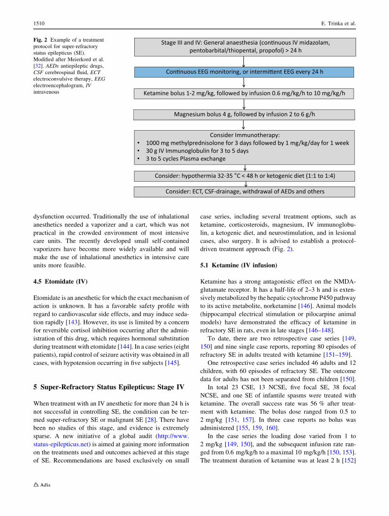
cases, also surgery. It is advised to establish a protocol-
driven treatment approach (Fig. 2).
5.1 Ketamine (IV infusion)
Ketamine has a strong antagonistic effect on the NMDA-
glutamate receptor. It has a half-life of 2–3 h and is exten-
sively metabolized by the hepatic cytochrome P450 pathway
to its active metabolite, norketamine [146]. Animal models
(hippocampal electrical stimulation or pilocarpine animal
models) have demonstrated the efficacy of ketamine in
refractory SE in rats, even in late stages [146–148].
To date, there are two retrospective case series [149,
150] and nine single case reports, reporting 80 episodes of
refractory SE in adults treated with ketamine [151–159].
One retrospective case series included 46 adults and 12
children, with 60 episodes of refractory SE. The outcome
data for adults has not been separated from children [150].
In total 23 CSE, 13 NCSE, five focal SE, 38 focal
NCSE, and one SE of infantile spasms were treated with
ketamine. The overall success rate was 56 % after treat-
ment with ketamine. The bolus dose ranged from 0.5 to
2 mg/kg [151, 157]. In three case reports no bolus was
administered [155, 159, 160].
In the case series the loading dose varied from 1 to
2 mg/kg [149, 150], and the subsequent infusion rate ran-
ged from 0.6 mg/kg/h to a maximal 10 mg/kg/h [150, 153].
The treatment duration of ketamine was at least 2 h [152]
Stage III and IV: General anaesthesia (con�nuous IV midazolam, pentobarbital/thiopental, propofol) > 24 h
Con�nuous EEG monitoring, or intermi�ent EEG every 24 h
Ketamine bolus 1-2 mg/kg, followed by infusion 0.6 mg/kg/h to 10 mg/kg/h
Magnesium bolus 4 g, followed by infusion 2 to 6 g/h
Consider Immunotherapy: • 1000 mg methylprednisolone for 3 days followed by 1 mg/kg/day for 1 week• 30 g IV Immunoglobulin for 3 to 5 days• 3 to 5 cycles Plasma exchange
Consider: hypothermia 32-35 °C < 48 h or ketogenic diet (1:1 to 1:4)
Consider: ECT, CSF-drainage, withdrawal of AEDs and others
Fig. 2 Example of a treatment
protocol for super-refractory
status epilepticus (SE).
Modified after Meierkord et al.
[32]. AEDs antiepileptic drugs,
CSF cerebrospinal fluid, ECT
electroconvulsive therapy, EEG
electroencephalogram, IV
intravenous
1510 E. Trinka et al.

and at most 27 days [150]. The time of SE until ketamine
administration was started ranged from a few hours [152]
up to 140 days [155]. A median of five medications (min
four, max nine) prior to ketamine were used.
With regard to adverse events, one patient suffered from
dysarthria, drooling, and appendicular ataxia after treat-
ment with ketamine; upon magnetic resonance imaging a
cerebellar atrophy was described. The authors concluded
this to be a neurotoxic effect of ketamine. In comparison
with other reports they used a higher loading—and a higher
infusion dose of ketamine (2 mg/kg and a maximum dose
of 7.5 mg/kg/h). After a follow-up of 21 months there was
a slight clinical improvement [157].
One patient suffered from hypertension, with systolic
blood pressure [220 mmHg for 10 min, after the initial
bolus of 0.5 mg/kg was administered [154]. In one case
series a patient had symptoms like propofol-infusion syn-
drome after 4 days of high-dose ketamine (4.5 mg/kg/h)
and midazolam, and recovered after discontinuation.
Another two patients developed supraventricular tachy-
cardia [150]. A systematic review evaluating efficacy in
pediatrics and adults showed that currently there exists
Oxford level 4, Grade C evidence to support the use of
ketamine for refractory SE. But they believe that there is a
potential benefit with low adverse effects of NMDA
antagonists—further prospective studies of early ketamine
administration are needed [159].
5.2 Magnesium (IV)
Magnesium sulphate probably has an antiepileptic action
through blocking the NMDA receptor. Magnesium sul-
phate has been used in SE since 1901, but has not gained
much attention until now. It is currently used as the drug of
choice in treating seizures occurring in eclampsia [161].
The evidence on its use in patients with SE is based on
single case reports, with some evidence of benefit [162,
163]. To date, no comparative study has been conducted to
assess its role in SE management. However, its infusion is
safe and without significant adverse events. It has been
suggested to administer magnesium as an initial IV bolus,
followed by a continuous infusion at a dose that increases
the serum level to *3.5 mmol/L [163].
5.3 Topiramate (Enteral)
Topiramate is a broad-spectrum AED with several mech-
anisms of action, including blockade of the ionotropic
glutamatergic AMPA receptor [164]. There is no com-
mercially available IV formulation, but it can be adminis-
tered enterally.
Evidence on the use of topiramate in super-refractory SE
is based on 95 patients reported to date in the literature
[124, 165]. The dose of topiramate used in studies ranged
between 2 and 25 mg/kg/day in children and up to
1600 mg/day in adults, leading to clinical seizure cessation
in 62/95 (65 %) of patients. Metabolic acidosis was the
most frequently reported side effect with its use.
5.4 Immunotherapy: Corticosteroids, IV
Immunoglobulins, Plasma Exchange
The recent discovery that super-refractory SE may be
caused by antibodies against neural cell receptors (voltage-
gated potassium and N-methyl-D-aspartate (NMDA)) and
evidence on the role of inflammation in epileptogenesis
[166–168] led to the increasing use of immunotherapy in
this stage of SE, even in the absence of any defined
immunologic disease. However, to date no single com-
parative study has been conducted to evaluate the efficacy
and tolerability of immunotherapies in super-refractory SE,
and evidence is based on more than 50 patients treated so
far [28]. In all reported cases, other therapies were also
introduced concurrently, so that it is extremely difficult to
definitively dissect the antiepileptic efficacy of
immunotherapy from that of other treatments.
The rationale for the use of immunotherapies is that in
some cases super-refractory SE without a clear underlying
cause might be due to occult immunologic diseases with
antibodies (not yet identified) directed against neural ele-
ments, which may also explain the persistence of the SE.
Consequently, even in the absence of an immunologic
underlying cause for the SE, a trial of high-dose steroids
(1 g of IV prednisolone/day for 3 days followed by 1 mg/
kg/day for *1 week) can be given. In case of no efficacy
within 2 days, either IV immunglobulins (at a dose of
0.4 g/kg over 5 days) or plasma exchange can be tried in
addition. If there is a response, treatment is continued with
long-term corticosteroids, repeated courses of IV
immunoglobulins and, later, other immunomodulatory
agents such as cyclophosphamide or rituximab [28].
6 Other Drugs Used in Status Epilepticus
6.1 Lacosamide (IV)
Lacosamide has been available since 2008 in the European
Union for the treatment of focal epilepsies [169–171].
Bioequivalence studies in healthy probands and adults with
epilepsy report a good tolerability of the IV solution [172,
173]. In animal models on SE, lacosamide demonstrated a
good efficacy [174]. First case reports and retrospective
case series on lacosamide were published shortly after
availability of the IV formulation [175–178]. There are in
total 19 studies (ten single case reports and nine case
Pharmacotherapy for Status Epilepticus 1511

series), reporting a total of 136 episodes of refractory SE
(50 % NCSE, 31 % focal SE, and 19 % CSE) treated with
lacosamide [176]. All retrospective case series included
patients with various forms of SE in different stages. The
most commonly used bolus dose was 400 mg, followed by
a daily dose of 200–400 mg lacosamide. The overall suc-
cess rate was 56 % (76/136). Adverse events were reported
in 25 % (34/136) of patients: mild sedation in 25 cases, one
patient with possible angioedema, two with allergic skin
reactions, four with hypotension, and one with pruritus.
One patient developed a third-degree atrioventricular (AV)
block and paroxysmal asystole [176]. There are also two
single case reports (one in a patient with NCSE, one with
neuropathy), reporting on AV-conduction abnormalities
associated with an application of the drug [179, 180]. There
was a small increase in PR interval at the end of the
infusion reported in the bioequivalence studies, but this
was judged to be clinically not relevant [172, 173].
6.2 Paraldehyde (IM, Rectal)
Paraldehyde is a drug with proven anticonvulsant proper-
ties both in animal models [181] and in humans [182]. It
acts through a mechanism not yet identified, and appears to
be safe with regard to cardiovascular tolerability [143]. The
rectal route, which is less painful and carries no risk of
sterile abscess, has largely replaced the traditional IM
injection [182].
In one pediatric study rectal paraldehyde was found to be
effective in terminating over 60 % of acute and prolonged
convulsive seizure episodes within 10 min of its adminis-
tration and without any documented adverse side effects
[182]. A similar finding (clinical seizure cessation within
10 min of its administration) was found in a subsequent
open-label randomized, controlled clinical trial comparing
IN lorazepam and IM paraldehyde in children with pro-
longed ([5 min) tonic-clonic seizures; no clinically
important cardiorespiratory events were reported with IM
paraldehyde [183]. Of note, in this study IN lorazepam was
more effective (i.e., less likely to require additional drugs to
terminate seizure), significantly safer, and cheaper than IM
paraldehyde in the treatment of acute tonic-clonic seizures.
6.3 Lidocaine (IV)
Lidocaine acts as a local anesthetic drug by inhibiting ionic
currents through voltage-gated sodium channels during
abnormal membrane depolarization [184]. Interestingly, IV
lidocaine has been widely used in Japan for the treatment
of CSE, although this drug has no official approval for the
management of this condition. Most studies assessing its
role in SE were performed in Japan [185, 186]. To date,
there are no large, double-blind, placebo-controlled studies
evaluating lidocaine in SE, although numerous case reports
and case series support its use [185–188]. Most of the
available data derive from patients refractory to multiple
AEDs. Furthermore, additional data supporting the use of
lidocaine in SE come from pediatric uncontrolled studies,
where this drug proved effective in controlling SE in
neonates not responding to barbiturates [187, 188].
Overall effectiveness of lidocaine in cessation of CSE
ranges between 35.8 % and 53 % [187–190]. Furthermore,
unlike other AEDs used for the treatment of SE carrying
the risk of respiratory depression, lidocaine has been
reported to reduce the rate of mechanical ventilation [187].
6.4 Chlormethiazole (IV)
Chlormethiazole (clomethiazole) is a thiazole derivative
acting by enhancing GABA activity, with subsequent
increased inhibitory neurotransmission. No controlled
studies have been conducted to evaluate its effectiveness
and tolerability in SE. To date, only a few reports of
patients with SE (mostly children) treated with IV
chlormethiazole are available [191–194], so that the evi-
dence supporting its use in SE is scarce. Several adverse
effects related to its use have been reported including
thrombophlebitis and respiratory depression [195].
6.5 Carbamazepine (Rectal, IV)
Carbamazepine has ILAE level A evidence for treatment of
adults with focal epilepsy. This drug is characterized by
poor water solubility, which represents an obstacle in the
development of a parenteral formulation. IV carba-
mazepine has been tested in animal models [196, 197],
showing rapid onset of anticonvulsant action and lack of
respiratory or cardiovascular adverse effects, and very
recently also in humans [198–200]. Novel IV formulations
with favorable pharmacokinetics have been developed
[201], but their use in clinical practice needs to be defini-
tively assessed.
To date, there is no study reporting on patients with SE
treated with IV carbamazepine. However, there is a case
series reporting on seven patients presenting with cluster
seizures or generalized tonic-clonic SE, who received a
rectally administered oral syrup of carbamazepine with
subsequent measurement of serum levels (the goal was to
achieve minimum therapeutic levels of total carbamazepine
[20 lmol/L) [202]. In all patients seizures had terminated
prior to administration of carbamazepine, but no patient
had a seizure recurrence after carbamazepine administra-
tion. This study therefore indicates the potential long-act-
ing efficacy of rectal carbamazepine in achieving
maintenance of freedom from seizures in patients with SE,
but does not support its use in terminating SE.
1512 E. Trinka et al.

7 Conclusions
In this article, we reviewed the pharmacotherapy of SE
according to a staged approach based on pragmatic defi-
nitions. Morbidity and mortality correlate with duration of
epileptic activity, the rapid identification of the cause of
SE, age, and comorbidity of the patients [9, 10, 203–206].
Indeed, if left untreated, SE is potentially fatal or can lead
to irreversible brain damage.
There is general agreement that treatment of status should
follow a staged treatment protocol, so that patients may
receive prompt and appropriate treatment [30]. Despite
conflicting data [207], clinical seizure cessation seems to be
clearly associated with adherence to treatment protocols
[208–210]. Yet many hospitals do not have these or do not
review and update them regularly. Randomized, controlled
trials show that IV lorazepam [40, 41, 81] or IM midazolam
[44, 211, 212] are the most efficient drugs in early-stage SE.
Midazolam buccal administration is a widely used
alternative first-line treatment for emergency management
of acute seizure disorders by non-medical personnel, prior
to admission to hospital. The IN route may also be used for
early SE, particularly in children, but its value compared
with the buccal or IM routes is less established. Compared
to lorazepam or midazolam, diazepam is more widely
available throughout the world, representing a valuable
alternative to lorazepam for early SE. However, compared
to lorazepam, diazepam undergoes a rapid redistribution
with subsequent shorter duration of action, and higher risk
of accumulation after repeated administration. Rectal dia-
zepam administration is a valuable therapeutic alternative
to IV diazepam or lorazepam when this method of
administration is not feasible, as it often occurs in pre-
hospital settings without skilled health-care personnel.
However, buccal or IN midazolam represent alternatives
that are more socially accepted than rectal diazepam.
Approximately 30–40 % of all patients do not respond to
benzodiazepines administered as first-line drugs, needing
further treatment with IV AEDs. Even with their use, a high
proportion of patients remain refractory, requiring the use of
anesthetics [81, 85, 213, 214]. IV phenytoin or phenobarbital
are widely used treatments for established SE. However,
phenytoin and phenobarbital are not efficacious in some epi-
lepsy syndromes and there are several concerns related to their
safety profile, as both drugs may cause cardiac arrhythmias,
hypotension, and respiratory depression, the latter being
aggravated by co-administration with benzodiazepines.
Hence, IV formulations of other AEDs, including val-
proate, levetiracetam, and lacosamide have generated
considerable interest as possible treatments for established
SE after failure of benzodiazepines [100, 112, 113, 176–
178, 215].
Classic randomized clinical trials satisfying contempo-
rary standards are extremely challenging to perform in the
setting of established SE. To date there are no class I data
to support treatment recommendations for most AEDs that
may be used for established, refractory, and super-refrac-
tory SE. The only partial exception concerns the evidence
for use of IV valproate in the treatment of established SE,
which has been shown to have a similar efficacy but a
better tolerability than phenytoin in a meta-analysis [37,
100]. A recent systematic review compared the relative
efficacy of five AEDs in treatment of stage II, benzodi-
azepine-resistant CSE [115]. Valproate was effective in
controlling SE in 75.7 % (95 % CI 63.7–84.8), levetirac-
etam in 68.5 %, (95 % CI 56.2–78.7), phenobarbital in
73.6 % (95 % CI 58.3–84.8), and phenytoin in 50.2 %
(95 % CI 34.2–66.1). Considered overall, cumulative data
from the literature are therefore consistent with valproate
and levetiracetam being a safe and effective therapeutic
alternative to phenobarbital and phenytoin for treatment of
established SE persisting despite conventional first-line
treatment with benzodiazepines. A high-class randomized,
controlled trial comparing valproate, levetiracetam, and
phenytoin for established SE (ESET Trial) is underway,
and the first results can be expected in the next few years
[216]. Limiting the methodological heterogeneity across
studies will be of the utmost importance in further research
in the treatment of SE: investigators should adopt uniform
definitions of SE and its different stages, provide individual
patient data, and report their results clearly and explicitly.
Compliance with Ethical Standards
Funding There was no funding related to the preparation of this
article.
Conflicts of interest Eugen Trinka has acted as a paid consultant to
Eisai, Ever Neuropharma, Biogen Idec, Medtronics, Bial, and UCB
and has received speakers’ honoraria from Bial, Eisai, GL Pharma,
GlaxoSmithKline, Boehringer, Viropharma, Actavis, and UCB
Pharma in the past 3 years. Eugen Trinka has received research
funding from UCB Pharma, Biogen Idec, Red Bull, Merck, the
European Union, FWF Osterreichischer Fond zur Wis-
senschaftsforderung, and Bundesministerium fur Wissenschaft und
Forschung. Eugen Trinka is also part of the investigators planning the
ESET-Trial and member of the Task Force on Classification of Status
Epilepticus of the ILAE. Julia Hofler has received speakers’ honoraria
from UCB Pharma and travel support from Eisai and GL Pharma.
Markus Leitinger has received travel grants from Medtronics.
Francesco Brigo has received speaker’s honoraria from UCB Pharma.
Open Access This article is distributed under the terms of the
Creative Commons Attribution-NonCommercial 4.0 International
License (http://creativecommons.org/licenses/by-nc/4.0/), which per-
mits any noncommercial use, distribution, and reproduction in any
medium, provided you give appropriate credit to the original
author(s) and the source, provide a link to the Creative Commons
license, and indicate if changes were made.
Pharmacotherapy for Status Epilepticus 1513

Appendix
Early Status Epilepticus: Stage I
Intravenous (IV) route available:
– Lorazepam 0.07 mg/kg (usually 4 mg) IV bolus
(maximum rate 2 mg/kg); if necessary can be
repeated once.
OR
– Diazepam 5–10 mg IV bolus (maximum rate 5 mg/
min); if necessary can be repeated once up to 20 mg.
OR
– Clonazepam 1 mg IV bolus (maximum rate 0.5 mg/
min); if necessary can be repeated once after 5 min.
If IV route is difficult or not possible:
– Midazolam 10 mg buccal (5 mg in the elderly or in
patients \50 kg); if necessary can be repeated once
after 10 min. Alternatively, use 10 mg/2 mL injection
via buccal route.
OR
– Midazolam 10 mg intramuscularly (5 mg in the
elderly or in patients \50 kg); if necessary can be
repeated once after 10 min.
OR
– Diazepam 10 mg rectal (5 mg in the elderly or in
patients \50 kg); if necessary can be repeated once
after 10 min.
Established Status Epilepticus: Stage II
– Phenobarbital 10 mg/kg (range 10–20) IV bolus
infusion at a max. rate of 100 mg/min.
OR
– Phenytoin 18 mg/kg (range 15–20) IV bolus infusion
at max. rate of 50 mg/min.
OR
– Fosphenytoin 15 mg PE/kg (range 15–20) IV bolus
infusion at max. rate of 100 mg PE/min.
OR
– Valproate 30 mg/kg (range 15–30) IV bolus infusion
at 3–6 mg/kg/min.
OR
– Levetiracetam* 30 mg/kg (range 30–60) IV bolus
infused over 10 min.
OR
– Lacosamide* 200–400 mg IV bolus infused over
3–5 min.
Refractory Status Epilepticus: Stage III
The following drugs need to be administered by a neu-
rointensivist/experienced anaesthetist in an intensive care
unit setting:
– Propofol 2 mg/kg IV bolus infusion, repeated if
necessary, and then followed by a continuous infusion
of 5–10 mg/kg/h initially, reducing to a dose suffi-
cient to maintain a burst-suppression pattern on the
EEG (usually 1–3 mg/kg/h).
OR
– Thiopental 100–250 mg IV bolus infusion giver over
20 s with further 50-mg boluses every 2–3 min until
seizure control, followed by a continuous IV infusion
at a dose sufficient to maintain a burst-suppression
pattern on the EEG (usually 3–5 mg/kg/h).
OR
– Pentobarbital 5–15 mg/kg IV bolus, followed by a
continuous IV infusion at a dose sufficient to maintain
a burst-suppression pattern on the EEG (usually
0.5–3 mg/kg/h).
OR
– Midazolam 0.1–0.3 mg/kg IV bolus infusion at max.
rate of 4 mg/min initially, followed by a continuous
IV infusion at a dose sufficient to maintain a burst-
suppression pattern on the EEG (usually 0.05–0.4 mg/
kg/h).
After seizure control has been achieved for at least
12 h, the drug dosage should be slowly reduced over a
further 12 h. If seizures recur, the anesthetic agent should
be administered again for another 12 h, and then with-
drawal attempted again. This cycle may need to be
repeated every 24 h until achievement of seizure control.
Continuous EEG monitoring is indicated to assess
level of anesthesia (burst-suppression pattern) and aboli-
tion of ictal discharges after drug withdrawal
Super-Refractory Status Epilepticus: Stage IV
No randomized, controlled trials are available in the lit-
erature to inform about the use of any drug in the treat-
ment of super-refractory status epilepticus.
Maintain the use of anesthetic drugs used in stage III.
1514 E. Trinka et al.

References
1. Lowenstein DH, Bleck T, Macdonald RL. It’s time to revise the
definition of status epilepticus. Epilepsia. 1999;40(1):120–2.
2. Bauer G, Trinka E. Nonconvulsive status epilepticus and coma.
Epilepsia. 2010;51(2):177–90.
3. Drislane FW. Presentation, evaluation, and treatment of non-
convulsive status epilepticus. Epilepsy Behav.
2000;1(5):301–14.
4. Trinka E, Hesdorffer D, Rossetti AO, Scheffer I, Shinnar S,
Shorvon S, Lowenstein D. A definition and classification of
status epilepticus - report of the task force on classification of
status epilepticus. Epilepsia. 2015. doi:10.1111/epi.13121.
5. Logroscino G, Hesdorffer DC, Cascino G, Annegers JF, Hauser
WA. Time trends in incidence, mortality, and case-fatality after
first episode of status epilepticus. Epilepsia. 2001;42(8):1031–5.
6. Logroscino G, Hesdorffer DC, Cascino G, Hauser WA, Coey-
taux A, Galobardes B, Morabia A, Jallon P. Mortality after a
first episode of status epilepticus in the united states and europe.
Epilepsia. 2005;46(Suppl 11):46–8.
7. Logroscino G, Hesdorffer DC, Cascino GD, Annegers JF,
Bagiella E, Hauser WA. Long-term mortality after a first epi-
sode of status epilepticus. Neurology. 2002;58(4):537–41.
8. Sokic DV, Jankovic SM, Vojvodic NM, Ristic AJ. Etiology of a
short-term mortality in the group of 750 patients with 920 epi-
sodes of status epilepticus within a period of 10 years
(1988–1997). Seizure. 2009;18(3):215–9.
9. Sutter R, Marsch S, Fuhr P, Ruegg S. Mortality and recovery
from refractory status epilepticus in the intensive care unit: A
7-year observational study. Epilepsia. 2013;54(3):502–11.
10. Leitinger M, Holler Y, Kalss G, Rohracher A, Novak HF, Hofler
J, Dobesberger J, Kuchukhidze G, Trinka E. Epidemiology-
based mortality score in status epilepticus (emse). Neurocrit
care. 2015;22(2):273–82.
11. DeLorenzo RJ, Pellock JM, Towne AR, Boggs JG. Epidemiol-
ogy of status epilepticus. J Clin Neurophysiol.
1995;12(4):316–25.
12. Waterhouse EJ, DeLorenzo RJ. Status epilepticus in older
patients: Epidemiology and treatment options. Drugs Aging.
2001;18(2):133–42.
13. Knake S, Rosenow F, Vescovi M, Oertel WH, Mueller HH,
Wirbatz A, Katsarou N, Hamer HM. Incidence of status
epilepticus in adults in germany: a prospective, population-
based study. Epilepsia. 2001;42(6):714–8.
14. Hesdorffer DC, Logroscino G, Cascino G, Annegers JF, Hauser
WA. Incidence of status epilepticus in rochester, minnesota,
1965-1984. Neurology. 1998;50(3):735–41.
15. Vignatelli L, Rinaldi R, Galeotti M, de Carolis P, D’Alessandro
R. Epidemiology of status epilepticus in a rural area of northern
italy: A 2-year population-based study. Eur J Neurol.
2005;12(11):897–902.
16. Vignatelli L, Tonon C, D’Alessandro R. Bologna Group for the
Study of Status E: Incidence and short-term prognosis of status
epilepticus in adults in bologna, italy. Epilepsia.
2003;44(7):964–8.
17. Chin RF, Neville BG, Scott RC. A systematic review of the
epidemiology of status epilepticus. Eur J Neurol.
2004;11(12):800–10.
18. Neville BG, Chin RF, Scott RC. Childhood convulsive status
epilepticus: epidemiology, management and outcome. Acta
Neurol Scand. 2007;115(4):21–4.
19. DeLorenzo RJ, Towne AR, Pellock JM, Ko D. Status epilepticus
in children, adults, and the elderly. Epilepsia. 1992;33(Suppl
4):S15–25.
20. Litt B, Wityk RJ, Hertz SH, Mullen PD, Weiss H, Ryan DD,
Henry TR. Nonconvulsive status epilepticus in the critically ill
elderly. Epilepsia. 1998;39(11):1194–202.
21. Sung CY, Chu NS. Status epilepticus in the elderly: etiology,
seizure type and outcome. Acta Neurologica Scandinavica.
1989;80:51–6.
22. Towne AR. Epidemiology and outcomes of status epilepticus in
the elderly. Int Rev Neurobiol. 2007;81:111–27.
23. Shorvon SD, Trinka E, Walker MC. The proceedings of the first
London colloquium on status epilepticus—University College
London, April 12–15, 2007. Introduction. Epilepsia.
2007;48(Suppl 8):1–3.
24. Wolf P, Trinka E, Bauer G. Absence status epilepticus: The first
documented case? Epilepsia. 2007;48(Suppl. 8):4–5.
25. Clark LP, Prout TP. Status epilepticus: a clinical and patho-
logical study in epilepsy [part1]. Am J Insanity.
1903;60:291–306.
26. Clark LP, Prout TP. Status epilepticus: a clinical and patho-
logical study in epilepsy [an artical in 3 parts.]. Am J Insanity.
1903;60:291–306, 260, 645–675, 261, 281–108.
Consider the use of:
– Ketamine* 1–3 mg/kg IV bolus, followed by a
continuous IV infusion at a dose sufficient to maintain
a burst-suppression pattern on the EEG (usually up to
5 mg/kg/h).
As second-line therapy [124] consider:
– Hypothermia* levels of hypothermia uncertain, usu-
ally target temperatures between 32 and 35 �Ccontinued in the first instance for 24–48 h;
– Magnesium infusion* dose of 2–6 g/h to obtain a
serum level of 3.5 mmol/L;
– Pyridoxine infusion* (in young children):
180–300 mg;
– Immunologic therapy* high-dose steroids (1 g/day in
adults) over 3 days and continued at lower doses
(1 mg/kg/day) over 1 week; in addition, course of IV
immunoglobulin (0.4 g/kg/day) over 5 days or plasma
exchange;
– Ketogenic diet*;
– Emergency neurosurgery* (including focal resection,
multiple subpial transection, corpus callosotomy, and
hemispherectomy, even in combination).
As third-line therapy [124] consider:
– Electroconvulsive therapy*;
– Cerebrospinal fluid drainage*.
*No randomized, controlled trials available in the lit-
erature to inform about the use of this drug/procedure in
this stage of status epilepticus.
Ferlisi and Shorvon [124].
Pharmacotherapy for Status Epilepticus 1515

27. Clark LP, Prout TP. Status epilepticus: a clinical and patho-
logical study in epilepsy [part 3]. Am J Insanity.
1904;61:81–108.
28. Shorvon S, Ferlisi M. The treatment of super-refractory status
epilepticus: a critical review of available therapies and a clinical
treatment protocol. Brain. 2011;134(Pt 10):2802–18.
29. Holtkamp M, Othman J, Buchheim K, Masuhr F, Schielke E,
Meierkord H. A ‘‘malignant’’ variant of status epilepticus. Arch
Neurol. 2005;62(9):1428–31.
30. Shorvon S, Baulac M, Cross H, Trinka E, Walker M. The drug
treatment of status epilepticus in europe: Consensus document
from a workshop at the first london colloquium on status
epilepticus. Epilepsia. 2008;49(7):1277–85.
31. Jones S, Pahl C, Trinka E, Nashef L. A protocol for the inhos-
pital emergency drug management of convulsive status epilep-
ticus in adults. Pract Neurol. 2014;14(3):194–7.
32. Meierkord H, Boon P, Engelsen B, Gocke K, Shorvon S, Tin-
uper P, Holtkamp M. European Federation of Neurological S:
Efns guideline on the management of status epilepticus in adults.
Eur J Neurol. 2010;17(3):348–55.
33. Janz D. Historical vignette: medical treatment of status epilep-
ticus. Epilepsia. 2013;54(Suppl 6):3–10.
34. Neligan A, Shorvon SD. The history of status epilepticus and its
treatment. Epilepsia. 2009;50(Suppl 3):56–68.
35. Prasad K, Al Roomi K, Krishnan PR, Sequeira R. Anticonvul-
sant therapy for status epilepticus. Cochrane Database Syst Rev.
2005;4:CD003723.
36. Prasad K, Krishnan PR, Al Roomi K, Sequeira R. Anticonvul-
sant therapy for status epilepticus. Br J Clin Pharmacol.
2007;63(6):640–7.
37. Brigo F, Storti M, Del Felice A, Fiaschi A, Bongiovanni LG. Iv
valproate in generalized convulsive status epilepticus: A sys-
tematic review. Eur J Neurol. 2012;19(9):1180–91.
38. Haynes RB. Of studies, syntheses, synopses, summaries, and
systems: the ‘‘5s’’ evolution of information services for evi-
dence-based healthcare decisions. Evid Based Med.
2006;11(6):162–4.
39. Trinka E. Benzodiazepines used primarily for emergency
treatment (diazepam, lorazepam, and midazolam). In: Shorvon
SD, Perucca E, Engel Jr J, editors. Treatment of epilepsies. 3rd
ed. Oxford: Blackwell Publishing; 2009. p. 470–84.
40. Alldredge BK, Gelb AM, Isaacs SM, Corry MD, Allen F, Ulrich
S, Gottwald MD, O’Neil N, Neuhaus JM, Segal MR, Lowen-
stein DH. A comparison of lorazepam, diazepam, and placebo
for the treatment of out-of-hospital status epilepticus. N Engl J
Med. 2001;345(9):631–7.
41. Leppik IE, Derivan AT, Homan RW, Walker J, Ramsay RE,
Patrick B. Double-blind study of lorazepam and diazepam in
status epilepticus. J Am Med Assoc. 1983;249:1452–4.
42. Appleton R, Sweeney A, Choonara I, Robson J, Molyneux E.
Lorazepam versus diazepam in the acute treatment of epileptic
seizures and status epilepticus. Dev Med Child Neurol.
1995;37(8):682–8.
43. Chamberlain JM, Okada P, Holsti M, Mahajan P, Brown KM,
Vance C, Gonzalez V, Lichenstein R, Stanley R, Brousseau DC,
Grubenhoff J, et al. Lorazepam vs diazepam for pediatric status
epilepticus: A randomized clinical trial. JAMA.
2014;311(16):1652–60.
44. Silbergleit R, Durkalski V, Lowenstein D, Conwit R, Pancioli A,
Palesch Y, Barsan W, Investigators N. Intramuscular versus
intravenous therapy for prehospital status epilepticus. The New
England journal of medicine. 2012;366(7):591–600.
45. Prasad M, Krishnan PR, Sequeira R, Al-Roomi K. Anticonvul-
sant therapy for status epilepticus. Cochrane Database Syst Rev.
2014;9:CD003723.
46. Wermeling DP, Miller JL, Archer SM, Manaligod JM, Rudy
AC. Bioavailability and pharmacokinetics of lorazepam after
intranasal, intravenous, and intramuscular administration. J Clin
Pharmacol. 2001;41(11):1225–31.
47. Anderson M, Tambe P, Sammons H, Mulla H, Cole R, Choonara
I. Pharmacokinetics of buccal and intranasal lorazepam in
healthy adult volunteers. Eur J Clin Pharmacol.
2012;68(2):155–9.
48. Arya R, Gulati S, Kabra M, Sahu JK, Kalra V. Intranasal versus
intravenous lorazepam for control of acute seizures in children:
A randomized open-label study. Epilepsia. 2011;52(4):788–93.
49. Cereghino JJ, Cloyd JC, Kuzniecky RI. Rectal diazepam gel for
treatment of acute repetitive seizures in adults. Arch Neurol.
2002;59(12):1915–20.
50. Cereghino JJ, Mitchell WG, Murphy J, Kriel RL, Rosenfeld
WE, Trevathan E. Treating repetitive seizures with a rectal
diazepam formulation: a randomized study. The north american
diastat study group. Neurology. 1998;51(5):1274–82.
51. Kriel RL, Cloyd JC, Pellock JM, Mitchell WG, Cereghino JJ,
Rosman NP. Rectal diazepam gel for treatment of acute repet-
itive seizures. The north american diastat study group. Pediatr
Neurol. 1999;20(4):282–8.
52. Dreifuss FE, Rosman NP, Cloyd JC, Pellock JM, Kuzniecky RI,
Lo WD, Matsuo F, Sharp GB, Conry JA, Bergen DC, Bell WE.
A comparison of rectal diazepam gel and placebo for acute
repetitive seizures. N Engl J Med. 1998;338(26):1869–75.
53. Remy C, Jourdil N, Villemain D, Favel P, Genton P. Intrarectal
diazepam in epileptic adults. Epilepsia. 1992;33(2):353–8.
54. Congdon PJ, Forysthe WI. Intravenous clonazepam in the
treatment of status epilepticus in children. Epilepsia.
1980;21:97–102.
55. Singh AN, Le Morvan P. Treatment of status epilepticus with
intravenous clonazepam. Prog Neuropsychopharmacol Biol
Psychiatry. 1982;6(4–6):539–42.
56. Navarro V, Dagron C, Demeret S, An K, Lamhaut L, Bolgert F,
Baulac M, Carli P. A prehospital randomized trial in convulsive
status epilepticus. Epilepsia. 2011;52(Suppl 8):48–9.
57. Patsalos PN, Bell DM, Richards G, Sander JWAS, Oxley JR,
Dhillion S, Cromarty J. Pharmacokinetic evaluation of intra-
venous and intramuscular midazolam in patients with epilepsy.
Epilepsia. 1991;32(suppl. 1):29.
58. Gathwala G, Goel M, Singh J, Mittal K. Intravenous diazepam,
midazolam and lorazepam in acute seizure control. Indian J
Pediatr. 2012;79(3):327–32.
59. Lahat E, Goldman M, Barr J, Bistritzer T, Berkovitch M.
Comparison of intranasal midazolam with intravenous diazepam
for treating febrile seizures in children: prospective randomised
study. BMJ. 2000;321(7253):83–6.
60. Mahmoudian T, Zadeh MM. Comparison of intranasal mida-
zolam with intravenous diazepam for treating acute seizures in
children. Epilepsy Behav. 2004;5(2):253–5.
61. Thakker A, Shanbag P. A randomized controlled trial of intra-
nasal-midazolam versus intravenous-diazepam for acute child-
hood seizures. J Neurol. 2013;260(2):470–4.
62. Fisgin T, Gurer Y, Tezic T, Senbil N, Zorlu P, Okuyaz C, Akgun
D. Effects of intranasal midazolam and rectal diazepam on acute
convulsions in children: Prospective randomized study. J Child
Neurol. 2002;17(2):123–6.
63. McIntyre J, Robertson S, Norris E, Appleton R, Whitehouse
WP, Phillips B, Martland T, Berry K, Collier J, Smith S,
Choonara I. Safety and efficacy of buccal midazolam versus
rectal diazepam for emergency treatment of seizures in children:
a randomised controlled trial. Lancet. 2005;366(9481):205–10.
64. Talukdar B, Chakrabarty B. Efficacy of buccal midazolam
compared to intravenous diazepam in controlling convulsions in
1516 E. Trinka et al.

children: a randomized controlled trial. Brain Dev.
2009;31(10):744–9.
65. Scott RC, Besag FM, Neville BG. Buccal midazolam and
rectal diazepam for treatment of prolonged seizures in child-
hood and adolescence: a randomised trial. Lancet.
1999;353(9153):623–6.
66. Mpimbaza A, Ndeezi G, Staedke S, Rosenthal PJ, Byarugaba J.
Comparison of buccal midazolam with rectal diazepam in the
treatment of prolonged seizures in ugandan children: a ran-
domized clinical trial. Pediatrics. 2008;121(1):e58–64.
67. Shah I, Deshmukh CT. Intramuscular midazolam vs intravenous
diazepam for acute seizures. Indian J Pediatr.
2005;72(8):667–70.
68. Carter C. Use of parenteral diphenylhydantoin (dilantin) sodium
in control of status epilepticus. Arch Neurol Psychiatry.
1958;79:136–7.
69. Wilder BJ, Ramsay RE, Willmore LJ, Feussner GF, Perchalski
RJ, Shumate JB. Efficacy of intravenous phenytoin in the
treatment of status epilepticus: kinetics of central nervous sys-
tem penetration. Ann Neurol. 1977:1;511–8.
70. Feddersen B, Trinka E. Status epilepticus. Nervenarzt.
2012;83(2):187–94.
71. Cranford RE, Leppik IE, Patrick B, Anderson CB, Kostick B.
Intravenous phenytoin in acute treatment of seizures. Neurology.
1979;29(11):1474–9.
72. Leppik IE, Patrick BK, Cranford RE. Treatment of acute sei-
zures and status epilepticus with intravenous phenytoin. In:
Escueta AVD, Wasterlain CG, Treiman DM, Porter RJ, editors.
Status epileptics: mechaisms of brain damage and treatment.
Advances in neurology, vol. 34. New York: Raven Press; 1983.
pp. 447–52.
73. Burneo JG, Anandan JV, Barkley GL. A prospective study of the
incidence of the purple glove syndrome. Epilepsia.
2001;42(9):1156–9.
74. O’Brien TJ, Cascino GD, So EL, Hanna DR. Incidence and
clinical consequence of the purple glove syndrome in patients
receiving intravenous phenytoin. Neurology.
1998;51(4):1034–9.
75. Appleton RE, Gill A. Adverse events associated with intra-
venous phenytoin in children: a prospective study. Seizure.
2003;12(6):369–72.
76. Chhabra P, Gupta N, Kaushik A. Compartment syndrome as a
spectrum of purple glove syndrome following intravenous
phenytoin administration in a young male: A case report and
review of literature. Neurology India. 2013;61(4):419–20.
77. McWilliam PKA. Intravenous phenytoin sodium in continuous
convulsions in children. Lancet. 1958;2:1147–1149.
78. Leppik IE, Patrick BK, Cranford RE. Treatment of acute sei-
zures and status epilepticus with intravenous phenytoin. Adv
Neurol. 1983;34:447–51.
79. Wallis W, Kutt H, McDowell F. Intravenous diphenyl hydantoin
treatment of acute repetitive seizures. Neurology.
1968;18:513–25.
80. von Albeert HH. A new phenytoin infusion concentrate for
status epilepticus. In: Escueta AVD, Wasterlain CG, Treiman
DM, Porter RJ, editors. Status epilepticus: mechanisms of brain
damage and treatment. Advances in neruology, vol. 34. New
York: Raven Press; 1983. pp. 453-456.
81. Treiman DM, Meyers PD, Walton NY, Collins JF, Colling C,
Rowan AJ, Handforth A, Faught E, Calabrese VP, Uthman BM,
Ramsay RE et al. A comparison of four treatments for gener-
alized convulsive status epilepticus. Veterans affairs status
epilepticus cooperative study group. N Engl J Med.
1998;339(12):792–8.
82. Misra UK, Kalita J, Patel R. Sodium valproate vs phenytoin in
status epilepticus: A pilot study. Neurology. 2006;67(2):340–2.
83. Agarwal P, Kumar N, Chandra R, Gupta G, Antony AR, Garg N.
Randomized study of intravenous valproate and phenytoin in
status epilepticus. Seizure. 2007;16(6):527–32.
84. Gilad R, Izkovitz N, Dabby R, Rapoport A, Sadeh M, Weller B,
Lampl Y. Treatment of status epilepticus and acute repetitive
seizures with i.V. Valproic acid vs phenytoin. Acta Neurol
Scand. 2008;118(5):296–300.
85. Trinka E. What is the relative value of the standard anticon-
vulsants: phenytoin and fosphenytoin, phenobarbital, valproate,
and levetiracetam? Epilepsia. 2009;50(Suppl 12):40–3.
86. Ramsay RE, Wilder BJ, Uthman BM, Garnett WR, Pellock JM,
Barkley GL, Leppik IE, Knapp LE. Intramuscular fosphenytoin
(cerebyx) in patients requiring a loading dose of phenytoin.
Epilepsy Res. 1997;28(3):181–7.
87. Wilder BJ, Campbell K, Ramsay RE, Garnett WR, Pellock JM,
Henkin SA, Kugler AR. Safety and tolerance of multiple doses
of intramuscular fosphenytoin substituted for oral phenytoin in
epilepsy or neurosurgery. Arch Neurol. 1996;53(8):764–8.
88. Lowenstein DH, Alldredge BK. Status epilepticus. N Engl J
Med. 1998;338(14):970–6.
89. Perucca E. Pharmacological and therapeutic properties of val-
proate: a summary after 35 years of clinical experience. CNS
Drugs. 2002;16(10):695–714.
90. Zaccara G, Franciotta D, Perucca E. Idiosyncratic adverse
reactions to antiepileptic drugs. Epilepsia. 2007;48(7):1223–44.
91. Genton P, Semah F, Trinka E. Valproic acid in epilepsy: preg-
nancy-related issues. Drug Saf. 2006;29(1):1–21.
92. Gerstner T, Busing D, Bell N, Longin E, Kasper JM, Kloster-
mann W, Hebing B, Hanefeld F, Eckel U, Hoffmann R, Bet-
tendorf U, et al. Valproic acid-induced pancreatitis: 16 new
cases and a review of the literature. J Gastroenterol.
2007;42(1):39–48.
93. Konig SA, Schenk M, Sick C, Holm E, Heubner C, Weiss A,
Konig I, Hehlmann R. Fatal liver failure associated with val-
proate therapy in a patient with friedreich’s disease: review of
valproate hepatotoxicity in adults. Epilepsia.
1999;40(7):1036–40.
94. Konig SA, Siemes H, Blaker F, Boenigk E, Gross-Selbeck G,
Hanefeld F, Haas N, Kohler B, Koelfen W, Korinthenberg R.
Severe hepatotoxicity during valproate therapy: an update and
report of eight new fatalities. Epilepsia. 1994;35(5):1005–15.
95. Bialer M, Hussein Z, Dubrovsky J, Raz I, Abramsky O. Phar-
macokinetics of valproic acid obtained after administration of
three oral formulations to humans. Isr J Med Sci.
1984;20(1):46–9.
96. Hussein Z, Mukherjee D, Lamm J, Cavanaugh JH, Granneman
GR. Pharmacokinetics of valproate after multiple-dose oral and
intravenous infusion administration: gastrointestinal-related
diurnal variation. J Clin Pharmacol. 1994;34(7):754–9.
97. Hussein Z, Patterson KJ, Lamm JE, Cavanaugh JH, Granneman
GR. Effect of infusion duration on valproate pharmacokinetics.
Biopharm Drug Dispos. 1993;14(5):389–99.
98. Honack D, Loscher W. Intravenous valproate: onset and dura-
tion of anticonvulsant activity against a series of electrocon-
vulsions in comparison with diazepam and phenytoin. Epilepsy
Res. 1992;13(3):215–21.
99. Walton NY, Treiman DM. Valproic acid treatment of experi-
mental status epilepticus. Epilepsy Res. 1992;12(3):199–205.
100. Trinka E, Hofler J, Zerbs A, Brigo F. Efficacy and safety of
intravenous valproate for status epilepticus: a systematic review.
CNS drugs. 2014;28(7):623–39.
101. Limdi NA, Faught E. The safety of rapid valproic acid infusion.
Epilepsia. 2000;41(10):1342–5.
102. Limdi NA, Knowlton RK, Cofield SS, Ver Hoef LW, Paige AL,
Dutta S, Faught E. Safety of rapid intravenous loading of val-
proate. Epilepsia. 2007;48(3):478–83.
Pharmacotherapy for Status Epilepticus 1517

103. Limdi NA, Shimpi AV, Faught E, Gomez CR, Burneo JG.
Efficacy of rapid iv administration of valproic acid for status
epilepticus. Neurology. 2005;64(2):353–5.
104. Kramer G, Bergmann A, Deshpande LS, Kınig S, Kurth C,
Kurlemann G, Loscher W, Luef G, Meierkord H, Noachtar S,
Pohlmann-Eden B, et al. Current place of intravenous valproic
acid in the treatment of generalized tonic-clonic status epilep-
ticus. Aktuelle Neurol. 2005;32(5):263–74.
105. Sinha S, Naritoku DK. Intravenous valproate is well tolerated in
unstable patients with status epilepticus. Neurology.
2000;55(5):722–4.
106. Embacher N, Karner E, Wanschitz J, Beer R, Trinka E. Acute
encephalopathy after intravenous administration of valproate in
non-convulsive status epilepticus. Eur J Neurol.
2006;13(10):e5–6.
107. Trinka E, Dobesberger J, Brıssner G, Walser G, Embacher N,
Unterberger I, Pittschieler S, Luef G, Haberlandt E, Aucken-
thaler A, Schmutzhard E, et al. Treatment of status epilepticus.
A retrospective analysis of 248 patients. Epilepsia.
2006;47(s3):16.
108. Gidal BE, Baltes E, Otoul C, Perucca E. Effect of levetiracetam
on the pharmacokinetics of adjunctive antiepileptic drugs: A
pooled analysis of data from randomized clinical trials. Epilepsy
Res. 2005;64(1–2):1–11.
109. Otoul C, Arrigo C, van Rijckevorsel K, French JA. Meta-anal-
ysis and indirect comparisons of levetiracetam with other sec-
ond-generation antiepileptic drugs in partial epilepsy. Clin
Neuropharmacol. 2005;28(2):72–8.
110. Patsalos PN. Clinical pharmacokinetics of levetiracetam. Clin
Pharmacokinet. 2004;43(11):707–24.
111. Mazarati AM, Baldwin R, Klitgaard H, Matagne A, Wasterlain
CG. Anticonvulsant effects of levetiracetam and levetiracetam-
diazepam combinations in experimental status epilepticus. Epi-
lepsy Res. 2004;58(2–3):167–74.
112. Trinka E. What is the evidence to use new intravenous AEDs in
status epilepticus? Epilepsia. 2011;52(Suppl 8):35–8.
113. Trinka E, Dobesberger J. New treatment options in status
epilepticus: a critical review on levetiracetam. Ther Adv Neurol
Dis. 2009;2(2):79–91.
114. Zelano J, Kumlien E. Levetiracetam as alternative stage two
antiepileptic drug in status epilepticus: a systematic review.
Seizure. 2012;21(4):233–6.
115. Yasiry Z, Shorvon SD. The relative effectiveness of five
antiepileptic drugs in treatment of benzodiazepine-resistant
convulsive status epilepticus: a meta-analysis of published
studies. Seizure. 2014;23(3):167–74.
116. Misra UK, Kalita J, Maurya PK. Levetiracetam versus lor-
azepam in status epilepticus: a randomized, open labeled pilot
study. J Neurol. 2012;259(4):645–8.
117. Shaner DM, McCurdy SA, Herring MO, Gabor AJ. Treatment of
status epilepticus: a prospective comparison of diazepam and
phenytoin versus phenobarbital and optional phenytoin. Neu-
rology. 1988;38:202–7.
118. Mayer SA, Claassen J, Lokin J, Mendelsohn F, Dennis LJ,
Fitzsimmons BF. Refractory status epilepticus: frequency, risk
factors, and impact on outcome. Arch Neurol. 2002;59(2):
205–10.
119. Holtkamp M, Othman J, Buchheim K, Meierkord H. Predictors
and prognosis of refractory status epilepticus treated in a neu-
rological intensive care unit. J Neurol Neurosurg Psychiatry.
2005;76(4):534–9.
120. Kalviainen R, Eriksson K, Parviainen I. Refractory generalised
convulsive status epilepticus : A guide to treatment. CNS Drugs.
2005;19(9):759–68.
121. Rossetti AO. Which anesthetic should be used in the treatment
of refractory status epilepticus? Epilepsia. 2007;48(Suppl
8):52–5.
122. Holtkamp M. The anaesthetic and intensive care of status
epilepticus. Curr Opin Neurol. 2007;20(2):188–93.
123. Claassen J, Hirsch LJ, Emerson RG, Mayer SA. Treatment of
refractory status epilepticus with pentobarbital, propofol, or
midazolam: a systematic review. Epilepsia. 2002;43(2):146–53.
124. Ferlisi M, Shorvon S. The outcome of therapies in refractory and
super-refractory convulsive status epilepticus and recommen-
dations for therapy. Brain. 2012;135(Pt 8):2314–28.
125. Niermeijer JM, Uiterwaal CS, Van Donselaar CA. Propofol in
status epilepticus: Little evidence, many dangers? J Neurol.
2003;250(10):1237–40.
126. Sutter R, Marsch S, Fuhr P, Kaplan PW, Ruegg S. Anesthetic
drugs in status epilepticus: risk or rescue? A 6-year cohort study.
Neurology. 2014;82(8):656–64.
127. Singhi S, Murthy A, Singhi P, Jayashree M. Continuous mida-
zolam versus diazepam infusion for refractory convulsive status
epilepticus. J Child Neurol. 2002;17(2):106–10.
128. Fernandez A, Lantigua H, Lesch C, Shao B, Foreman B, Sch-
midt JM, Hirsch LJ, Mayer SA, Claassen J. High-dose mida-
zolam infusion for refractory status epilepticus. Neurology.
2014;82(4):359–65.
129. Zhan RZ, Qi S, Wu C, Fujihara H, Taga K, Shimoji K. Intra-
venous anesthetics differentially reduce neurotransmission
damage caused by oxygen-glucose deprivation in rat hip-
pocampal slices in correlation with n-methyl-d-aspartate recep-
tor inhibition. Crit Care Med. 2001;29(4):808–13.
130. Marik PE. Propofol: therapeutic indications and side-effects.
Current Pharm Des. 2004;10(29):3639–49.
131. Marik PE. Propofol: an immunomodulating agent. Pharma-
cotherapy. 2005;25(5 Pt 2):28S–33S.
132. Zarovnaya EL, Jobst BC, Harris BT. Propofol-associated fatal
myocardial failure and rhabdomyolysis in an adult with status
epilepticus. Epilepsia. 2007;48(5):1002–6.
133. Mayette M, Gonda J, Hsu JL, Mihm FG. Propofol infusion syn-
drome resuscitation with extracorporeal life support: a case report
and review of the literature. Ann Intensiv Care. 2013;3(1):32.
134. Mirrakhimov AE, Voore P, Halytskyy O, Khan M, Ali AM.
Propofol infusion syndrome in adults: a clinical update. Crit
Care Res Pract. 2015. doi:10.1155/2015/260385.
135. Rossetti AO, Milligan TA, Vulliemoz S, Michaelides C, Bert-
schi M, Lee JW. A randomized trial for the treatment of
refractory status epilepticus. Neurocrit Care. 2011;14(1):4–10.
136. Prabhakar H, Bindra A, Singh GP, Kalaivani M. Propofol versus
thiopental sodium for the treatment of refractory status epilep-
ticus. Cochrane Database Syst Rev. 2012;8:CD009202.
137. Lowenstein DH, Aminoff MJ, Simon RP. Barbiturate anesthesia
in the treatment of status epilepticus. Clinical experience of 14
patients. Neurology. 1988;38:395–400.
138. Melamed R, Bar-Yosef S, Shakhar G, Shakhar K, Ben-Eliyahu
S. Suppression of natural killer cell activity and promotion of
tumor metastasis by ketamine, thiopental, and halothane, but not
by propofol: Mediating mechanisms and prophylactic measures.
Anesth Analg. 2003;97(5):1331–9.
139. Stover JF, Stocker R. Barbiturate coma may promote reversible
bone marrow suppression in patients with severe isolated trau-
matic brain injury. Eur J Clin Pharmacol. 1998;54(7):529–34.
140. Tian J, Chau C, Hales TG, Kaufman DL. Gaba(a) receptors
mediate inhibition of t cell responses. J Neuroimmunol.
1999;96(1):21–8.
141. Kofke WA, Young RSK, Davis P, Woelfel SK, Gray L, Johnson
D, Gelb A, Meeke R, Warner DS, Pearson KS, Gibson JR et al.
1518 E. Trinka et al.

Isoflurane for refractory status epilepticus: a clinical series.
Anesthesiology. 1989;71:653–9.
142. Mirsattari SM, Sharpe MD, Young GB. Treatment of refractory
status epilepticus with inhalational anesthetic agents isoflurane
and desflurane. Arch Neurol. 2004;61(8):1254–9.
143. Rossetti AO. Novel anesthetics and other treatment strategies for
refractory status epilepticus. Epilepsia. 2009;50(Suppl 12):51–3.
144. van den Heuvel I, Wurmb TE, Bottiger BW, Bernhard M. Pros
and cons of etomidate—more discussion than evidence? Curr
Opin Anaesthesiol. 2013;26(4):404–8.
145. Yeoman P, Hutchinson A, Byrne A, Smith J, Durham S. Eto-
midate infusions for the control of refractory status epilepticus.
Intensiv Care Med. 1989;15:255–9.
146. Borris DJ, Bertram EH, Kapur J. Ketamine controls prolonged
status epilepticus. Epilepsy Res. 2000;42(2–3):117–22.
147. Kapur J, Coulter DA. Experimental status epilepticus alters
gamma-aminobutyric acid type a receptor function in ca1
pyramidal neurons. Ann Neurol. 1995;38(6):893–900.
148. Mazarati AM, Wasterlain CG. N-methyl-D-asparate receptor
antagonists abolish the maintenance phase of self-sustaining
status epilepticus in rat. Neurosci Lett. 1999;265(3):187–90.
149. Synowiec AS, Singh DS, Yenugadhati V, Valeriano JP,
Schramke CJ, Kelly KM. Ketamine use in the treatment of
refractory status epilepticus. Epilepsy Res.
2013;105(1–2):183–8.
150. Gaspard N, Foreman B, Judd LM, Brenton JN, Nathan BR,
McCoy BM, Al-Otaibi A, Kilbride R, Fernandez IS, Mendoza L,
Samuel S, et al. Intravenous ketamine for the treatment of
refractory status epilepticus: a retrospective multicenter study.
Epilepsia. 2013;54(8):1498–503.
151. Hsieh CY, Sung PS, Tsai JJ, Huang CW. Terminating prolonged
refractory status epilepticus using ketamine. Clin Neurophar-
macol. 2010;33(3):165–7.
152. Kofke WA, Bloom MJ, Van Cott A, Brenner RP. Electrographic
tachyphylaxis to etomidate and ketamine used for refractory
status epilepticus controlled with isoflurane. J Neurosurg
Anesthesiol. 1997;9(3):269–72.
153. Kramer AH. Early ketamine to treat refractory status epilepticus.
Neurocrit Care. 2012;16(2):299–305.
154. Pruss H, Holtkamp M. Ketamine successfully terminates
malignant status epilepticus. Epilepsy Res.
2008;82(2–3):219–22.
155. Robakis TK, Hirsch LJ. Literature review, case report, and
expert discussion of prolonged refractory status epilepticus.
Neurocrit Care. 2006;4(1):35–46.
156. Sheth RD, Gidal BE. Refractory status epilepticus: response to
ketamine. Neurology. 1998;51(6):1765–6.
157. Ubogu EE, Sagar SM, Lerner AJ, Maddux BN, Suarez JI, Werz
MA. Ketamine for refractory status epilepticus: a case of pos-
sible ketamine-induced neurotoxicity. Epilepsy Behav.
2003;4(1):70–5.
158. Yeh PS, Shen HN, Chen TY. Oral ketamine controlled refrac-
tory nonconvulsive status epilepticus in an elderly patient. Sei-
zure. 2011;20(9):723–6.
159. Zeiler FA, Kaufmann AM, Gillman LM, West M, Silvaggio J.
Ketamine for medically refractory status epilepticus after elec-
tive aneurysm clipping. Neurocrit Care. 2013;19(1):119–24.
160. Walker MC, Howard RS, Smith SJ, Miller DH, Shorvon SD,
Hirsch NP. Diagnosis and treatment of status epilepticus on a
neurological intensive care unit. QJM. 1996;89(12):913–20.
161. Duley L, Henderson-Smart DJ, Chou D. Magnesium sulphate
versus phenytoin for eclampsia. Cochrane Database Syst Rev.
2010;10:CD000128.
162. Storchheim F. Status epilepticus treated by magnesium sulphate,
injected intravenously. J Am Med Assoc. 1933;101:1313–4.
163. Visser NA, Braun KP, Leijten FS, van Nieuwenhuizen O,
Wokke JH, van den Bergh WM. Magnesium treatment for
patients with refractory status epilepticus due to polg1-muta-
tions. J Neurol. 2011;258(2):218–22.
164. Shank RP, Gardocki JF, Streeter AJ, Maryanoff BE. An over-
view of the preclinical aspects of topiramate: pharmacology,
pharmacokinetics, and mechanism of action. Epilepsia.
2000;41(Suppl 1):S3–9.
165. Hottinger A, Sutter R, Marsch S, Ruegg S. Topiramate as an
adjunctive treatment in patients with refractory status epilepti-
cus: an observational cohort study. CNS drugs.
2012;26(9):761–72.
166. Vezzani A. Epilepsy and inflammation in the brain: overview
and pathophysiology. Epilepsy Curr. 2014;14(1 Suppl):3–7.
167. Vezzani A, Aronica E, Mazarati A, Pittman QJ. Epilepsy and
brain inflammation. Exp Neurol. 2013;244:11–21.
168. Vezzani A, Balosso S, Aronica E, Ravizza T. Basic mechanisms
of status epilepticus due to infection and inflammation.
Epilepsia. 2009;50(Suppl 12):56–7.
169. Chung S, Ben-Menachem E, Sperling MR, Rosenfeld W,
Fountain NB, Benbadis S, Hebert D, Isojarvi J, Doty P. Exam-
ining the clinical utility of lacosamide: pooled analyses of three
phase ii/iii clinical trials. CNS Drugs. 2010;24(12):1041–54.
170. Ben-Menachem E, Biton V, Jatuzis D, Abou-Khalil B, Doty P,
Rudd GD. Efficacy and safety of oral lacosamide as adjunctive
therapy in adults with partial-onset seizures. Epilepsia.
2007;48(7):1308–17.
171. Ben-Menachem E. Lacosamide: an investigational drug for
adjunctive treatment of partial-onset seizures. Drugs Today.
2008;44(1):35–40.
172. Krauss G, Ben-Menachem E, Mameniskiene R, Vaiciene-Mag-
istris N, Brock M, Whitesides JG, Johnson ME, Group SPS.
Intravenous lacosamide as short-term replacement for oral
lacosamide in partial-onset seizures. Epilepsia.
2010;51(6):951–7.
173. Biton V, Rosenfeld WE, Whitesides J, Fountain NB, Vaiciene
N, Rudd GD. Intravenous lacosamide as replacement for oral
lacosamide in patients with partial-onset seizures. Epilepsia.
2008;49(3):418–24.
174. Wasterlain CG, Stohr T, Matagne A. The acute and chronic
effects of the novel anticonvulsant lacosamide in an experi-
mental model of status epilepticus. Epilepsy Res.
2011;94(1–2):10–7.
175. Kellinghaus C, Berning S, Besselmann M. Intravenous lacosa-
mide as successful treatment for nonconvulsive status epilepti-
cus after failure of first-line therapy. Epilepsy Behav.
2009;14(2):429–31.
176. Hofler J, Trinka E. Lacosamide as a new treatment option in
status epilepticus. Epilepsia. 2013;54(3):393–404.
177. Kellinghaus C, Berning S, Immisch I, Larch J, Rosenow F,
Rossetti AO, Tilz C, Trinka E. Intravenous lacosamide for
treatment of status epilepticus. Acta Neurol Scand.
2011;123(2):137–41.
178. Hofler J, Unterberger I, Dobesberger J, Kuchukhidze G, Walser
G, Trinka E. Intravenous lacosamide in status epilepticus and
seizure clusters. Epilepsia. 2011;52(10):e148–52.
179. Kellinghaus C. Lacosamide as treatment for partial epilepsy:
mechanisms of action, pharmacology, effects, and safety. Ther
Clinical Risk Manag. 2009;5:757–66.
180. Krause LU, Brodowski KO, Kellinghaus C. Atrioventricular
block following lacosamide intoxication. Epilepsy Behav.
2011;20(4):725–7.
181. Kubova H, Rejchrtova J, Redkozubova O, Mares P. Outcome of
status epilepticus in immature rats varies according to the par-
aldehyde treatment. Epilepsia. 2005;46(Suppl 5)38–42.
Pharmacotherapy for Status Epilepticus 1519

182. Rowland AG, Gill AM, Stewart AB, Appleton RE, Al Kharusi
A, Cramp C, Yeung LK. Review of the efficacy of rectal par-
aldehyde in the management of acute and prolonged tonic-clonic
convulsions. Arch Dis Child. 2009;94(9):720–3.
183. Ahmad S, Ellis JC, Kamwendo H, Molyneux E. Efficacy and
safety of intranasal lorazepam versus intramuscular paraldehyde
for protracted convulsions in children: An open randomised
trial. Lancet. 2006;367(9522):1591–7.
184. Bernhard CG, Bohm E, Hojeberg S. A new treatment of status
epilepticus; intravenous injections of a local anesthetic (lido-
caine). AMA Archiv Neurol Psychiatry. 1955;74(2):208–14.
185. Hattori H, Yamano T, Hayashi K, Osawa M, Kondo K, Aihara
M, Haginoya K, Hamano S, Izumi T, Kaneko K, Kato I, et al.
Effectiveness of lidocaine infusion for status epilepticus in
childhood: a retrospective multi-institutional study in japan.
Brain Dev. 2008;30(8):504–12.
186. Kobayashi O, Iwasaki Y, Yamanouchi H, Sugai K. A case of
refractory status epilepticus associated with aberrant intracranial
shunt tube: efficacy of lidocaine in the determination of the
epileptic focus. No To Hattatsu. 1994;26(4):329–34.
187. Yildiz B, Citak A, Ucsel R, Karabocuoglu M, Aydinli N, Uzel
N. Lidocaine treatment in pediatric convulsive status epilepti-
cus. Pediatr Int. 2008;50(1):35–9.
188. Walker IA, Slovis CM. Lidocaine in the treatment of status
epilepticus. Acad Emerg Med. 1997;4(9):918–22.
189. Hamano S, Sugiyama N, Yamashita S, Tanaka M, Hayakawa M,
Minamitani M, Yoshinari S, Eto Y. Intravenous lidocaine for
status epilepticus during childhood. Dev Med Child Neurol.
2006;48(3):220–2.
190. Sugiyama N, Hamano S, Mochizuki M, Tanaka M, Eto Y.
Efficacy of lidocaine on seizures by intravenous and intra-
venous-drip infusion. No To Hattatsu. 2004;36(6):451–4.
191. Garcia Garcia MJ, Lopez-Herce Cid J, Martinez Bermejo A,
Perdikidi Olivieri L, Rubio de la Iglesia F, Hernandez Gonzalez
J. Teatment of refractory status convulsivus with chlormethia-
zole. An Esp Pediatr. 1988;29(4):324–6.
192. Harvey PK, Higenbottam TW, Loh L. Chlormethiazole in
treatment of status epilepticus. Br Med J. 1975;2(5971):603–5.
193. Martinovic Z, Jovic N. Chlormethiazole in the treatment of
complex partial epileptic status in childhood. Srp Arh Celok
Lek. 1991;119(1–2):53–7.
194. Miller P, Kovar I. Chlormethiazole in the treatment of neonatal
status epilepticus. Postgrad Med J. 1983;59(698):801–2.
195. Miller P, Kovar I. Chlormethiazole in the treatment of neonatal
status epilepticus. Postgrad Med J. 1983;59:801–2.
196. Loscher W, Honack D. Intravenous carbamazepine: comparison
of different parenteral formulations in a mouse model of con-
vulsive status epilepticus. Epilepsia. 1997;38(1):106–13.
197. Loscher W, Honack D, Richter A, Schulz HU, Schurer M,
Dusing R, Brewster ME. New injectable aqueous carbamazepine
solution through complexing with 2-hydroxypropyl-beta-cy-
clodextrin: tolerability and pharmacokinetics after intravenous
injection in comparison to a glycofurol-based formulation.
Epilepsia. 1995;36(3):255–61.
198. Kelmann RG, Kuminek G, Teixeira HF, Koester LS. Prelimi-
nary study on the development of nanoemulsions for carba-
mazepine intravenous delivery: an investigation of drug
polymorphic transition. Drug Dev Ind Pharm. 2008;34(1):53–8.
199. Kelmann RG, Kuminek G, Teixeira HF, Koester LS. Carba-
mazepine parenteral nanoemulsions prepared by spontaneous
emulsification process. Int J Pharm. 2007;342(1–2):231–9.
200. Ahmed GF, Brundage RC, Marino SE, Cloyd JC, Leppik IE,
Pennell PB, Ramsay RE, Birnbaum AK. Population pharma-
cokinetics of unbound and total drug concentrations following
intravenously administered carbamazepine in elderly and
younger adult patients with epilepsy. J Clin Pharmacol.
2013;53(3):276–84.
201. Franco V, Iudice A, Grillo E, Citraro R, De Sarro G, Russo E.
Perspective on the use of perampanel and intravenous carba-
mazepine for generalized seizures. Exp Opin Pharmacother.
2014;15(5):637–44.
202. Patel V, Cordato DJ, Malkan A, Beran RG. Rectal carba-
mazepine as effective long-acting treatment after cluster sei-
zures and status epilepticus. Epilepsy Behav. 2014;31:31–33.
203. Rossetti AO, Logroscino G, Bromfield EB. A clinical score for
prognosis of status epilepticus in adults. Neurology.
2006;66(11):1736–8.
204. Rossetti AO, Novy J, Ruffieux C, Olivier P, Foletti GB, Hayoz
D, Burnand B, Logroscino G. Management and prognosis of
status epilepticus according to hospital setting: a prospective
study. Swiss Med Wkly. 2009;139(49–50):719–23.
205. Sutter R, Kaplan PW, Marsch S, Hammel EM, Ruegg S, Ziai
WC. Early predictors of refractory status epilepticus: an inter-
national two-center study. Eur J Neurol. 2015;22(1):79–85.
206. Towne AR, Pellock JM, Ko D, DeLorenzo RJ. Determinants of
mortality in status epilepticus. Epilepsia. 1994;35(1):27–34.
207. Rossetti AO, Alvarez V, Januel JM, Burnand B. Treatment
deviating from guidelines does not influence status epilepticus
prognosis. J Neurol. 2013;260(2):421–8.
208. Aranda A, Foucart G, Ducasse JL, Grolleau S, McGonigal A,
Valton L. Generalized convulsive status epilepticus manage-
ment in adults: a cohort study with evaluation of professional
practice. Epilepsia. 2010;51(10):2159–67.
209. Scholtes FB, Renier WO, Meinardi H. Generalized convulsive
status epilepticus: Pathophysiology and treatment. Pharm World
Sci. 1993;15(1):17–28.
210. Vignatelli L, Rinaldi R, Baldin E, Tinuper P, Michelucci R,
Galeotti M, de Carolis P, D’Alessandro R. Impact of treatment
on the short-term prognosis of status epilepticus in two popu-
lation-based cohorts. J Neurol. 2008;255(2):197–204.
211. Silbergleit R, Lowenstein D, Durkalski V, Conwit R, Investi-
gators N. Lessons from the rampart study—and which is the best
route of administration of benzodiazepines in status epilepticus.
Epilepsia. 2013;54(Suppl 6)74–7.
212. Silbergleit R, Lowenstein D, Durkalski V, Conwit R, Neuro-
logical Emergency Treatment Trials I. Rampart (rapid anticon-
vulsant medication prior to arrival trial): a double-blind
randomized clinical trial of the efficacy of intramuscular mida-
zolam versus intravenous lorazepam in the prehospital treatment
of status epilepticus by paramedics. Epilepsia. 2011;52(Suppl
8):45–7.
213. Mayer SA, Claassen J, Lokin J, Mendelsohn F, Dennis LJ,
Fitzsimmons BF. Refractory status epilepticus: frequency, risk
factors, and impact on outcome. Arch Neurol.
2002;59(2):205–10.
214. Novy J, Logroscino G, Rossetti AO. Refractory status epilepti-
cus: a prospective observational study. Epilepsia.
2010;51(2):251–6.
215. Kellinghaus C, Berning S, Stogbauer F. Intravenous lacosamide
or phenytoin for treatment of refractory status epilepticus. Acta
Neurol Scand. 2014;129(5):294–9.
216. Bleck T, Cock H, Chamberlain J, Cloyd J, Connor J, Elm J,
Fountain N, Jones E, Lowenstein D, Shinnar S, Silbergleit R,
et al. The established status epilepticus trial 2013. Epilepsia.
2013;54(Suppl 6):89–92.
217. Chamberlain JM, Altieri MA, Futterman C, Young GM, Och-
senschlager DW, Waisman Y. A prospective, randomized study
comparing intramuscular midazolam with intravenous diazepam
for the treatment of seizures in children. Pediatr Emerg Care.
1997;13(2):92–4.
1520 E. Trinka et al.

218. Chen WB, Gao R, Su YY, Zhao JW, Zhang YZ, Wang L, Ren
Y, Fan CQ. Valproate versus diazepam for generalized con-
vulsive status epilepticus: a pilot study. Eur J Neurol.
2011;18(12):1391–6.
219. Malamiri RA, Ghaempanah M, Khosroshahi N, Nikkhah A,
Bavarian B, Ashrafi MR. Efficacy and safety of intravenous
sodium valproate versus phenobarbital in controlling convulsive
status epilepticus and acute prolonged convulsive seizures in
children: a randomised trial. Eur J Paediatr Neurol.
2012;16(5):536–41.
220. Mehta V, Singhi P, Singhi S. Intravenous sodium valproate
versus diazepam infusion for the control of refractory status
epilepticus in children: a randomized controlled trial. J Child
Neurol. 2007;22(10):1191–7.
221. McCormick EM, Lieh-Lai M, Knazik S, Nigro M. A prospective
comparison of midazolam ans lorazepam in the initial treatment
of status epilepticus in the pediatric patient. Epilepsia.
1999;40(s7):160.
Pharmacotherapy for Status Epilepticus 1521