Embed Size (px)
Citation preview
Biochemical Pharmacology 117 (2016) 68–77
Contents lists available at ScienceDirect
Biochemical Pharmacology
journal homepage: www.elsevier .com/locate /b iochempharm
Pharmacological properties of microneurotrophin drugs developed fortreatment of amyotrophic lateral sclerosis
http://dx.doi.org/10.1016/j.bcp.2016.08.0010006-2952/� 2016 Elsevier Inc. All rights reserved.
⇑ Corresponding author at: Neurodegeneration Therapeutics, Inc., 3050A BerkmarDrive, Charlottesville, VA 22901, United States.
E-mail address: [email protected](J.P. Bennett Jr.).
URL: http://www.NDTherapeutics.org (J.P. Bennett).
James P. Bennett Jr. a,b,c,d,⇑, Laura C. O’Brien a,b, David G. Brohawn a,e
a Parkinson’s Disease Research Center, Virginia Commonwealth University, Richmond, VA, United StatesbDepartment of Physiology and Biophysics, Virginia Commonwealth University, Richmond, VA, United StatescDepartment of Neurology, Virginia Commonwealth University, Richmond, VA, United StatesdNeurodegeneration Therapeutics, Inc., Charlottesville, VA, United StateseDepartment of Human Genetics, Virginia Commonwealth University, Richmond, VA, United States
a r t i c l e i n f o
Article history:Received 26 May 2016Accepted 1 August 2016Available online 3 August 2016
Keywords:MicroneurotrophinALSiPSC-derived motor neuronRNA-sequencingGene ontology
a b s t r a c t
Microneurotrophins (MNT’s) are small molecule derivatives of dehydroepiandrosterone (DHEA) and donot have significant interactions with sex steroid receptors. MNT’s retain high-affinity binding to proteintyrosine kinase (Trk) receptors and can mimic many pleiotropic actions of neurotrophin (NT) proteins onneurons. MNT’s offer therapeutic potential for diseases such as amyotrophic lateral sclerosis (ALS) wheremotor neurons (MN) degenerate.MNT’s cross artificial membranes mimicking the blood–brain barrier, are not major substrates for ABC
(ATP-binding cassette) transporters and are metabolized rapidly by mouse but more slowly by humanhepatocytes. A lead MNT (BNN27) and its mono-oxidation metabolites enter mouse brain rapidly.RNA-sequencing measured gene expression profiles of human H9eSC-(embryonic stem cell)-derived orCTL (control) subject iPSC-(induced pluripotential stem cell)-derived MN’s exposed to NT proteins orMNT molecules. Expression ratios (relative to DMSO (dimethylsulfoxide) vehicle) were calculated, andthe resulting top 500 gene lists were analyzed for Gene Ontology (GO) grouping using DAVID(Database for Annotation, Visualization and Integrated Discovery). The MNT’s BNN20, BNN23, andBNN27 showed overlap of GO terms with NGF (nerve growth factor) and BDNF (brain-derived neu-rotrophic factor) in the H9eSC-derived MN’s. In the iPSC-derived MN’s two (BNN20, BNN27) showedoverlap of GO terms with NGF or BDNF. Each NT protein had GO terms that did not overlap with anyMNT in the MN cell lines.
� 2016 Elsevier Inc. All rights reserved.
1. Introduction
Amyotrophic lateral sclerosis (ALS) is a neurodegenerative dis-ease (NDD) of adults first described in Western medicine by Char-cot in the late 19th century [1]. ALS is usually rapidly fatal withdeath, in the absence of artificial ventilation, due to respiratoryfailure within 2–5 years of diagnosis. Pathologically, ALS developsfrom the accelerated dysfunction and death of upper and lowermotor neurons, leading to spasticity, weakness, muscle atrophy,dysphagia and ventilatory failure.
Most ALS arises ‘‘sporadically” without known autosomal muta-tions (sporadic ALS, sALS), with �5–10% of cases arising in families
(familial ALS, fALS) from point mutations in at least 20 knowngenes or hexanucleotide expansions in the non-coding sequencebetween alternate 50 exons in transcripts from C9orf72 [2,3]. Thisgene is associated with 9p-linked ALS and frontotemporal demen-tia (FTD [3,4]). Abnormally expanded C9orf72 is the most commonknown cause of fALS (�40%), is found in up to 11% of patients withsALS, and appears to interfere with RNA processing, export fromthe nucleus and protein translation [2–4].
There are no meaningful disease-altering therapies for sALS inspite of multiple large drug trials to determine efficacy. There aremany potential reasons for these failures, including the possibilitythat sALS is a syndrome, like in manymalignancies, where multipledifferent molecular genetic etiologies can yield similar clinical phe-notypes. If true, then a search for a single universal therapy target-ing one gene or pathway has limited chances for success, and abetter approach could include drugs with pleiotropic effects onmultiple neuronal survival pathways.
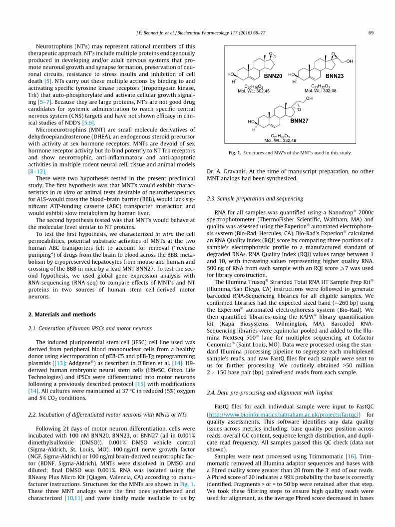
Fig. 1. Structures and MW’s of the MNT’s used in this study.
J.P. Bennett Jr. et al. / Biochemical Pharmacology 117 (2016) 68–77 69
Neurotrophins (NT’s) may represent rational members of thistherapeutic approach. NT’s include multiple proteins endogenouslyproduced in developing and/or adult nervous systems that pro-mote neuronal growth and synapse formation, preservation of neu-ronal circuits, resistance to stress insults and inhibition of celldeath [5]. NTs carry out these multiple actions by binding to andactivating specific tyrosine kinase receptors (tropomyosin kinase,Trk) that auto-phosphorylate and activate cellular growth signal-ing [5–7]. Because they are large proteins, NT’s are not good drugcandidates for systemic administration to reach specific centralnervous system (CNS) targets and have not shown efficacy in clin-ical studies of NDD’s [5,6].
Microneurotrophins (MNT) are small molecule derivatives ofdehydroepiandrosterone (DHEA), an endogenous steroid precursorwith activity at sex hormone receptors. MNTs are devoid of sexhormone receptor activity but do bind potently to NT Trk receptorsand show neurotrophic, anti-inflammatory and anti-apoptoticactivities in multiple rodent neural cell, tissue and animal models[8–12].
There were two hypotheses tested in the present preclinicalstudy. The first hypothesis was that MNT’s would exhibit charac-teristics in in vitro or animal tests desirable of neurotherapeuticsfor ALS-would cross the blood–brain barrier (BBB), would lack sig-nificant ATP-binding cassette (ABC) transporter interaction andwould exhibit slow metabolism by human liver.
The second hypothesis tested was that MNT’s would behave atthe molecular level similar to NT proteins.
To test the first hypothesis, we characterized in vitro the cellpermeabilities, potential substrate activities of MNTs at the twohuman ABC transporters felt to account for removal (‘‘reversepumping”) of drugs from the brain to blood across the BBB, meta-bolism by cryopreserved hepatocytes from mouse and human andcrossing of the BBB in mice by a lead MNT BNN27. To test the sec-ond hypothesis, we used global gene expression analysis withRNA-sequencing (RNA-seq) to compare effects of MNT’s and NTproteins in two sources of human stem cell-derived motorneurons.
2. Materials and methods
2.1. Generation of human iPSCs and motor neurons
The induced pluripotential stem cell (iPSC) cell line used wasderived from peripheral blood mononuclear cells from a healthydonor using electroporation of pEB-C5 and pEB-Tg reprogrammingplasmids ([13]; Addgene�) as described in O’Brien et al. [14]. H9-derived human embryonic neural stem cells (H9eSC, Gibco, LifeTechnologies) and iPSCs were differentiated into motor neuronsfollowing a previously described protocol [15] with modifications[14]. All cultures were maintained at 37 �C in reduced (5%) oxygenand 5% CO2 conditions.
2.2. Incubation of differentiated motor neurons with MNTs or NTs
Following 21 days of motor neuron differentiation, cells wereincubated with 100 nM BNN20, BNN23, or BNN27 (all in 0.001%dimethylsulfoxide (DMSO)), 0.001% DMSO vehicle control(Sigma-Aldrich, St. Louis, MO), 100 ng/ml nerve growth factor(NGF, Sigma-Aldrich) or 100 ng/ml brain-derived neurotrophic fac-tor (BDNF, Sigma-Aldrich). MNTs were dissolved in DMSO anddiluted; final DMSO was 0.001%. RNA was isolated using theRNeasy Plus Micro Kit (Qiagen, Valencia, CA) according to manu-facturer instructions. Structures for the MNTs are shown in Fig. 1.These three MNT analogs were the first ones synthesized andcharacterized [10,11] and were kindly made available to us by
Dr. A. Gravanis. At the time of manuscript preparation, no otherMNT analogs had been synthesized.
2.3. Sample preparation and sequencing
RNA for all samples was quantified using a Nanodrop� 2000cspectrophotometer (ThermoFisher Scientific, Waltham, MA) andquality was assessed using the Experion� automated electrophore-sis system (Bio-Rad, Hercules, CA). Bio-Rad’s Experion� calculatedan RNA Quality Index (RQI) score by comparing three portions of asample’s electrophoretic profile to a manufactured standard ofdegraded RNAs. RNA Quality Index (RQI) values range between 1and 10, with increasing values representing higher quality RNA.500 ng of RNA from each sample with an RQI score P7 was usedfor library construction.
The Illumina Truseq� Stranded Total RNA HT Sample Prep Kit�
(Illumina, San Diego, CA) instructions were followed to generatebarcoded RNA-Sequencing libraries for all eligible samples. Weconfirmed libraries had the expected sized band (�260 bp) usingthe Experion� automated electrophoresis system (Bio-Rad). Wethen quantified libraries using the KAPA� library quantificationkit (Kapa Biosystems, Wilmington, MA). Barcoded RNA-Sequencing libraries were equimolar pooled and added to the Illu-mina Nextseq 500� lane for multiplex sequencing at CofactorGenomics� (Saint Louis, MO). Data were processed using the stan-dard Illumina processing pipeline to segregate each multiplexedsample’s reads, and raw FastQ files for each sample were sent tous for further processing. We routinely obtained >50 million2 � 150 base pair (bp), paired-end reads from each sample.
2.4. Data pre-processing and alignment with Tophat
FastQ files for each individual sample were input to FastQC
(http://www.bioinformatics.babraham.ac.uk/projects/fastqc/) forquality assessments. This software identifies any data qualityissues across metrics including: base quality per position acrossreads, overall GC content, sequence length distribution, and dupli-cate read frequency. All samples passed this QC check (data notshown).
Samples were next processed using Trimmomatic [16]. Trim-momatic removed all Illumina adaptor sequences and bases witha Phred quality score greater than 20 from the 30 end of our reads.A Phred score of 20 indicates a 99% probability the base is correctlyidentified. Fragments > or = to 50 bp were retained after that step.We took these filtering steps to ensure high quality reads wereused for alignment, as the average Phred score decreased in bases
70 J.P. Bennett Jr. et al. / Biochemical Pharmacology 117 (2016) 68–77
toward the 30 end of reads across samples decreased (data notshown).
We next used the Burrows-Wheeler Aligner (BWA [17]), to cal-culate insert size metrics for each sample (including average sizeand standard deviation) to improve Tophat2 alignment. Once thesemetrics were obtained, we aligned each sample’s reads to the hg38human reference transcriptome then genome using Tophat2 [18].The hg38 reference transcriptome and genome were derived from
Illumina iGenomes UCSC hg38 directory (https://support.illu-
mina.com/sequencing/sequencing_software/igenome.html).
2.5. Cufflinks for gene abundance estimates
Gene and transcript abundances for all samples were estimated
using Cufflinks v2.2.1 (http://cufflinks.cbcb.umd.edu/howitworks.
html). We opted to mask all rRNA, tRNA, and mitochondrial RNAmapped reads from FPKM calculations, as these RNA speciesaccounted for different proportions of total mapped reads in eachsample. We only considered reads aligning to known genes (com-patible hits norm flag) in our FPKM calculations, as we suspectedthe number of novel transcripts varies across samples. We alsoused the genome bias and multi-hits correction flags, as recom-mended by the Tuxedo Suite developers.
2.6. Determination of P-gp substrate properties of microneurotrophindrugs (Cyprotex�, Watertown, MA Table 1)
Madin Darby Canine Kidney (MDCK) epithelial cells stablytransfected with the human MDR1 gene (the gene encoding forthe efflux protein P-glycoprotein, P-gp) are seeded on a Mul-tiscreenTM plate (MilliporeSigma, Billerica, MA) and form a conflu-ent monolayer over 4 days prior to the experiment. To verify theMDCK-MDR1 cell monolayers are properly formed, aliquots ofthe cell buffers are analyzed by fluorescence to determine thetransport of the impermeable dye Lucifer Yellow. Any deviationsfrom control values are reported.
For apical to basolateral (A? B) permeability, the test agent inthe absence or presence of 100 lM verapamil (a P-gp inhibitor)was added to the apical (A) side and the amount of permeationis determined on the basolateral (B) side; for basolateral to apical(B? A) permeability, the test agent in the absence and presenceof 100 lM verapamil was added to the B side and the amount ofpermeation was determined on the A side. The A-side buffer
Table 1MDCK-MDR1 Permeability and P-gp Substrate Identification.
Test Article TestConc.
AssayDuration
Mean A? B Papp(10�6 cm s�1)
Ranitidine 10 lM 2 h 0.22Warfarin 10 lM 2 h 28.2Loperamide 10 lM 2 h 1.1Loperamide + 100 lM
verapamil10 lM 2 h 46.6
BNN-20 10 lM 2 h 3.3BNN-20 + 100 lM verapamil 10 lM 2 h 4.4
BNN-27 10 lM 2 h 9.2BNN-27 + 100 lM verapamil 10 lM 2 h 9.6
BNN-23 10 lM 2 h 7.6BNN-23 + 100 lM verapamil 10 lM 2 h 8.7
Papp: apparent permeability rate coefficient; Efflux Ratio: Papp (B? A)/Papp (A? B)Conclusion: All 3 drugs exhibit moderate (or moderate-high) permeability rates across Mverapamil did not alter this minor efflux.
* Low post-assay recovery in the A? B transport direction; Low recovery may be duearticle.
contains 100 lM Lucifer yellow dye, in Transport Buffer (1.98 g/Lglucose in 10 mM HEPES, 1� Hank’s Balanced Salt Solution) pH7.4, while the B-side buffer has Transport Buffer at pH 7.4.MDCK-MDR1 cells are incubated with these buffers for 2 h, andthe receiver side buffer is removed for analysis by LC–MS/MS(using propranolol as an analytical internal standard).
The rate of passage of the test compounds through this cellmonolayer barrier from apical to basolateral (A? B) in this bi-directional transport assay is used to determine the apparent per-meability coefficient (Papp) = (dQ/dt)/(C0A) where dQ/dt is rate ofpermeation, C0 is initial concentration of test agent, and A is thearea of monolayer (0.11 cm2).
The basolateral to apical (B? A) to apical to basolateral (A? B)apparent permeability coefficient ratio
Re ¼ PappðB ! AÞPappðA ! BÞ
is used to determine the extent of efflux of the test compounds. Allexperiments were carried out in duplicate and averaged results areshown.
2.7. Determination of BBB-PAMPA permeability properties ofmicroneurotrophin drugs (Cyprotex�, Table 2)
Compounds (10 mM) in 100% DMSOwere dissolved in transportbuffer (pH 7.4) to a final concentration of 10 lM (finalDMSO = 0.1%). The filter membrane was coated with 4 ll of a20 mg/ml porcine brain lipid in dodecane. Three hundred (300)ll of the compound solution was added to the donor well. Theacceptor well was filled with 200 ll of transport buffer. The accep-tor filter plate was carefully placed on to the donor plate to create asandwich. The plate was left undisturbed for 18 h. Samples of thedonor and acceptor wells were analyzed by LC/MS/MS and theeffective permeability (Pe) was calculated using the followingequation: Data are expressed as permeability (Pe):
logPe ¼ f�ððVDVAÞ=ðVD þ VAÞAtÞlnf1� ½drug�A=½drug�Eggwhere Pe is the permeability, VD and VA are the volumes of thedonor and acceptor compartments, A is the area of the membrane,t is the incubation time, and A and E subscripts on the concentrationterm refer to the acceptor and equilibrium concentrations, respec-tively. All experiments were performed in quadruplicate and aver-aged results are shown.
Mean B? A Papp(10�6 cm s�1)
EffluxRatio
Comment
2.1 9.5 Low permeability35.1 1.2 High permeability71.9 65.4 P-gp efflux control48.4 1.1 P-gp substrate + inhibitor
8.0 2.410.7 2.4 BNN-20 is not a P-gp
substrate*
33.7 3.739.5 4.1 BNN-27 is not a P-gp
substrate30.7 4.057.3 6.6 BNN-23 is not a P-gp
substrate*
DR1-MDCK epithelial monolayers with only minor efflux evident. The inclusion of
to low aqueous solubility, high lipophilicity, and/or non-specific binding of the test
Table 2BBB-PAMPA Permeability.
TestArticle
AssayDuration
Mean Pe⁄
(x10�6 cm s�1)Recovery(%)
Comment
Atenolol 18 h 0.0000 111 Low permeabilitycontrol
Verapamil 18 h 34.9 42.2 High permeabilitycontrol
BNN-20 18 h 7.9 35.4 High permeabilityBNN-27 18 h 12.7 93.0 High permeabilityBNN-23 18 h 14.0 94.3 High permeability
Pe⁄: Effective Permeability coefficient.Conclusion: All 3 drugs exhibit high permeability rates across PAMPA-BBB mem-branes, predicting a high probability of passive BBB permeability.
J.P. Bennett Jr. et al. / Biochemical Pharmacology 117 (2016) 68–77 71
2.8. Determination of breast cancer resistance protein (bcrp)properties of microneurotrophin drugs (Cyprotex�, Table 3)
Caco-2 cells (human epithelial colorectal adenocarcinoma cells)grown in tissue culture flasks are trypsinized, suspended in med-ium, and the suspensions were applied to wells of a Millipore 96well Caco-2 plate. The cells are allowed to grow and differentiatefor 21 days. For Apical to Basolateral (A? B) permeability, the drugis added to the apical (A) side and amount of permeation is deter-mined on the basolateral (B) side; for Basolateral to Apical (B? A)permeability, the drug is added to the B side and the amount ofpermeation is determine on the A side. To verify a compound is aBCRP substrate, 50 lM of novobiocin is added to both chambersduring the equilibration step and for the duration of the assay.The A-side buffer contains 100 lM Lucifer yellow dye, in TransportBuffer (1.98 g/L glucose in 10 mM HEPES, 1� Hank’s Balanced SaltSolution) pH 7.4, and the B-side buffer is Transport Buffer, pH 7.4.Caco-2 cells are incubated with these buffers for 2 h, and the recei-ver side buffer is removed for analysis by LC–MS/MS. To verify thecell monolayers are properly formed, aliquots of the cell buffers areanalyzed by fluorescence to determine the transport of the imper-meable dye Lucifer Yellow. Any deviations from control values arereported.
Data are expressed as permeability Papp
Papp ¼ dQ=dtC0A
where dQ/dt is rate of permeation, C0 is initial concentration of testagent, and A is the area of monolayer. All experiments were per-formed in duplicate and averaged results are shown.
Table 3Caco-2 Cell Permeability and BCRP Substrate Identification.
Test Article TestConc.
AssayDuration
Mean A? B Papp(10�6 cm s�1)
ranitidine 10 lM 2 h 0.46warfarin 10 lM 2 h 21.5Estrone 3-sulfate 10 lM 2 h 0.52Estrone 3-sulfart + 50 lM
novobiocin10 lM 2 h 2.2
BNN-20 10 lM 2 h 1.7BNN-20 + 50 lM novobiocin 10 lM 2 h 2.2
BNN-27 10 lM 2 h 4.0BNN-27 + 50 lM novobiocin 10 lM 2 h 6.4
BNN-23 10 lM 2 h 1.9BNN-23 + 50 lM novobiocin 10 lM 2 h 3.0
Conclusion: All 3 drugs exhibit moderate permeability rates across Caco-2 epithelial monthe overall permeability profiles of these compounds.
* Low post-assay recovery in the A? B transport direction; Low recovery may be due t
2.9. In vitro and in vivo metabolism of BNN27
In vitro metabolism of BNN27 was carried out using cryopre-served hepatocytes from mice or humans. BNN27 was incubatedin duplicate with primary, cryopreserved hepatocytes at 37 �C.The cells were thawed; viable cells counted, and equilibratedaccording to the supplier’s directions. After 30 min equilibrationat 37 �C with gentle agitation, BNN27 was added into the cell sus-pension to give the desired final concentration of 3 lM. The cellsuspension was incubated at 37 �C as described above. At the indi-cated times, samples were removed and mixed with an equal vol-ume of ice-cold stop solution (methanol). In parallel, blankhepatocytes in the absence of BNN27 was incubated for 120 minand is used as a control to show the presence of peaks derived fromthe hepatocytes. Stopped reactions were incubated at least tenminutes on ice, and an additional volume of water was added.The samples are centrifuged to remove precipitated protein, andthe supernatants were analyzed by LC–MS/MS.
In vivo brain penetration and metabolism studies of BNN27used adult CD-1 mice (n = 4) given 50 mg/kg IP of BNN27 dissolvedin 10% DMSO; 90% of 10% hydroxypropyl-b-cyclodextrin in water.30 min after injection the brains were removed and divided in halfsagitally. One set of brain samples was homogenized in PBS buffer(2 ml per gram of tissue, on ice) and homogenates were mixedwith 3 volumes of methanol containing an analytical internal stan-dard (diclofenac) to extract the compound. Samples were cen-trifuged to remove precipitated protein and supernatantsanalyzed by LC–MS/MS. The second set of brain samples washomogenized in ice-cold absolute ethanol (2 ml per gram of tissue)and then stored overnight at�20 �C to extract. The following day, 3volumes of methanol containing an analytical internal standard(diclofenac) was added, and samples were vortexed and cen-trifuged to remove precipitated protein, followed by LC–MS/MSanalysis of the supernatants.
The analyte signal was optimized for each compound by elec-trospray ionization (ESI) in positive ionization mode. An MS2 scanwas used to optimize the precursor ion mass, and an SIM scan wasused to optimize the fragmenter voltage. Product ion analysis wasused to identify the best fragment and collision energy for analysis.Following MRM method optimization, a test injection was per-formed using the column/gradient conditions shown below. Aqualitative ionization ranking was assigned indicating the com-pound’s ease of ionization.
Samples were analyzed by LC–MS/MS using a SCIEX QTrap 5500mass spectrometer coupled with an Agilent 1290 HPLC Infinity ser-
Mean B? A Papp(10�6 cm s�1)
EffluxRatio
Comment
3.0 6.5 Low permeability ctl16.2 0.75 High permeability ctl18.2 35.0 BCRP efflux control2.2 1.0 BCRP substrate + inhibitor
3.7 2.22.0 0.90 BNN-20 is not a BCRP
substrate*
6.0 1.53.3 0.52 BNN-27 is not a BCRP
substrate*
3.7 1.94.6 1.5 BNN-23 is not a BCRP
substrate*
olayers with no efflux evident. The inclusion of novobiocin did not significantly alter
o low aqueous solubility, high lipophilicity, and/or non-specific binding of the drug.
72 J.P. Bennett Jr. et al. / Biochemical Pharmacology 117 (2016) 68–77
ies, a CTC PAL chilled autosampler, all controlled by Analyst soft-ware. After separation on a C18 reverse phase HPLC column(Acquity UPLC HSS T3, 1.8, 2.1 � 50 mm) using an acetonitrile-water gradient system, peaks were analyzed by mass spectrometry(MS) using ESI ionization in MRM mode. HPLC retention times (Rt)of metabolites varied within 5% or less from each other.
2.10. Data analysis
All graphs, curve fitting and statistical analyses were performedwith GraphPad Prism� for Macintosh, v 5.0–7.0. All RNA seq bioin-formatics processing was performed using Unix code on MacintoshOSX v.10� MacPro computers with 8 or 12 core processors and64 GB RAM.
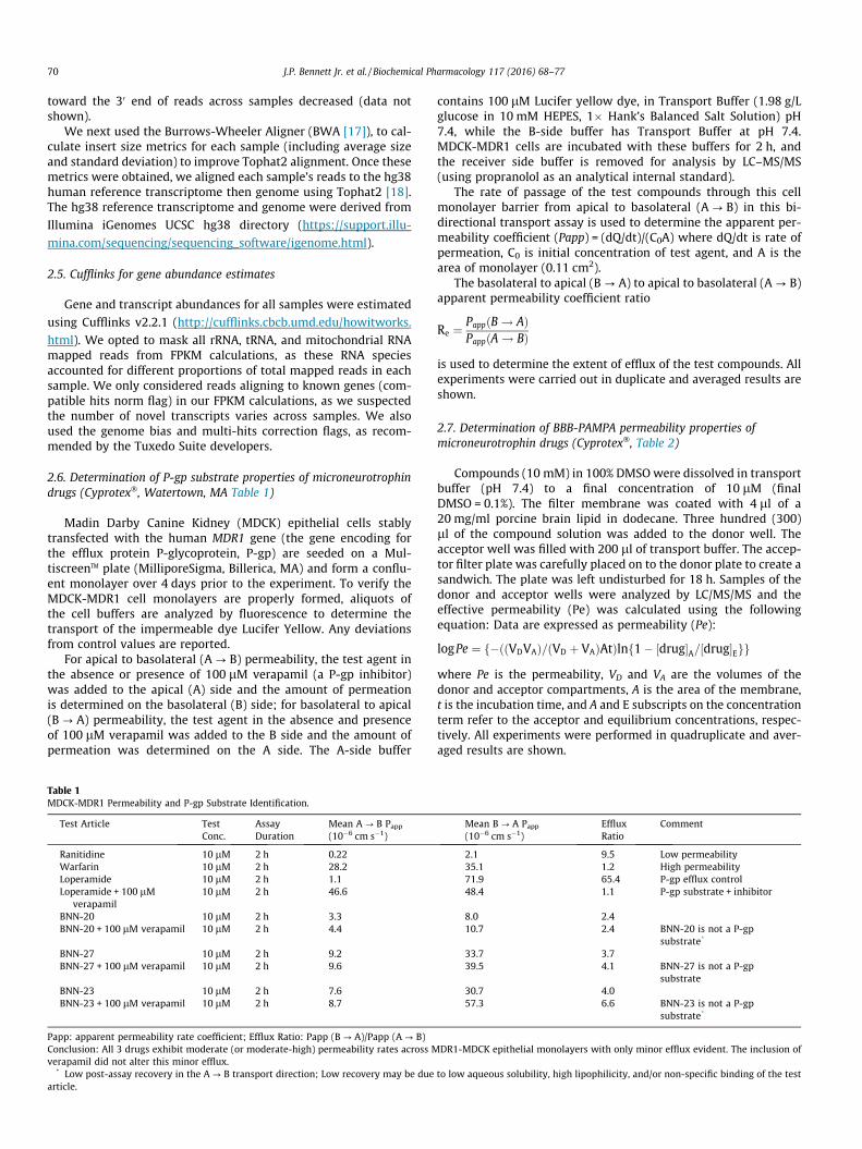
Fig. 2. BNN27 is metabolized rapidly by mouse cryopreserved hepatocytes (top)but more slowly by human cryopreserved hepatocytes (bottom). Experiments werecarried out with 10 lM initial [BNN27] and at 37�. [BNN27] in the supernatant wasanalyzed by LC/MS/MS at m/z of 315. Data were fit to one- or two-phase decaycurves in Prism�.
3. Results
3.1. MNT’s show very good cell permeability and are not majorsubstrates for blood–brain barrier ABC transporters
Tables 1–3 show that none of the three MNTs tested in vitrowere major substrates for human ABC transporters P-gp [19–22](Table 1) or BCRP [22,23] (Table 3). The efflux rates (B? A) ofBNN20 and BNN27 were reduced <50% by inclusion of novobiocin,suggesting weak interaction with BCRP transporter.
All three MNTs had high cellular permeability in a PAMPA assay(parallel artificial membrane permeability assay) (Table 2) inwhich the movement of drugs through a lipid-rich artificial mem-brane is measured. The effective permeability coefficients (Pe) forthe three MNT’s tested ranged from 7.9 to 14.0 (�10�6 cm/s), withthe high permeability CTL (verapamil) having a Pe of 34.9, and thelow permeability CTL having Pe of 0.00. These findings suggest thatMNTs will cross the BBB rapidly and will not be pumped out of thebrain by P-gp and will be pumped very little if at all by BRCP. Bothare known ABC transporters active at the BBB.
3.2. The lead MNT BNN27 is rapidly metabolized by mouse hepatocytesbut more slowly metabolized by human hepatocytes to likely singlehydroxylation species
Fig. 2 shows that cryopreserved mouse hepatocytes rapidlymetabolized BNN27 whereas human cryopreserved hepatocytesmore slowly metabolized BNN27. Both experiments were carriedout over 2 h. Fig. 3 shows metabolic profiling of BNN27 followingincubation with mouse (upper) or human (lower) cryopreservedhepatocytes. The detection was carried out at a mass/charge (m/z) ratio of 331, which represents the parent drug (m/z = 315) + 16to scan for possible single mono-oxidation (likely hydroxylation)metabolites. At least 7 metabolites of BNN27 were identified inmouse hepatocyte cultures, whereas 3–4 metabolites of BNN27were found in human hepatocyte cultures at much lower levels(�5-fold) (metabolite M1 present at very low levels). The individ-ual metabolites were not structurally identified in this study.
3.3. BNN27 and its metabolites can be detected in mouse brain 30 minafter injection
Fig. 4 shows that within 30 min of IP injection (50 mg/kg) ofBNN27, 6 mono-oxidation metabolites are detectable in mousebrain (n = 4 mice examined; shown are the results from mouse 2which were very representative of the group). BNN27 levels inwhole brain, assayed by LC/MS/MS at m/z of 315, 30 min after50 mg/kg IP injection, were 8230 ± 1995 (SEM) ng/gm with PBS/methanol extraction, and 9856 ± 1719 (SEM) ng/gm with ethanolextraction.
3.4. H9 eSC/NSC-derived motor neurons and Ctl iPSC/NSC-derivedmotor neurons have very similar gene expression
Fig. 5 shows that at the gene expression level, H9 eSC/NSC-derived motor neurons and Ctl iPSC/NSC-derived motor neuronswere very similar to each other. There was an excellent correlation(Pearson r = 0.92) of the log2FPKM values for 8904 gene pairs fromthe two MN cell lines.
3.5. Gene expression analyses reveal many similarities among MNT’sand NT’s effects on NSC-derived motor neurons
We used Cufflinks2 to acquire FPKM (Fragments Per Kilobase ofexon per Million reads) values in which NSC’s made from H9 eSC/NSC’s or iPSC’s derived from PBMC’s of a 62 year-old control indi-vidual without any neurological illness were differentiated intomotor neurons for 21 days, then exposed in duplicate for 24 h toneurotrophin proteins (NGF, BDNF) at 100 ng/ml or MNT’sBNN20, BNN23, BNN27 (see Fig. 1) at 100 nM, or to 0.001% DMSOvehicle. Total RNA was then extracted, analyzed for quality, bar-coded sequencing libraries were made and quantitated, followedby multiplex, paired-end read RNA sequencing. The reads werechecked for quality using FastQC�, trimmed of bar codes usingTrimmomatic� [16], then aligned against the hg38 version of the
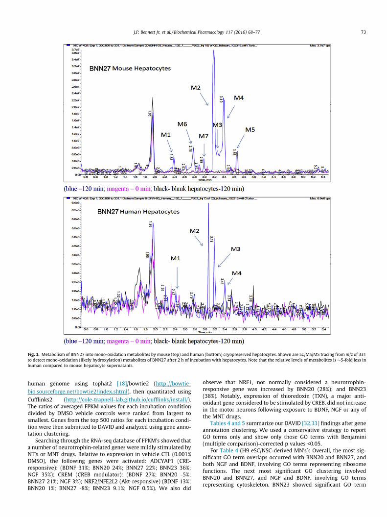
Fig. 3. Metabolism of BNN27 into mono-oxidation metabolites by mouse (top) and human (bottom) cryopreserved hepatocytes. Shown are LC/MS/MS tracing fromm/z of 331to detect mono-oxidation (likely hydroxylation) metabolites of BNN27 after 2 h of incubation with hepatocytes. Note that the relative levels of metabolites is �5-fold less inhuman compared to mouse hepatocyte supernatants.
J.P. Bennett Jr. et al. / Biochemical Pharmacology 117 (2016) 68–77 73
human genome using tophat2 [18]/bowtie2 (http://bowtie-
bio.sourceforge.net/bowtie2/index.shtml), then quantitated using
Cufflinks2 (http://cole-trapnell-lab.github.io/cufflinks/install/).The ratios of averaged FPKM values for each incubation conditiondivided by DMSO vehicle controls were ranked from largest tosmallest. Genes from the top 500 ratios for each incubation condi-tion were then submitted to DAVID and analyzed using gene anno-tation clustering.
Searching through the RNA-seq database of FPKM’s showed thata number of neurotrophin-related genes were mildly stimulated byNT’s or MNT drugs. Relative to expression in vehicle CTL (0.001%DMSO), the following genes were activated: ADCYAP1 (CRE-responsive): (BDNF 31%; BNN20 24%; BNN27 22%; BNN23 36%;NGF 35%); CREM (CREB modulator): (BDNF 27%; BNN20 -5%;BNN27 21%; NGF 3%); NRF2/NFE2L2 (Akt-responsive) (BDNF 13%;BNN20 1%; BNN27 -8%; BNN23 9.1%; NGF 0.5%). We also did
observe that NRF1, not normally considered a neurotrophin-responsive gene was increased by BNN20 (28%); and BNN23(38%). Notably, expression of thioredoxin (TXN), a major anti-oxidant gene considered to be stimulated by CREB, did not increasein the motor neurons following exposure to BDNF, NGF or any ofthe MNT drugs.
Tables 4 and 5 summarize our DAVID [32,33] findings after geneannotation clustering. We used a conservative strategy to reportGO terms only and show only those GO terms with Benjamini(multiple comparison)-corrected p values <0.05.
For Table 4 (H9 eSC/NSC-derived MN’s): Overall, the most sig-nificant GO term overlaps occurred with BNN20 and BNN27, andboth NGF and BDNF, involving GO terms representing ribosomefunctions. The next most significant GO clustering involvedBNN20 and BNN27, and NGF and BDNF, involving GO termsrepresenting cytoskeleton. BNN23 showed significant GO term
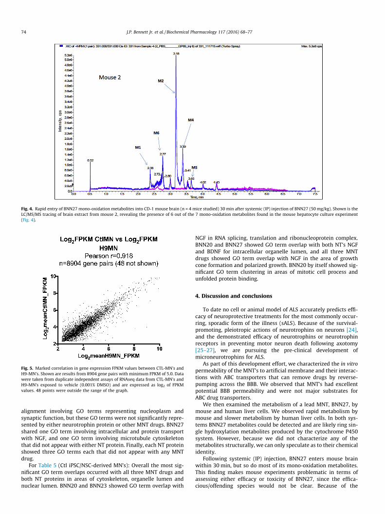
Fig. 4. Rapid entry of BNN27 mono-oxidation metabolites into CD-1 mouse brain (n = 4 mice studied) 30 min after systemic (IP) injection of BNN27 (50 mg/kg). Shown is theLC/MS/MS tracing of brain extract from mouse 2, revealing the presence of 6 out of the 7 mono-oxidation metabolites found in the mouse hepatocyte culture experiment(Fig. 4).
Fig. 5. Marked correlation in gene expression FPKM values between CTL-MN’s andH9-MN’s. Shown are results from 8904 gene pairs with minimum FPKM of 5.0. Datawere taken from duplicate independent assays of RNAseq data from CTL-MN’s andH9-MN’s exposed to vehicle (0.001% DMSO) and are expressed as log2 of FPKMvalues. 48 points were outside the range of the graph.
74 J.P. Bennett Jr. et al. / Biochemical Pharmacology 117 (2016) 68–77
alignment involving GO terms representing nucleoplasm andsynaptic function, but these GO terms were not significantly repre-sented by either neurotrophin protein or other MNT drugs. BNN27shared one GO term involving intracellular and protein transportwith NGF, and one GO term involving microtubule cytoskeletonthat did not appear with either NT protein. Finally, each NT proteinshowed three GO terms each that did not appear with any MNTdrug.
For Table 5 (Ctl iPSC/NSC-derived MN’s): Overall the most sig-nificant GO term overlaps occurred with all three MNT drugs andboth NT proteins in areas of cytoskeleton, organelle lumen andnuclear lumen. BNN20 and BNN23 showed GO term overlap with
NGF in RNA splicing, translation and ribonucleoprotein complex.BNN20 and BNN27 showed GO term overlap with both NT’s NGFand BDNF for intracellular organelle lumen, and all three MNTdrugs showed GO term overlap with NGF in the area of growthcone formation and polarized growth. BNN20 by itself showed sig-nificant GO term clustering in areas of mitotic cell process andunfolded protein binding.
4. Discussion and conclusions
To date no cell or animal model of ALS accurately predicts effi-cacy of neuroprotective treatments for the most commonly occur-ring, sporadic form of the illness (sALS). Because of the survival-promoting, pleiotropic actions of neurotrophins on neurons [24],and the demonstrated efficacy of neurotrophins or neurotrophinreceptors in preventing motor neuron death following axotomy[25–27], we are pursuing the pre-clinical development ofmicroneurotrophins for ALS.
As part of this development effort, we characterized the in vitropermeability of the MNT’s to artificial membrane and their interac-tions with ABC transporters that can remove drugs by reverse-pumping across the BBB. We observed that MNT’s had excellentpotential BBB permeability and were not major substrates forABC drug transporters.
We then examined the metabolism of a lead MNT, BNN27, bymouse and human liver cells. We observed rapid metabolism bymouse and slower metabolism by human liver cells. In both sys-tems BNN27 metabolites could be detected and are likely ring sin-gle hydroxylation metabolites produced by the cytochrome P450system. However, because we did not characterize any of themetabolites structurally, we can only speculate as to their chemicalidentity.
Following systemic (IP) injection, BNN27 enters mouse brainwithin 30 min, but so do most of its mono-oxidation metabolites.This finding makes mouse experiments problematic in terms ofassessing either efficacy or toxicity of BNN27, since the effica-cious/offending species would not be clear. Because of the
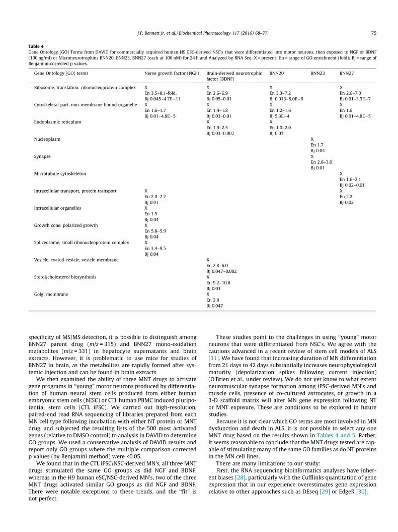
Table 4Gene Ontology (GO) Terms from DAVID for commercially acquired human H9 ESC-derived NSC’s that were differentiated into motor neurons, then exposed to NGF or BDNF(100 ng/ml) or Microneurotrophins BNN20, BNN23, BNN27 (each at 100 nM) for 24 h and Analyzed by RNA Seq. X = present; En = range of GO enrichment (fold); Bj = range ofBenjamini-corrected p values.
Gene Ontology (GO) terms Nerve growth factor (NGF) Brain-derived neurotrophicfactor (BDNF)
BNN20 BNN23 BNN27
Ribosome, translation, ribonucleoprotein complex XEn 3.5–8.1-fold,Bj 0.045–4.7E�11
XEn 2.6–6.9Bj 0.05–0.01
XEn 3.3–7.2Bj 0.013–8.0E�6
XEn 2.6–7.0Bj 0.01–3.3E�7
Cytoskeletal part, non-membrane bound organelle XEn 1.6–1.7Bj 0.01–4.8E�5
XEn 1.4–1.8Bj 0.03–0.01
XEn 1.2–1.6Bj 5.3E�4
XEn 1.6Bj 0.01–4.8E�5
Endoplasmic reticulum XEn 1.9–2.5Bj 0.03–0.002
XEn 1.0–2.0Bj 0.03
Nucleoplasm XEn 1.7Bj 0.04
Synapse XEn 2.6–3.0Bj 0.01
Microtubule cytoskeleton XEn 1.6–2.1Bj 0.02–0.01
Intracellular transport, protein transport XEn 2.0–2.2Bj 0.01
XEn 2.2Bj 0.02
Intracellular organelles XEn 1.5Bj 0.04
Growth cone, polarized growth XEn 5.8–5.9Bj 0.04
Spliceosome, small ribonucleoprotein complex XEn 3.4–9.5Bj 0.04
Vesicle, coated vesicle, vesicle membrane XEn 2.8–6.0Bj 0.047–0.002
Sterol/cholesterol biosynthesis XEn 9.2–10.8Bj 0.03
Golgi membrane XEn 2.8Bj 0.047
J.P. Bennett Jr. et al. / Biochemical Pharmacology 117 (2016) 68–77 75
specificity of MS/MS detection, it is possible to distinguish amongBNN27 parent drug (m/z = 315) and BNN27 mono-oxidationmetabolites (m/z = 331) in hepatocyte supernatants and brainextracts. However, it is problematic to use mice for studies ofBNN27 in brain, as the metabolites are rapidly formed after sys-temic injection and can be found in brain extracts.
We then examined the ability of three MNT drugs to activategene programs in ‘‘young” motor neurons produced by differentia-tion of human neural stem cells produced from either humanembryonic stem cells (hESC) or CTL human PBMC induced pluripo-tential stem cells (CTL iPSC). We carried out high-resolution,paired-end read RNA sequencing of libraries prepared from eachMN cell type following incubation with either NT protein or MNTdrug, and subjected the resulting lists of the 500 most activatedgenes (relative to DMSO control) to analysis in DAVID to determineGO groups. We used a conservative analysis of DAVID results andreport only GO groups where the multiple comparison-correctedp values (by Benjamini method) were <0.05.
We found that in the CTL iPSC/NSC-derived MN’s, all three MNTdrugs stimulated the same GO groups as did NGF and BDNF,whereas in the H9 human eSC/NSC-derived MN’s, two of the threeMNT drugs activated similar GO groups as did NGF and BDNF.There were notable exceptions to these trends, and the ‘‘fit” isnot perfect.
These studies point to the challenges in using ‘‘young” motorneurons that were differentiated from NSC’s. We agree with thecautions advanced in a recent review of stem cell models of ALS[31]. We have found that increasing duration of MN differentiationfrom 21 days to 42 days substantially increases neurophysiologicalmaturity (depolarization spikes following current injection)(O’Brien et al., under review). We do not yet know to what extentneuromuscular synapse formation among iPSC-derived MN’s andmuscle cells, presence of co-cultured astrocytes, or growth in a3-D scaffold matrix will alter MN gene expression following NTor MNT exposure. These are conditions to be explored in futurestudies.
Because it is not clear which GO terms are most involved in MNdysfunction and death in ALS, it is not possible to select any oneMNT drug based on the results shown in Tables 4 and 5. Rather,it seems reasonable to conclude that the MNT drugs tested are cap-able of stimulating many of the same GO families as do NT proteinsin the MN cell lines.
There are many limitations to our study:First, the RNA sequencing bioinformatics analyses have inher-
ent biases [28], particularly with the Cufflinks quantitation of geneexpression that in our experience overestimates gene expressionrelative to other approaches such as DEseq [29] or EdgeR [30].
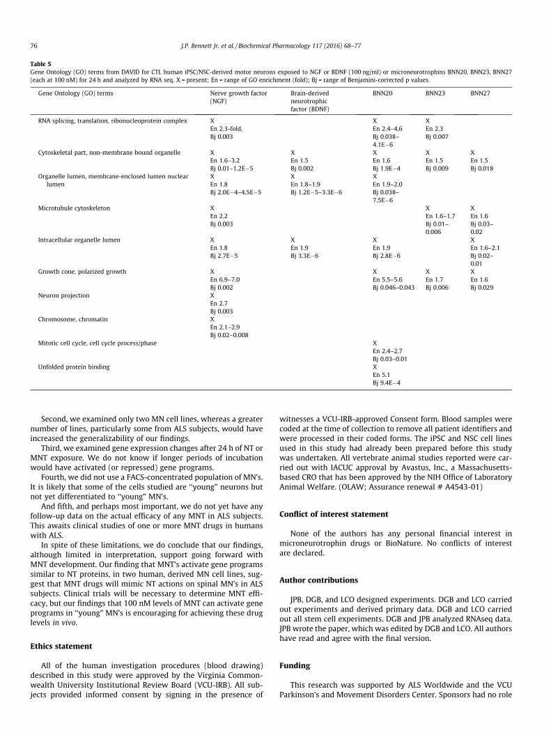
Table 5Gene Ontology (GO) terms from DAVID for CTL human iPSC/NSC-derived motor neurons exposed to NGF or BDNF (100 ng/ml) or microneurotrophins BNN20, BNN23, BNN27(each at 100 nM) for 24 h and analyzed by RNA seq. X = present; En = range of GO enrichment (fold); Bj = range of Benjamini-corrected p values.
Gene Ontology (GO) terms Nerve growth factor(NGF)
Brain-derivedneurotrophicfactor (BDNF)
BNN20 BNN23 BNN27
RNA splicing, translation, ribonucleoprotein complex XEn 2.3-fold,Bj 0.003
XEn 2.4–4.6Bj 0.038–4.1E�6
XEn 2.3Bj 0.007
Cytoskeletal part, non-membrane bound organelle XEn 1.6–3.2Bj 0.01–1.2E�5
XEn 1.5Bj 0.002
XEn 1.6Bj 1.9E�4
XEn 1.5Bj 0.009
XEn 1.5Bj 0.018
Organelle lumen, membrane-enclosed lumen nuclearlumen
XEn 1.8Bj 2.0E�4–4.5E�5
XEn 1.8–1.9Bj 1.2E�5–3.3E�6
XEn 1.9–2.0Bj 0.038–7.5E�6
Microtubule cytoskeleton XEn 2.2Bj 0.003
XEn 1.6–1.7Bj 0.01–0.006
XEn 1.6Bj 0.03–0.02
Intracellular organelle lumen XEn 1.8Bj 2.7E�5
XEn 1.9Bj 3.3E�6
XEn 1.9Bj 2.8E�6
XEn 1.6–2.1Bj 0.02–0.01
Growth cone, polarized growth XEn 6.9–7.0Bj 0.002
XEn 5.5–5.6Bj 0.046–0.043
XEn 1.7Bj 0.006
XEn 1.6Bj 0.029
Neuron projection XEn 2.7Bj 0.003
Chromosome, chromatin XEn 2.1–2.9Bj 0.02–0.008
Mitotic cell cycle, cell cycle process/phase XEn 2.4–2.7Bj 0.03–0.01
Unfolded protein binding XEn 5.1Bj 9.4E�4
76 J.P. Bennett Jr. et al. / Biochemical Pharmacology 117 (2016) 68–77
Second, we examined only two MN cell lines, whereas a greaternumber of lines, particularly some from ALS subjects, would haveincreased the generalizability of our findings.
Third, we examined gene expression changes after 24 h of NT orMNT exposure. We do not know if longer periods of incubationwould have activated (or repressed) gene programs.
Fourth, we did not use a FACS-concentrated population of MN’s.It is likely that some of the cells studied are ‘‘young” neurons butnot yet differentiated to ‘‘young” MN’s.
And fifth, and perhaps most important, we do not yet have anyfollow-up data on the actual efficacy of any MNT in ALS subjects.This awaits clinical studies of one or more MNT drugs in humanswith ALS.
In spite of these limitations, we do conclude that our findings,although limited in interpretation, support going forward withMNT development. Our finding that MNT’s activate gene programssimilar to NT proteins, in two human, derived MN cell lines, sug-gest that MNT drugs will mimic NT actions on spinal MN’s in ALSsubjects. Clinical trials will be necessary to determine MNT effi-cacy, but our findings that 100 nM levels of MNT can activate geneprograms in ‘‘young” MN’s is encouraging for achieving these druglevels in vivo.
Ethics statement
All of the human investigation procedures (blood drawing)described in this study were approved by the Virginia Common-wealth University Institutional Review Board (VCU-IRB). All sub-jects provided informed consent by signing in the presence of
witnesses a VCU-IRB-approved Consent form. Blood samples werecoded at the time of collection to remove all patient identifiers andwere processed in their coded forms. The iPSC and NSC cell linesused in this study had already been prepared before this studywas undertaken. All vertebrate animal studies reported were car-ried out with IACUC approval by Avastus, Inc., a Massachusetts-based CRO that has been approved by the NIH Office of LaboratoryAnimal Welfare. (OLAW; Assurance renewal # A4543-01)
Conflict of interest statement
None of the authors has any personal financial interest inmicroneurotrophin drugs or BioNature. No conflicts of interestare declared.
Author contributions
JPB, DGB, and LCO designed experiments. DGB and LCO carriedout experiments and derived primary data. DGB and LCO carriedout all stem cell experiments. DGB and JPB analyzed RNAseq data.JPB wrote the paper, which was edited by DGB and LCO. All authorshave read and agree with the final version.
Funding
This research was supported by ALS Worldwide and the VCUParkinson’s and Movement Disorders Center. Sponsors had no role

J.P. Bennett Jr. et al. / Biochemical Pharmacology 117 (2016) 68–77 77
in experimental design, data analysis, manuscript preparation ordecision to publish.
Acknowledgements
The authors thank Ms. Paula Keeney for assistance with cell cul-ture, Drs. A. Gravanis and C. Neophytou for providing the threeMNT analogs tested, colleagues at CoFactor Genomics� for carryingout the Illumina RNA sequencing, and Dr. B. Press and colleagues atCyprotex� for carrying out the permeability, ABC transporter andBNN27 metabolism assays. Primary RNA sequencing data aredeposited at NCBI’s Sequence Read Archive (SRA) under the studyaccession number SRP064478.
References
[1] A. Al-Chalabi, O. Hardiman, The epidemiology of ALS: a conspiracy of genes,environment and time, Nat. Rev. Neurol. 9 (2013) 617–628.
[2] A.E. Renton, A. Chio, B.J. Traynor, State of play in amyotrophic lateral sclerosisgenetics, Nat. Neurosci. 17 (2014) 17–23.
[3] M. Prudencio, V.V. Belzil, R. Batra, C.A. Ross, T.F. Gendron, L.J. Pregent, et al.,Distinct brain transcriptome profiles in C9orf72-associated and sporadic ALS,Nat. Neurosci. 18 (2015) 1175–1182.
[4] A. Jovicic, J. Mertens, S. Boeynaems, E. Bogaert, N. Chai, S.B. Yamada, et al.,Modifiers of C9orf72 dipeptide repeat toxicity connect nucleocytoplasmictransport defects to FTD/ALS, Nat. Neurosci. 18 (2015) 1226–1229.
[5] J.P. Steiner, A. Nath, Neurotrophin strategies for neuroprotection: are theysufficient? J. Neuroimmune Pharmacol. 9 (2014) 182–194.
[6] I. Mocchetti, M. Brown, Targeting neurotrophin receptors in the centralnervous system, CNS Neurol. Disord. Drug Targets 7 (2008) 71–82.
[7] I.G. Onyango, J.Y. Ahn, J.B. Tuttle, J.P. Bennett Jr., R.H. Swerdlow, Nerve growthfactor attenuates oxidant-induced beta-amyloid neurotoxicity in sporadicAlzheimer’s disease cybrids, J. Neurochem. 114 (2010) 1605–1618.
[8] I. Charalampopoulos, V.I. Alexaki, C. Tsatsanis, V. Minas, E. Dermitzaki, I.Lasaridis, et al., Neurosteroids as endogenous inhibitors of neuronal cellapoptosis in aging, Ann. N. Y. Acad. Sci. 1088 (2006) 139–152.
[9] I. Charalampopoulos, E. Remboutsika, A.N. Margioris, A. Gravanis,Neurosteroids as modulators of neurogenesis and neuronal survival, TrendsEndocrinol. Metab. 19 (2008) 300–307.
[10] T. Calogeropoulou, N. Avlonitis, V. Minas, X. Alexi, A. Pantzou, I.Charalampopoulos, et al., Novel dehydroepiandrosterone derivatives withantiapoptotic, neuroprotective activity, J. Med. Chem. 52 (2009) 6569–6587.
[11] A. Gravanis, T. Calogeropoulou, V. Panoutsakopoulou, K. Thermos, C.Neophytou, I. Charalampopoulos, Neurosteroids and microneurotrophinssignal through NGF receptors to induce prosurvival signaling in neuronalcells, Sci. Signal. 5 (2012). pt8.
[12] I. Pediaditakis, I. Iliopoulos, I. Theologidis, N. Delivanoglou, A.N. Margioris, I.Charalampopoulos, et al., Dehydroepiandrosterone: an ancestral ligand ofneurotrophin receptors, Endocrinology 156 (2015) 16–23.
[13] S.N. Dowey, X. Huang, B.K. Chou, Z. Ye, L. Cheng, Generation of integration-freehuman induced pluripotent stem cells from postnatal blood mononuclear cellsby plasmid vector expression, Nat. Protoc. 7 (2012) 2013–2021.
[14] L.C. O’Brien, P.M. Keeney, J.P. Bennett Jr., Differentiation of human neural stemcells into motor neurons stimulates mitochondrial biogenesis and decreasesglycolytic flux, Stem Cells Dev. 24 (2015) 1984–1994.
[15] M.W. Amoroso, G.F. Croft, D.J. Williams, S. O’Keeffe, M.A. Carrasco, A.R. Davis,et al., Accelerated high-yield generation of limb-innervating motor neuronsfrom human stem cells, J. Neurosci. 33 (2013) 574–586.
[16] A.M. Bolger, M. Lohse, B. Usadel, Trimmomatic: a flexible trimmer for Illuminasequence data, Bioinformatics (Oxford, England) 30 (2014) 2114–2120.
[17] H. Li, R. Durbin, Fast and accurate long-read alignment with Burrows-Wheelertransform, Bioinformatics (Oxford, England) 26 (2010) 589–595.
[18] D. Kim, G. Pertea, C. Trapnell, H. Pimentel, R. Kelley, S.L. Salzberg, TopHat2:accurate alignment of transcriptomes in the presence of insertions, deletionsand gene fusions, Genome Biol. 14 (2013) R36. doi: 10.1186/gb-2013-14-4-r36.
[19] E.M. Leslie, R.G. Deeley, S.P. Cole, Multidrug resistance proteins: role of P-glycoprotein, MRP1, MRP2, and BCRP (ABCG2) in tissue defense, Toxicol. Appl.Pharmacol. 204 (2005) 216–237.
[20] D.S. Miller, Regulation of P-glycoprotein and other ABC drug transporters atthe blood-brain barrier, Trends Pharmacol. Sci. 31 (2010) 246–254.
[21] A.L. Bartels, Blood-brain barrier P-glycoprotein function in neurodegenerativedisease, Curr. Pharm. Des. 17 (2011) 2771–2777.
[22] S. Agarwal, A.M. Hartz, W.F. Elmquist, B. Bauer, Breast cancer resistanceprotein and P-glycoprotein in brain cancer: two gatekeepers team up, Curr.Pharm. Des. 17 (2011) 2793–2802.
[23] K. Natarajan, Y. Xie, M.R. Baer, D.D. Ross, Role of breast cancer resistanceprotein (BCRP/ABCG2) in cancer drug resistance, Biochem. Pharmacol. 83(2012) 1084–1103.
[24] O. Schulte-Herbruggen, A. Braun, S. Rochlitzer, M.C. Jockers-Scherubl, R.Hellweg, Neurotrophic factors – a tool for therapeutic strategies inneurological, neuropsychiatric and neuroimmunological diseases? Curr. Med.Chem. 14 (2007) 2318–2329.
[25] S. Wiese, S. Jablonka, B. Holtmann, N. Orel, R. Rajagopal, M.V. Chao, et al.,Adenosine receptor A2A-R contributes to motoneuron survival bytransactivating the tyrosine kinase receptor TrkB, Proc. Natl. Acad. Sci. U.S.A.104 (2007) 17210–17215.
[26] H.U. Saragovi, E. Hamel, A. Di Polo, A neurotrophic rationale for the therapy ofneurodegenerative disorders, Curr. Alzheimer Res. 6 (2009) 419–423.
[27] S. Morcuende, R. Munoz-Hernandez, B. Benitez-Temino, A.M. Pastor, R.R. de laCruz, Neuroprotective effects of NGF, BDNF, NT-3 and GDNF on axotomizedextraocular motoneurons in neonatal rats, Neuroscience 250 (2013) 31–48.
[28] Z.H. Zhang, D.J. Jhaveri, V.M. Marshall, D.C. Bauer, J. Edson, R.K. Narayanan,et al., A comparative study of techniques for differential expression analysis onRNA-Seq data, PLoS One 9 (2014) e103207.
[29] M.I. Love, W. Huber, S. Anders, Moderated estimation of fold change anddispersion for RNA-seq data with DESeq2, Genome Biol. 15 (2014) 550.
[30] M.D. Robinson, D.J. McCarthy, G.K. Smyth, EdgeR: a bioconductor package fordifferential expression analysis of digital gene expression data, Bioinformatics(Oxford, England) 26 (2010) 139–140.
[31] S. Sances, L.I. Bruijn, S. Chandran, K. Eggan, R. Ho, J.R. Klim, et al., Modeling ALSwith motor neurons derived from human induced pluripotent stem cells, Nat.Neurosci. 16 (2016) 542–553.
[32] W. Huang da, B.T. Sherman, R.A. Lempicki, Systematic and integrative analysisof large gene lists using DAVID bioinformatics resources, Nat. Protoc. 4 (2009)44–57.
[33] W. Huang da, B.T. Sherman, R.A. Lempicki, Bioinformatics enrichment tools:paths toward the comprehensive functional analysis of large gene lists, NucleicAcids Res. 37 (2009) 1–13.