Embed Size (px)
Citation preview
بسم اهللا الرحمن الرحيمANTIMICROBIAL AND
PHARMACOLOGICAL PROPERTIES OF SOME MEDICINAL PLANTS
By
MANAL IBRAHIM ALI ABDALLA
B.V. Sc UNIVERSITY OF KHARTOUM
Supervisor
Dr. Afaf Izzeldin Abuelgasim
Faculty of Veterinary Medicine University of Khartoum
Co-Supervisor
Prof. Abd Alwahab Hassan Mohamed
Medicinal and Aromatic Plants Research Institutes,
Khartoum
A THESIS
Submitted in accordance with the requirements of the
University of Khartoum for the Degree of
Master of Veterinary science
March- 2008

i
Dedication
I dedicate this thesis to my loving parents
Who painted my bootsteps to the path of learning
To my sisters, brothers and my lovely son Abdullah
The real meaning of the life, for thoughtfulness,
Support and encouragement.
Manal Ibrahim.

ii
ACKNOWLEDGEMENTS
I would like to express my gratitude and sincere appreciation to my
supervisor Dr. Afaf Izzeldin Abuelgasim Department of pathology,
University of Khartoum, for her guidance, encouragement and patience,
and my special gratitude is to my Co- supervisor Prof. Abd Alwahab
Hassan Mohamed Department of Pharmacology and Toxicology,
Medicinal and Aromatic Plants Research Institute Khartoum, for his
constant encouragement, support, patience and enthusiasm, help and
invaluable remarks throughout the research . I am sincerely grateful to
Prof. Aiesha Zohier Almagboul Director of Medicinal and Aromatic
Plants Research Institute for her helps, patience and encouragement.
Thanks are extended to the technical staff and other supporting
personnel of the Medicinal and Aromatic Plants Research Institute
Khartoum.
I am grateful to Faculty of Veterinary Medicine and University of
Khartoum for facilities placed at my disposal through out the duration of
this work, special thanks to the staff of department of pathology.

iii
LIST OF CONTENTS
PageDEDICATION i ACKNOWLEDGEMENT ii LIST OF CONTENT iii LIST OF TABLES v LIST OF FIGURES vi ABSTRACT viii ARABIC ABSTRACT ix INTRODUCTION 1 CHAPTER ONE: LIETERATURE REVIEW 3 1.1. Plants as source of drugs 4 1.2 Antimicrobial plants 6 1.3 Toxicity of the medicinal plants 9 1.4 Pharmacological studies 10
1.4.1 Cholinergic agonists and antagonists 10 1.4.2 Cholinesterase inhibitor 11
1.4.3 Atropine 12 1.4.4 Histamine 13 1.4.5 Chlorphenramine 13 1.4.6 5- Hydroxytryptamine (5-HT) 14 1.4.7 Cyproheptadine (Periactin) 16
1.5 Plants used in this study 16 1.5.1 Monechma ciliatum 16 1.5.2 Lepidium sativum 18 1.5.3 Linum usitatissimum 19
CHAPTER TWO: MATERIALS AND METHODS 25 2.1 Materials and Experimental Designs 25
2.1.1 Plants 25 2.1.2 Animals 25 2.1.3 Microbes 25 2.1.4 Toxicity of Methanolic extract of Monechma
ciliatum leaves 26 2.1.5 Experimental design 26 2.1.6 Parameters 26
2.2 Methods 26 2.2.1 Preparation of the plants extraction 26 2.2.2 Testing for Antimicrobial activities 27 2.2.2.1 Testing for Antibacterial activities 27 2.2.2.2 Testing for Antifungal activities 28
iv
Page 2.2.3 Pharmacological experiments 28 2.2.3.1 Rabbit jejunum 28 2.2.3.2 Guinea pig ileum 29 2.2.4.3 Rat fundus 29 2.2.4.4 Rat uterus 30 2.2.5 Blood samples 30 2.2.5.1 White blood cell count 30 2.2.5.2 Differential leucocytes count 31 2.2.6 Biochemical Analysis 31 2.2.6.1 Total Bilirubin 31 2.2.6.2 Total Cholesterol 31 2.2.6.3 Total Protein 32 2.2.6.4 Total Albumin 33 2.2.7 Histopathological methods 33 2.2.8 Statistical analysis 33
CHAPTER THREE: RESULTS 34 3.1 Antimicrobial activities of the plants 34
3.1.1 Antimicrobial activities of Monechma ciliatum stems against standard organisms 34
3.1.2 Antimicrobial activities of Monechma ciliatum leavess against standard organisms 34
3.1.3 Antimicrobial activities of Linum usitatissimum seeds against standard organisms 35
3.1.4 Antimicrobial activities of Lepidiu sativum seeds against standard organisms 36
3.2 Pharmacological activities of methanol extract of Monechma ciliatum leaves in different isolated 47
3.2.1 Rabbit jejunum 47 3.2.2 Guinea pig ileum 47 3.2.3 Rat fundus 47 3.2.4 Rat uterus 47
3.3 Effect of methanol extract of Monechma ciliatum in rats 55 3.3.1 Clinical finding 55 3.3.2 Haematological changes 55 3.3.3 Postmortem Finding 55 3.3.4 Histopathological findings 55 3.3.5 Serum constituents changes 55
CHAPTERFOUR: DISCUSSION 63 CONCLUSION AND RECOMMENDATION 69 REFERENCES 70

v
List of tables
Table Page
1 Antimicrobial activity of Monelchma ciliatum stems extracts against standard organisms 37
2 Antimicrobial activity of Monelchma ciliatum leaves extracts against standard organisms 38
3 Antimicrobial activity of Linum usitatissimum seeds extracts against standard organisms 39
4 Antimicrobial activity of Lepidium sativum seeds extracts against standard organisms 40
5 White blood cell and differential count of rats
treated with methanolic extract of Monechma ciliatium leaves
56
6 Serum constituents values in rats treated with methanol extract of Monelchma ciliatum leaves 58
vi
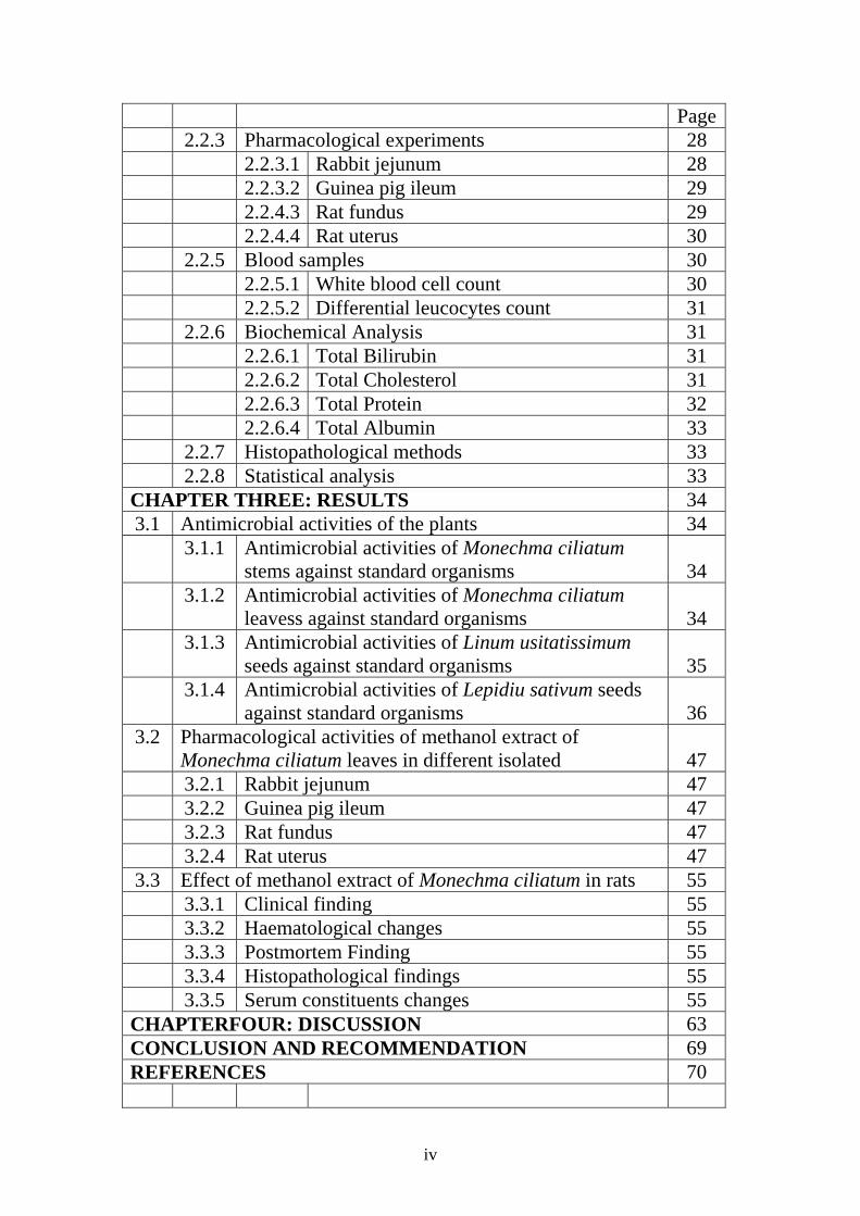
List of Figures
Page No. Figure 20 Monechema ciliatum leaves 1 21 Monechema ciliatum leaves and fruits 2 22 Lepidium sativum whole plant3
22 Lepidium sativum leaves4
23 Lepidium sativum seeds 5 24 Linum usitatissimum plant 6 24 Linum usitatissimum seeds 7
41 Antimicrobial activity of methanolic extract of Monelchma ciliatum leaves on Escherichia coli
8
42 Antimicrobial activity of methanolic extract of Monelchma ciliatum leaves on Staphylococcus aureus
9
43 Antimicrobial activity of methanolic extract of Monelchma ciliatum leaves on Candida albicans
10
44 Antimicrobial activity of methanolic extract of Monelchma ciliatum leaves on Pseudomonas aeruginosa
11
45 Antimicrobial activity of methanolic extract of Monelchma ciliatum leaves on Bacillus subtilis
12
46 Antimicrobial activity of methanolic extract of Monelchma ciliatum leaves on Aspergillus niger
13
47 Effect of methanolic extract of Monelchma ciliatum leaves on isolated Rabbit jejunum
14
49 Effect of methanolic extract of Monelchma ciliatum leaves on isolated rabbit jejunum
15
50 Effect of methanolic extract of Monelchma ciliatum leaves on isolated Guinea pig ileum
16
51 Effect of methanolic extract of Monelchma ciliatum leaves on isolated Guinea pig ileum
17
52 Effect of methanolic extract of Monelchma ciliatum leaves on isolated Guinea pig ileum
18
53 Effect of methanolic extract of Monelchma ciliatum leaves on isolated Rat fundus
19
vii
54 Effect of methanolic extract of Monelchma ciliatum leaves on isolated Rat uterus
20
57 White blood cell count in rats given methanol extract of Monechma ciliatum.
21
57 Differential count in rats given methanol extract of Monechma ciliatum.
22
59 Changes in serum protein in rats given methanolic extract of Monechema ciliatum leaves
23
59 Changes in serum albumin in rats given methanolic extract of Monechema ciliatum leaves
24
60
Changes in serum cholesterol in rats given methanolic extract of Monechema ciliatum leaves
25
60 Changes in serum Bilirubin in rats given methanolic extract of Monechema ciliatum leaves
26
61
Liver of a rat given methanolic extract of
Monechma ciliatum leaves
27
61
Intestine of a rat given methanolic extract of
Monechma ciliatum leaves
28
62
Kidney of a rat given methanolic extract of
Monechma ciliatum leaves
29
viii
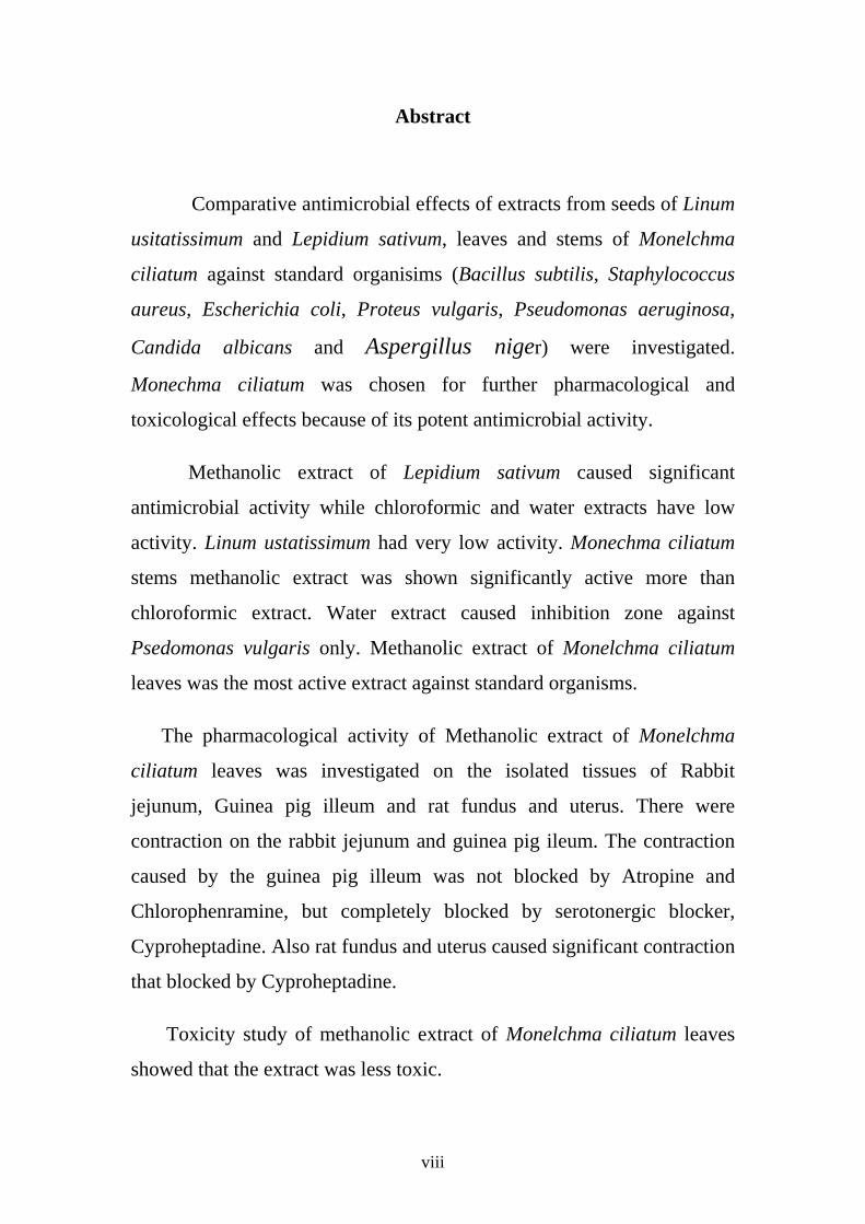
Abstract
Comparative antimicrobial effects of extracts from seeds of Linum
usitatissimum and Lepidium sativum, leaves and stems of Monelchma
ciliatum against standard organisims (Bacillus subtilis, Staphylococcus
aureus, Escherichia coli, Proteus vulgaris, Pseudomonas aeruginosa,
Candida albicans and Aspergillus niger) were investigated.
Monechma ciliatum was chosen for further pharmacological and
toxicological effects because of its potent antimicrobial activity.
Methanolic extract of Lepidium sativum caused significant
antimicrobial activity while chloroformic and water extracts have low
activity. Linum ustatissimum had very low activity. Monechma ciliatum
stems methanolic extract was shown significantly active more than
chloroformic extract. Water extract caused inhibition zone against
Psedomonas vulgaris only. Methanolic extract of Monelchma ciliatum
leaves was the most active extract against standard organisms.
The pharmacological activity of Methanolic extract of Monelchma
ciliatum leaves was investigated on the isolated tissues of Rabbit
jejunum, Guinea pig illeum and rat fundus and uterus. There were
contraction on the rabbit jejunum and guinea pig ileum. The contraction
caused by the guinea pig illeum was not blocked by Atropine and
Chlorophenramine, but completely blocked by serotonergic blocker,
Cyproheptadine. Also rat fundus and uterus caused significant contraction
that blocked by Cyproheptadine.
Toxicity study of methanolic extract of Monelchma ciliatum leaves
showed that the extract was less toxic.
ix
وحةملخص األطر
أجريت هذه الدراسة على ثالثة نباتات طبية بذور الكتان ،حـب االرشـاد وسـيقان
وأوراق المحلب األسود وذلك لمعرفة نشاطاتها ضد الميكروبات، ومن ثـم اختيـار أفـضل
. وسميته )الفارماكولوجي(مستخلص فعال إلختبار نشاطه الدوائي
Staphylococcus الميكروبات القياسية المكـورة الذهبيـة تم إختبار النباتات على
aureus االشـرنكية القولونيـة ،Escherichia coli، Pseudomonas aeruginosa ،
Bacillus subtilis،Proteus vulgaris ، Aspergillus niger وCandida
albicans.
ـ عف نـشاط أثبتت التجربة نشاط المستخلص الميثيلي لنبـات حـب الرشـاد و ض
المستخلص المائي والكلوروفورمي له ضد الميكروبات القياسية، بينما كـان نـشاط جميـع
مستخلصات نبات بذرة الكتان التي اختبرت ضعيفة األثـر مقارنـة بمستخلـصات النبـاتين
.األخريين
أما أوراق نبات المحلب األسود كانت أكثر نشاطا خاصة المستخلص الميثيلـي الـذي
أعطي أفضل النتائج وقد أعطت سيقان نبات المحلب األسود نشاطاًًًًًً متوسطاً ضد الميكروبـات
.القياسية
ي تم إختبار المستخلص الميثيلي ألوراق نبات المحلب األسود علي األنسجة المعزولة وه
.عفج األرنب، أمعاء خنزير غينيا والجزء الفؤادي للمعدة والرحم من الجرز األبيض
قام المستخلص بتحفيز كل من معدة األرنب وأمعاء خنزير غينيا . لمعرفة خصائصه الدوائية
حيث لم يتمكن األتروبين والكلوروفنرامين من قفل هذا األثر بينما تم قفـل هـذا اإلنقبـاض
.سبروهبتادينبواسطة ال
كما تم اختبار المستخلص الميثيلي ألوراق نبات المحلب األسود علي معـدة ورحـم
الجرز حيث أعطي المستخلص انقباضا في كل من النسيجين والذي تم قفلـه كليـة بواسـطة
.السبروهيبتادين
نبـات أثبتت التجربة التي تمت في الجرزان لقياس سمية المستخلص الميثيلي ألوراق
. المحلب األسود ان المستخلص قليل السمية
1
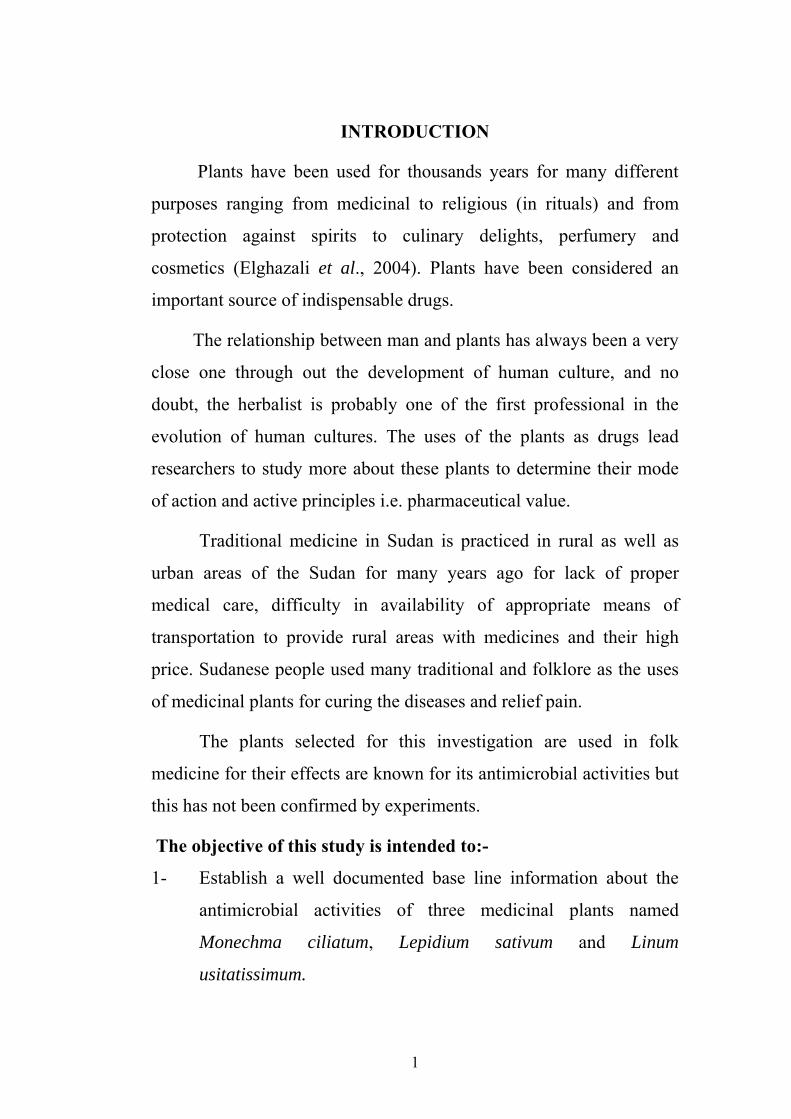
INTRODUCTION
Plants have been used for thousands years for many different
purposes ranging from medicinal to religious (in rituals) and from
protection against spirits to culinary delights, perfumery and
cosmetics (Elghazali et al., 2004). Plants have been considered an
important source of indispensable drugs.
The relationship between man and plants has always been a very
close one through out the development of human culture, and no
doubt, the herbalist is probably one of the first professional in the
evolution of human cultures. The uses of the plants as drugs lead
researchers to study more about these plants to determine their mode
of action and active principles i.e. pharmaceutical value.
Traditional medicine in Sudan is practiced in rural as well as
urban areas of the Sudan for many years ago for lack of proper
medical care, difficulty in availability of appropriate means of
transportation to provide rural areas with medicines and their high
price. Sudanese people used many traditional and folklore as the uses
of medicinal plants for curing the diseases and relief pain.
The plants selected for this investigation are used in folk
medicine for their effects are known for its antimicrobial activities but
this has not been confirmed by experiments.
The objective of this study is intended to:-
1- Establish a well documented base line information about the
antimicrobial activities of three medicinal plants named
Monechma ciliatum, Lepidium sativum and Linum
usitatissimum.

2
2- To select the most potent antimicrobial plant extract and to test
their pharmacological activity in isolated organs.
3- To investigate the toxicity of this selected plant.

3
CHAPTER ONE
LITERATURE REVIEW
The use of plants for treatment of various diseases, as specific
against magic and for religious ceremonies is universal, and has been
practiced for many years; even up to now where many people are
treated by modern drugs, in East Africans, both literate and illiterate
people still use local plants as drugs in many conditions (Kokwaro,
1976).
Chinese medicinal plants, which have been used for centuries,
are still being widely used today within the framework of health care
services. A recent nation wide survey has shown that there are 7295
species of plants used medicinally in China (Ciba Found, 1994).
However, he reported that in an ethno pharmacological investigation
of 100 species 15 species with a high proportion of therapeutic agents
was found. He also reported that medicinal plants are an important
source for new drug development in China. Up to now about 200 new
drugs have been developed directly or indirectly from medicinal
plants. Nearly one half of the new drugs are from a single medicinal
plant, or its active principle and synthetic derivatives, or active
fraction or even the total extract.
Through antimicrobial agents of synthetic origin and those
made by fermentation processes, commonly used in therapeutics, no
use of antibiotic of higher plant origin has been reported. However,
plants may represent a potential source of antibiotic (Aizenman,
1978).

4
Furthermore, in the various health care systems of the world,
there are no accurate data available to assess the value and extent of
use of plants or their derived active principles. However, the World
Health Organization has estimated that 80% or more than four billion
inhabitants of the world rely primarily on traditional medicine for
primary health care need. It can be presumed that major part of
traditional medicine involved the use of plant extracts and their
derived active principles, though their use is not always verified by
scientific means (Aizenman, 1978).
For many years, Africans people have developed a store of
empirical information concerning the therapeutic values of Africans
plants (Kokwaro, 1976). He also stated that, some of the plants used
for direct or specific treatment of particular disease are worth further
investigation in order to determine their chemical composition and
their mechanism of action.
In the Sudan medicinal folklore passed from one generation to
another but was never documented (Elghzali et al., 1997).
1.1. Plants as source of drugs:-
Acaia nilotica is traditionally used to treat sore throat, colds,
bronchitis, pneumonia, opthalmia, diarrhoea, dysentery, leprosy
venereal diseases and haemorrhages. Aqueous extract of fruits showed
activity against bacteria and fungi (Abdel Nabi et al., 1992). Elgazali
et al. (2003) reported that, maceration of the roots of Acaia
polyacantha is used for diarrhoea and vomiting.
Momordica cabrei is used for treatment of malaria and
cutaneus abcess (Adjanohoum, et al., 1991). Awatif et al. (2001)

5
found that Khaya sengalensis is used to treat malaria and for wound
healing.
Some medicinal plants can destroy helminthic parasites by
different mechanisms. Ananas comosus, Pineapple (Bromeliaceae)
contains the proteolytic enzyme bromelain, and can destroy the
parasite through lysis (Oliver-Bever, 1986). He also reported that
other plants can act specifically on cestodes, especially those
containing cucurbitine as from Cucurbita pepo, Pumkin and
shammam (Curcurbitaceae) and saponins from Opilia cettidifolia.
The powder of the whole plant of Barleria ruellioides mixed with
Gossypium seeds are used externally against scabies (Elgazali et al.,
2003). They also reported that decoction of the barks of Anogessus
leiocarpus are used against cough while maceration of whole plant of
Clitoria ternatea is used against constipation and Dicoma tomentosa
Cass leaves used to relief tooth pains.
Vernolepin and vernodalin were isolated from leaves of
Vernonia amygdalina, both compounds exhibited activity against a
panel of bacteria (Almagboul et al., 1997).
The decoction of the barks of Gynocarpus jacquinii is used
against cancer and renal troubles whereas the powdered fruits mixed
with sesame oil for reheumatism (Elgazali et al, 2003). The same
authors found that, maceration of whole plant of Polycarpea
corymbosa is used as antifungal remedy .Wissadula rostrata the
powdered seeds mixed with (Alkohl) are used to treat conjunctivitis
and eye infection.
6
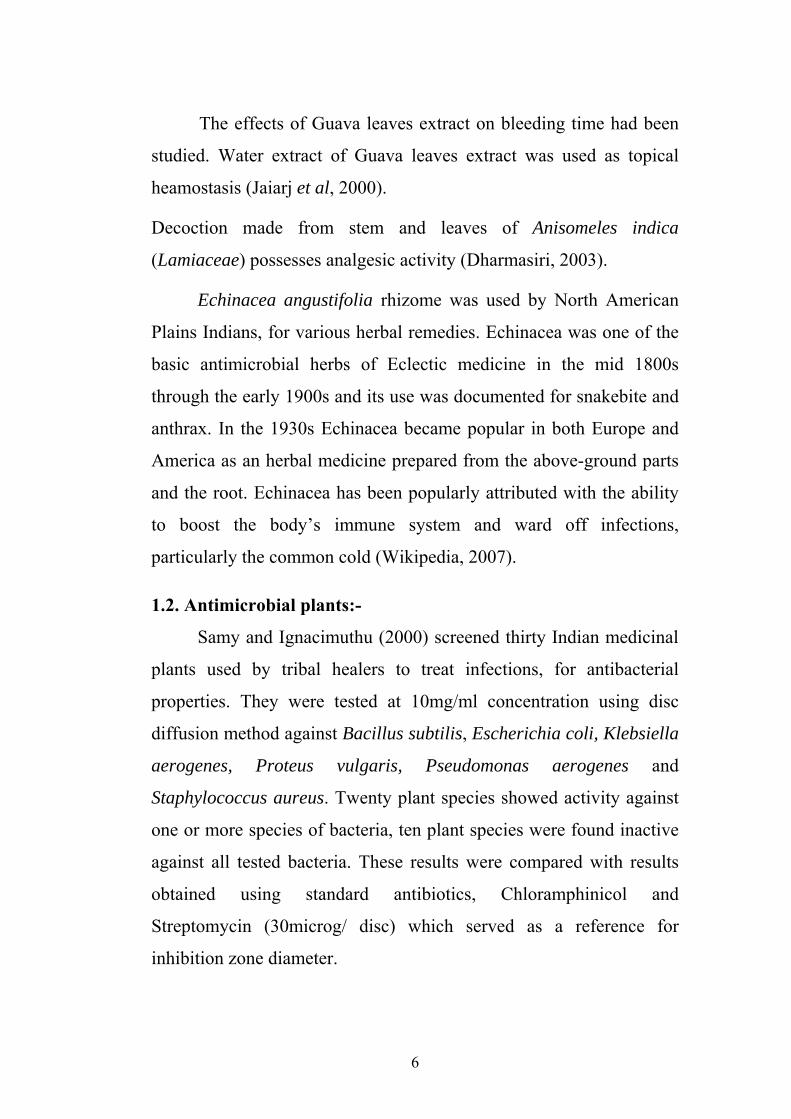
The effects of Guava leaves extract on bleeding time had been
studied. Water extract of Guava leaves extract was used as topical
heamostasis (Jaiarj et al, 2000).
Decoction made from stem and leaves of Anisomeles indica
(Lamiaceae) possesses analgesic activity (Dharmasiri, 2003).
Echinacea angustifolia rhizome was used by North American
Plains Indians, for various herbal remedies. Echinacea was one of the
basic antimicrobial herbs of Eclectic medicine in the mid 1800s
through the early 1900s and its use was documented for snakebite and
anthrax. In the 1930s Echinacea became popular in both Europe and
America as an herbal medicine prepared from the above-ground parts
and the root. Echinacea has been popularly attributed with the ability
to boost the body’s immune system and ward off infections,
particularly the common cold (Wikipedia, 2007).
1.2. Antimicrobial plants:-
Samy and Ignacimuthu (2000) screened thirty Indian medicinal
plants used by tribal healers to treat infections, for antibacterial
properties. They were tested at 10mg/ml concentration using disc
diffusion method against Bacillus subtilis, Escherichia coli, Klebsiella
aerogenes, Proteus vulgaris, Pseudomonas aerogenes and
Staphylococcus aureus. Twenty plant species showed activity against
one or more species of bacteria, ten plant species were found inactive
against all tested bacteria. These results were compared with results
obtained using standard antibiotics, Chloramphinicol and
Streptomycin (30microg/ disc) which served as a reference for
inhibition zone diameter.
7
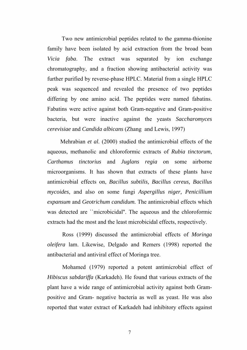
Two new antimicrobial peptides related to the gamma-thionine
family have been isolated by acid extraction from the broad bean
Vicia faba. The extract was separated by ion exchange
chromatography, and a fraction showing antibacterial activity was
further purified by reverse-phase HPLC. Material from a single HPLC
peak was sequenced and revealed the presence of two peptides
differing by one amino acid. The peptides were named fabatins.
Fabatins were active against both Gram-negative and Gram-positive
bacteria, but were inactive against the yeasts Saccharomyces
cerevisiae and Candida albicans (Zhang and Lewis, 1997)
Mehrabian et al. (2000) studied the antimicrobial effects of the
aqueous, methanolic and chloroformic extracts of Rubia tinctorum,
Carthamus tinctorius and Juglans regia on some airborne
microorganisms. It has shown that extracts of these plants have
antimicrobial effects on, Bacillus subtilis, Bacillus cereus, Bacillus
mycoides, and also on some fungi Aspergillus niger, Penicillium
expansum and Geotrichum candidum. The antimicrobial effects which
was detected are ``microbicidal''. The aqueous and the chloroformic
extracts had the most and the least microbicidal effects, respectively.
Ross (1999) discussed the antimicrobial effects of Moringa
oleifera lam. Likewise, Delgado and Remers (1998) reported the
antibacterial and antiviral effect of Moringa tree.
Mohamed (1979) reported a potent antimicrobial effect of
Hibiscus sabdariffa (Karkadeh). He found that various extracts of the
plant have a wide range of antimicrobial activity against both Gram-
positive and Gram- negative bacteria as well as yeast. He was also
reported that water extract of Karkadeh had inhibitory effects against

8
the standard organism namly, Staphylococcus aureus, Escherichia
coli, Pseudomonas aeruginosa and Bacillus subtils. Waffaa (1989)
found that all concentrations of the water extract of karkadeh possess
antibacterial activity. The diameter of the inhibitory zones increased
with the increase in concentration.
A number of essential oils from Mongolian aromatic plants are
claimed to have antimicrobial activities. The essential oil of
Dracocephalum foetidum, a popular essential oil used in Mongolian
traditional medicine, was examined for its antimicrobial activity.
The essential oil of Dracocephalum foetidum exhibited strong
antimicrobial activity against most of these pathogenic bacteria and
yeast strains by both the agar diffusion method and the minimum
inhibitory concentration (MIC) assay (MIC rang was 26-2592
microg/ml). Interestingly, Dracocephalum foetidum even showed
antimicrobial activity against methicillin-resistant Staphylococcus
aureus (MRSA) strains (Lee, et al, 2007).
Molina-Salinas et al. (2007) evaluate the potential antimicrobial
activity of 14 plants used in northeast Mexico for the treatment of
respiratory diseases, against drug-sensitive and drug-resistant strains
of Streptococcus pneumoniae, Staphylococcus aureus, Haemophilus
influenzae and Mycobacterium tuberculosis. Organic and aqueous
extracts were tested against these bacterial strains. The aqueous
extracts showed no antimicrobial activity, whereas most of the organic
extracts presented antimicrobial activity against at least one of the
drug-resistant microorganisms tested. Methanol-based extracts from
the roots and leaves of Leucophyllum frutescens and ethyl ether
extract from the roots of Chrysanctinia mexicana showed the greatest
9
antimicrobial activity against the drug-resistant strain of
Mycobacterium tuberculosis.
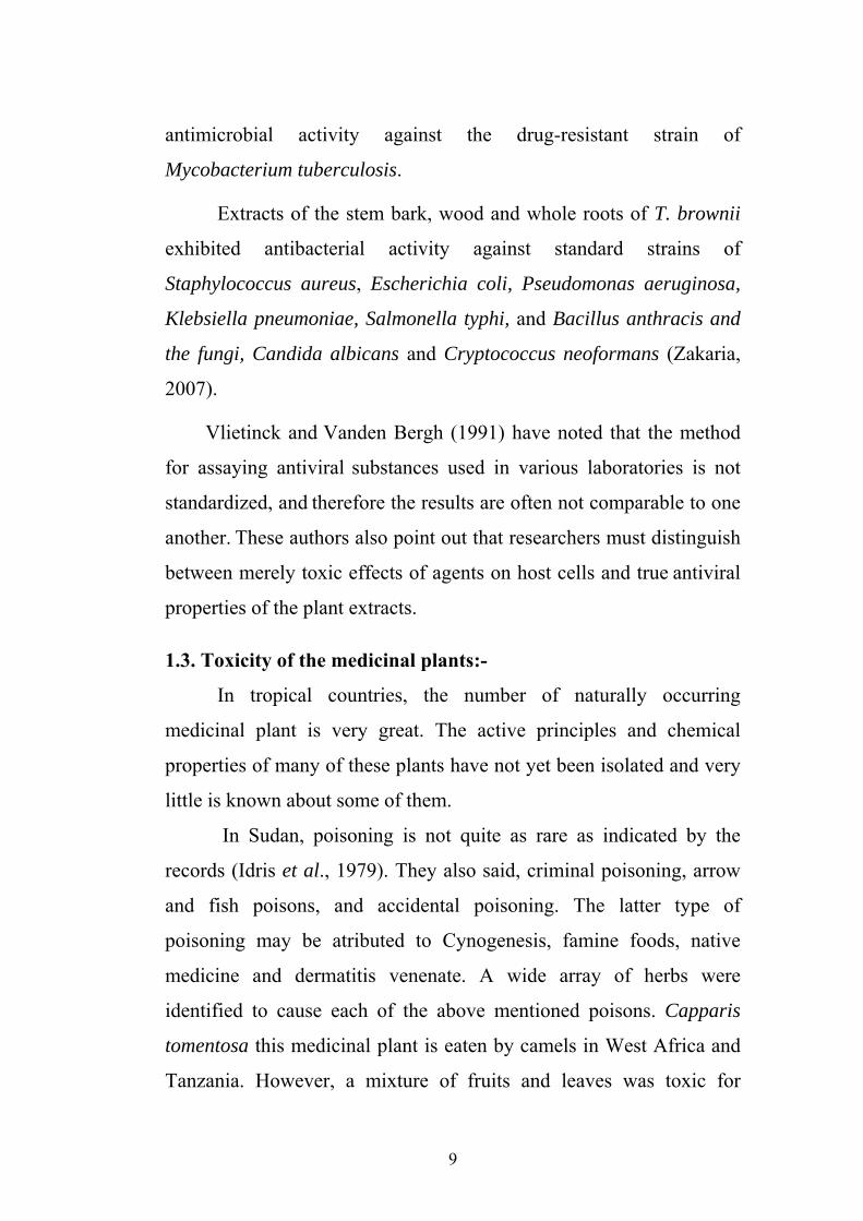
Extracts of the stem bark, wood and whole roots of T. brownii
exhibited antibacterial activity against standard strains of
Staphylococcus aureus, Escherichia coli, Pseudomonas aeruginosa,
Klebsiella pneumoniae, Salmonella typhi, and Bacillus anthracis and
the fungi, Candida albicans and Cryptococcus neoformans (Zakaria,
2007).
Vlietinck and Vanden Bergh (1991) have noted that the method
for assaying antiviral substances used in various laboratories is not
standardized, and therefore the results are often not comparable to one
another. These authors also point out that researchers must distinguish
between merely toxic effects of agents on host cells and true antiviral
properties of the plant extracts.
1.3. Toxicity of the medicinal plants:-
In tropical countries, the number of naturally occurring
medicinal plant is very great. The active principles and chemical
properties of many of these plants have not yet been isolated and very
little is known about some of them.
In Sudan, poisoning is not quite as rare as indicated by the
records (Idris et al., 1979). They also said, criminal poisoning, arrow
and fish poisons, and accidental poisoning. The latter type of
poisoning may be atributed to Cynogenesis, famine foods, native
medicine and dermatitis venenate. A wide array of herbs were
identified to cause each of the above mentioned poisons. Capparis
tomentosa this medicinal plant is eaten by camels in West Africa and
Tanzania. However, a mixture of fruits and leaves was toxic for
10
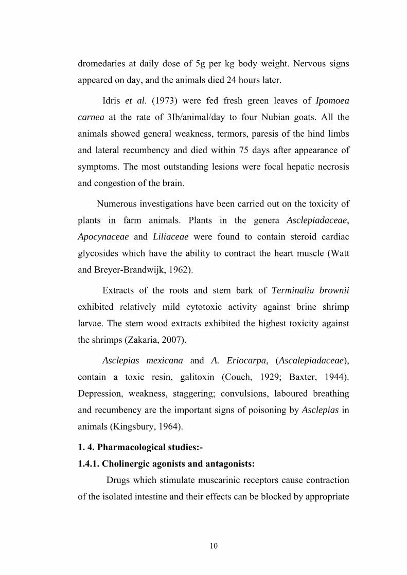
dromedaries at daily dose of 5g per kg body weight. Nervous signs
appeared on day, and the animals died 24 hours later.
Idris et al. (1973) were fed fresh green leaves of Ipomoea
carnea at the rate of 3Ib/animal/day to four Nubian goats. All the
animals showed general weakness, termors, paresis of the hind limbs
and lateral recumbency and died within 75 days after appearance of
symptoms. The most outstanding lesions were focal hepatic necrosis
and congestion of the brain.
Numerous investigations have been carried out on the toxicity of
plants in farm animals. Plants in the genera Asclepiadaceae,
Apocynaceae and Liliaceae were found to contain steroid cardiac
glycosides which have the ability to contract the heart muscle (Watt
and Breyer-Brandwijk, 1962).
Extracts of the roots and stem bark of Terminalia brownii
exhibited relatively mild cytotoxic activity against brine shrimp
larvae. The stem wood extracts exhibited the highest toxicity against
the shrimps (Zakaria, 2007).
Asclepias mexicana and A. Eriocarpa, (Ascalepiadaceae),
contain a toxic resin, galitoxin (Couch, 1929; Baxter, 1944).
Depression, weakness, staggering; convulsions, laboured breathing
and recumbency are the important signs of poisoning by Asclepias in
animals (Kingsbury, 1964).
1. 4. Pharmacological studies:-
1.4.1. Cholinergic agonists and antagonists:
Drugs which stimulate muscarinic receptors cause contraction
of the isolated intestine and their effects can be blocked by appropriate
11
antagonists such as atropine. Contractions can also be induced by
stimulation of parasympathetic ganglia with nicotinic agonists, which
causes the postganglionic neuron to fire and these results in the release
of acetylcholine at the neuroeffector cell junction. These effects can
antagonize by muscarinic antagonists as well as ganglion blockers
such as hexamethonium (Lan kitchen, 1984).
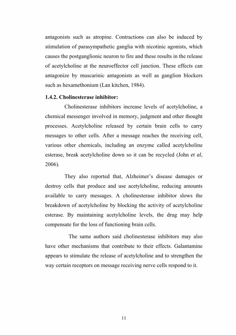
1.4.2. Cholinesterase inhibitor:
Cholinesterase inhibitors increase levels of acetylcholine, a
chemical messenger involved in memory, judgment and other thought
processes. Acetylcholine released by certain brain cells to carry
messages to other cells. After a message reaches the receiving cell,
various other chemicals, including an enzyme called acetylcholine
esterase, break acetylcholine down so it can be recycled (John et al,
2006).
They also reported that, Alzheimer’s disease damages or
destroy cells that produce and use acetylcholine, reducing amounts
available to carry messages. A cholinesterase inhibitor slows the
breakdown of acetylcholine by blocking the activity of acetylcholine
esterase. By maintaining acetylcholine levels, the drug may help
compensate for the loss of functioning brain cells.
The same authors said cholinesterase inhibitors may also
have other mechanisms that contribute to their effects. Galantamine
appears to stimulate the release of acetylcholine and to strengthen the
way certain receptors on message receiving nerve cells respond to it.

12
1.4.3. Atropine:
Derived primarily from Atropa belladonna, atropine is a
typical alkaloid; it is insoluble in water but soluble in alcohol, and
reacts with acids to form salts, e.g. atropine sulphate and
hydrochloride, which are more frequently, used then the parent
alkaloid.
In addition to pure atropine salts used in the form of injection,
the alkaloid is commonly used as the relatively crude belladonna-
plant preparations. The more popular preparations are the powdered
leaf, the tincture and a soft extract.
Following absorption the effects of atropine can be divided
into two groups;
1. The action on the central nervous system; in normal therapeutic
doses no cerebral effect is apparent. Intravenously in small over
doses atropine has a stimulant and sometimes convulsing action
on the cerebral cortex. Larger doses will excessively stimulate
the medulla and still larger doses will affect the spinal cord.
Excessive over dosage produces cortical and medullar
depression, coma and death.
2. The action on the peripheral nervous system.
This is one of paralysis of muscarinic part of the
parasympathetic division of the autonomic nervous system. The
muscarinic effects of acetylcholine are seen when the postganglionic
parasympathetic receptors are activated, e. g. by the alkaloids
muscarinic or pilocarpine. The paralysis is not complete when single
therapeutic doses are used, but even partial blocking of the

13
parasympathetic impulses allows greater action of the sympathetic
system (Brander and pugh, 1977).
1.4.4. Histamine:-
The same authors reported that, histamine is widely
distributed in plants and is found in animal foodstuffs (silage) and
digesta. Histamine occurs in an inactive form in all tissues, but
especially in the lungs, skin, intestinal and muscular tissues. It is
formed from amino acid histidine by decarboxylation mediated by an
enzyme histidine decarboxylase: bacteria decomposition of proteins
also produces histamine and fairly high concentrations exist in septic
foci where tissue destruction is in progress. When administrated by
intra dermal injection, it elicits what is called the triple –response. The
small vessels at the site of injection dilate, and then the neighboring
vessels dilate via an axon reflex of a sensory nerve. Finally, as plasma
escapes through the walls of the most widely dilated vessels, the
central area of the reaction becomes edematous and appears raised and
blanched.
Histamine can stimulate nerve-endings is well remembered
from the itch and pain of a nettle sting and it has been suggested that
histamine is the peripheral mediator for sensations of itch and pain. It
has also been suggested that wherever tissue is damaged there is a
production of histamine which causes either local or general shock.
1.4.5. Chlorphenramine:
Chlorpheniramine is part of a series of antihistamines
including pheniramine and its halogenated derivatives and others
including fluorpheniramine, chlorpheniramine, dexchlorpheniramine,
brompheniramine, dexbrompheniramine, deschlorpheniramine and

14
dipheniramine. Chlorphenamine has antidepressant properties,
inhibiting reuptake of the neurotransmitter serotonin (Bruce, 2005).
1.4.6. 5-Hydroxytryptamine (5-HT):
This base Known as serotonin was identified as
vasoconstrictor released from platelets lyses in the coagulation
process. It is now established that considerable quantities of 5HT
occur in the argentaffin cells of the gastro- intestinal tract. It also
occurs in the mast cells of rats, mice and bovines in which species it is
released during anaphylaxis. It is present in other tissues and is
believed to be a transmitter substance in CNS.
5-HT is produced in the body by the hydroxylation of
tryptophan to 5hydroxytrptophan which is then decarboxylated to 5-
HT. The blood 5-HT is stored in platelets and these, just like granules
of argentaffin and mast cells can accumulate 5-HT against a
concentration gradient. Elimination of 5HT by a monoamine oxidase
catalysed oxidation to 5hydroxyindolacetic acid or by acetylation to
yield N-acetyl-5hydroxytryptamine and excretion in the urine.
The actions of 5HT are constriction of blood vessels,
especially in the lungs, and contraction of the smooth muscle of
intestine, uterus and bronchial tree. Nerves and ganglion cells may be
stimulated and adrenaline is released from the adrenal medulla
(Brander and Pugh, 1977).
Tryptamine and its hydroxyl derivative are powerful
radioprotective amines in vivo, but they also give rise to physiological
changes (Van den brenk and Kathleen, 1958). Bacq and Alexander
(1955) state that tryptamine is not a vasoconstrictor, but is
nevertheless as good a radiation protector of mice as 5-

15
hydroxytryptamine. However, tryptamine causes intense smooth
muscle contraction, similar to its hydroxyl derivative.
1.4.7. Cyproheptadine (Periactin): Cyproheptadine is an antihistamine that similar to other
antihistamines with which we are more familiar. Histamine, a
biochemical mediator of inflammation, works by binding to histamine
receptors. The histamine receptors we usually want to inactivate in
combating allergic reactions are called H1 receptors and this is the
type inactivated by cyproheptadine (Wendy et al, 2005).
They also reported that cyproheptadine also has some other
properties of interest. It also antagonizes serotonin, a neurotransmitter,
in the brain. This leads to an increase in appetite and often is the
reason this medication is used, rather than for its antihistamine effects.
Cyproheptadine has been used in the treatment of Cushing's disease,
where there is excess cortisone production but not with reliable
success.
1.5. Plants used in this study:
1.5.1. Monechma ciliatum
Monechma ciliatum belongs to the family Acanthaceae,
locally known as "Black Mahlab". It is an annual hispid-scabrous
almost glabrous herb 1-2 ft. high; woody below-leave linear or
narrowly linear-lanceolate, flowers cream-white with purple and
orange streaks, 2-lipped, in short spikes bracts pectinate with long stiff
white bristles. Seeds with a tuft of rigid thick hairs at the hilum and a
similar tuft at the other end (Fig.1and 2) (Andrews, 1956).
According to Show (1973) Monechma ciliatum is
approximately native of tropical Africa and India. It is widely

16
distributed from Senegal to Cameroon and east word to central and
southern states of the Sudan and south through E. Africa to Malawi
and Zambia (Wichens, 1976). It is also grown in Nigeria (Uguru
et al., 1998).
In Botswana, they believed that Monechma cilaitum played an
important role as a medicine; it is used for general body pain, liver,
bowel trouble and sterility in women (Inga Hedberg and Stengard,
1989).
In the Sudan Monechma ciliatum is widely distributed in
Kassala, Southern Blue Nile, Kordofan, Upper Nile, Bahr Elghazal
and Bahr El Jebel States (Broun and Massey, 1929).
Black Mahlab is a famous medicinal plant in western Sudan,
especially in Gebel Mara Area .It’s seeds are used as an effective
laxative and contain an essential oil which emits a sweet and pleasant
odour. It is also used in traditional Sudanese fragrances, lotions and
other cosmetics used for wedding preparations and child birth.
Phytochemical and pharmacological characters on Monechma
ciliatum was studied by Uguru and Evans (2000). The leaves of M.
ciliatum were found to contain alkaloids, glycosides, proteins, tannins
and saponins. The hot methanol extract of the leaves was found to
have potent oxytocic effect, which may constitute an amino acidic
derivative.
Uguru et al. (1998) investigated the utrotonic properties of
methanol extract of Monechma ciliatum, the oxytocic activity of hot
methanolic extract of leaves of M. ciliatum was compared with other
uterine stimulants like Ergometrine, Oxytocin, 5-Hydroxy tryptamine,
17
Acetyl Choline and Prostaglandins (E2 and F2alpha) in the presence
of some antagonists in an attempt to explain the mechanism of action
of the extract. Atropine partially blocked the effect of hot methanolic
extract. Indomethacin inhibited the effects of hot methanolic extract as
well as all drugs, except the Prostaglandins and Acetyl Choline .D-600
blocked the effect of all the drugs including hot methanolic extract.
Methyserigide antagonised only the effect of 5-Hydroxy
tryptamine and partially blocked Ergometrine. Prolonged treatment
altered the uterine musculature and the activity profile of the drugs.
These results suggest that the hot methanolic extract may be
acting by more than one mechanism to contract the uterus and this
explains the mechanism of the anti implantation activity of the plant.
Ayoub and David (1984) stated that Monechma ciliatum had
anticancer constituents.
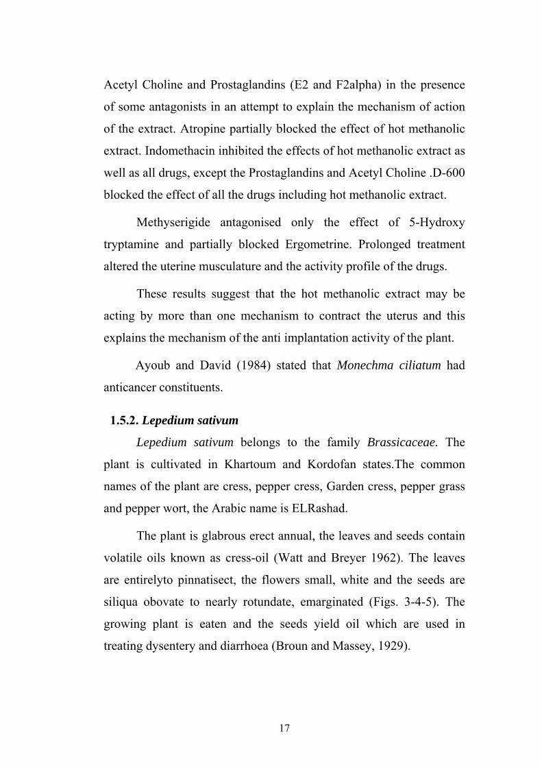
1.5.2. Lepedium sativum
Lepedium sativum belongs to the family Brassicaceae. The
plant is cultivated in Khartoum and Kordofan states.The common
names of the plant are cress, pepper cress, Garden cress, pepper grass
and pepper wort, the Arabic name is ELRashad.
The plant is glabrous erect annual, the leaves and seeds contain
volatile oils known as cress-oil (Watt and Breyer 1962). The leaves
are entirelyto pinnatisect, the flowers small, white and the seeds are
siliqua obovate to nearly rotundate, emarginated (Figs. 3-4-5). The
growing plant is eaten and the seeds yield oil which are used in
treating dysentery and diarrhoea (Broun and Massey, 1929).

18
The chemical constituents of the plant include phenolic
compounds (Ozeker and Esiyok, 1999), imidazole alkaloids (Maier,
et al., 1998), hydroxylated glutamic acids (Bell et al., 1981) sinigrin,
K-salt (Thies, 1988); flavonoid compounds (Paszkowski and Kremer,
1988), sterols (Bettach et al., 1997) and Benzyl glucosinolate
degradation protect (Gil and Macleod, 1980). Lepidium sativum
contained glucotropaeolin as reported by Songsak and Lockwood
(2002).
Phytochemical screening of Lepidium sativum studied by Nuha
(2006) showed that the plant was positive for Triterpenses, Alkaloids,
Flavonoids, Tannins and Cumarins but negative for Saponins,
Cynogenicglycoside and Anthraquinoneglycoside
The plant is traditionally used in Indian lactating women as well
as in cases of diarrhoea and dysentery. The seeds of Lepidium sativum
are used for treatment of fracture healing in Saudi traditional medicine
(Ahsan et al., 1989). The mucilage in the outer seed is used as
substitute for tragacanth and Gum Arabic (Mathews et al., 1993).
Lepedium sativum was toxic to Nubian goats (Amani,
1995). The signs in goats of Lepidium sativum poisoning were bloat,
loss in condition and recombency.
1.5.3. Linum usitatissimum:-
The plant Linum usitatissimum belongs to the family Linaceae.
The plant distributed in the Mediterranean Sea area and was
commonly cultivated in North Africa. The Arabic name of the plant is
Kettan, Berber, Tifert and Delkmouch. And English name is
Commonfax (Kokwaro, 1976). (Fig 6 and 7).
19
Traditionally the plant was used to treat fever. Seeds used as a
laxative, soothing and pain –relieving. 'Infusion of seeds used for
inflammation of digestive and urinary tracts, antidiarrhoea, often
mixed with Althaea flowers. Seeds used in preparation of cataplasms
for their emollient prosperities against boils and inflammations
(Kokwaro, 1976).The plant is considered one of the richest dietary
source of soluble fiber mucilage and that 50g high lingolenic acid flax
seed per day for 4weeks is palatable, safe and nutritionally beneficial
in human by raising n-3 fatty acids in plasma and erythrocytes and
decreasing post-prandial glucose responses (Cunnane et al., 1993).
Linum usitatissimum contains linamarin which contains a peptide,
linatine and has an antipyridoxine action in chickens (Humphreys,
1988).
Bakhiet and Adam (1995) studied the response of Bovans chicks
to low dietary levels of Linum usitatissimum seeds. They found that
the effect of 10-20 % Linum usitatissimum seeds in their normal diet
for 6weeks increased the growth rate in spite of the development of
centrilobular hepatocyte fatty vacuolation and an inflammatory cell
response in the intestinal lamina propria and renal cortex. These
effects were correlated with alteration in some hepatorenal function
and heamo-atolgy parameters.
Furthermore Linum usitatissimum was toxic to Nubian goats
(Amani, 1995). The main clinical signs of Linum usitatissimum
poisoning in Nubian goats were locometor disturbances, diarrhoea,
and loss in condition, dullness and recumbence.

20
Fig (1): Monechma ciliatum leaves

21
Fig (2): Monechma ciliatum leaves, stems and fruit

22
Fig (3): Lepidium sativum (shoot system)
Fig (4): Lepidium sativum leaves

23
Fig (5): Lepidium sativum seeds

24
Fig (6): Linum usitatissimum L. plant
Fig (7): Linum usitatissimum L. seeds

25
CHAPTER TWO
MATERIALS AND METHODS
2.1. Materials and Experimental Designs
2.1.1. Plants
Three plants were used in this experiment; Monechma ciliatum
(leaves and stems) and Lepidium sativum were collected from
Medicinal and Aromatic Plants Research Institute Farm. Linum
usitatissimum was supplied by herbalist.
The plants were authenticated by the scientist of the Medicinal
and Aromatic Plants Research Institute.
2.1.2. Animals
Rabbits, local strain Oryctolagus cuniculus, weighing 1-3 Kg
were brought from market- Khartoum North Guinea pigs, weighing
about ½-1½kg and Wister albino rats weighing about 80-120gms
were obtained from the Laboratory animal's house, Medicinal and
Aromatic Plants Research Institute, National Center for Research,
Khartoum.
Jejunum was dissected from rabbits while ileum from Guinea
pig. Uterus and fundic part of stomach were isolated from rat. The
animals were scarified.
2.1.3. Microbes
The negative organisms used are Escherichia coli (NCTC
8196), Proteus vulgaris (ATCC 6380) and Pseudomonas aeruginosa
(ATCC 27853), while the gram positive organisms were Bacillus
subtilis (NCTC 8236) and Staphylococcus aureus (ATCC 25923).

26
Two fungi Aspergillus niger (ATCC 9763) and Candida albicans
(ATCC 7596) are used. All organisms are obtained from National
Collection of Culture Type Colindale (NCTC) England and American
Type Culture Collection (ATCC) Rockville Mary land, USA.
2.1.4. Toxicity of Monechma ciliatum leaves methanolic extract in albino rats:-
Twelve Wister albino rats of different sexes weighing from
80-110 gm, obtained from Medicinal and Aromatic Plants Research
Institute were used. They were kept in cages within the premises of
MARPI. The rats were allowed one week adaptation periods.
2.1.4.1. Experimental design: -
At the end of the adaptation period, the rats were weighed and
divided into two groups 6 rats in each group. The first group was kept
as a control and the other group received a dose of 100mg /kg body
weight (B wt) daily of methanolic extract of Monechma ciliatum
leaves for 21 days.
2.1.4.2. Parameters:-
Blood sample were collected after slaughter for serum
analysis. Sera were analyzed for total Cholesterol, Bilirubin, Protein
and Albumin. Samples from the liver, heart, kidneys, lung, brain,
intestine and spleen were collected immediately after slaughter and
fixed in 10% formalin for histopathology.
2.2. Methods
2.2.1. Preparation of the plants extraction
150gm of the dried leaves and 50gm of stem of Monechma
ciliatum 160gm from dried seeds of Lepidium sativum and 50gm from
dried seeds of Linum usitatissimum were coarsely powdered and

27
extracted according to Harborne, (1984). They were left for 20 hours
soaked in Chloroform in Soxhlet apparatus. The Chloroform extract
was filtered and evaporated under reduced pressure using Rota-Vap.
The extracted plant material was then air-dried, repacked in the
Soxhlet and exhaustively extracted with Methanol. The methanolic
extract was filtered and again evaporated under reduced pressure
using Rota-Vap.
The yield percentages were calculated after the traces of the
solvent were air dried as follows:
Weight of extract Weight of plant X 100
2.2.2. In vitro testing for antimicrobial activity:-
2.2.2.1. Testing for antibacterial activity:-
The cup –plate –agar diffusion method (Kavanagh, 1972) was
adopted with some minor modifications, to assess the antibacterial
activity of the extracts.
Three ml of each of the five standardized bacterial stock
suspensions (108-109 C.F.U. /ml) were thoroughly mixed with 300ml
of sterile melted nutrient agar maintained at 45ºC.
20 ml aliquots of the inoculated nutrient agar were distributed into
sterile Petri-dishes. The agar was left to set for 5-10 minutes. The
plates were divided into two halves, two cups in each half (10mm in
diameter) using a sterile cork borer (No. 4). The agar disks were
removed. Alternate cups were filled with 0.1ml samples of each of the
extracts using Transfer adjustable volume automatic microtitre pipette,
and allowed to diffuse at room temperature for 2 hour.
The plates were then incubated in the up right position at 37oC
for 18 hours.

28
Two replicates were carried out for each extract against each of
the tested organisms.
Simultaneously positive controls involving the addition of the
respective solvents instead of the extracts were carried out separately.
After incubation the diameters of the resultant growth inhibition zones
were measured and the mean values were tabulated.
2.2.2.2. Testing for antifungal activity:-
The antifungal activity was measured by a method similar for
testing antibacterial activity. For the antifungal activity sabouraud
dextrose agar was used.
The inoculated medium was incubated at 25ºC for two days for
the Candida albicans and three days for Aspergillus niger.
2.2.3. Pharmacological Experiments:-
The animals were starved for 24 hours before killing. Animals
were killed by dislocating the neck, and then exsanguinated and the
abdomen was opened.
2.2.3.1. Rabbit jejunum:-
The preparation of the isolated jejunum is based on the method
of Finkleman (1930). The jejuna area of the intestine in the rabbit was
located out and then cut into isolated segments about 4cm in length.
The parts of jejunum were transferred to a Petri dish containing
Tyrode solution, the mesentery and fat surrounding the muscle were
trimmed away and thread was passed through one wall of the jejunum
at both top and bottom. The bottom thread was attached to the tissue
holder and the mounted tissue was transferred to the organ bath and
attached to an isotonic transducer. The tissue was left for 45 minutes for
29
adaptation to the new environment and washed several times by out
flow.
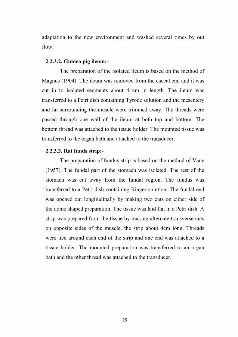
2.2.3.2. Guinea pig ileum:-
The preparation of the isolated ileum is based on the method of
Magnus (1904). The ileum was removed from the caecal end and it was
cut in to isolated segments about 4 cm in length. The ileum was
transferred to a Petri dish containing Tyrode solution and the mesentery
and fat surrounding the muscle were trimmed away. The threads were
passed through one wall of the ileum at both top and bottom. The
bottom thread was attached to the tissue holder. The mounted tissue was
transferred to the organ bath and attached to the transducer.
2.2.3.3. Rat funds strip:-
The preparation of fundus strip is based on the method of Vane
(1957). The fundal part of the stomach was isolated. The rest of the
stomach was cut away from the fundal region. The fundus was
transferred to a Petri dish containing Ringer solution. The fundal end
was opened out longitudinally by making two cuts on either side of
the dome shaped preparation. The tissue was laid flat in a Petri dish. A
strip was prepared from the tissue by making alternate transverse cuts
on opposite sides of the muscle, the strip about 4cm long. Threads
were tied around each end of the strip and one end was attached to a
tissue holder. The mounted preparation was transferred to an organ
bath and the other thread was attached to the transducer.

30
2.2.3.4. Rat uterus:-
The preparation of isolated rat uterus is based on the method of
De Jalon et al. (1945). A female rat was injected with 0.1mg/kg
stilboesterole intramuscularly 24 hours before the removal of uterus.
The two uterine horns were exposed by pulling a side the
intestine. Each horn was free from surrounding fat and mesenteric
attachments. Each horn was cut out separately, and transferred to a
Petri dish containing De Jalons Ringer. The two horns were divided
by making a transverse cut, in to four preparations 3-4cms. The tissue
will work quite adequately as a tube, a longitudinal cut was made and
the tissue was set up as a sheet of muscle. Threads were passed
through one wall of the uterus at both top and bottom. The bottom
thread was attached to the tissue holder, and was transferred to an
isolated organ bath and the top thread was attached to the transducer.
2.2.5. Blood samples:-
Blood samples were collected from rat during slaughter either
into dry clean bottles containing EDTA (Ethylene – diamine tetra
acetic acid) as anticoagulant for hematological studies or into clean
test tubes with out anticoagulant for sera. The tubes were left for 45
minutes and then sera were separated by centrifugation at the
centrifuge (HETTICH EBA 35 centrifuge). The sera were stored at -
4ºc till analysis.
2.2.5.1. White blood cells Count (WBC):-
Leukocytes were counted according to Schalm (1965) using
Neubauer haemocytometer for white blood cell counts. Turk's solution
(1% glacial acetic acid, tinged with gentian violet) was used as
diluents.
31
2.2.5.2. Differential leucocytes count:-
Blood films on clean slides were stained with leishman's stain.
The Battlement method was used for different leukocyte count. 100
cells were counted in each blood smear and average percentages were
recorded.
2.2.6. Biochemical Analysis:-
The biochemical analysis of bilirubin, cholesterol, protein and
albumin were estimated to determination of serum constituents.
2.2.6.1. Total Bilirubin:-
The bilirubin in the serum was measured as stated by
Jendrassik and Grof (1938).
Test principle:-
Bilirubin is coupled with diazotised sulfanilic acid in presence
of caffeine to give azodye. The optical density was measured by
JENWAY 6305 UV/VIS spectrophotometer at wave length 540 nm.
Total bilirubin calculated as follow:-
Total bilirubin (mg/dl) =Absorbance of sample – Absorbance of
sample blank.
2.2.6.2. Total cholesterol:-
Serum total cholesterol was measured by enzymatic
colorimetric test according to Richmond (1973). Its concentration was
estimated using commercial kit.
Test principle:-
The principle of assay depends on released of cholesterol
from its esters after enzymatic hydrolysis and oxidation to
cholestenone plus H2O2 – The indicator, quinonemine, is formed as a
result of reaction between hydrogen peroxide (H2O2) and 4- amino
antipyrine in the presence of peroxidase (POD).

32
Cholesterol ester + H2O Cholesterol Cholesterol + Fatty acids Esterase Cholesterol + H2O Cholesterol Cholestenone + H2O2
Oxidase
H2O2+ 4AP + Phenol Peroxidase Quinoneimine + H2O
Cholesterol concentration was estimated as follows:-
Cholesterol (mg/dl) =
Where the concentration of the standard is 200 mg/dl.
2.2.6.3. Total Protein:-
Biuet reagent was used to determine the total serum protein
concentration as described by Wiechselbaum (1946).
Test principle:-
The method is based on the reaction of protein with copper
sulphate in the presence of sodium hydroxide and potassium sodium
tartrate (Rochelle salt) which keeps cupric hydroxide in solution for
development of blue colour.
A spectrophotometer (Corning Halstead, U.K) was used at a
wavelength of 540 nm and values were calculated as follows:-
Total Protein (gm/dl) =
Where the concentration of the standard is 8 gm/dl
A sample A standard X concentration of standard
A sample A standard X concentration of standard

33
2.2.6.4. Albumin:-
Serum albumin concentration was measured by the use of
Bromo- Cresol Green method as described by Doumas et al. (1971).
Test principle:-
The principle of this method is that the measurement of
serum albumin is based on its quantitative binding to the indicator,
bromo-cresol green (BCG). The sera and reagents were mixed and
incubated at 20-25ºc. The absorbance was measured against the
reagent at 578 nm using a spectrophotometer. Albumin concentration
was calculated as follows:-
Serum albumin (g/dl) =
Where the concentration of the standard is 5gm/dl
2.2.7. Histopathological methods
Slices of tissues from lungs, small intestine, heart, liver,
kidneys, spleen and brain of rats after slaughter were fixed in 10%
formalin, embedded in paraffin wax, sectioned at 5µm and stained
with haematoxlylin and eosin (Drury and Wallington, 1980).
2.2.8. Statistical analysis:-
The student t- test was used for significance of data
analysis according to procedure described by Mendenhall (1971).
A sample A standard X concentration of standard
34
CHAPTER THREE
RESULTS
3.1. The antimicrobial activities of the plants:-
3.1.1. The antimicrobial activity of Monechma ciliatum (stems) extracts against standard organisms are presented in Table (1):-
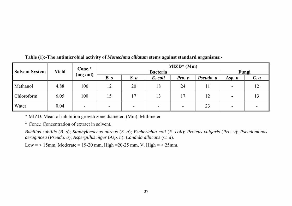
The methanolic extract of Monechma ciliatum stems was
showed high antimicrobial activity against Proteus vulgaris (24) and
Staphylococcus aureus (20), moderate activity against Escherchia coli
(18) and low inhibition zone against Bacillus subtilis (12),
Pseudomonas aeruginosa (12) and Candida albicans (12).
The Chloroform extract of Monechma ciliatum stems showed
moderate activity against Staphylococcus aureus (17), Proteus
vulgaris, and Bacillus subtilis (15). It had low activity against
Escherchia coli (13), Pseudomonas aeruginosa (13) and Candida
albicans (13).
The water extract of Monechma ciliatum stems, had high
antimicrobial activity against Pseudomonas aeruginosa (23), while the
activity was absent against all other standard organisms tested.
Aspergillus niger was resistant to the all plant extracts.
3.1.2. The antimicrobial activity of Monechma ciliatum leaves extracts against standard organisms are presented in Table (2) and Fig (8-13):-
The antimicrobial activity of methanolic extract of Monechma
ciliatum leaves was moderate against Proteus vulgaris (19) and
Escherchia coli (18), low against Bacillus subtilis (17),

35
Pseudomonas aeruginosa (17) and Staphylococcus aureus (17). The
activity was absent aganst Candida albicans and Aspergillus niger.
The chloroform extract of Monechma ciliatum leaves caused
high inhibition zone against Staphylococcus aureus (23) and low
against Bacillus subtilis (15) and Candida albicans (14). However, the
extract showed low activity against Escherchia coli (13), Proteus
vulgaris (13) and Pseudomonas aeruginosa (13).
Water extract of Monechma ciliatum leaves caused moderate
activity against Pseudomonas aeruginosa (17) and low inhibition zone
activity against Bacillus subtilis (13) and Staphylococcus aureus (13).
The activity was absent against Escherchia coli, Proteus Vulgaris,
Candida albicans and Aspergillus niger (Table 4).
3.1.3. The antimicrobial activity of Linum usitatissimum seeds extracts against standard organisms is presented in Table (3):-
The methanolic extract of Linum usitatissimum caused an
inhibition growth zone diameter. It was moderate in Pseudomonas
aeruginosa (18) and Bacillus subtilis (16), low against Proteus
vulgaris (15), Staphylococcus aureus (14) and Escherchia coli (13).
The activity was absent against Candida albicans and Aspergillus
niger.
The antimicrobial activity of Chloroform extract of Linum
usitatissimum was low against Pseudomonas vulgaris (13) and
inactive against Bacillus subtilis, Staphylococcus aureus, Escherchia
coli, Proteus vulgaris, Aspergillus niger and Candida albicans.
Water extracts showed low activity against Candida albicans
(14) while there was no activity against other organisms.

36
3.1.4. The antimicrobial activity of Lepidium sativum seeds extracts against standard organisms is presented in Table (3):-
The methanolic extract of Lepidium sativum had high inhibition
against Candida albicans (21) and Proteus vulgaris (20). The
antimicrobial activity of the extract was low against Bacillus subtilis
(13), Staphylococcus aureus (13), Escherchia coli (13) and
Pseudomonas aeruginosa (13). The extract was inactive against
Aspergillus niger.
The chloroform extract of Lepidium sativum had very low
activity against Staphylococcus aureus (12) and Pseudomonas
aeruginosa (12) and the activity was absent against Bacillus subtilis,
Escherchia Coli, Proteus Vulgaris, Candida albicans and Aspergillus
niger.
The antimicrobial activity of water extract of Lepidium sativum
was low against Bacillus subtilis (15) and Escherchia coli (15). The
extract had no effect against Staphylococcus aureus, Proteus Vulgaris,
Candida albicans and Aspergillus niger.
37
Table (1):-The antimicrobial activity of Monechma ciliatum stems against standard organisms:-
MIZD* (Mm) Bacteria Fungi Solvent System Yield Conc.*
(mg /ml) B. s S. a E. coli Pro. v Pseudo. a Asp. n C. a
Methanol 4.88 100 12 20 18 24 11 - 12
Chloroform 6.05 100 15 17 13 17 12 - 13
Water 0.04 - - - - - 23 - -
* MIZD: Mean of inhibition growth zone diameter. (Mm): Millimeter * Conc.: Concentration of extract in solvent. Bacillus subtilis (B. s); Staphylococcus aureus (S .a); Escherichia coli (E .coli); Proteus vulgaris (Pro. v); Pseudomonas aeruginosa (Pseudo. a); Aspergillus niger (Asp. n); Candida albicans (C. a). Low = < 15mm, Moderate = 19-20 mm, High =20-25 mm, V. High = > 25mm.

38
Table (2):-The antimicrobial activity of Monechma ciliatum Leaves against the standard organisms:-
MIZD* (Mm) Bacteria Fungi
Solvent System Yield % Conc.*
mg/ml B. s S. a E .coli Pro. v Pseudo. a Asp. n C. a
Methanol 12.43 100 17 16 18 19 17 - -
Chloroform 11.13 100 15 23 13 13 13 - 14
Water 0.09 - 13 12 - - 17 - -
* MIZD: Mean of inhibition growth zone diameter. (Mm): Millimeter * Conc.: Concentration of extract in solvent.
Bacillus subtilis (B. s); Staphylococcus aureus (S. a); Escherichia coli (E. coli); Proteus vulgaris (Pro. v); Pseudomonas aeruginosa (Pseudo. a); Aspergillus niger (Asp. n); Candida albicans (C. a). Low = < 15mm, Moderate = 19-20 mm, High =20-25 mm, V. High = > 25mm.

39
Table (3):-The antimicrobial activity of Linum usitatissimum against the standard organisms:-
MIZD* (Mm) Bacteria Fungi
Solvent System
Yield %
Conc.* mg /ml
B. s S. a E. coli Pro. v Pseudo. a Asp. n C. a
Methanol 4.6 100 16 14 13 15 18 - -
chloroform 28.5 100 - - - - 13 - -
Water 0.041 - - - - - - - 14
* MIZD: Mean of inhibition growth zone diameter. (Mm): Millimeter * Conc.: Concentration of extract in solvent.
Bacillus subtilis (B. s); Staphylococcus aureus (S. a); Escherichia coli (E. coli); Proteus vulgaris (Pro. v); Pseudomonas aeruginosa (Pseudo. a); Aspergillus niger (Asp. n); Candida albicans (C. a). Low= <15mm, Moderate= 19-20mm, High= 20-25mm, V. High= >25.25.

40
Table (4):-The antimicrobial activity of Lepidium sativum against the standard organisms:
MIZD* (Mm) Bacteria Fungi
Solvent System
Yield %
Conc.* mg/ml
B. s S. a E. coli Pro. v Pseudo. a Asp. n C. a
Methanol 9.82 100 13 13 13 20 13 - 21
chloroform 9.0 100 - 11 - - 12 - -
Water 0.03 - 15 - 15 - - - -
* MIZD: Mean of inhibition growth zone diameter. (Mm): Millimeter. * Conc.: Concentration of extract in solvent.
Bacillus subtilis (B. s); Staphylococcus aureus (S. a); Escherichia coli (E. coli); Proteus vulgaris (Pro. v); Pseudomonas aeruginosa (Pseudo. a); Aspergillus niger (Asp. n); Candida albicans (C. a). Low = < 15mm, Moderate = 19-20 mm, High =20-25 mm, V. High = > 25m

41
Fig (8): Antimicrobial activity of methanol and chloroform extract of Monechma ciliatum leaves on Escherichia coli.
• M = methanol extract • Ch = chloroform extract

42
Fig (9): Antimicrobial activity of methanol and chloroform extract of Monechma ciliatum leaves on Staphylococcus aureus.
• M = methanol extract • Ch = chloroform extract

43
Fig (10): Antimicrobial activity of methanol and chloroform extract
of Monechma ciliatum leaves on Candida albicans.
• M = methanol extract • Ch = chloroform extract

44
Fig (11): Antimicrobial activity of methanol and chloroform extract
of Monechma ciliatum leaves on Pseudomonas aeruginosa.
• M = methanol extract • Ch = chloroform extract

45
Fig (12): Antimicrobial activity of methanol and chloroform extract of Monechma ciliatum leaves on Bacillus subtilis.
• M = methanol extract • Ch = chloroform extract

46
Fig (13): Antimicrobial activity of methanol and chloroform extract of Monechma ciliatum leaves on Aspergillus niger.
• M = methanol extract • Ch = chloroform extract
47
3.2. The pharmacological activity of Methanolic extract of Monechma ciliatum leaves on different isolated tissues:-
3.2.1. Rabbit jejunum:
The methanolic extract of Monechma cilitum leaves caused
significant contraction in rabbit jejunum at different doses (1.6, 3.2, 6.4
mg/ml) (Fig. 14). Similar contraction in rabbit jejunum caused by doses
(0.4, 1, 2, 0.8 mg/ml) (Fig. 15).
3.2.2. Guinea pig illeum:-
On the isolated Guinea pig illeum, the plant extract showed
contraction at submaximum dose 3.2 mg/ml. The contraction was
refractory to both the muscarinic blocker Atropine (Fig. 16) and the
antihistamine antagonist Chlorophenramine (Fig. 17), but effectively
blocked by the serotonergic blocker, Cyproheptadine at dose of 4µg/ml
(Fig. 18).
3.2.3. Rat fundus:-
The rat fundus strip is highly sensitive to 5-Hydroxytriptamine
(5HT). 5HT (1µg/ml) stimulated the rat fundus strip while extract of
Monechma ciliatum leaves stimulated it at (3.2 mg/ml) (Fig. 19). This
contracting activity of both extract and serotonin was effectively blocked
by Cyproheptadine (2µg/ml).
3.2.4. Rat uterus:-
The methanolic extract of Monechma ciliatum leaves at a
concentration of 3.2 mg/ml caused contraction on isolated rat uterus. This
effect was completely blocked by Cyproheptadine (2µg/ml) (Fig. 20).
In a similar manner the contraction produced by standard (5HT)
was blocked completely by Cyproheptadine (2µg/ml).

48

49

50

51

52

53

54

55
3.3. Effect of methanolic extract of Monechma ciliatum leaves in
rats:-
3.3.1. Clinical findings:-
There were no clinical signs observed in experimental rats treated
with 100mg/kg through out the experiment.
3.3.2. Haematological changes:-
The means WBC and differential count are shown in Table (5).
The WBC and differential count showed no significant changes.
3.3.3. Postmortem Findings:-
There were no postmortem changes noticed in the treated group.
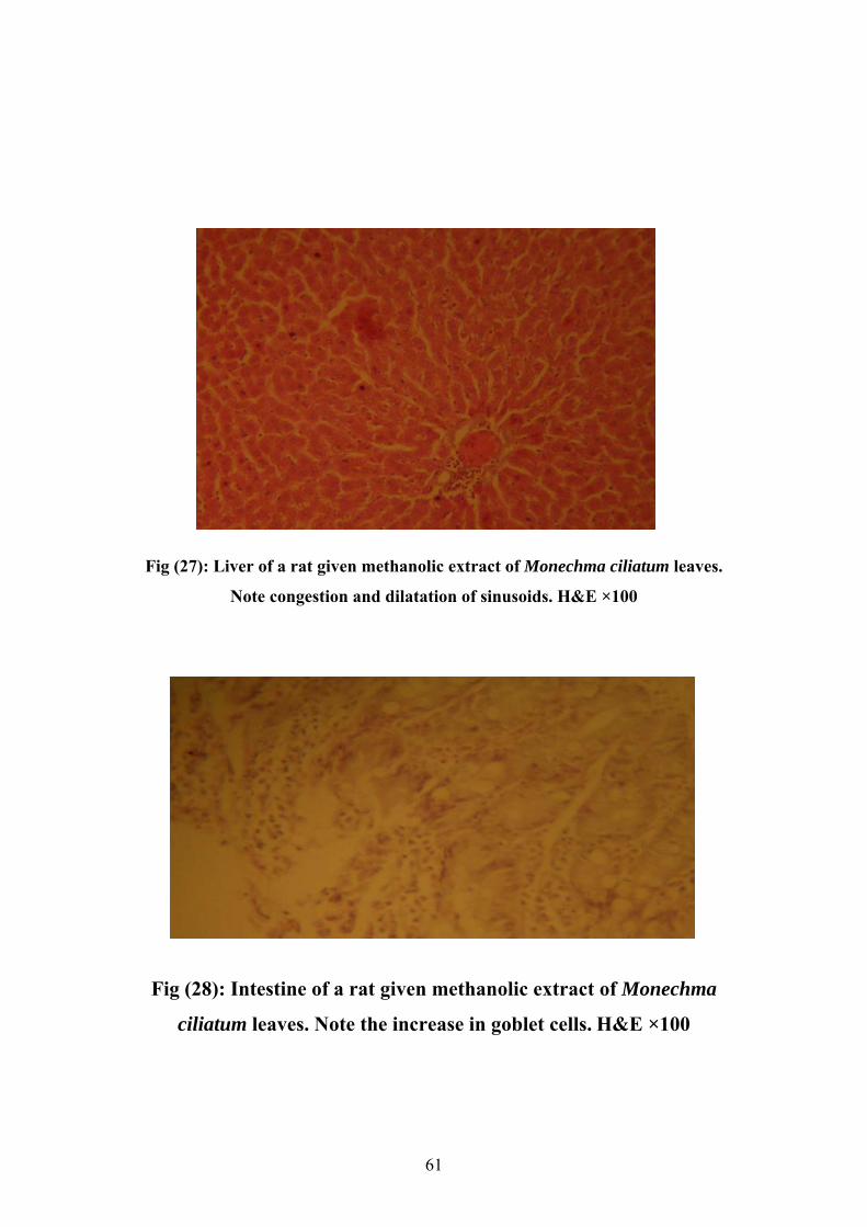
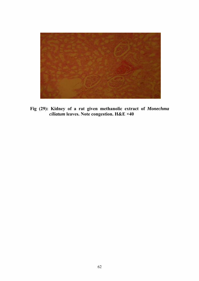
3.3.4. Histopathological findings:-
The rats treated with methanolic extract of Monechma ciliatum
leaves at (100mg/kg) showed congestion of livers, Kidneys, brains and
spleens. In some rats the liver showed dilatation of sinusoids, some
treated rats showed increase in goblet cells in the intestine. Fig (27- 28-
29).
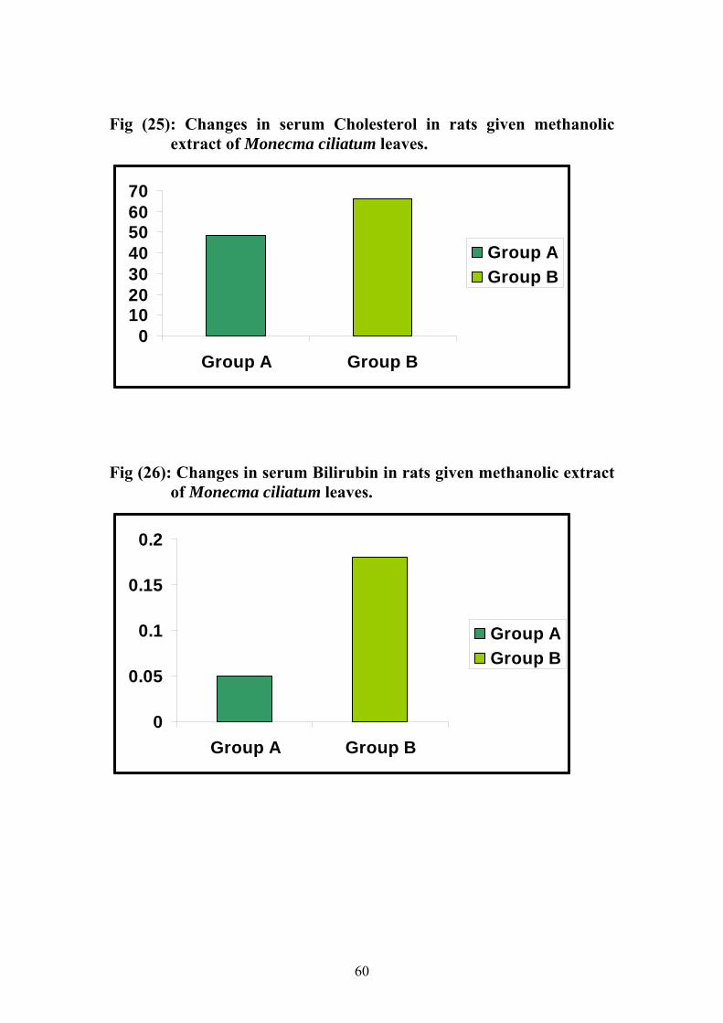
3.3.5. Serum constituents change:-
The mean levels of bilirubin, cholesterol, total protein and
albumin were shown in Table (6). There was significant increase (p <
0.05) in the values of total protein. In the other hand there were no
significant changes in cholesterol, albumin and bilirubin concentration.

56
Table (5): White blood cell and differential count of rats treated with
methanolic extract of Monechma ciliatium leaves
Group
WBC Lymphocytes
(%) Neutrophils
(%)
Eosinophils
(%)
Basophils
(%)
Monocytes
(%)
Group (A) 2.5×10³ + 170.78 42.83 + 1.7 18.50 + 1.7 18.17 +1.7 1.17+ 1.7 17.67 +1.7
Group (B) 2.3 ×10³ + 600.9 42.30 + 2.51 21.70 + 2.88 17.00 + 2.6 1.00 + 1.00 18.00 + 2.65
Group A: Treated with methanolic extract of Monechma ciliatum leaves
100mg/kg B wt.
Group B: control.
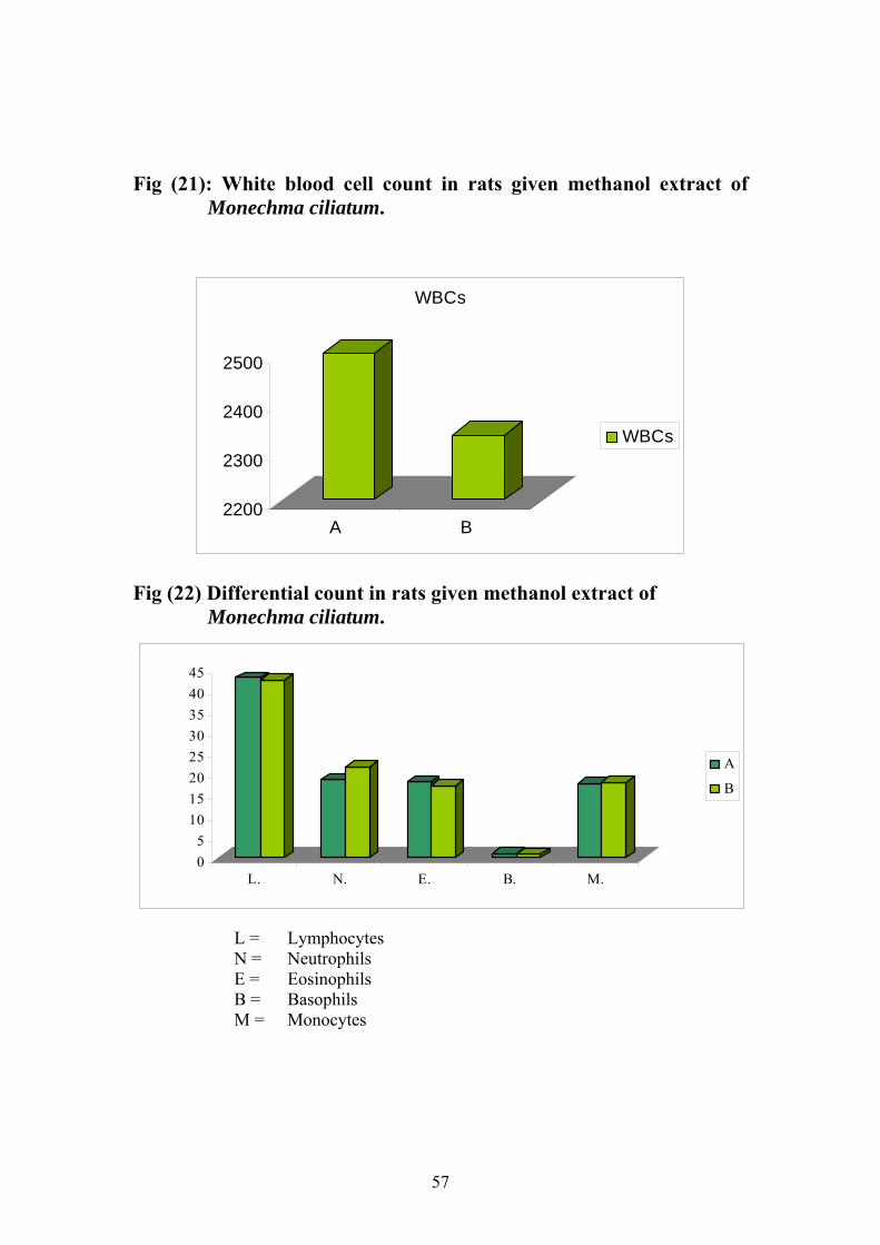
57
Fig (21): White blood cell count in rats given methanol extract of
Monechma ciliatum.
2200
2300
2400
2500
A B
WBCs
WBCs
Fig (22) Differential count in rats given methanol extract of Monechma ciliatum.
05
1015202530354045
L. N. E. B. M.
A
B
L = Lymphocytes N = Neutrophils E = Eosinophils B = Basophils M = Monocytes

58
Table (6): Serum constituents values in rats treated with methanolic extract of Monechma ciliatum leaves.
* P< 0.05
Group A: Treated with methanolic extract of Monechma ciliatum leaves
100mg/kg B wt.
Group B: control.
Group Total Protein gm/dl
(Means + S. E.)
Albumin gm/dl
(Means + S. E.)
Cholesterol mg/dl
(Means + S. E.)
Bilirubin mg/dl
(Means + S. E.)
Group A 11.9+2.65* 2.35 + 1.26 48.5 + 17.7 0.05 + 0.034
Group B 7.35+ 0.65 2.9 + 0.92 66.00 + 10.00 0.18 + 0.173

59
Fig (23) Changes in serum Protein in rats given methanolic extract of Monecma ciliatum leaves.
02468
101214
Group A Group B
Group AGroup B
Fig (24): Changes in serum Albumin in rats given methanolic extract
of Monecma ciliatum leaves.
00.5
11.5
22.5
33.5
Group A Group B
Group AGroup B
60
Fig (25): Changes in serum Cholesterol in rats given methanolic
extract of Monecma ciliatum leaves.
010203040506070
Group A Group B
Group AGroup B
Fig (26): Changes in serum Bilirubin in rats given methanolic extract
of Monecma ciliatum leaves.
0
0.05
0.1
0.15
0.2
Group A Group B
Group AGroup B
61
Fig (27): Liver of a rat given methanolic extract of Monechma ciliatum leaves.
Note congestion and dilatation of sinusoids. H&E ×100
Fig (28): Intestine of a rat given methanolic extract of Monechma
ciliatum leaves. Note the increase in goblet cells. H&E ×100
62
Fig (29): Kidney of a rat given methanolic extract of Monechma
ciliatum leaves. Note congestion. H&E ×40

63
CHAPTER FOUR
DISCUSSION
In this study the methanol, chloroform and water extracts of
Lepidium sativum, Linum usitatissimum seeds and Monechma ciliatum
leaves and stems were tested to verify their antimicrobial activities
against standard organisms. The present investigation has shown that
Monechma ciliatum leaves extracts were the most effective against
standard organisms than the other tested plants extracts. All five standard
bacteria (Bacillus subtilis, Staphylococcus aureus, Escherichia coli,
Proteus vulgaris and Pseudomonas aeruginosa) were sensitive to the
methanol extract of Monechma ciliatum leaves and showed moderate
inhibitory zone sizes, while chloroform extract of Monechma ciliatum
leaves shown low activity against standard bacteria except
staphylococcus aureus which has higher inhibitory zone size. Water
extract was shown low inhibitory zone on Bacillus subtilis,
Staphylococcus aureus and Pseudomonas aeruginosa, when Escherichia
coli, Proteus vulgaris, Aspergillus niger and Candida albicans were
resistant to this extract. The Aspergillus niger was inactive to all
Monechma ciliatum leaves extracts. From these findings, it is clear that
the methanol extract of Monechma ciliatum leaves, as an antimicrobial
agent, is more effective than the chloroform and water extracts.
Mohamed (1979) reported similar result by using Hibiscus sabdariffa
(Karkadeh) which showed a potent antimicrobial effect. He found that an
aqueous, ethanol, 1% methanol hydrochloric acid, ethereal, chloroform
and petroleum ether extracts have a wide range of antimicrobial activity
against both Gram- positive and Gram- negative bacteria as well as yeast.
64
Methanol extract of Lepidium sativum showed high activity against
Gram-negative Proteus vulgaris and the yeast Candida albicans, but low
against Bacillus subtilis, Staphylococcus aureus, Escherichia coli and
Pseudomonas aeruginosa. The chloroform extract of Lepidium sativum
was inactive against all standard organisms tested. Aspergillus niger was
resistant to this plant extracts. Asma (2003) recorded similar effects in her
study of Lepidium sativum seeds for its antimicrobial activities. She
found that the methanol extract of Lepidium sativum was the most active
extract. The clinical isolates exhibited low susceptibility compared with
the standard organisms.
In the present investigation the Linum usitatissimum seeds extracts
showed very low antimicrobial activity against standard organisms
compared with the other plants extracts tested in this study. Methanol
extract of Linum usitatissimum caused inhibitory zone sizes in Gram
positive and Gram-negative bacteria. The fungi Aspergillus niger and
Candida albicans did not show any effect to methanolic and chloroform
extracts. Water extract of Linum usitatissimum had low inhibitory against
Candica albicans, the activity was absent against Aspergillus niger and
almost five standard bacteria.
In the other hand the topical wound healing properties of Canisep
cream containing oils from Linum usitatissimum was studied by Amresh
et al (2005). This study was conducted on 32 surgical wounds of 8 goats.
Wounds were subjected to topical application of cream. Granulation
tissue appeared but scab never appeared. This result suggested that
Canisep cream had antimicrobial property which makes Linum
usitatissimum a suitable for wound healing. They may indicate that Linum
usitatissimum has a synergistic effect as antimicrobial properties.

65
In this study the methanolic extract of Monechma ciliatum stems
was found to be more effective as an antimicrobial agent than
chloroformic and water extracts. This may be due to the fact that the
active constituent which had antimicrobial affect dissolved in methanol
rather than chloroform. The water extract had no inhibitory zone which
means that the active ingredients were insoluble in water. The fungi
responded less to Monechma ciliatum stems extracts which indicated that
the plant had very little antifungal component. Monechma ciliatum leaves
extracts are more effective than Monechma ciliatum stems this explain
that Monechma ciliatum plant had different component between the parts
of the plant.
In this study the investigations revealed that the methanolic extract
of Monechma ciliatum leaves contained pharmacologically active
components. Small doses of the extract produced contraction on Rabbit
jejunum which was increased immediately with increase of the dose.
ElTayeb (1996) had found that the aqueous extract of Hyphaene thebaica
(Doum) also stimulated the rabbit jejunum. It also caused significant
contraction at a dose of 3.2mg/ml on isolated Guinea pig ileum. The
contraction was neither due to cholinomimitic activity because, it was not
blocked by the muscarinic blocker Atropine and nor due to histamine
receptors that the antihistamine blocker Chlorophenramine did not block
the effect which blocked the standard Histamine. However, the effect was
blocked by Cyproheptadine a drug which antagonized the action of both
(serotonin) 5-Hydroxytryptamine (Gyermek, 1961) and Histamine (H1)
receptors agonists (D Arcy, 1963).This suggested that the contraction
could be probably due to 5- Hydroxytryptamine like substance. Afra a
(2002) reported similar result by water extract of Amobrosia maritime
66
which induced contraction on isolated guinea pig at dose of (20mg/ml)
this stimulant action was blocked completely by 2µg/ml cyprohyptadine.
Serotonin causes increased gastrointestinal motility and contraction
of isolated strips of intestine, this being partly due to direct effect on
smooth muscle cells and partly due to indirect effect on enteric neuron
(Rang and Dale, 1987).
The fundus strip was also sensitive to serotonin (Vane, 1957). 5-
Hydroxytryptamine (1µg/ml) produced contraction on the rat fundus
while methanol extract of Monechma ciliatum leaves (3.2 mg/ml) caused
similar contraction to that produced by 5-Hydroxytryptamine.
Furthermore, the contraction of both the extract and 5-
Hydroxytryptamine were completely blocked by Cyproheptadine. Einas
(2000) reported that the decoction of Striga hermonthica stimulated the
isolated rat fundus strip at (2mg/ml); this stimulant effect was blocked
partially by (2µg/ml) cyprohyptadine.
Moreover, the extract of Monechma ciliatum leaves showed
significant contraction on isolated rat uterus at dose of 3.2 mg/ml, similar
contraction was caused by the standard 5-Hydroxytryptamine. This
contracting activity on the rat uterus was blocked with Cyproheptadine at
dose of 2µg/ml. Uguru et al. (1998) in their investigated of the utrotonic
properties of methanol extract of Monechma ciliatum leaves, the
contracted activity of hot methanolic extract of leaves of Monechma
ciliatum was compared with other uterine stimulants like Ergometrine,
Oxytocin, 5-Hydroxy tryptamine, Acetyl Choline and Prostaglandins
(E2 and F 2α) in the presence of some antagonists in an attempt to explain
the mechanism of action of the extract.
Atropine blocked partially the effect of hot methanolic extract.
Indomethacin inhibited the effects of hot methanolic extract. They
67
suggested that the hot methanolic extract may be acting by more than one
mechanism to contract the uterus. The present finding which showed that
contracting activities of rat uterus blocked by Cyproheptadine and this
similar to which Uguru et al (1998) suggested.
Ronald (1994) stated that serotonin is widely distributed in animals
and plants, occurring in vertebrates, fruits and venoms. A number of
congeners of serotonin are also found in nature and have been shown to
possess variety of peripheral and central nervous activities. The epidermis
of growing potions of Panicum virgatum roots was found to contain
cytoplasmic inclsions. Evidence was presented that these epidermal
inclusions were largely an amino compound similar to 5-
Hydroxytriptamine (Lewis, 1974).
A herbal drug (Chot- san) improved performance in mice with
procephalic ischaemia, disrupted passive avoidance and this effect was
antagonized by NAN-190, a serotonin IA receptor antagonist (Yuzurihara
et al., 1999).
Serotonin is found in mushrooms and plants, including fruits and
vegetables (Jerome and Ellen, 1985). They reported that the highest
values of 25 - 400 mg/kg have been found in nuts of the walnut (Juglans)
and hickory (Carya) genus. Serotonin concentrations of 3 - 30 mg/kg
have been found in plantain, pineapple, banana, kiwifruit, plums, and
tomatoes. Moderate levels from 0.1 - 3 mg/kg have been found in a wide
range of tested vegetables. Serotonin is one compound of the poison
contained in the stinging hairs of the stinging nettle (Urtica dioica). It
should be noted serotonin does not cross the blood-brain barrier unlike its
precursors 5-HTP or tryptophan. Several plants contain serotonin together
with a family of related tryptamines that are methylated at the amino
(NH2) and hydroxy (OH) groups, are N-oxides, or miss the OH group.
68
Examples are plants from the Anadenanthera genus that are used in the
hallucinogenicyopo snuff.
Toxicological experiment was done on methanol extract of
Monechma ciliatum leaves on albino rats. The daily oral administration of
the extract at 100mg/kg was not lethal and less toxic. The investigation
showed that the extract had low toxicity on the rat, there was no any signs
indicated to toxicity. This result makes the Plant wide safety for its uses.
69
CONCLUSION AND RECOMMENDATION
1- On the light of the present study it is concluded that the three plants
had ability to inhibit the effects of standard organisms or act as an
antimicrobial agents in variable inhibition ranges.
2- The stimulatory effect of methanolic extract of Monechma
ciliatum leaves on isolated rabbit jejunum and contraction on the
guinea pig ileum may be due to the presence of 5-
Hydroxytryptamine like components.Contraction on the isolated rat
uterus suggested that the plant may contain oxytosic proprieties.
3- The toxicity results of Monechma ciliatum methanol extract
showed that it is not toxic and safer to use.
According to above mention results we recommend the
following:-
1. Further antimicrobial studies on clinical isolated and standard
antibiotics to know specifically the mode of action of the plants
studies.
2. Biological guided fractionation of the extracts to isolate the
different active components is recommended.
3. Pharmacological and toxicological investigation on Lebidium
sativum seeds, Monechma ciliatum stems and Linum usitatissimum
seeds to know the activities of their extracts and mode of action.

70
REFERENCES
Abdel Nabi, O.M; Reisinger, E.C; Reinthaler, F.F; Still, F; Eibel, U;
Krejs, G.J. (1992). Journal of Ethnopharmacology, 37(1):77-79.
Adjanohoum, E; Ahyi, M.R.A; Ake Assi, L; Dramane, K; Elewude, J.A; Fadoju, S.O; Gbile, Z.O; Goudote-; E; Johnson, C.L.A; Keita, A; Morakinyo, O; Ojewole, J.A.O; Olatunji, A.O; Sofowora, E.A (1991). Traditional Medicine and Pharmacopoeia.
Afra a, S.M. (2002). Pharmacological Study of Plants with Putative Hypotensive Activity. M.Sc. Thesis Faculty of Veterinary Medicine, University of Khartoum.
Ahsan, S.K.; Tarig, M; Ageel, M; Alyanya, M. A; Shah, A.H (1989). Studies on some herbaldrugs used in fracture healing. International Journal of Crude-Drug Research, 27:4, 235-239.
Aizenman, B.E. (1978). Higher Plants as a source For Preparation of new antibiotics Microbial Zh. (Kiev) 40:233.
Al Magboul, A.Z.; Bashir, A.K.; Khalid, S.A.; farouk, A. (1997). Antimicrobial activity of vernolepin and vernodalin. Fitoterapia, 68(1):83-84.
Amani, B.A. (1995) .Studies on Medicinal plants Linum usitatissimum and Lepidium sativum. Department of Veterinary Medicine, Pharmacology and Toxicology University of Khartoum.
Amresh, K; Dass, L.L; Samir, S; Anwar, T. (2005). Birsa Agricultural University, Ranchi (Jharkhand), India. Indian Journal of veterinary surgery Vol. (26) (No. 1) 28- 30.
Andrews, E.W. (1956). The Flowering Plant of the Anglo –Egyptian Sudan. Published by T. Buracle & Co. LTD.
Asma, A.M. (2003). Antimicrobial activity of Lepidium sativum. Sudan University of Science and Technology – College of Post Graduate Studies. For Higher Diploma in clinical Microbiology – Khartoum.
Awatif, A.E; Aisha, Z.A; Mohamed, A.O; Mahagoub, S.T (2001). Sudanese Plants used in folkloric medicine: screening for antibacterial activity. Fitoterapia. 72pp 810-817.
71
Ayoub, S.M. and David, G.I; (1984). Kingston, Department of chemistry, Virginia polytecbnic Institute and state University, Blacks burg.J. Of Natural Prodects, 47(5): 875- 876.
Bacq, Z. M. and Alexander, P. (1955). "Fundamentals in Radiobiology" Butterworths Science. Pub., London.
Bakheit, A.O. and Adam, S.E.I. (1995). Response of Bovans chicks to low dietary levels of Linum usitatissimum. Veterinary and Human Toxicology, 37 (6), 534- 536.
Baxter, C.M. (1944). Broad Leaf milk weed poisoning. Cornell Veterinarian, 34, 256.
Bell, E.A; Meiei, L.K; Sorensen, H (1981). Hydroxylated glutamic acids in phlox, Lepidium and Rheum species. Phytochemestry, 20:9, 2213-2216.
Bettach, N; Banoul, J; Delmas, M. (1997). Comparative Study of seeds of Crucifers of genera Lepidium eruca, Diploaxis and Sinapis Characterization of Sterols.
Brander, G. C. and Pugh, D.M. (1977). Veterinary Applied Pharmacology and Therapeutics. Nutritional Research Center, Walton Oaks, Tad worth, Surrey. Special Professor of Animal Health, University of Nottingham. Bailliere Tindall- London.
Broun, A.F. and Massey, R.E. (1929). Flora of the Sudan, Wellington House, Buckingham Gate. London, p: 347.
Bruce, G. C. (2005). Psychopharmacology: the S-DTM therapeutic model - self-diagnosis, 65: 823-828 Medical Hypotheses, self-treatment.
Ciba Found Symp. (1994); Ethnopharmacological investigation of Chinese medicinal plants. 185:169-73; discussion173-7.
Couch, J. F. (1929). Experiments with extracts from the wooly pod milk weed (Asclepias eriocarpa) American Journal of Pharmacology, 101, 815.
Cunnane, S. C; Ganguli, S; Menard, C; Liede, A. C; Hamadeh, M. J; Chen, Z. Y; Wolever, T. M. and Jenkins, D. J. (1993). High alpha- Linolenic acid flax seed (Linum usitatissimum): some nutritional properties in humans. British Journal of Nutrition, 69, 443.
72
D Arcy, P.F. (1963). The testing of antihistamines practioner, 190: 20-28.
Delgado, J.N. and Remers, W.A. (1998). Text Book of Organic Medicinal and Pharmaceutical Chemistry. Philadelphia, PA: Lippincott-Raven Publishers.
Dharmasiri, M.G; Ratnasooriya, M.I; Thabrew (2003). Pharmaceutical Biology, Journal of Pharmacognosy; 41: (1) 37- 44.
Doumas, B. T., Waston, W. A. and Biggs, H. G. (1971). Determination of albumin concentration in blood. Clinical Chemistry. Acta, 31: 87.
Drury, R.A.B. and Wallington, E.A. (1980). Carleton’s Histological Technique. Fifth edition, Oxford, New York, Toronto.
EL-Gazali, G.E.B., Abdalla, W.E., Khalid, H.E., Khalafalla, M.M., Abeer, A.H., (2003). Medicinal Plants of the Sudan. Part 5. Medicinal Plants of the Ingassana Area. Institute of Medicinal and Aromatic Plants, Khartoum, Sudan.
EL-Gazali, G.E.B., Abdalla, W.E., Awatif, A.B.E., Aisha, Z.I.E., Abeer, A.H., (2004). Medicinal &Aromatic Plants. Institute of Medicinal and Aromatic Plants, Khartoum, Sudan.
EL-Gazali, G.E.B., El Tohami, M.S., Awatif, A.B.E., Abdalla, W.E., Mohammed, M.G., (1997). Medicinal Plants of Northern Kordofan. Institute of Medicinal and Aromatic Plants ,Khartoum,Sudan.
El- Tayep, I.M. (1996). Phytochemical Screening. Phytochemical and Pharmacological Studies on the Plant Hyphaene the bacia L.M.Sc. Thesis University of Khartoum p.48- 52.
Einas, S.A. (2000). Pharmacology of plants of Putative Hypoglycaemic Activities. Thesis for master degree. Faculty of Veterinary Science. University of Khartoum.
Finkleman, B. (1930). On the nature of inhibition in the intestine. Journal of physiology. London. 70, 145-157.
Gil, V. and Macleod, A.L; (1980). The benzyl glucosinolate degradation in Lepidium sativum: effect of plants age and time of autolysis. Phytochemistry, 19:7, 1365- 1368.

73
Gyermek, L. (1961). 5-Hydroxytryptamine antagonists, Pharmacology Rev, B: 399-439.
Horborne, J.B. (1984). Phytochemical method (Guide to modern techniques plant analysis). 2nd Edition, Richard clay Ltd, Great Britain, Bungay Suffolk pp 4-6.
Humphreys, D. J. (1988). Veterinary Toxicology, 3rd Ed. Bailliere- Tindall, London, England.
Idris O.F;Salih, Y.M; Wahbi, A.G.A; AbdelGadir, S.E; Cockrill, W.R (1979). Toxicity of Capparis tomentosa for camels. The Camiled: an all-purpose animal.Vol. (1): 532- 544, The Khartoum Work Shop on camels, Library: C.B. Agric. Economic, Oxford.
Idris, O.F; Tartour, G.; Adam, S.E.I; Obeid, H.M. (1973). Toxicity to goats of Ipomoea carnea. Tropical Animal Health and Production, 5(2): 119-123.
Inga Hedberg and Frants Stangard, (1989). Traditional Medicine in Botswana. Traditional Medicinal Plants. I pelegeny Publisher Sweden.
Jaiarj, P; Wongkrajang, Y; Thongpraditchote, S; Peungvich, P; Bunyapraphatsara, N; Opartkiattikul, N; (2000). Department of physiology, Faculty of pharmcy, Mahidol University, Bankok, Thailand.
Jendrassik, L. and Grof, P. (1938). Total and unconjugated bilirubin. Biochemistry. z. 297, 4.
Jerome, M. F and Ellen, M. L. (1985). Serotonin content of foods: effect on urinary excretion of 5-hydroxyindoleacetic acid Am. Journal of Clinical Nutrition. 42(4):639-643.
John, C; Morris, M.D., Friedman (2006). FDA- approved cholinesterase inhibitors. Professor of Neurology Washington University, St. Louis, Missouri.
Kavanagh, F. (1972). Analytical Microbiology. Vol. 11. Academic Press (Pub.). Newyork and London.
Kingsbury, J. M. (1964). Poisonous Plants of United States and Canada. Prentice- Hall, N. J.

74
Kokwaro, J.O. (1976). Medicinal Plants of East Africa. Nairobi University.
Lan Kitchen (1984). Cholinergic agonists and antagonists. Text book of in vitro practical Pharmacology. Back well scientific publication. Oxford London, Edinburgh Boston Melbourne. P. 41.
Lee, S.B; Cha, K.H; Kim, S.N; Altantsetseg, S.; Shatar, S; Sarangerel, O; Nho, C.W (2007). The antimicrobial activity of essential oil from Dracocephalum foetidum against pathogenic microorganisms. Journal of Microbiology. 45 (1):53-7.
Lewis, RF. (1974). Ultra structural and histochemical characteristics the primary root epidermis of Panicum virgatum. Dissertation Abstracts International 35 (2) 675.
Magnus (1904). Agonists and antagonists on guinea-pig ileum. Text book of in vitro practical pharmacology. Back well scientific publication. Oxford London Edinburgh Boston Melbourne. P.45.
Maier, U.H; Gundlash, H; Zenk, M.H; (1998). Seven imidazole alkaloids from Lepidium sativum. Phytochemistry, 49 (6): 1791- 1795.
Mathews, S; Singhal, R.S; Kulkarni, P.R. (1993). Some Phsiochemical characteristics of Lepidium sativum seeds. Nahrung, 37 (1): 69- 71.
Mehrabian S.; Majd A.; Majd I. (2000). Antimicrobial effects of three plants (rubia tinctorum, carthamus tinctorius and juglans regia) on some airborne microorganisms Volume 16, Numbers 3-4, 2000, pp. 455-458(4).
Mendenhall, W. (1971). Introduction to probability and statistics. Third edition. Wad sworth Publishing company Inc., California, U.S.A.
Mohamed, W.K. (1979). A phytochemical investigation of certain pigment bearing plants. A thesis presented for degree of Master of Pharmaceutical Science in Pharmacognosy. Pharmacognosy Department, Faculty of Pharmacy, Cairo University, Cairo, Egypt.
Molina-Salinas, G.M; Perez-Lopez, A; Becerril-Montes, P; Salazar-Aranda, R, Said-Fernandez, S. (2007). Journal of Ethnopharmacology. Feb 12; 109(3):435-441.
75
Nuha, H. S. (2006). Hepatoprotective activities of some Sudanese medicinal plants. M.V. Sc. Thesis, University of Khartoum. Sudan.
Oliver-Bever, B.E.P; (1986). Medicinal Plants in Tropical West Africa, Cambridge. University Press Cambridge, England.
Ozeker, E. and Esiyok, D. (1999). Identification of phenolic compounds in seeds of different rocket species (Eruca saliva and Diplotaxin tenuifolia) and Land cress (Lepidium sativum).Crui ferae-News letter, No. 21,21- 22.
Paszkowski, W.I. and Kremer, R.J. (1988). The Biological activity and tentative identification of flavonoid components (Abatilon Thephrasti Medik) seed coat. J. of Chemical Ecology, 14: (7) 1573- 1582.
Rang, H.P. and Dale, M.M. (1987). Pharmacological effects of serotonin. Textbook of Pharmacology, English Language book Society / Churchill. Livingstone. P. 263 - 264.
Richmond, W. (1973). Clinical chemistry 19, 1350.
Ronald, F. Borne. (1994). Serotonin: The neurotransmitter for gos. Drug Topics October, P. 108.
Ross, I.A., (1999). Medicinal Plants of the World. Chemical Constituents, Traditional and Modern Medicinal uses. Totowa, NJ: Humana Press.
Samy, R.P. and Ignacimuthu, S. (2000). Entomology Research Institute, Loyola College Chennai, India. Journal of Ethnopharmacol; 69(1): 63- 71.
Schalm, O.W. (1965). Veterinary Haematology 4th ed. Philadelphia.
Show, H.K. (1973). Adictionary of the flowering plant and Ferns. English Edition .Cambridge.
Songsak, T. and Lockwood, G.B. (2002). Glucosinolates of seven medicinal plants from Thailand. Journal of Fitoterapia 73: 209-216.
Thies, W. (1988).Isolation of sinigrin and glucotropaeolin from cruci ferous seeds –fat-science and technology, 71: 8, 311- 314 No 80, 115- 120.

76
Uguru, M.O. and Evans, F. (2000). Phytochemical and pharmacological studies on Monechma ciliatum. Journal of Ethnopharmacol, 73(1-2): 289- 92.
Uguru, M.O; Okwuasaba, F.K; Ekwenchi, E.E; Uguru, V.E. (1998). Uterotonic properties of the methanol extract of Monechma ciliatum. Journal Ethnopharmacol, 62(3): 303-8.University of Jos, Nigeria.
Vane, J.R. (1957). A sensitive method for assay of 5-HT, Brit. Journal of Pharmacology. 12, 344. Dis- Tox.
Van den brenk, H. A. S. and Kathleen, E. (1958). Radioprotective action of 5-Hydroxy-tryptamine. Radiobiological Research Unit, Cancer Institute Board, 483 Little Lonsdale Street, Melbourne, Victoria.
Vlietinck, A. J., and D. A. Vanden Berghe. (1991). Ethnopharmacology contribute to the development of antiviral drugs. Journal of Ethnopharmacol. 32:141-153.
Waffaa, M.S. (1989). Some Pharmacological Aspects of Hibiscus sabdariffa (Karkadeh). Thesis for degree of Master of Veterinary Science. Department of Medicine, Pharmacology and Toxicology. Faculty of veterinary science University of Khartoum.
Watt, J.M. and Breyer-Brandmuijic, M.G. (1962).Medicinal and Poisonous Plants of Southern and Eastern Africa, second edition, Livingston Ltd., Edinburgh.
Wendy, C., Brooks, D. V. M. and Dip, A.B.V.P. (2005). Cyproheptadine (Periactin). Educational Director, Veterinary Partner. Com.
Wichens, G.E. (1976). The Flora of Jebel Mara (Sudan Republic) and its Geographical Affinities. London Her Majest.
Wiechselbaum, T. E. (1946). An accurate and rapid method for the determination of proteins in small amounts of blood serum and plasma. American journal of clinical pathology, 16, 40.
Wikipedia.org/wiki/Echinacea (2007).
http://www.botanical.com/botanical/mgmh/e/echina01.html

77
Yuzurihara, M; Goto, K; Sugimoto, A; Komatsu, Y; Shimada, Y; Terasawa, K. (1999). Effect of Choto- san, a kampo medicine, on impairment of passive avoidance performance in mice. Phytotherapy- Research. 13: (3) 233-235.
Zakaria, H.M., Mainen, J. M., Pax, J.M., Modest, C.K. and Ramadhani, S.O. (2007). Institute of Traditional Medicine, Muhimbili University College of Health Sciences, P.O. Box 65001, Dar esSalaam, Tanzania.
Zhang Y. and Lewis K. (1997). Fabatins: new antimicrobial plant peptides. FEMS Microbiol Lett. 1; 149(1):59-64. Links Department of Biology, Massachusetts Institute of Technology, Cambridge 02139, USA.
![Pharmacological management for agitation and … Professionals/Pharmacological... · [Intervention Review] Pharmacological management for agitation and aggression in people with acquired](https://img.pdfslide.us/doc/110x75/5a9dcaaa7f8b9a0d5a8c29c1/pharmacological-management-for-agitation-and-professionalspharmacologicalintervention.jpg)