Embed Size (px)
Citation preview

S9
into different facial spaces, including the maxillary sinus15, infratemporal5,10, pterygomandibular6,14, lateral pharyn-geal16,17, lateral cervical18, pterygopalatine19, buccal20, sublin-gual9,21, and submandibular1-3 spaces. The maxillary sinus and submandibular spaces are the two most commonly affected. Since so few cases have been published, no consensus has yet been reached in terms of the optimum management protocol that can be applied in all circumstances22,23.
Here, we present a case of displacement into the subman-dibular space and review related published cases. To this end, the PubMed-MEDLINE and Google Scholar databases were searched for papers about iatrogenic displacement of the mandibular third molar tooth or tooth fragment into the submandibular space. The date range extended through 2017. The search was performed using the keywords ‘displacement’, ‘submandibular space’, ‘mandibular third molar’, ‘acciden-tal’, ‘complication’, and combinations thereof.
II. Case Report
A 48-year-old female patient was referred to Oral and Maxillofacial Surgery Clinic with complaints of headache, paresthesia on the right lateral region of the tongue, and taste impairment. She had undergone a mandibular third molar ex-traction performed by a general practitioner 3 months earlier.
I. Introduction
Removal of the third molar remains one of the most com-mon oral and maxillofacial surgical procedure and is associ-ated with low morbidity1-5. However, like all surgeries, surgi-cal removal of the third molar may be associated with several complications. These complications are more common in the mandible than in the maxilla, with reported incidences rang-ing from 2.6% to 30.9%1,3,6-11. Accidental displacement of the third molar tooth or root fragment into into different anatomi-cal spaces is a very rare complication of third molar surgery and may be accompanied by severe tissue damage, psy-chological distress, and medico-legal conditions12-14. While iatrogenic displacement is a well-known phenomenon, little information related to this complication is available in the lit-erature1,7. A few cases have previously reported displacement
CASE REPORT
Damla TorulDepartment of Oral and Maxillofacial Surgery, Faculty of Dentistry, Ondokuz Mayis University, Kurupelit, Atakum, Samsun 55139, TurkeyTEL: +90-362312-1919 FAX: +90-36245-76032E-mail: [email protected]: http://orcid.org/0000-0003-2323-606X This is an open-access article distributed under the terms of the Creative Commons Attribution Non-Commercial License (http://creativecommons.org/licenses/by-nc/4.0/), which permits unrestricted non-commercial use, distribution, and reproduction in any medium, provided the original work is properly cited.
CC
Persistent lingual paresthesia caused by a displaced tooth fragment: a case report and literature review
Damla Torul1, Dilara Kazan1, Mehmet Cihan Bereket1, Rifat Karli2
1Department of Oral and Maxillofacial Surgery, Faculty of Dentistry, Ondokuz Mayis University, 2Department of Otorhinolaryngology, Faculty of Medicine, Ondokuz Mayis University, Samsun, Turkey
Abstract (J Korean Assoc Oral Maxillofac Surg 2017;43 Suppl 1:S9-13)
Accidental displacement of the third molar tooth or its fragment into the anatomical spaces is a rare but potentially serious complication. The most common sites of mandibular third molar displacement are the sublingual, submandibular, and pterygomandibular spaces. Removal of a displaced tooth or its fragments from these spaces may be difficult due to poor access and the vital structures involved in these spaces; therefore, removal may result in permanent damage. This article is intended to provide a concise update of the reported cases of submandibular displacement and to present a case of intraoral management of mandibular third molar root fragments that were displaced into the submandibular space.
Key words: Tooth extraction, Third molar, Displacement, Paresthesia[paper submitted 2017. 2. 17 / revised 2017. 4. 13 / accepted 2017. 5. 7]
Copyright © 2017 The Korean Association of Oral and Maxillofacial Surgeons. All rights reserved.
https://doi.org/10.5125/jkaoms.2017.43.S1.S9pISSN 2234-7550·eISSN 2234-5930
J Korean Assoc Oral Maxillofac Surg 2017;43 Suppl 1:S9-13
S10
tooth or fragments into the submandibular space is very rare, with a reported incidence of less than 1%2. The first case of iatrogenic submandibular displacement was reported by Ho24 in 1980; to date, only 12 reports have been pub-
Panoramic radiography showed the presence of a radiopaque mass similar to that of a tooth root.(Fig. 1) Cone-beam com-puted tomography (CBCT) scans were obtained for detailed radiographic examination and revealed high density areas in the submandibular region.(Fig. 2) A surgical operation was planned under general anesthesia. Potential complications were explained to the patient and written consent was ob-tained.
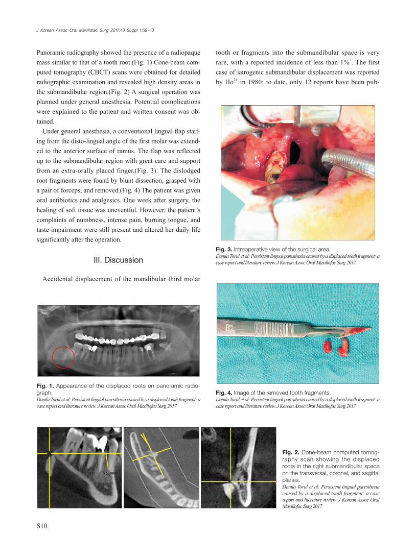
Under general anesthesia, a conventional lingual flap start-ing from the disto-lingual angle of the first molar was extend-ed to the anterior surface of ramus. The flap was reflected up to the submandibular region with great care and support from an extra-orally placed finger.(Fig. 3). The dislodged root fragments were found by blunt dissection, grasped with a pair of forceps, and removed.(Fig. 4) The patient was given oral antibiotics and analgesics. One week after surgery, the healing of soft tissue was uneventful. However, the patient’s complaints of numbness, intense pain, burning tongue, and taste impairment were still present and altered her daily life significantly after the operation.
III. Discussion
Accidental displacement of the mandibular third molar
Fig. 1. Appearance of the displaced roots on panoramic radio-graph.Damla Torul et al: Persistent lingual paresthesia caused by a displaced tooth fragment: a case report and literature review. J Korean Assoc Oral Maxillofac Surg 2017
Fig. 2. Cone-beam computed tomog-raphy scan showing the displaced roots in the right submandibular space on the transversal, coronal, and sagittal planes.Damla Torul et al: Persistent lingual paresthesia caused by a displaced tooth fragment: a case report and literature review. J Korean Assoc Oral Maxillofac Surg 2017
Fig. 3. Intraoperative view of the surgical area.Damla Torul et al: Persistent lingual paresthesia caused by a displaced tooth fragment: a case report and literature review. J Korean Assoc Oral Maxillofac Surg 2017
Fig. 4. Image of the removed tooth fragments.Damla Torul et al: Persistent lingual paresthesia caused by a displaced tooth fragment: a case report and literature review. J Korean Assoc Oral Maxillofac Surg 2017

Persistent lingual paresthesia caused by a displaced tooth fragment
S11
lished1,3,4,7,22,24-30.(Table 1) In most of the cases, the major predisposing factors contributing to displacement were a thin lingual plate and uncontrolled maneuvering due to a lack of basic surgical knowledge. However, number of patient and operator related factors have also been reported to potentially contribute to displacement1,3,4,22,25.
The submandibular space is a complex triangular compart-ment that is bounded superiorly by the mylohyoid muscle, anteriorly by the medial surface of the mandible and the skin, and inferiorly by the superficial fascia and platysma muscle. It encloses vital anatomic structures such as the submandibu-lar salivary gland, facial artery/vein, lingual nerve, and hy-poglossal nerve31. The submandibular space also freely com-municates superiorly with the sublingual space and inferiorly with the lateral pharyngeal space26. Therefore, accidental displacement of a mandibular third molar root or fragments into this space poses a risk of iatrogenic injury to vital struc-tures. Moreover, the root or fragments could potentially be displaced further into deeper facial spaces. Such deeper dis-placements usually require additional invasive approaches.
A displaced tooth may remain asymptomatic or cause pain, swelling, paresthesia, and trismus. The effects of a displaced tooth depend on its size, location, and whether or not there is an infection1. Solanki et al.25 reported a case of mandibular third molar displacement into the submandibular space that remained asymptomatic for two years. In a case reported by Anand and Patil4, dysaesthesia occurred immediately in the lip after an unsuccessful surgery performed by a general prac-titioner. Aznar-Arasa et al.21 reported 6 cases of sublingual displacement; five of the six patients remained asymptomatic. According to Aznar-Arasa et al.21, the symptoms are closely related to the size of the displaced fragment and displacement tends to trigger symptoms when the size of the fragment ex-ceeds 5 mm. In the present case, both medial and distal root fragments of the third molar tooth exceeding 5 mm were dis-placed into the submandibular fossa and caused paresthesia on the right lateral region of the tongue after the first surgery. Due to the severe psychological distress of the patient, the case management was dramatically complicated.
Once the complication occurs, it is important to rigorously evaluate the severity of the condition. Any attempt to retrieve the displaced tooth or fragment without adequate visibility and surgical/anatomical knowledge may result in further dis-placement into deeper facial spaces, iatrogenic injury to the vital structures, and life-threatening events such as deep neck infection, mediastinites, and airway compromise26,27. Based on the reported cases, it can be concluded that the majority of T
able
1. R
evie
w o
f pub
lishe
d ca
ses
of s
ubm
andi
bula
r di
spla
cem
ent
Aut
hor
(yea
r)G
ende
r/
age
(yr)
Side
Dis
plac
ed
frag
men
tL
ingu
al p
late
fr
actu
reSy
mpt
oms
App
roac
hT
imin
gO
pera
tor
Post
oper
ativ
e co
mpl
icat
ion
Ho24
(19
80)
Gra
ndin
i et a
l.27 (
1993
)Y
eh28
(20
02)
Yeh
28 (
2002
)Y
eh28
(20
02)
Ozy
uvac
i et a
l.29 (
2003
)O
lusa
nya
et a
l.30 (
2008
)K
ambu
rogl
u et
al.7 (
2010
)N
usra
th a
nd B
anks
26 (
2010
)A
nand
and
Pat
il4 (20
13)
Kos
e et
al.22
(20
14)
Kos
e et
al.22
(20
14)
Jolly
et a
l.3 (20
14)
Ade
yem
i et a
l.1 (20
16)
Sola
nki e
t al.25
(20
16)
Sola
nki e
t al.25
(20
16)
Pres
ent c
ase
ND
M/3
1N
DN
DN
DN
DN
DF/
46F/
26M
/38
F/30
M/3
4F/
42M
/24
F/46
F/26
F/48
ND R ND
ND
ND
ND
ND R R R R R R R L L R
Too
thT
ooth
Roo
tR
oot
Too
thT
ooth
Too
thR
oot
Roo
tT
ooth
Too
thR
oot
Roo
tT
ooth
Roo
tT
ooth
Roo
t
ND – ND
ND
ND
ND
ND – – + – – + + – + –
ND
Pain
, tri
smus
, sw
ellin
gN
DN
DT
rism
usN
DN
DPa
in tr
ism
us, s
wel
ling
Swel
ling,
pai
n, tr
ism
us-
Pain
tris
mus
, sw
ellin
gPa
in tr
ism
us, s
wel
ling
Pain
tris
mus
,Pa
in tr
ism
us, s
wel
ling
Pain
, sw
ellin
gPa
inPa
rest
hesi
a, ta
ste
impa
irm
ent
ND
LA
/IO
LA
+CS/
IOL
A+C
S/IO
GA
/IO
ND
ND
GA
/IO
GA
/IO
GA
/IO
GA
/ND
LA
/ND
LA
/IO
GA
/EO
LA
+CS/
EO
LA
/IO
GA
/IO
ND
DE
ND
ND
ND
ND
ND IE DE IE IE IE DE
DE
DE
DE
DE
ND S ND
ND
ND
ND
GP
GP
GP
GP
GP
GP
GP
GP
GP
GP
GP
ND
Non
eN
one
Non
eN
one
Non
eN
one
Non
eN
one
Non
eN
one
Tem
pora
ry L
N p
ares
thes
iaN
one
Non
eN
one
Tem
pora
ry L
N p
ares
thes
iaPe
rsis
tent
LN
par
esth
esia
(ND
: no
dat
a, M
: m
ale,
F:
fem
ale,
R:
righ
t, L
: le
ft,
LA
: lo
cal
anes
thes
ia,
CS:
con
scio
usne
ss s
edat
ion,
GA
: ge
nera
l an
esth
esia
, IO
: in
trao
ral,
EO
: ex
trao
ral,
DE
: de
laye
d ex
trac
tion,
IE
: im
med
iate
ex
trac
tion,
S: s
urge
on, G
P: g
ener
al p
ract
ition
er, L
N: l
ingu
al n
erve
)Da
mla T
orul
et al:
Per
sisten
t ling
ual p
ares
thesia
caus
ed by
a dis
place
d too
th fra
gmen
t: a c
ase r
epor
t and
liter
ature
review
. J K
orea
n Asso
c Ora
l Max
illofac
Surg
2017

J Korean Assoc Oral Maxillofac Surg 2017;43 Suppl 1:S9-13
S12
fragment and also has severe psychological distress, as in the present case, surgical removal of the fragment should be performed after explaining all possible complications to the patient.
Access to the submandibular space is difficult because of the anatomical structures enclosed by this space31. Many surgical approaches, including trans-oral approaches via gen-eral4,7,22,26 or local anesthesia3,25,27 and extra-oral approaches via local25 or general anesthesia1 have been reported. In 2002, Yeh28 reported a modified technique using a combination of intraoral and extraoral approaches to facilitate the retrieval of displaced teeth and fragments. In addition to the conventional lingual flap extended to the first molar, a hemostat was insert-ed to stabilize the fragment via a skin incision that was made in the submandibular region. Moreover, recent advancements in biotechnology computer-assisted navigation systems have enabled retrieval of displaced teeth in a matter of minutes32,33. In the present case, adequate access and visualization were achieved with a preoperative CBCT scan and by carefully reflecting a deep lingual flap up to the submandibular region and supporting the flap extra-orally with digital pressure.
In conclusion, trans-operative displacement of a tooth or tooth fragment into the facial spaces is a rare but potentially serious condition, commonly requiring a second surgery. A second major procedure may cause further complications or worsen existing ones, as in our case. Thus, the best way to manage a surgical complication is prevention. This can be achieved by thorough evaluation of all significant risk fac-tors and careful execution of the surgical procedure. Also, for optimal surgical outcomes, operation should be preferably performed by maxillofacial surgeons who are highly familiar-ized with the surgical morphology of the head and neck.
Conflict of Interest
No potential conflict of interest relevant to this article was reported.
ORCID
Damla Torul, http://orcid.org/0000-0003-2323-606XDilara Kazan, http://orcid.org/0000-0002-7471-8758Mehmet Cihan Bereket, http://orcid.org/0000-0003-0578-
7087Rifat Karli, http://orcid.org/0000-0002-2845-6558
iatrogenic displacements were caused by practitioners with a lack of surgical expertise. Adeyemi et al.1 reported a case of submasseteric abscess that occurred 1 month after subman-dibular displacement. The present case showed persistent dysaesthesia caused by an unsuccessful first surgical attempt by a practitioner. Moreover, the operator may be faced with legal implications related to this unsuccessful surgery12. Thus, it is prudent to stop the operation, inform the patient about the condition, and refer the patient to a maxillofacial surgeon as soon as possible22.
On the other hand, this complication can occasionally oc-cur even in the hands of the most experienced surgeons32. Grandini et al.27 reported a case in which a dental surgeon caused the mandibular third molar to displace into the sub-mandibular space and failed to retrieve the tooth for 6 hours, leading to severe tissue injury. Thus, in addition to surgical knowledge and experience, adequate pre-surgical planning, clinical and radiographic examinations, adequate access and visibility, proper technique, and controlled use of force are crucial for reducing the incidence and legal problems of this complication7.
Advanced imaging techniques are useful for thorough case evaluation when complications occur. Computed tomography is considered the most suitable technique and enables the exact location and size of the displaced tooth or tooth frag-ment to be pinpointed3. However, CBCT scanning, which has the advantage of low radiation exposure, can be used prefer-ably if the fragment or tooth is displaced into deep spaces9. Panoramic, postero-anterior, submento-vertex, and occlusal views, in addition to image intensifiers, are among the other imaging modalities that can also be used to determine the location of displaced teeth or fragments26. We used CBCT to evaluate the fragment locations and sizes because this tech-nique is already available in our Oral and Maxillofacial Sur-gery Clinic.
No clear consensus has been reached regarding the retriev-al time of displaced teeth or their fragments. Some authors have suggested delaying tooth or fragment retrieval to allow fibrosis to develop12,17, but others have claimed that delay-ing fragment removal can result in severe pain, infection, and further migration of the root or root fragment into deep facial spaces7,27. These authors suggest removal of the tooth or fragment as soon as possible. We conclude that if the frag-ment is small, is not accompanied by any symptoms, and is in a deep position, then it is better to keep the fragment under observation to prevent further tissue injuries. However, if the patient has symptoms associated with the displaced tooth or

Persistent lingual paresthesia caused by a displaced tooth fragment
S13
References
1. Adeyemi MO, James O, Lawal AO, Fadeyibi SO. Iatrogenic dis-placement of impacted mandibular third molar into the subman-dibular space complicated by submasseteric abscess. Afr J Trauma 2016;5:19-22.
2. Brauer HU. Unusual complications associated with third molar sur-gery: a systematic review. Quintessence Int 2009;40:565-72.
3. Jolly SS, Rattan V, Rai SK. Intraoral management of displaced root into submandibular space under local anaesthesia: a case report and review of literature. Saudi Dent J 2014;26:181-4.
4. Anand R, Patil PM. Accidental displacement of third molars; report of three cases, review of literature and treatment recommendations. Oral Surgery 2013;6:2-8.
5. Primo BT, Stringhini DJ, Klüppel LE, da Costa DJ, Rebellato LBR, de Moraes RS. Delayed removal of maxillary third molar displaced into the infratemporal fossa. Rev Esp Cir Oral Maxillo-fac 2014;30:78-81.
6. Tumuluri V, Punnia-Moorthy A. Displacement of a mandibular third molar root fragment into the pterygomandibular space. Aust Dent J 2002;47:68-71.
7. Kamburoglu K, Kursun S, Oztas B. Submandibular displacement of a mandibular third molar root during extraction: a case report. Cases J 2010;3:8.
8. De Biase A, Guerra F, Giordano G, Salucci S, Solidani M. Surgical removal of a left lower third molar root after iatrogenic displace-ment in soft tissue. Case report. Minerva Stomatol 2005;54:389-93.
9. Silveira RJ, Garcia RR, Botelho TL, Franco A, Silva RF. Acciden-tal displacement of third molar into the sublingual space: a case report. J Oral Maxillofac Res 2014;5:e5.
10. Shahakbari R, Mortazavi H, Eshghpour M. First report of acci-dental displacement of mandibular third molar into infratemporal space. J Oral Maxillofac Surg 2011;69:1301-3.
11. Selvi F, Cakarer S, Keskin C, Ozyuvaci H. Delayed removal of a maxillary third molar accidentally displaced into the infratemporal fossa. J Craniofac Surg 2011;22:1391-3.
12. Huang IY, Chen CM, Chang SW, Yang CF, Chen CH, Chen CM. Surgical management of accidentally displaced mandibular third molar into the pterygomandibular space: a case report. Kaohsiung J Med Sci 2007;23:370-4.
13. Khan M, Mehboob B, Kundi JA. Pattern and management of iatro-genic displacement of teeth in maxillofacial anatomical spaces. Pak Oral Dent J 2015;35:186-9.
14. Xavier CB, Gonçalves FR, Batista SH, Veras Filho Rde O, Vogt BF. Spontaneous migration of third molar following displacement to pterygomandibular fossa. J Oral Maxillofac Surg 2011;69:1004-7.
15. Huang IY, Chen CM, Chuang FH. Caldwell-Luc procedure for re-trieval of displaced root in the maxillary sinus. Oral Surg Oral Med Oral Pathol Oral Radiol Endod 2011;112:e59-63.
16. Ertas U, Yaruz MS, Tozoğlu S. Accidental third molar displace-
ment into the lateral pharyngeal space. J Oral Maxillofac Surg 2002;60:1217.
17. Esen E, Aydoğan LB, Akçali MC. Accidental displacement of an impacted mandibular third molar into the lateral pharyngeal space. J Oral Maxillofac Surg 2000;58:96-7.
18. Gay-Escoda C, Berini-Aytés L, Piñera-Penalva M. Accidental displacement of a lower third molar. Report of a case in the lateral cervical position. Oral Surg Oral Med Oral Pathol 1993;76:159-60.
19. Ozer N, Uçem F, Saruhanoğlu A, Yilmaz S, Tanyeri H. Removal of a maxillary third molar displaced into pterygopalatine fossa via in-traoral approach. Case Rep Dent 2013. doi: 10.1155/2013/392148.
20. Kocaelli H, Balcioglu HA, Erdem TL. Displacement of a maxillary third molar into the buccal space: anatomical implications apropos of a case. Int J Oral Maxillofac Surg 2011;40:650-3.
21. Aznar-Arasa L, Figueiredo R, Gay-Escoda C. Iatrogenic displace-ment of lower third molar roots into the sublingual space: report of 6 cases. J Oral Maxillofac Surg 2012;70:e107-15.
22. Kose I, Koparal M, Güneş N, Atalay Y, Yaman F, Atilgan S, et al. Displaced lower third molar tooth into the submandibular space: Two case reports. J Nat Sci Biol Med 2014;5:482-4.
23. Roshanghias K, Peisker A, Zieron JO. Maxillary tooth displace-ment in the infratemporal fossa. Dent Res J (Isfahan) 2016;13:373-5.
24. Ho KH. Retrieval of a molar from the submandibular space--a case report. Singapore Dent J 1980;5:61-2.
25. Solanki R, Khangwal M, Kumar D, Goel M. Retrieval of man-dibular third molar tooth accidentally displaced in submandibular space: series of two cases. Indian J Dent 2016;7:105-8.
26. Nusrath MA, Banks RJ. Unrecognised displacement of mandibular molar root into the submandibular space. Br Dent J 2010;209:279-80.
27. Grandini SA, Barros VM, Salata LA, Rosa AL, Soares UN. Com-plications in exodontia--accidental dislodgment to adjacent ana-tomical areas. Braz Dent J 1993;3:103-12.
28. Yeh CJ. A simple retrieval technique for accidentally displaced mandibular third molars. J Oral Maxillofac Surg 2002;60:836-7.
29. Ozyuvaci H, Firat D, Tanyel C. Accidental displacement of a man-dibular third molar: a case report. Quintessence Int 2003;34:278-80.
30. Olusanya AA, Akadiri OA, Akinmoladun VI. Accidental displace-ment of mandibular third molar into soft tissue: a case report. Afr J Med Med Sci 2008;37:77-80.
31. Bonala N, Kishan TV, Sri Pavani B, Murthy PV. Accessory belly of digastric muscle presenting as a submandibular space mass. Med J Armed Forces India 2015;71(Suppl 2):S506-8.
32. Campbell A, Costello BJ. Retrieval of a displaced third molar us-ing navigation and active image guidance. J Oral Maxillofac Surg 2010;68:480-5.
33. Guo Y, Xu DD, Lv K, Wan QL, Li ZB, Li Z. Use of computer-assisted navigation in the retrieval of accidentally displaced third molars. J Oral Maxillofac Surg 2016;74:889-94.