Embed Size (px)
Citation preview
7/21/2019 pernix una archea fantastica
http://slidepdf.com/reader/full/pernix-una-archea-fantastica 1/8
INTERNATIONALOURNALF SYSTEMATICACTERIOLOGY,ct. 1996, p.
1070-1077
0020-7713/96/$04.00+0
Copyright 996, International Union of Microbiological Societies
Vol. 46,
No.
4
Aeropyrumpemix
gen. nov.,
sp.
nov., a Novel
Hyp erthermophilic Archaeon Growing
Tem peratures up
to 100°C
Aerobic
at
YOSHI HI KO SAKO,' NORIMICHI NOMURA, ' ARITSUN E UCHIDA, ' YU ZABU RO ISHIDA, '
HI R OYUKI MOR I I ,2 YOSUKE KOGA,2 TOS HIH IRO HOAKI,3-t AND TADASHI MAR UYAMA3
Laboratory
of
Marine Microbiology, Department
of
Applied Bioscience, Graduate School
of
Agriculture, Kyoto
University, Kyoto
606-01,
Department
of
Chemistry, School
of
Medicine, University
of
Occupational
and Environmental Health, Kitakyushu 807 and Marine Biotechnology Institute,
Shimizu Laboratories, Shimizu, Shizuoka
424
Japan
A novel aerobic hyperthermophilic archaeon was isolated from a coastal solfataric vent at Kodakara-Jim a
Island, Japan. The new isolate, strain K1, is the first strictly aerobic organism growing at temperatures up to
100°C. It grows optimally at 90 to 95 C, pH 7.0, and a salinity of 3.5%. The cells are spherical shaped and 0.8
to 1.2 p m in diameter. Various proteinaceous complex compounds served as substrates during aerobic growth.
Thiosulfate stimulates growth without producing H,S. The core lipids consist solely of C,,-isopranyl archae-
ol(glycero1 diether). The
G + C
content of the genomic DNA is 67 mol%. Phylogenetic analysis based on 1 6 s
rRNA sequence indicates that strain
K 1
is a new member of Crenarchaeota. On the basis of our results, the
name Aeropyrum
pe rn h
gen. nov., sp. nov. is proposed (type strain: K1; JCM 9820 .
Proposal of the Arc haea (originally called arch aeba cteria ) as
a discrete domain
(57,
58) shed new light on the central prob-
lems of both early evolution of life and prokaryotic systematics.
Although this concept is now generally accepted because of the
several biochemical features peculiar to the Archaea, it is still
a matter of controversy how the Archaea domain is phyloge-
netically related to the other
two
domains, the Eucarya (eu-
karyote) and the Bacteria (eubacteria)
(9).
This problem stem s partially from an essential lack of infor-
mation on the deepest (earliest) branches within the universal
p hylogenetic trees. F or instance, a deep missing branch
might dec rease th e reliability
of
the deep-branching topologies
of
the phylogenetic trees.
In
order to obtain additional infor-
mation pertinent to this problem, we aimed to isolate organ-
isms that might be representative
of
the d eep missing branches.
Interestingly, it was pointed out that the deepest an d short-
est branches within the universal phylogenetic trees are dom-
inated by hyperthermophiles, which grow optimally at tem per-
atures above 80°C (1, 2, 3, 55) . During the past decade, many
new hyperthermophiles w ere isolated from solfataric fields and
submarine volcanic vents
(43).
Those include the hyperther-
mophilic meth anog enic archa ea, the archaeal sulfate reducers,
the hyperthermophilic So-m etabolizers, and the gene ra Ther-
motogales
and
AquifRx
within the
Bacteria.
However, since
ox-
y,pen availability in the h ydrothe rmal environm ents is low be-
c.ause of poo r solubility, most stud ies
of
life in these ecosystems
have been restricted to anaerobic organisms (17, 20).
Recently, we succeeded in th e isolation and cultivation of a
novel type of the strictly aerobic hyperthermophilic archaeon,
growing optimally at temperatures above 90°C. In this report,
we
characterize the new isolate and propose that it should be
classified in a new genus,
Aeropyrum.
Corresponding author. Phone: 81-75-753-6218. Fax: 81-75-753-
t
Present address: Biotechnology Research Department, Taisci Cor-
6226. Electronic mail address: [email protected].
poration, Narashino, Chiba 275, Japan.
MATERIALS AND METHODS
Collections of samples. Hot sedimentary materials and venting water were
collected at a coastal solfataric thermal vent in Kodakara-Jima Island (29 13'N,
129'20'E) during a cruise of the research vesscl Sugen-mum in 1993. Anaerobic
hyperthermophiles were isolated from this field (16, 17, 28). In situ temperature
was determ ined with a mercury-filled C elsius thermometer
(0
to 200 C), and the
pH was estimated by a pH meter (Toa Electronics, Tokyo, Japan). A fter collec-
tion, the samples were brought back to th e laboratory, cooled on ice, and stored
at 4°C until enrichment.
Culture conditions. The base for all media described in this paper is the
synthetic scawater JS, which was prepared from Jamarine
S
synthetic sea salts
(Jamarine Laboratory, Osaka , Japa n) according to the manufacturer 's instruc-
tions. Unless specified otherwise, the new isolate was grown in the standard
medium referred t o as JXT, containing (per liter of JS) 1g of yeast extract (Difco
Laboratories, Detroit, Mich.), 1 g of trypticase peptone (BBL Microbiology
Systems, Cockeysville, Md.), and 1 g of Na,S,O, SH,O, pH 7.0 (adjusted with
HCI at room temperature). For enrichment cultures and isolation, JX medium,
prep ared by omitting Na,S,O, 5H,O from JX T medium , was use d.
Cul tures were routinely grown in screw-ca pped test tubes (180 by 18 mm)
containing
10
ml of medium a nd incubated a t 90°C in a forced convection oven
(FC-610; Advantec, Tokyo, Japan) without shaking. In order to maintain aerob ic
culture conditions, the gas phase of the closed test tubes was not evacuated and
thc atmospheric air (100 kPa) was confined. Resazurin (1 p,g/ml) was used
as a
redox indicator.
Batch cultures were grown in cotton-plugged 2-liter Erlenmeyer flasks con-
taining 500 ml of medium and incubated at 90°C in an air bath rotary shaker
(RGS-
32.TT; Sanki Seiki, Osaka, Japan) with vigorous shaking (180 rpm).
Substrate utilization. In an attempt to find carbon substra tes which would
sustain growth and to test the optimum c oncentration for growth, each of the
following organic substances (obtained from Nacalai Tesque, Kyoto, Japan,
except whe re noted otherwise) was adde d to 10 ml of the
JS
base, supplemented
with 0.1% (wtivol) Na,S,O, 5H,O, in conc entra tions (wt/vol) of 0.05, 0.1, 0.2,
and 0.4%:
D-(
-)-ribose,
L-(
+)-arabinose,
D-(
+)-xylose,
D-(
+)-glucose,
L-( -1-
glucose,
u-(
+)-galactose,
D-(
-)-fructose, L-( -)-fucose, L-( +)-rhamnose, D-( +)-
mannose, maltose, lactose, sucrose, D-(
+
-ccllobiose, D-( +)-melibiose, D-( +)-
raffinose, cellulose, starch, D-sorbitol, u-mannitol, glycerol, acetate, alanine plus
glycine, 20 different L-amino acids, yeast extract (Difco), tryptone (Difco),
nu-
trient broth (Difco), Casamino Acids (vitamin free; Difco), and trypticase pep-
tone (BBL). To test the various sulfur compounds, the Na,S,O, in JXT medium
was replaced with 10 mM (each) Na,S04, Na,SO,, So, cysteine, or m ethionine.
These cu ltures were incubated at 90°C without shaking.
Sulfide analysis. The sulfide production during growth was detected qualita-
tivcly as described by Huber et al.
(19).
Oxygen requirement for growth. In order to ensure strict anaerobiosis, the
anaerobic techniques described by Balch and Wolfe
(4)
were employed. Oxygen
was reduced by adding 0.05% (wtivol) Na,S 9H,O. Micro aerobic cultu re con -
ditions were achieved as described by Huber et al. (21) with minor modifications.
Prior to autoclaving, 10 ml of medium was dispensed into a 120-ml glass bottle
(Schott Glaswerke, Mainz, Germany) that was tightly sealed with butyl rubber
1070
7/21/2019 pernix una archea fantastica
http://slidepdf.com/reader/full/pernix-una-archea-fantastica 2/8
VOL.46, 1996 AEROPYRUM P E N I X , A NOVEL ARCHAEON
1071
stoppers, and the gas phase was exchanged with either H2 -C 02 80:20,
300
kPa)
o r N, (loo%, 300 kPa). In both experiments, the state of oxygenation was
monitored by resazurin (1 pg/ml), and cultures were incubated at 90°C without
shaking.
Light and electron microscopy. Cells were inspected with a Nikon Optipho t
XF -NT differential interfere nce light microscope equi ppe d with an oil immers ion
objective of 100/1.25. Transmis sion electron microscopy of negatively stained
cells was carried out as described by Zillig et al.
(59).
The cells were negatively
stained with 1 to 2% (wt/vol) uranyl acetate and observed in a Hitachi H-7000
transmission electron microscope at an accelerating voltage of 75 kV. For ultra-
thin sectioning, cells were fixed in JX T med ium with 2% (vol/vol) glutaraldehyde
and postfixed with 1% OsO,. The fixed cells were then dehydrated with ethanol
and embedded in Quetol 812 (Nisshin EM, Tokyo, Japan) epoxy resin. Thin
sections were do uble contrasted with uranyl acetate and lead citrate. Th e spec-
imens were observed with a JEO L 1200EX I electron microscope operated at 80
kV.
Determination of growth. Growth was determine d either by measurement of
the optical density at 660 nm or by direct cell counting by using a Tho ma
chamber (dep th, 0.02 mm). A good c orrelation was found between these
two
methods in exponential and stationary phase cultures. In the standard cul ture
conditions, an OD,,,, of
1
corresponded to a cell density of about
1.4
x 10' cells
per ml.
To study the growth characteristics, the batch culture technique dcscribed
above was applied. At temperatures above 100 C, screw-capped 1-liter glass
bottles (Schott Glaswcrke) were used to prevent the evaporation of the medium.
The p H dependency of growth was studied in JX T mcdium with various buffer
systems at a concentration of 20 mM, e.g., sodium succinatc (below pH 5.0):
MES [2-(N-morpholino) ethanesulfonic acid] (pH
5.5
to 6.5), HEPES [N-(2-
hydroxycthyl) piperazine-N'-2-cthanesulfonicacid] (pH 7.0 to 8.0), and BTP
{ 1,3-bis [tris (hydroxymethyl) m ethylamino] propane} (pH 8.5 or above). The
pH of the medium was adjusted by the addition of HCI or NaO H at room
temperature. Growth at different salinities was determined by using 2X JS
diluted approp riately in the medium. D oubling times were calculated by using
linear regression analysis from 3 to 5 points along the logarithmic part of the
resulting growth curves.
Antibiotic sensitivity.
Sensitivity to the a ntibiotics chloramphe nicol, am picillin,
vancomycin, and cycloserine (all from Sigma Chemical, St. Louis, Mu.) was
tested at a concentration of 100 Fg/ml. Th e tem perature for these tests was 70°C.
Preliminary experiments with the bacterium Thermus aqitaticus (ATCC 25104)
had indicated that the antibiotics were effective at 70°C.
Lipid analysis. Total lipid was extracted from the cells as described by Bligh
and Dyer (8) and analyzed by two-dimensional thin-layer chromatography (2D-
TLC) performed
on
silica gel 60 plates (Art. 5721: Merck, Darms tadt, Gcrman y).
Solvents included chloroform-methanol-7 M aque ous amm onia (65:35:8, vol/
volivol) in the first dimension and chloroform-methanol-acetic acid-water
(85:
30:15:5, vol/vo ~vo l/vol ) n the sec ond dimension. Polar lipid composition was
determine d by measu rcm ent of the phos pho rus (6) of individual lipid spots on
the 2D-TLC chromatogram.
Core lipids were prepared by splitting off the polar head groups by acid
methanolysis
(5%
HCI-methanol at 100°C for 2 h) of the tota l lipid extracts and
separated by TLC with the solvent light petroleum-diethyl ether-acetic acid
(50:50:1, vol/vol/vol). Hydrocarbon chains were p repa red f rom thc core lipids by
hydroiodic acid degra datio n followed by LiA lH, reduction a s previously re-
ported (31) and identified by gas chromatography-mass spectrometry and elec-
tron impact mass spectrometry employing instrumentation and conditions as
previously described (33). The polar head groups obtained in the aqueou s frac-
tion of the acid methanolys is were fu rthe r acid hydrolyzed
1
M H CI at 100°C for
3 h or 4 M HCI at 100°C for 16 h) and acctylated. Th e acetylated hydrolysate was
analyzed by gas liquid chromatography as described previously (31). Glycero-
phospho ric csters prepa red from the total lipid extracts by dealkylation with BCI,
(32) were analyzed by cellulose TLC (Art. 5716, Merck) (32) with the solvent
phcnol-water (100:38, vol/vol). Authentic samples
of
archaeol and caldarchaeol
(30) were prepared from total lipids of Methanobacterium therinoautotrophicum
or
Sulfolobus solfataricus.
DNA preparation. Cell pellets (about 1 g) were suspended in 10 ml
of
NE T
buffer
(50
mM Tris-HCI,
150
mM NaCI, 100 mM Na,-EDTA [pH 8.01) contain-
ing 2% (wtivol) sodium dodecyl sulfate and 0.1 mg of proteinase K (Nacalai
Tesque ) per ml and incubated at 65°C for 30 min. Thc crude genomic DNA was
prepared by the method
of
Samb rook et al. (38) and was purificd by a protocol
which employed cetyltrimethyl am mo nium bromide (29) and by CsCl dcnsity-
gradient centrifugation.
DNA base composition. The G + C contcnt of the genomic DNA was deter-
mined by high-performance liquid chromatography (27) with a DNA-GC kit
(Yamasa Shoyu, Tokyo, Japan). Calf thymus D NA (42 mol% G +C ; Sigma) (26)
and nonmethylated DN A from bacteriophage (49.8 mol% G+C ; Takara
Shuzo, Kyoto, Japan) (39) were used as references.
1 6 s
rRNA preparation.
16s rRNA was preparcd essentially according to
Traub et al.
(47)
with modifications as follows. The exponentially growing cells
(about
1
g) were suspended in
20
ml of buffer I [ lo m M Tr is-HCI , 10 mM
Mg(CH,COO),, 60 mM NH,CI,
6
mM 2-merca ptoethano l, pH 7.81 and 20 ml of
glass beads (type
I;
Sigma) was added. Th c suspension was vortexed for 15 min
at 4°C and centrifuged twice at 30,000
X
g for 30 min. The supernatant was
removed a nd recentrifuged at 105,000 X g for 6 h to precipitate the ribosomes.
The pellet was resuspended in buffer I and treated with RNase-free DNase I
(Boehringer, Mannheim, Gcrmany) at a concentration of
2
pg/ml. rR NAs were
obtained after three extractions with phenol-chloroform followed by two ethanol
precipitations and separated by a
5
to 20% sucrose density gradient ccntrifuga-
tion at 100,000 X g for 17 h (TS T 28.38 rotor; Kontron Instruments, Milan, Italy).
Fractions containing 1 6s rRNA were pooled. Purity of the 16s rRN A was
checked electrophoretically.
1 6 s
rRNA analysis.
A cDNA clone of the
16s
rRNA was obtained af tcr
reverse transcription and PCR amplification. The oligonucleotide 1521R (5'-A
GG TGA ITC AG CC GC AG GTT ), complementary to the 3 '- ta il conserved re-
gion of the archaeal 16s rRN A, was employed to prim e first-strand synthesis with
Superscript
I1
RNas eH- (GIB CO Laboratories, Grand Island, N.Y.). The re-
verse transcription reaction was performed according to the manufacturer 's
instructions, and the product was amplified by PCR with Arch21F (5'-TTCCG
G l T G A T C C Y G C C G G A ) (10) and 1521R as p rimers. Thirty-five amplification
cycles of 90 s at
96 C,
1 min at 62 T , and 2 min at 72°C were performed. T hc
double-stranded PCR product was cloned directly into the pCRII vector
( In-
vitrogen, San Diego, Calif.) to yield plasmid pNA4. T he nucleo tide sequence was
determined by the dideoxy sequencing method (40) by using a set of primers
complcmcntary to conserved regions located along the archaeal 16 s rRNA. The
sequen ce was aligned to a collection of arch aeal
16s
rRNA sequences (DNA
Data Bank
of
Japan [DDBJ], National Institute of Genetics, Shizuoka, Japan).
Num bers of base substitutions per site were estimated by the meth od of Tajima
and Nei (45), and the phylogenetic tree was inferred on the basis of the neighbor-
joining method (36). The resulting tree was teste d by using bootstrap analysis
Nucleotide sequence accession number. The 16 s rRNA se quence of isolate K1
will appear in the DDBJ, EMBL, and GenBank nucleotide sequence databases
with the accession number D83259.
(14).
RESULTS
Enrichment and isolation of the aerobic hyperthermophile.
In order to enrich aerobic hyperthermophiles, 10 ml of JX
medium was inoculated with approximately
1
g of sample in
the laboratory. T he sample was a m ixture
of
sandy sediment
and clear seawater. Microbial mat material was not visible in
the sample. The original temperatu re was
98
to 103 C, and the
pH was 6.9. The enrichment was performed at
90°C
in screw-
capped test tubes with air as the gas phase (100 kPa) without
shaking. Within
2
days, turbidity caused by cell growth was
observed. This growth consisted of a mixed population of var-
ious sizes
of
rods an d cocci. During this enrichm ent culture,
resazurin remained red, and sulfide was not produced. All
positive enrichment cultures could be successfully transferred
in the sam e medium.
In order to obtain pure cultures, a dilution-to-extinction
technique was employed
5 ) .
After the cell density of an en-
richment culture reached approximately
lo7
cells per ml,
5
separ ate dilution series were con ducted in w hich
5
1:lO dilu-
tions of the culture were followed by 20
1:3
dilutions. Each
dilution in the dilution-to-extinction series was carried out in
triplicate and incubated for at least 7 days. The culture in the
tube showing growth at the highest dilution was designated
isolate
K1,
which usually grew u p to about
lo7
cells per ml in
4 days. The purity of this culture was routinely confirmed by
microscopic examinations and by restriction fragment length
polymorphism (RFLP) analysis of the PCR-amplified
16s
rRN A gene fragm ent (data not shown). For further investiga-
tions, JX medium was replaced by JXT medium, in which a
higher final cell density was obtained as described below. In
this medium, the cell yield in batch culture was about 1 g/liter
(wet weight) in stationary phas e. Packed cell masses exhibited
a brown color.
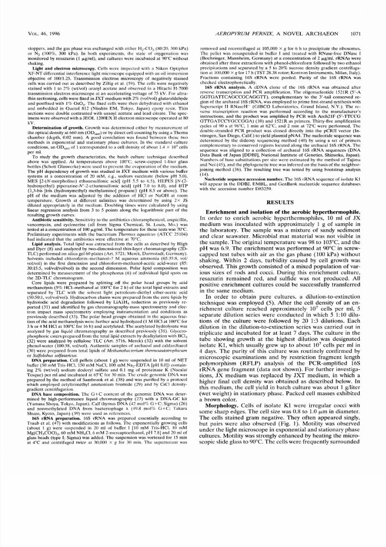
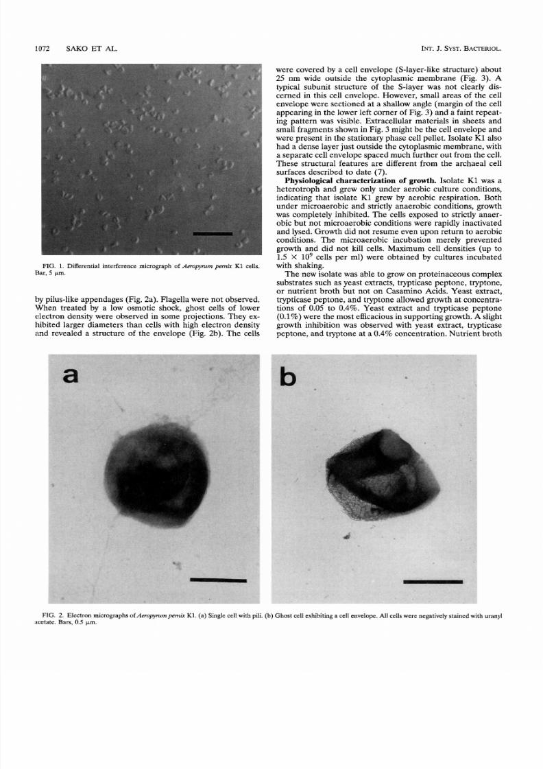
Morphology. Cells of isolate K l were irregular cocci with
some s har p edges. Th e cell size was
0.8
to
1.0
(-l.m n diameter.
The cells stained gram negative. They often appeared singly,
but pairs were also observed (Fig.
1).
Motility was observed
unde r the light microscope in exponential and stationary phase
cultures. Motility was strongly enh ance d by heating the m icro-
scopic slide glass to 90°C. Th e cells wer e frequently surro unde d
7/21/2019 pernix una archea fantastica
http://slidepdf.com/reader/full/pernix-una-archea-fantastica 3/8
18 72 SAKO ET
AL.
INT.
J.
SYST.
BACTERIOL.
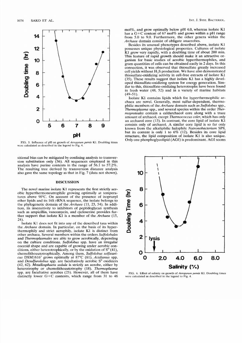
were covered by a cell envelope (S-layer-like structure) about
25
nm wide outside the cytoplasmic membrane (Fig.
3). A
typical subunit structure of the S-layer was not clearly dis-
cerned in this cell envelope. However, small areas of the cell
envelope were sectioned at a shallow angle (margin of the cell
appearing in the lower left corner of Fig.
3)
and a faint repeat-
ing pattern was visible. Extracellular materials in sheets and
small fragments shown in Fig.
3
might be the cell envelope and
were present in the stationary phase cell pellet. Isolate K1 also
had a dense layer just outside the cytoplasmic membrane, with
a separate cell envelope spaced much further out from the cell.
These structural features are different from the archaeal cell
surfaces described to date (7).
Physiological characterization
of
growth. Isolate
K1
was a
heterotroph and grew only under aerobic culture conditions,
indicating that isolate K1 grew by aerobic respiration. Both
under microaerobic and strictly anaerobic conditions, growth
was completely inhibited. The cells exposed to strictly anaer-
obic but not microaerobic conditions were rapidly inactivated
and lysed. Growth did not resume even upon return to aerobic
conditions. The microaerobic incubation merely prevented
growth and did not kill cells. Maximum cell densities (up to
1.5 X lo9 cells per ml) were obtained by cultures incubated
with shaking.
The new isolate was able to grow on proteinaceous complex
substrates such as yeast extracts, trypticase peptone, tryptone,
or nutrient broth but not on Casamino Acids. Yeast extract,
trypticase peptone, and tryptone allowed growth at concentra-
tions of 0.05 to 0.4%. Yeast extract and trypticase peptone
(0.1%)were the most efficacious in supporting growth.A slight
growth inhibition was observed with yeast extract, trypticase
peptone, and tryptone at a 0.4% concentration. Nutrient broth
FIG. 1. Differential interference micrograph of eropymrn
pemir
K1 cells.
Bar, km.
by pilus-like appendages (Fig. 2a). Flagella were not observed.
When treated by a low osmotic shock, ghost cells of lower
electron density were observed in some projections. They ex-
hibited larger diameters than cells with high electron density
and revealed a structure of the envelope
(Fig. 2b).
The cells
FIG. 2. Electron micrographsof erujyzmpemkK1. (a) Single cell with pili. (b) Ghost cell exhibiting a cell envelope. All cells were negatively stained with uranyl
acetate. Bars, 0.5 Fm.

7/21/2019 pernix una archea fantastica
http://slidepdf.com/reader/full/pernix-una-archea-fantastica 4/8
VOL.
46, 1996
5
40
30
20
10
AEROPYRUM PE WI X, A
NOVEL
ARCHAEON
1073
-
-
-
-
-
FIG. 3. Ultrathin section
of Aeropyrum pemrjr
K1. Bar,
0.2
pm
concentrations of over 0.1% inhibited growth. Any single car-
bohydrate, organic acid, or amino acid tested within the con-
centration range of
0.05
to 0.4% did not suffice as a sole carbon
and energy source. The growth yield was stimulated about
eightfold by thiosulfate. However, thiosulfate was not required
for growth. H,S was not formed during growth in either the
presence or absence of thiosulfate. The growth yield was not
stimulated by other sulfur-containing compounds such as sul-
fate, sulfite,
So,
sulfide, cysteine, or methionine.
Isolate K1 grew well between 70 and 9TC, and cell division
still occurred at
100°C.
No growth was observed at
68
or
102°C.
The optimum growth temperature was 90 to 95 C, with a
doubling time of 200 min (Fig.
4).
The optimum pH for growth
was around 7.0. Growth was obtained in a pH range from 5.0
to 9.0 (Fig. 5). Metabolism of the medium components did not
cause the initial medium pH to change significantly during
these experiments (data not shown). The optimum salinity in
the medium was found to be approximately 3.5 , which was
the same level of ionic strength as JXT medium (Fig.
6).
Growth was observed at a salinity from 1.8 to 7.0%. At a
salinity of 1.5% in the medium, rapid cell lysis occurred, as
indicated by the presence of ghost cells upon electron micro-
scopic examination and by the decreased turbidity of the cul-
ture.
Sensitivity to antibiotics.
Isolate
K1
was insensitive to 100-
&ml concentrations of ampicillin, vancomycin, and cyclo-
serine but was completely inhibited in the presence of the same
concentration of chloramphenicol.
Lipids.
The 2D-TLC pattern of the total lipid of isolate
K1
appeared to be different from those of the archaeal total lipids
investigated to date. At least five polar lipids were detected.
Among these five polar lipids, one phosphoglycolipid (desig-
nated as AGI) and one phospholipid (designated as
AI)
were
predominant. Assuming that each lipid contains one phos-
phate moiety, the content of AGI and A1 in the total lipid
was 91 mol% and 9 mol%, respectively. The other three polar
lipids (one phosphoglycolipid and
two
glycolipids) were pres-
ent at trace levels.
The hydrocarbon chain prepared from the core lipid was
identified by gas chromatography-mass spectrometry as C,-
isoprenoid, and the structure of the glycerol diether core lipid
was identified as disesterterpanyl (C,,, CZ5)glycerol. AGI con-
tained glucose and inositol and AI contained inositol as polar
head group components. Glycerophosphoinositol was detected
as a sole phosphorus-containing product of the BC1, treatment
of AGI or AI. The complete structures of the polar lipids and
their core portions will be published elsewhere.
DNA
base composition.
The genomic DNA
of
isolate
K1
had
a G +C content of 67 mol% as calculated by direct analysis of
the nucleosides.
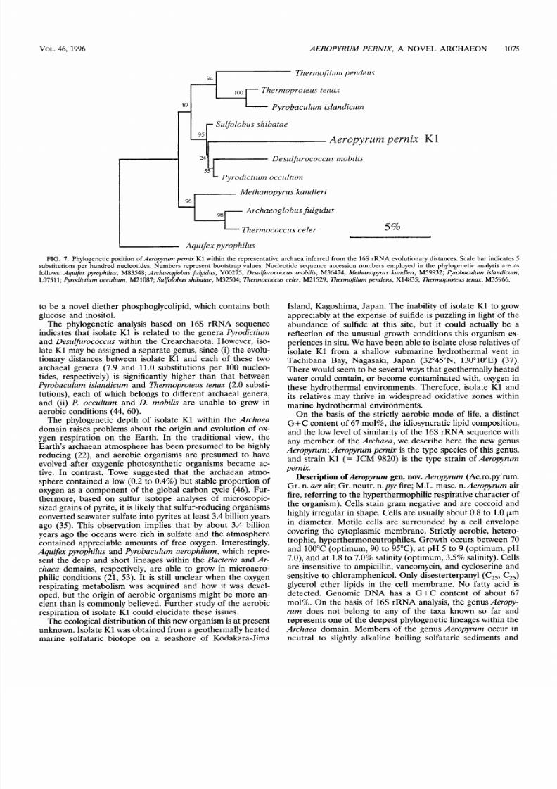
Phylogenetic analysis.
The G+C contents of the
16s
rRNA
sequences employed in this analysis ranged from 62.7 to
68.7
mol%, and it was therefore assumed that they were relatively
free from the variation of biased base compositions. Evolution-
ary distances were estimated by the comparison of represen-
tative archaeal
16s
rRNA sequences. Isolate
K1
presented
considerably close relationships to
Pyrodictium occultum
and
Desulfirococcusmobilis
as indicated by estimated exchanges of
7.9 and
11.0
nucleotides per
100
positions, respectively.
A phylogenetic tree was inferred on the basis of these evo-
lutionary distances by using the neighbor-joining method (Fig.
7).
Isolate
K1
represents a deep lineage within the Crenarcha-
eota. The cluster consisting of isolate
K1,
odictium occul-
tum,
and
Desulfurococcus mobilis
was specifically associated
with
Sulfolobus shibatae,
with a 95% confidence in a bootstrap
analysis of this tree on the basis of
100
resamplings. However,
the branching of isolate
K l
more deeply than
Pyrodictium
occultum
or
Desulfurococcus mobilis
is given with a low level of
confidence (24%). The level might be increased by isolating
and sequencing the
16s
rRNA of more species within the
Crenarchaeota, a work which is currently under way in our
laboratories.
Furthermore, this tree topology was contrasted to that in-
ferred from transversion distances, since the effect of compo-
I
0
I
I
I
7 80 90
100
Temperature ( C)
FIG.
4.
Optimum growth temperature of Aeropyrum pemk K1. Doubling
times were calculated from the slopes of the growth curves.
7/21/2019 pernix una archea fantastica
http://slidepdf.com/reader/full/pernix-una-archea-fantastica 5/8
1074 SAKO ET AL.
INT.J.
SYST.
BACTERIOL.
14
12
10
8
6
4
2
l l l l l l l l l l
5 6 7 8 9
PH
FIG. 5. Influence
of
pH on growth of
Aeropyrum
pemir K1. Doubling times
were calculated as describcd in the legend to Fig. 4.
sitional bias can be mitigated by confining analysis to transver-
sion substitution only (56). All sequences employed in this
analysis have purine contents in the range of 56.1 to 57.2%.
The resulting tree derived by transversion distance analysis
also gave the same topology as that in Fig. 7 (data not shown),
DISCUSSION
The novel marine isolate K1 represents the first strictly aer-
obic hyperthermoneutrophile growing optimally at tempera-
tures above 90°C. On account of the presence of isopranyl
ether lipids and its 16s rRNA sequence, the isolate belongs to
the phylogenetic domain of the Archaea (11, 23, 54). In addi-
tion, its insensitivity to inhibitors of peptidoglycan synthesis
such as ampicillin, vancomycin, and cycloserine provides fur-
ther support that isolate
K1
is a member of the
Archaea
(15,
24).
Isolate K1 does not fit into any of the described taxa within
the Archaea domain. In particular, on the basis of its hyper-
thermophily and strict aerophily, isolate K1 is distinct from
other archaea. Several members within the orders
Sulfolobales
and Thennoplasmales are able to grow aerobically, depending
on the culture conditions. Sulfolobus spp. have an irregular
coccoid shape and are capable of growing under aerobic con-
ditions, either heterotrophically, or by the oxidation of
So
(41),
chemolithoautotrophically.
Among them, Sulfolobus
solfatari-
cus DSM1616' grows optimally at 87°C (61). Acidianus spp.
and Desulfurolobus spp. are facultatively aerobic So oxidizers
(42,
6 2 ) .
Metallosphaera sedula is strictly an aerobe, either by
heterotrophy or chemolithoautotrophy (18). Themoplasma
spp. are facultative aerobes (25). However, all
of
them have
distinctly lower G+C contents, which range from 31 to 46
mol%, and grow optimally below pH 4.0, whereas isolate K1
has a G+C content of 67 mol% and grows within a pH range
from 5.0 to
9.0.
Furthermore, the other genera within the
Archaea domain consist of obligate anaerobes.
Besides its unusual phenotypes described above, isolate K1
possesses unique physiological properties. Cultures of isolate
K1 grow very rapidly, with a doubling time
of
about
200
min.
This feature
of
rapid growth should make it an attractive or-
ganism for basic studies
of
aerobic hyperthermophiles, and
gram quantities of cells can be obtained easily in
2
days. In this
connection, it was observed that thiosulfate greatly increased
cell yields without H,S production. We have also demonstrated
thiosulfate-oxidizing activity in cell-free extracts of isolate K1
(35). These results suggest that isolate K1 has a highly devel-
oped thiosulfate-oxidizing system for energy generation. Sim-
ilar to this, thiosulfate-oxidizing heterotrophs have been found
in fresh water (48,
52)
and in a variety
of
marine habitats
Isolate K1 contains lipids which for hyperthermophilic ar-
chaea are novel. Generally, most sulfur-dependent, thermo-
philic members of the Archaea domain such as
Sulfolobus
spp.,
Thennoplasma
spp., and several species within the order
Ther-
moproteales contain a caldarchaeol core along with a trace
amount of archaeol, except Thermoc occus celer,which has only
an archaeol core (13). In contrast, the core lipid of isolate K1
consists only of archaeol. A similar core lipid
is so
far only
known from the alkaliphilic halophile Natronobacterium SP8,
but its content is only 1 to 6% (12). Besides its core lipid
structure, the lipid composition
of
isolate K1 is also unique.
Only one phosphoglycolipid (AGI) is predominant. AGI seems
(49-5 1).
10
8
6
4
2 0
4.0
6.0 8 0
Salinity ( )
FIG. 6. Effect
of
salinity on growth
of
Aeropylum pem k K1. Doubling times
were calculated
as
described in the legend to Fig. 4.
7/21/2019 pernix una archea fantastica
http://slidepdf.com/reader/full/pernix-una-archea-fantastica 6/8
VOL.46,
1996
A E R O PHWM
PERNIX , A NOVEL ARCHAEON
1075
9J
hermofiliim pendens
r l hermoproteiis tenax
Pyro bnciihim islandiciim
Siilfolobiis shibatae
Aeropyrum
pernix K I
?
Desiilfurococciis mobilis
Pyrodictium occiilhim
Methanopyriis kandleri
Archaeoglobiis jiilgidus
5%
Thermococciis celer
I
Aqiiifex pyrophiliis
FIG.
7. Phylogenetic position of
Aeropymm pernix K1
within the representative arch aea inferred from the
16s
rRN A evolutionary distances. Scale bar indicates
5
substitutions per hundred nucleotides. Numbers represent bootstrap values. Nucleotide sequence accession numbers employed in the phylogenetic analysis are as
follows:
Aquifex pyrophilus,
M83548;
Archaeoglobus fulgidus,
Y00275;
Desulfurococcus mobilis,
M36474;
Methanopytus kandleri,
M59932;
Qrobacu lum islandicum,
LO7511; Qrodictium occultum, M21087; Sulfolobus shibatae, M32504; Thermococcus celer, M21529; Thermofilurn pend ens, X14835; Themo proteus tenax, M35966.
to be a novel diether phosphoglycolipid, which contains both
glucose and inositol.
The phylogenetic analysis based on 16 s rRN A sequence
indicates that isolate K1 is related to the genera Pyrodictium
and Desulfurococcus within the Crearchaeota. However, iso-
late K1 may be assigned a separate genus, since (i) the evolu-
tionary distances between isolate K1 and each of these two
archaeal genera (7.9 a nd 11.0 substitutions pe r 100 nucleo-
tides, respectively) is significantly higher than that between
Pyrobaculum islandicum and Thermoproteus tenax (2.0 substi-
tutions), each of which belongs to different archaeal genera,
and (ii) P. occultum and D. mobilis are unable to grow in
aerobic conditions (44,
60).
The phylogenetic depth of isolate K1 within the
Archaea
domain raises problems abo ut th e origin and evolution of
ox-
ygen respiration on the Earth. In the traditional view, the
Earth's archaean atmosphere has bee n presumed to be highly
reducing (22), and aerobic organisms are presumed to have
evolved after oxygenic photosynthetic organisms became ac-
tive. In contrast, Towe suggested that the archaean atmo-
sphere contained a low (0.2 to 0.4%) but stable proportion of
oxygen as a component of the global carbon cycle (46). Fur-
thermore, based on sulfur isotope analyses
of
microscopic-
sized grains of pyrite, it is likely that sulfur-reduc ing organ isms
converted seawater sulfate into pyrites a t least 3.4 billion years
ago (35). This observation implies that by about 3.4 billion
years ago the oceans were rich in sulfate and the atmosphere
contained appreciable amounts of free oxygen. Interestingly,
Aquifex
pyrophilus and Pyrobaculum aerophilum, which repre -
sent the deep and short lineages within the Bacteria and Ar-
chaea domains, respectively, are able to grow in microaero-
philic conditions (21, 53). It is still unclear when the oxygen
respirating metabolism was acquired and how it was devel-
oped, but the origin
of
aerobic organisms might be more an-
cient than is commonly believed. Furthe r study of th e aerobic
respiration of isolate K1 could elucidate these issues.
Th e ecological distribution of this new organism is a t present
unknown. Isolate K1 was obtained from a geothermally heate d
marine solfataric biotope on a seashore of Kodakara-Jima
Island, Kagoshima, Japan . The inability of isolate K1 to grow
appreciably at the expense
of
sulfide is puzzling in light
of
the
abundance
of
sulfide at this site, but it could actually be a
reflection of the unusual growth conditions this organism ex-
periences in situ. We have bee n able to isolate close relatives of
isolate K1 from a shallow submarine hydrothermal vent in
Tachiban a Bay, Nagasaki, Japa n (32 45'N, 130'10'E) (37).
Th ere would seem to be several ways tha t geothermally heated
water could contain, or become contaminated with, oxygen in
these hydrothermal environments. Therefore, isolate K1 and
its relatives may thrive in widespread oxidative zones within
marine hydrothermal environments.
On the basis of th e strictly aerobic mode
of
life, a distinct
G + C content of 67 mol% , the idiosyncratic lipid composition,
and the low level of similarity of the
16s
rRN A sequence with
any member of the Archaea, we describe here the new genus
Aeropyrum; Aeropy mm pernix is the type species of this genus,
and strain K1
(=
JCM 9820) is the type strain of Aeropyrurn
pernix.
Description
of
eropymm gen. nov.
Aeropyrum (Ae.ro.py'rum.
Gr . n. aer air; Gr . neutr. n.pyr fire; M.L. masc. n. Aeropyrum air
fire, referring to t he hy perthermo philic respirative ch aracter of
the organism). Cells stain gram negative and are coccoid and
highly irregular in shape. Cells are usually about
0.8
to
1.0 p m
in diameter. Motile cells are surrounded by a cell envelope
covering the cytoplasmic membrane. Strictly aerobic, hetero-
trophic, hyperthermoneutrophiles. Growth occurs between 70
and 100°C (optimum, 90 to 95 C), at pH
5
to 9 (optimum, pH
7.0), and a t
1.8
to 7.0% salinity (optimum,
3.5%
salinity). Cells
are insensitive to ampicillin, vancomycin, and cycloserine and
sensitive to chloram phenico l. Only disesterterpan yl (C,,, CZ5)
glycerol ether lipids in the cell membrane. No fatty acid is
detected. Genomic DN A has a G + C content of about 67
mol%. On the basis of
16s
rRN A analysis, the genus Aeropy-
rum does not belong to any of the taxa known
so
far and
represents on e of the d eepest phy logenetic lineages within the
Archaea domain. Members of the genus Aeropyrum occur in
neutral to slightly alkaline boiling solfataric sediments and
7/21/2019 pernix una archea fantastica
http://slidepdf.com/reader/full/pernix-una-archea-fantastica 7/8
1076 SAKO ET
AL.
INT.
. SYST.BACTERIOL.
waters in marine environments. The type species is Aeropyrum
pernix.
Description
of Aeropyrum pernix
sp. nov.
Aeropyrum pernix
(per'nix.
L.
adj.pernix nimble). Cells are gram negative and ir-
regular coccoid. Cells are usually about 0.8 to 1.0 pm in diam-
eter. Vigorous motility is evident by light microscopy either at
room temperature or at 90°C. Cells frequently have pili-like
appendages. The envelope surrounding the cells is about
25
nm wide. Packed cells are brown. Strictly aerobic, heterotro-
phic, hyperthermoneutrophiles. Growth occurs between 70
and 100°C (optimum, 90 to 95 C), at pH 5 to
9
(optimum, pH
7.0), and at
1.8
to 7.0% salinity (optimum,
3.5%
salinity). No
growth is detected at 68 or
102°C.
Cells lyse by low osmotic
shock (below 1.5% salinity). Thiosulfate stimulates growth.
Oxygen serves
as
a possible electron acceptor. During growth,
H,S is not produced in either the presence or absence of thio-
sulfate. The core lipids consist solely of disesterterpanyl (C,,,
C2,
glycerol ether lipids. The polar lipids consist of five com-
ponents, but only phosphoglycolipid
AGI;
91
)
(containing
inositol and glucose) and phospholipid
AI;
9%) are predom-
inant. Genomic
DNA
has a G+C content of 67 mol%. By 16s
rRNA sequence comparisons, the evolutionary distances (es-
timated changes per 100 nucleotides) from
P.
occultum and D .
nzobilis
are 7.9 and 11.0, respectively. Isolate was obtained
from hot sedimentary materials and venting waters at a coastal
solfataric vent in Kodakara-Jima Island, Japan. The type strain
is
Aeropyrum pernix K1, JCM 9820, Japan Collection of Micro-
organisms, The Institute of Physical and Chemical Research
(RIKEN), Wako-shi, Saitama, Japan.
ACKNOWLEDGMENTS
We thank the crew of the research vessel Sogen-man for collecting
samples. We are grateful to Iw ao Furusawa (Kyoto University, Japa n)
and Tadaaki Yoshida (K ureha Chemical Industry Co. Ltd., Japan ) for
electron microscopy.
This work was supported in part by a Grant-in-Aid for Scientific
Research (no. 07556048 and 5405) from the Ministry of Education,
Science and Culture of Japan. Norimichi Nomura was supported by
the R esearch Fellowship of the Jap an Society for the Promotion of
Science for Y oung Scientists.
REFERENCES
1. Achenbach-Richter, L.,
R.
Gupta, K.
0
Stetter, and C. R Woese. 1987.
Were the original eubacteria thermophiles? Syst. Appl. Microbiol. 934-39.
2. Achenb ach-Richter , L., R. Gupta, W. Zillig, and C. R Woese. 1988. Rooting
the archaebacterial tree: the pivotal role of
The mc occ us ce ler
in archae-
bacterial evolution. Syst. Appl. Microbiol. 10231-240.
3. Achenbach-Richter, L.,
K. 0
Stetter, and C. R. Woese. 1987. A possible
biochemical missing link among archaebacteria. Nature (London) 322348-
349.
4.
Balch, W. E., and R.
S.
Wolfe.
1976. New approach to the cultivation of
methanogenic bacteria:
2-mercaptoethanesulfonic
acid (HS-CoM)-depen-
dent growth of Methanobacterium ruminantiurn in a pressurized atmosphere.
Appl. Environ. Microbiol. 32781-791.
5.
Baross,
J.
A.
1995. Isolation, growth, and maintenance of hyperthermo-
philes, p. 15-23. In F. T. Robb and A. R. Place (ed.), Archaea, a laboratory
manual, thermophiles. Cold Spring Harbor Laboratory, Cold Spring Harbor,
N.Y.
6. Bartlett, G. R 1959. Phosphorus assay in column chromatography.
J.
Biol.
Chem. 234466468.
7.
Baum eister, W., and G. Lembcke.
1992. Structural features of archaebacte-
rial cell envelopes.
J.
Bioenerg. Biomembr. 24567-575.
8.
Bligh, E. G., and W. J. Dyer.
1959. A rapid method of total lipid extraction
and purification. Can. J.
Biochem. Physiol. 32911-917.
9. Cavalier-Sm ith, T. 1992. Bacteria and eukaryotes. Nature (London) 356570.
10. DeLong, E. F. 1992. Archaea in coastal marine environments. Proc. Natl.
Acad. Sci. USA 895685-5689.
11
De Rosa, M., and
A.
Gambacorta.
1988. The lipids of archaebacteria. Prog.
Lipid Res. 22153-157.
12.
De Rosa, M ., A. Gam bacorta, B. Nicolau s, and W. D. Grant.
1983. A C,,, C
diether core lipid from archaebacterial haloalkaliphiles. J. Gen. Microbiol.
1292333-2337.
13.
De Rosa, M., A. Gamb acorta, A. Trincone, A. Basso, W. Zillig, and I. Ho lz.
1987. Lipids of Thermococcus celer, a sulfur-reducing archaebacterium:
structure and biosynthesis. Syst. Appl. Microbiol. 91-5.
14.
Felsenstein,
J.
1985. Confidence limits
on
phylogenies: an approach using
bootstrap. Evolution 39:783-791.
15. Hilpert, R., J. Winter, W. Hammes, and 0 Kandler.
1981. The sensitivity of
archaebacteria to antibiotics. Zentralbl. Bakteriol. Parasitenkd. Infektionskr.
Hyg. Abt.
1
Orig. Reihe C 211-20.
16.
Hoaki, T., M. Nishijima, M. Kato, K. Adachi, S. Mizobuchi, N. Hanzawa,
and T. Maruyama.
1994. Growth requirements of hyperthermophilic sulfur-
dependent heterotrophicarchaea isolated from a shallow submarine geother-
mal system with reference to their essential amino acids. Appl. Environ.
Microbiol. 602898-2904.
17. Hoaki, T., M. Nishijima, H. Miyashita, and
T.
Maruyama. 1995. Dense
community of hyperthermophilic sulfur-dependent heterotrophs in a geo-
thermally heated shallow submarine biotope near Kodakara-Jima Island,
Kagoshima, Japan. Appl. Environ. Microbiol. 61:1931-1937.
18. Huber, G., C. Spinnler, A. Gambacorta, and K. 0 Stetter. 1989. Metallo-
spaera sedula
gen. and sp. nov. represents a new genus of aerobic, metal-
mobilizing, thermoacidophilic archaebacteria. Syst. Appl. Microbiol. 1238-
47.
19.
Huber,
R.,
T.
A.
Langworthy, H. Konig, M. Thomm, C.
R.
Woese, U. B.
Sleytr, and K.
0
Stetter.
1986.
Thermotoga maritirna
sp. nov. represents a
new genus of unique extremely thermophilic eubacteria growing up to 90°C.
Arch. Microbiol. 144324-333.
20.
Huber, R., P. Stoffers, J. L. Cheminee, H. H . Richnow, and K. 0 Stetter.
1990. Hyperthermophilic archaebacteria within the crater and open-sea
plume of erupting Macdonald Seamount. Nature (London) 345:179-182.
21. Huber, R.
T.
Wilharm, D. Huber, A. Trincone, S. Burggraf, H. K onig, R.
Rachel, I. R ockinger, H. Fricke, and K.
0
Stetter. 1992.Aquifex pyrophilus
gen. nov. represents a novel group of marine hyperthermophilic hydrogen-
oxidizing bacteria. Syst. Appl. Microbiol. 15340-351.
22.
Kasting, J. F.
1987. Theoretical constraints
on
oxygen and carbon dioxide
concentrations in the Precambrian atmosphere. Precambrian Res. 34:205-
229.
23. Koga, Y., M. Nishihara, H. Morii, and M. Akagawa-Matsushita. 1993. Ether
polar lipids of methanogenic bacteria: structures, comparative aspects, and
biosyntheses. Microbiol. Rev. 52164-182.
24. Konig, H., and K.
0
Stetter. Archaeobacteria, p. 2171-2173. In
J. T.
Staley,
M. P. Bryant,
N.
Pfennig, and
J. G.
Holt (ed.), Bergey's manual of systematic
bacteriology, vol. 3. The Williams & Wilkins Co., Baltimore.
25.
Langworthy, T. A., and P. F. Smith.
1989. Cell wall-less archaebacteria, p.
2233-2236.
In J.
T. Staley, M. P. Bryant, N. Pfennig, and J.
G.
Holt (ed.),
Bergey's manual of systematic bacteriology, vol. 3. The Williams
&
Wilkins
Co., Baltimore.
26.
Marmur, J., and P. Doty.
1962. Determination of the base composition of
deoxyribonucleic acid from its thermal denaturation temperature.
J.
Mol.
Biol. 5109-118.
27.
Mesbah, M., U. Premachandran, and W. B. Whitman.
1989. Precise mea-
surement of the
G
C content of deoxyribonucleic acid by high-performance
liquid chromatography. Int.
J.
Syst. Bacteriol. 39159-167.
28.
Morikawa, M.,
Y.
Izawa, N. Rashid, T. Hoaki, and T. Imanaka.
1994. Puri-
fication and characterization of a thermostable thiol protease from a newly
isolated hyperthermophilic
Fyrococcus
sp. Appl. Environ. Microbiol. 6 0
4559466.
29. Murray, M.
G.,
and
W.
F. Thompson. 1980. Rapid isolation
of
high molec-
ular-weight plant
DNA.
Nucleic Acids Res. 8432111325.
30.
Nishihara, M., H. Morii, and Y. Koga.
1987. Structure determination of a
quartet of novel tetraether lipids from
Methanobacterium therrnoautotrophi-
cum. J
Biochem. 101:1007-1015.
31. Nishihara, M., H. M orii, and Y. Koga. 1989. Heptads of polar ether lipids
of
an archaebacterium,
Methanobacterium thermoautotrophicurn:
structure and
biosynthetic relationship. Biochemistry 2895-102.
32.
Nishihara, M., and
Y.
Koga.
1988. Quantitative conversion
of
diether or
tetraether phospholipids to glycerophosphoesters by dealkylation with boron
trichloride: a tool for structural analysis of archaebacterial lipids. J. Lipid
Res. 29384-388.
33. Nishihara, M., and
Y.
Koga. 1991. Hydroxyarchaetidylserineand hydrox-
yarchaetidyl-rnyo-inositol n
Methano sarcina barkeri:
polar lipids with a new
ether core portion. Biochim. Biophys. Acta 1082211-217.
34.
Nomura, N., and
Y.
Sako.
Unpublished data.
35. Ohmoto, H., T. Kanegawa, and D. R. Lowe.
1993. 3.4-billion-year-oldbio-
genic pyrites from Barberton, South Africa: sulfur isotope evidence. Science
36.
Saitou, N., and M. Nei.
1987. The neighbor-joining method: a new method
37.
Sako, Y.,
T.
Kogishi, N. Nomura, and Y. Ishida.
Unpublished data.
38. Sambrook,
J.,
E.
F.
Fritsch, and T. Maniatis. 1989. Molecular cloning: a
laboratory manual, 2nd ed., p. 9.14-9.23. Cold Spring Harbor Laboratory,
Cold Spring Harbor,
N.Y.
39. Sanger,
F.,
A. R. Coulson, G. F. Hong,
0 F.
Hill, and G. B. Petersen. 1982.
Nucleotide sequence of bacteriophage DNA.
J.
Mol. Biol. 162:729-773.
262555-5.57.
for
reconstructing phylogenetic trees. Mol. Biol. Evol. 4:406-425.
7/21/2019 pernix una archea fantastica
http://slidepdf.com/reader/full/pernix-una-archea-fantastica 8/8
VOL.
46,
1996
AEROPYRUM PEWIX,
A NOVEL ARCHAEON
1077
40.
Sanger, F.,
S.
Nicklen, and
A.
R. Coulson.
1977. DNA sequencing with
chain-terminating inhibitors. Proc. Natl. Acad. Sci. USA 745463-5467.
41.
Segerer,A and
K.
0 Stetter.
1989. Genus
I. Sulfolobus
Brock, Brock, Belly
and Weiss 1972,66* ,p. 2250-2251.
In J.
T. Staley, M. P. Bryant, N. Pfennig,
and
J. G.
Holt (ed.), Bergey's manual of systematic bacteriology, vol. 3. The
Williams
&
Wilkins
Co. ,
Baltimore.
42.
Segerer, A., and K. 0 Stetter.
1989. Genus
11. Acidianus
Segerer, Neuner,
Kristjansson and Stetter 1986, 562 , p. 2251-2253.
In
J. T. Staley, M. P.
Bryant, N. Pfennig, and
J. G.
Holt (ed.), Bergey's manual of systematic
bacteriology, vol.
3.
The Williams Wilkins
Co.
altimore.
43.
Stetter, K. O., G. Fiala,
G.
Huber,
R
Huber, and A.. Segerer.
1990. Hyper-
thermophilic microorganisms. FEMS Microbiol. Rev. 75117-124.
44.
Stetter, K.
O., H.
Konig, and E. Stackebrandt.
1983.
Pyrodictium
gen. nov.,
a new genus of submarine disc-shaped sulphur reducing archaebacteria
growing optimally at 105°C. Syst. Appl. Microbiol. 4535-551.
45.
Tajima,
F.,
and M. Nei.
1984. Estimation of evolutionary distance between
nucleotide sequences. Mol. Biol. Evol. 1:269-285.
46.
Towe, K. M.
1990. Aerobic respiration in the Archaean? Nature (London).
34854-56.
47. Traub, S., S. Mizushima , C. V. Lowry, and M. Nomura. 1971. Reconstitution
of ribosomes from subribosomal components. Methods Enzymol. 20391-407.
48.
Tuttle, J.
H. 1980. Thiosulfate oxidation and tetrathionate reduction by
intact cells of marine pseudomonad
16B.
Appl. Environ. Microbiol. 391159-
1166.
49.
Tuttle, J. H., and H. W. Jannasch.
1972. Occurrence and types of
Thioba-
cillus-like bacteria in the sea. Limnol. Oceanogr. 17532-543.
50. Tuttle,
J.
H., and H. W. Jannasch. 1973. Sulfide and thiosulfate-oxidizing
bacteria in anoxic marine basins. Mar. Biol. 2064-70.
51.
Tuttle, J.
H.,
and
H.
W. Jannasch.
1977. Thiosulfate stimulation of microbial
dark assimilation of carbon dioxide in shallow marine waters. Microb. Ecol.
49-25.
52.
Veinstein,
M.
B.
1976. On taxonomy of
Thiobacillus trautweinii.
Microbi-
ologiya 45: 137-141.
53.
Volkel, P., R. Huber, E. Drobner, R. Rachel, S. Burggraf, A. Trincone, and
K. 0 Stetter.
1993.
Pyrobaculum aerophilum
sp. nov., a novel nitrate-reduc-
ing hyperthermophilic archaeum. Appl. Environ. Microbiol. 592918-2926.
54.
Winker, S., and C. R. Woese.
1991.
A
definition of the domains Archaea,
Bacteria, and Eucarya in terms of small subunit ribosomal RNA character-
istics. Syst. Appl. Microbiol. 14:305-310.
55. Woese, C. R.
1987. Bacterial evolution. Microbiol. Rev. 51:221-271.
56.
Woese, C. R., L. Achenbach,
P.
Rouviere, and L. Mandelco.
1991. Archaeal
phylogeny: reexamination of the phylogenetic position
of
Archaeoglobus
fulgidus
in light of certain composition-induced artifacts. Syst. Appl. Micro-
biol. 14364-371.
57.
Woese, C. R., and G . E. Fox.
1977. Phylogenetic structure of the prokaryotic
domain: the primary kingdoms. Proc. Natl. Acad. Sci.
USA
745088-5090.
58.
Woese, C. R.,
0
Kandler, and M. L. Wheelis.
1990. Towards a natural system
of organisms: proposal for the domains Archaea, Bacteria, and Eucarya.
Proc. Natl. Acad. Sci. USA 87:4576-4579.
59.
Zillig , W.,
I.
Holz,
D.
Janekovic, H.-P. Klenk,
E.
Imsel,
J.
Trent, S. Wunderl,
V. H. Forjaz,
R
Coutinho, and
T.
Ferreira.
1990.
Hyperthemus butylicus,
a
hyperthermophilic sulfur-reducing archaebacterium that ferments peptides.
J . Bacteriol. 1723959-3965.
60.
Zillig, W., K. 0 Stetter, D. Prangishvilli, W. Schafer, S. Wunderl,
D.
Janek-
ovic, I. Holz, and P. Palm.
1982.
Desulfurococcaceae,
the second family of the
extremely thermophilic, anaerobic, sulfur-respiring
Thermoproteules.
Zen-
tralbl. Bakteriol. Parasitenkd. Infektionskr. Hyg. Abt. 1Orig. Reihe
C
3304-
317.
61.
Zillig, W.,
K.
0 Stetter, S. Wunderl, W. Schulz, H. Priess, and I. Scholz.
1980. The
Sulfolobus- Culdanefla
group: taxonomy on the basis of the
structure of DNA-dependent RNA polymerases. Arch. Microbiol. 125259-
269.
62.
Zillig, W., S . Yeats, I. Holz,
A.
Bock, M. Rettenberger, F. Gropp, and G .
Simon.
1986.
Desulfirolobus ambivalens,
gen. nov., sp. nov., an autotrophic
archaebacterium facultatively oxidizing or reducing sulfur. Syst. Appl. Mi-
crobiol. 8197-203.