Embed Size (px)
Citation preview

PERIPHERAL NERVOUS SYSTEM

PNS
Provides the relays to the central nervous system. It picks up sensory information from the environment and translates it to the CNS.
The stimulus received by the PNS can be classified into five types.

Classification of stimulus
Mechanoreceptors respond to mechanical force such as touch, pressure (including BP), vibration, and stretch.

Thermo receptors- are sensitive to temperature changes.

Photoreceptors- such as those of the retina and the eye, respond to light energy.

Chemorecptors- respond to chemicals in chemical solutions. (molecules that can be smelled, or tasted, etc)

Noicreceptors- respond to stimulus that result from pain. Such as loud noise, extreme cold, pressure, and inflammatory chemicals.

Classification by location
Exteroceptors- sensitive to stimuli that occurs outside of the body. Needless to say, these receptors are found mostly on the body surface.

Interoceptors- respond to stimulus within the body. They monitor everything from chemical change, tissue stretch, and temperature. They can make us feel pain, hunger, and discomfort, but we are usually unaware of it.

Proprioceptors- Located in the skeletal muscles and joints , tendons and ligaments, and in connective tissue that covers the skeleton and muscle only.
They serve the same function as interoceptors.

Complexity
Most PNS structures are simple, meaning that they are usually just a modified dendritic cell.
The Few that are complex however, are what we associated as our sensory organs like the eyes.

Sensory Integration: from sensation to perception
Human survival depends not only on sensation, but the perception of that sensation.
For example If you get smacked on the arm, you sense pressure, but you perceive that pressure as pain.
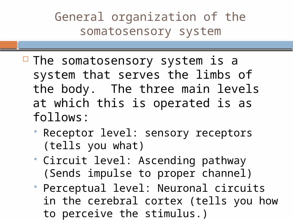
General organization of the somatosensory system
The somatosensory system is a system that serves the limbs of the body. The three main levels at which this is operated is as follows: Receptor level: sensory receptors (tells you
what) Circuit level: Ascending pathway (Sends
impulse to proper channel) Perceptual level: Neuronal circuits in the
cerebral cortex (tells you how to perceive the stimulus.)

Structural Classification of nerves
A nerve is a cordlike organ that is part of the peripheral nervous system. The consist of parallel bundles of peripheral axons, enclosed by successive wrappings of connective tissue.
Regeneration of Nerve Fibers
1. When a nerve is severed or crushed, Wallerian degeneration (cell destruction and the destruction of cells around the severed cell) occurs.
2. After debris is disposed of, Shwann cells proliferate and macrophages are released into the injury site.
3. Then the regenerated axons will sprout across the gap and to their original contacts.

Cranial nerves
There are 12 sets of nerves that associate themselves with the brain.
They are: Olfactoral Optic Oculomotor Trochlear Trigeminal Abducens Facial Vestibulocochlear Glossopharyngeal Vagus Accessory Hypoglossal
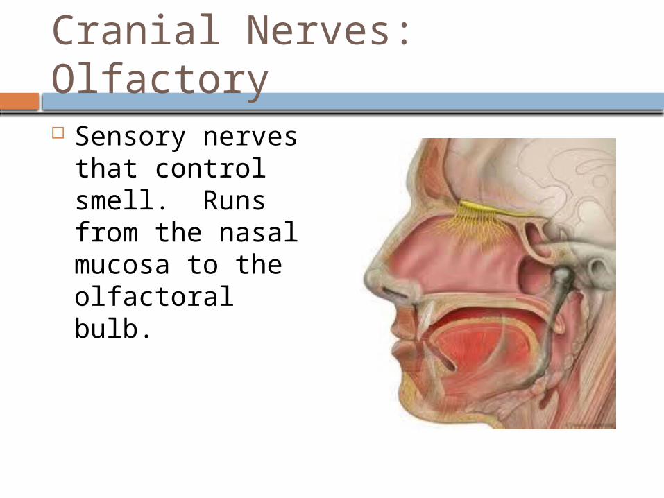
Cranial Nerves: Olfactory
Sensory nerves that control smell. Runs from the nasal mucosa to the olfactoral bulb.
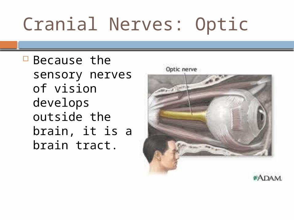
Cranial Nerves: Optic
Because the sensory nerves of vision develops outside the brain, it is a brain tract.
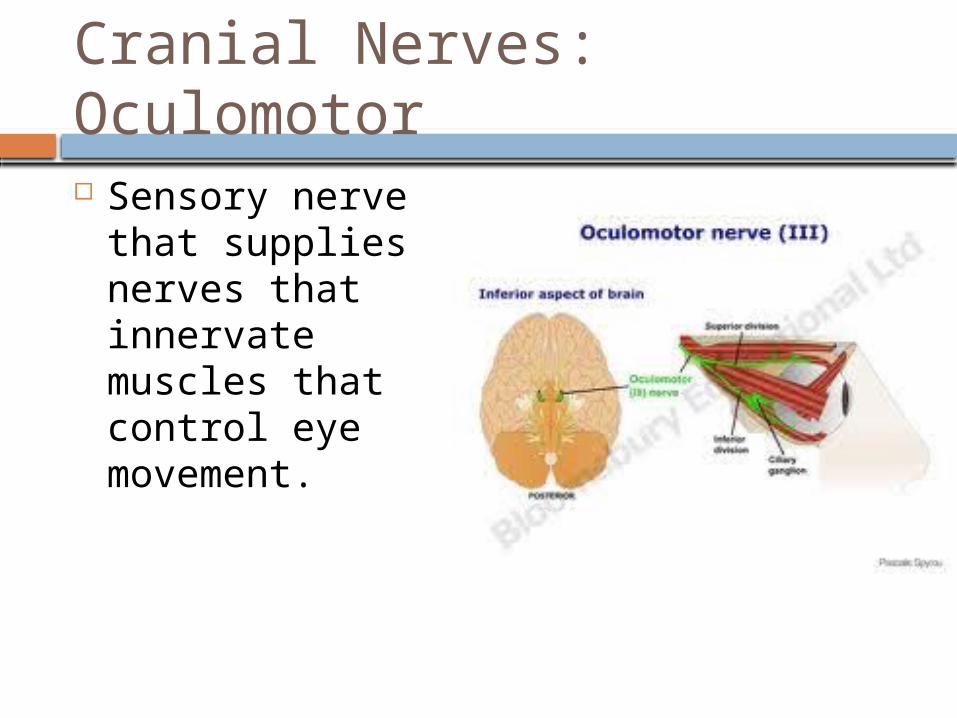
Cranial Nerves: Oculomotor
Sensory nerve that supplies nerves that innervate muscles that control eye movement.

Cranial Nerves: Trochlear
Means “pulley” and innervates muscles that are in a pulley shaped muscle in the orbit (eye area).
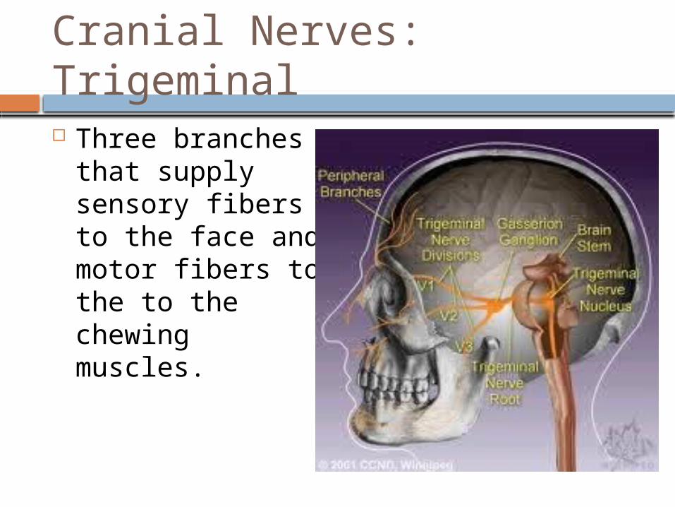
Cranial Nerves: Trigeminal
Three branches that supply sensory fibers to the face and motor fibers to the to the chewing muscles.

Cranial Nerves: Abducens
Controls the muscles that abduct the eyeball (move it laterally)

Cranial Nerves: Facial
A large nerve that innervates the muscles of the face. (and other things as well)

Cranial Nerves: Vestibulocochlear
Sensory nerve for hearing and balance. A.K.A. the auditory nerve.

Cranial Nerves: Glossopharyngeal
Means “tongue and pharynx”. Innervates the tongue and throat.

Cranial Nerves: Vagus
Means “wanderer” The only cranial nerve that extends to the thorax and the abdomen.
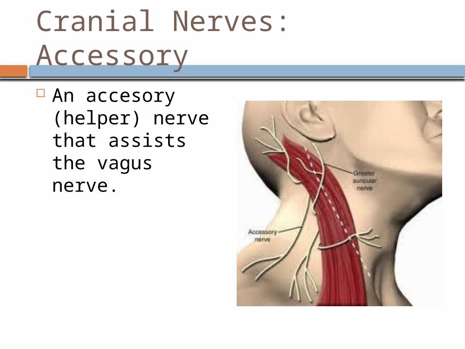
Cranial Nerves: Accessory
An accesory (helper) nerve that assists the vagus nerve.

Cranial Nerves: Hypoglossal
Runs inferior to the tongue and innervates the tongue muscles.

Spinal nerves
There are 31 pairs of spinal nerves that innervate the body. Each of those spinal nerves contain thousands of nerves. There are 8 pairs of cervical spinal
nerves There are 12 pairs of thoracic spinal
nerves There are 5 pairs of lumbar spinal
nerves There are 5 pairs of sacral spinal
nerves There is 1 pair of coccygeal nerves.

Nerve plexus
Are bundles of nerves that occur in the cervical, thoracic, lumbar, and sacral regions that are designed to innervate primarily the limbs.

Branches of the cervical plexus: Cutaneous branches
Branch Lesser occipital
C2 Greater auricular
C2, C3 Transverse
Cervical C2, C3
Supraclavicular C3, C4
Structure served Skin on
posterolateral aspect of neck
Skin of ear, skin over parotid gland
Skin on the anterior aspect of neck
Skin of the shoulder and the clavicular region

Branches of the cervical plexus: motor branches
Branch Ansa Cervicalis
C1-C3 Segmental and other
muscular branches C1-C5
Phrenic C3-C5
Structure Served Infrahyoid muscle
of the neck Deep muscles of
the neck, portions of the scalenes, levator, scapulae, trapezius, and sternonucleiodmastoid muscles
Diaphragm

Branches of the brachial plexus
Nerve Musculocutaneous
C5-C7
Median: (2) branches MedialC8-T1 lateral C5-C7
Structure served Muscular branches;
flexor muscles in anterior arm In cutaneous branches: controls skin on arm.
Flexor muscles of the forearm. Digits of the fingers.
Cutaneous: skin of 2/3 of hand and palm.

Nerve Ulnar
C8-T1
Structure served Flexor muscles of
the anterior forearm; interisitc muscles of the lateral palm. Cutaneous branches: skin of the posteriolateral surface of the entire limb

Nerve Radial
C5-C8, T1
Structure Served Posterior muscles
of the arm and forearm; most intrinsic muscles of the hands. Cutaneous: posteriolateral surface of the hand.

Nerve Axillary
C5,C6
Dorsal Scapular C5
Structure Served Deltoid and teres
minor muscles. Cutaneous: some skin of the shoulder
Rhomboid muscles and levator scapulae.

Nerve Long Thoracic
C5-C7 Subscapular
C5, C6 Suprascapular
C5, C6
Pectoralis C5-T1
Structure Served Serratus anterior
muscle
Teres major and subscapularis muscle
Shoulder joint; supraspinatus and infraspinas muscles
Pectoralis major and minor muscles

Branches of the Lumbar plexus Nerve Femoral
L2-L4
Obturator L2-L4
Structure served Skin of anterior and
medial thigh. Medial leg and foot. Hip, knees, and joints.
Motor to adductor magnus, longus, and brevis, obturator, skin for medial thigh, and for hip and knee joints

Nerve Lateral femoral
cutaneous. L2, L3
Iliohypogastric L1
Structure served Skin of lateral
thigh; some sensory branches to peritoneum.
Skin of lower abdomen and hip; muscles of anterolateral abdominal wall

Nerve Ilioiguinal
L1
Structure Served Skin of external
genitalia, and proximal medial aspect of the thigh; inferior abdominal muscles

Nerve Genitofemoral
Structure Served Skin of scrotum in
males, of labia majora in females, and of anterior thigh; inferior to middle portion of inguinal region; cremaster muscles in males.

Branches of sacral plexus
Nerve Sciatic nerve
Tibial (including sural, medial and lateral plantar, and medial calcaneal branches) L4-S3
Structure Served Cutaneous
branches: to skin of posterior surface of leg and sole of foot
Motor branches: posterior of adductor magnus, triceps surae, tibulis

Common fibular (superficial and deep branches) L4-S2
Cutaneous: to skin of anterior and lateral surface of the leg and dorsum of foot
Motor branches: short head of biceps femoralis of the thigh; extensor muscles of the toes.

Nerve Superior Gluteal
L4, L5, S1
Inferior Gluteal L5-S2
Structure served Motor branch to
gluteus medius and minimus
Motor branch to gluteus maximus

Posterior femoral cutaneous S1-S3
Pudendal S2-S4
Skin of the buttock, and posterior thigh
Supplies most of the skin muscles, anus, clitoris, labia, vaginal mucosa (females), scrotom and penis in males.