Embed Size (px)
Citation preview
1
13Peripheral Nervous System (PNS)
Overview

2
Peripheral Nervous System (PNS)
PNS – all neural structures outside the brain and spinal cord
Includes sensory receptors, peripheral nerves, associated ganglia, and motor endings
Provides links to and from the external environment
3
Sensory Receptors
Structures specialized to respond to stimuli
Activation of sensory receptors results in depolarizations that trigger impulses to the CNS
The realization of these stimuli, sensation and perception, occur in the brain
4
13Peripheral Nervous System (PNS)
Sensory Receptor
5
Classification by Stimulus Type
Mechanoreceptors – respond to touch, pressure, vibration, stretch, and itch
Thermoreceptors – sensitive to changes in temperature
Photoreceptors – respond to light energy (e.g., retina)
Chemoreceptors – respond to chemicals (e.g., smell, taste, changes in blood chemistry)
Nociceptors – sensitive to pain-causing stimuli
6
Classification by Location: Exteroceptors
Respond to stimuli arising outside the body
Found near the body surface
Sensitive to touch, pressure, pain, and temperature
Include the special sense organs (eyes, ears, nose, and tongues)
7
Classification by Location: Interoceptors
Respond to stimuli arising within the body
Found in internal viscera (organ) and blood vessels
Sensitive to chemical changes, stretch, and temperature changes
8
Classification by Location: Proprioceptors
Respond to degree of stretch of the organs they occupy
Found in skeletal muscles, tendons, joints, ligaments, and connective tissue coverings of bones and muscles
Constantly “advise” the brain of one’s movements
9
Receptors are structurally classified as either simple or complex
Complex receptors are special sense organs
Most receptors are simple and include encapsulated and unencapsulated varieties
Classification by Structural Complexity
10
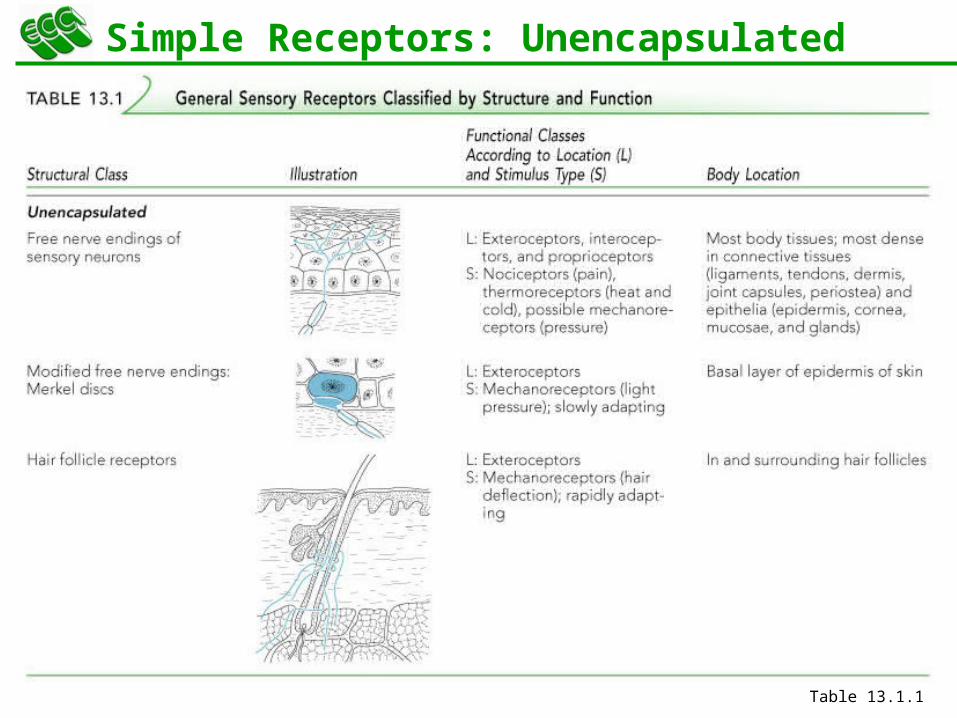
Simple Receptors: Unencapsulated
Free dendritic nerve endings
Respond chiefly to temperature and pain
Merkel (tactile) discs
Hair follicle receptors
11
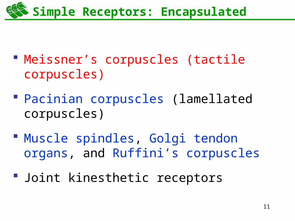
Simple Receptors: Encapsulated
Meissner’s corpuscles (tactile corpuscles)
Pacinian corpuscles (lamellated corpuscles)
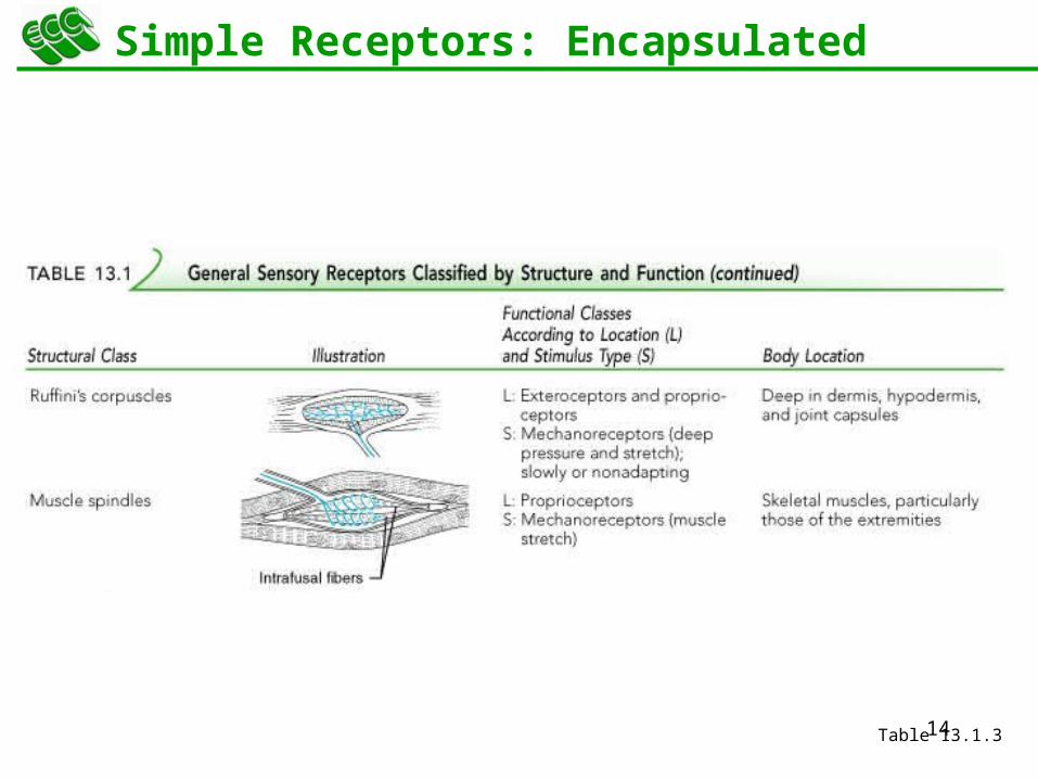
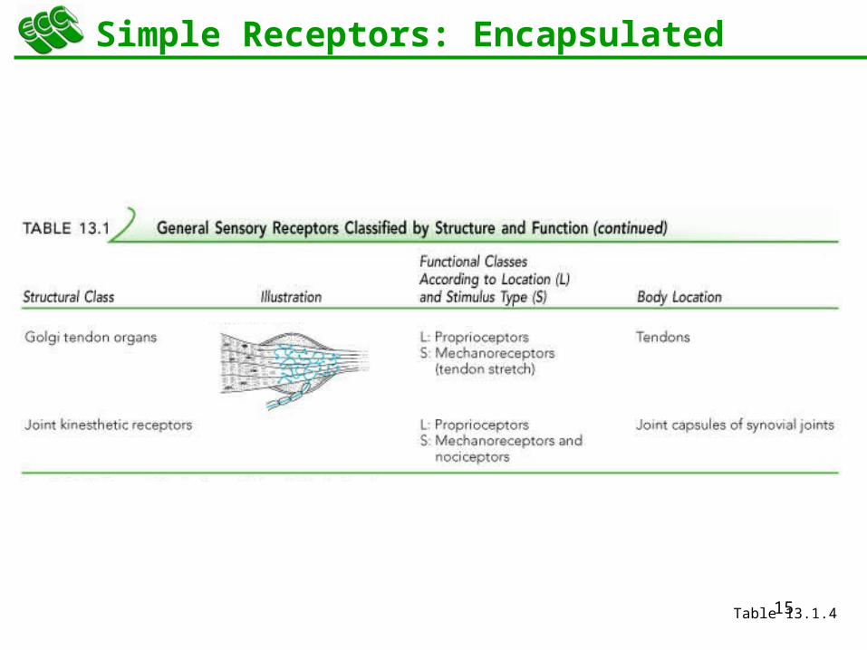
Muscle spindles, Golgi tendon organs, and Ruffini’s corpuscles
Joint kinesthetic receptors
12
Simple Receptors: Unencapsulated
Table 13.1.1

13
Simple Receptors: Encapsulated
Table 13.1.2
14
Simple Receptors: Encapsulated
Table 13.1.3
15
Simple Receptors: Encapsulated
Table 13.1.4
16
13Peripheral Nervous System (PNS)
From Sensation to Perception

17
From Sensation to Perception
Survival depends upon sensation and perception
Sensation is the awareness of changes in the internal and external environment
Perception is the conscious interpretation of those stimuli
18
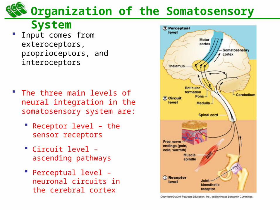
Organization of the Somatosensory System
Input comes from exteroceptors, proprioceptors, and interoceptors
The three main levels of neural integration in the somatosensory system are:
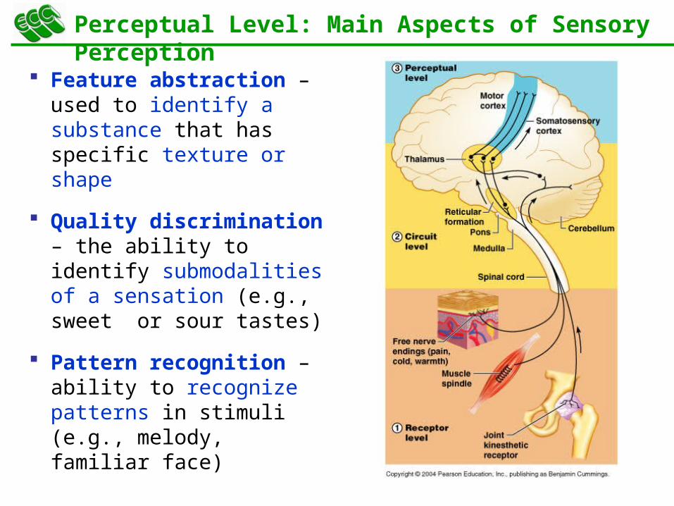
Receptor level – the sensor receptors
Circuit level – ascending pathways
Perceptual level – neuronal circuits in the cerebral cortex
19
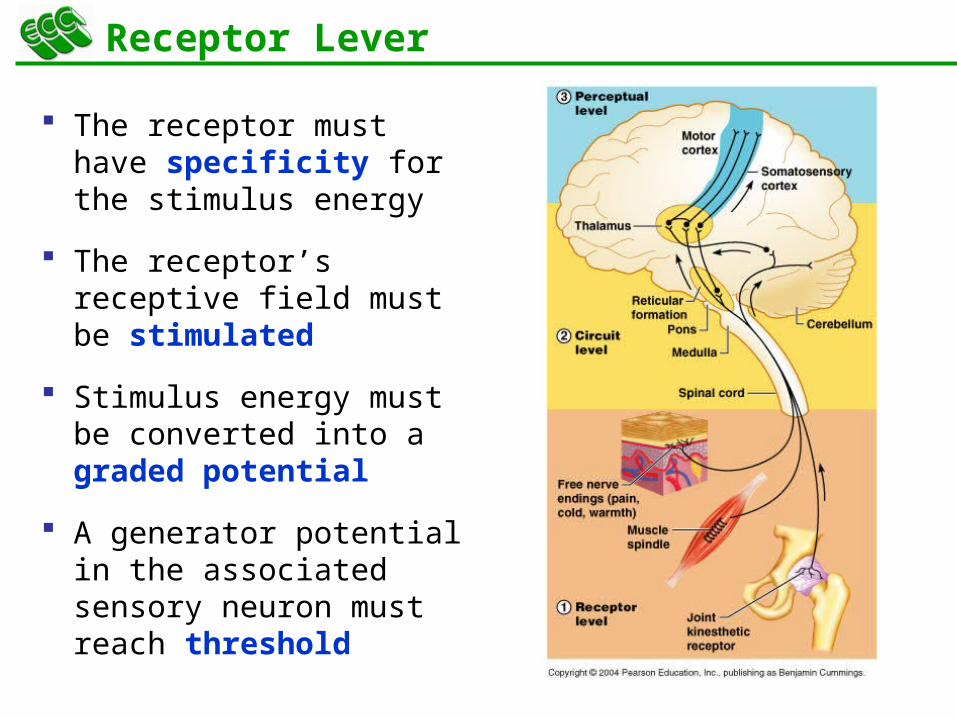
Receptor Lever
The receptor must have specificity for the stimulus energy
The receptor’s receptive field must be stimulated
Stimulus energy must be converted into a graded potential
A generator potential in the associated sensory neuron must reach threshold
20
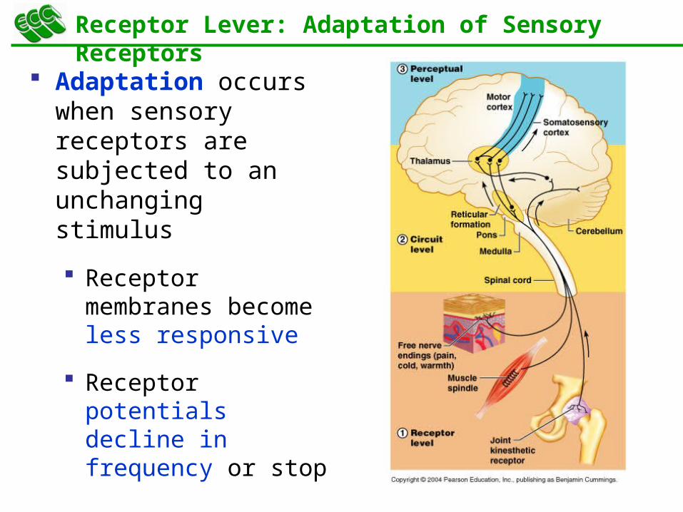
Receptor Lever: Adaptation of Sensory Receptors
Adaptation occurs when sensory receptors are subjected to an unchanging stimulus
Receptor membranes become less responsive
Receptor potentials decline in frequency or stop
21
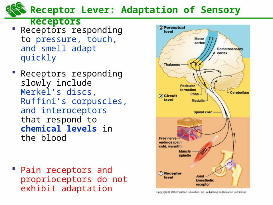
Receptor Lever: Adaptation of Sensory Receptors
Receptors responding to pressure, touch, and smell adapt quickly
Receptors responding slowly include Merkel’s discs, Ruffini’s corpuscles, and interoceptors that respond to chemical levels in the blood
Pain receptors and proprioceptors do not exhibit adaptation
22
Circuit Level
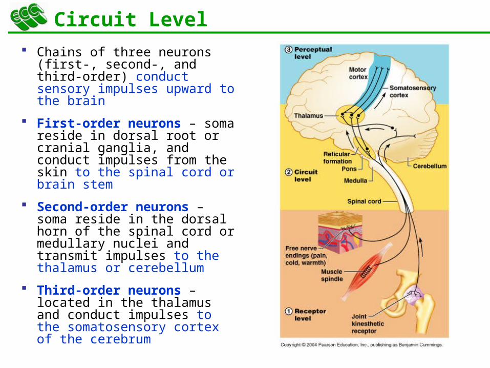
Chains of three neurons (first-, second-, and third-order) conduct sensory impulses upward to the brain
First-order neurons – soma reside in dorsal root or cranial ganglia, and conduct impulses from the skin to the spinal cord or brain stem
Second-order neurons – soma reside in the dorsal horn of the spinal cord or medullary nuclei and transmit impulses to the thalamus or cerebellum
Third-order neurons – located in the thalamus and conduct impulses to the somatosensory cortex of the cerebrum
23
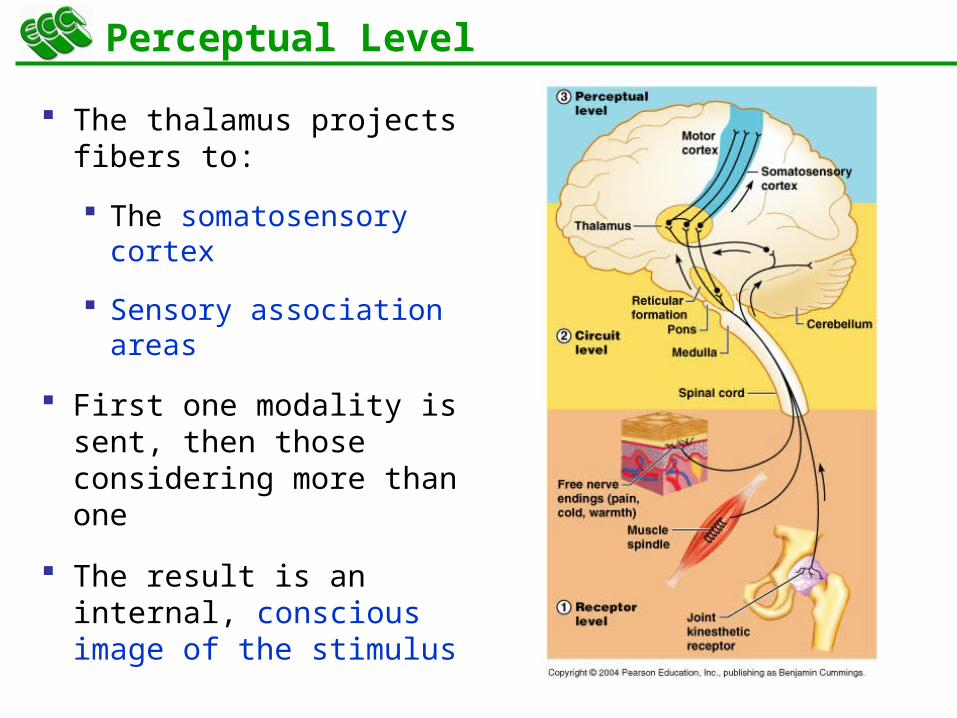
Perceptual Level
The thalamus projects fibers to:
The somatosensory cortex
Sensory association areas
First one modality is sent, then those considering more than one
The result is an internal, conscious image of the stimulus
24
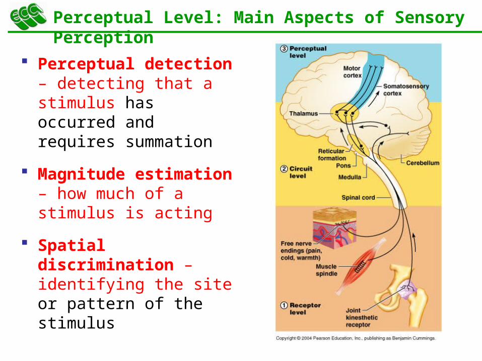
Perceptual Level: Main Aspects of Sensory Perception
Perceptual detection – detecting that a stimulus has occurred and requires summation
Magnitude estimation – how much of a stimulus is acting
Spatial discrimination – identifying the site or pattern of the stimulus
25
Perceptual Level: Main Aspects of Sensory Perception
Feature abstraction – used to identify a substance that has specific texture or shape
Quality discrimination – the ability to identify submodalities of a sensation (e.g., sweet or sour tastes)
Pattern recognition – ability to recognize patterns in stimuli (e.g., melody, familiar face)
26
13Peripheral Nervous System (PNS)
Nerve
27
Structure of a Nerve
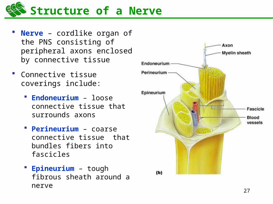
Nerve – cordlike organ of the PNS consisting of peripheral axons enclosed by connective tissue
Connective tissue coverings include:
Endoneurium – loose connective tissue that surrounds axons
Perineurium – coarse connective tissue that bundles fibers into fascicles
Epineurium – tough fibrous sheath around a nerve

29
Classification of Nerves
Sensory and motor divisions
Sensory (afferent) – carry impulse to the CNS
Motor (efferent) – carry impulses from CNS
Mixed – sensory and motor fibers carry impulses to and from CNS;
most common type of nerve
30
Peripheral Nerves
Mixed nerves – carry somatic and autonomic (visceral) impulses
The four types of mixed nerves are:
Somatic afferent and somatic efferent
Visceral afferent and visceral efferent
Peripheral nerves originate from the brain or spinal column
31
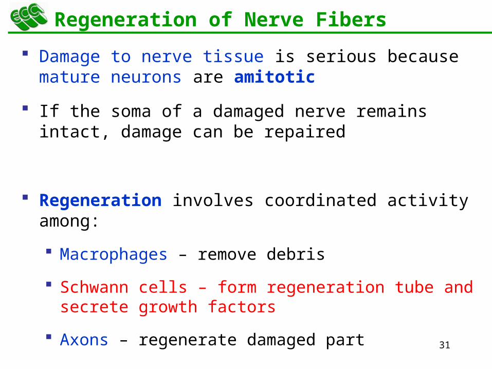
Regeneration of Nerve Fibers
Damage to nerve tissue is serious because mature neurons are amitotic
If the soma of a damaged nerve remains intact, damage can be repaired
Regeneration involves coordinated activity among:
Macrophages – remove debris
Schwann cells – form regeneration tube and secrete growth factors
Axons – regenerate damaged part
32
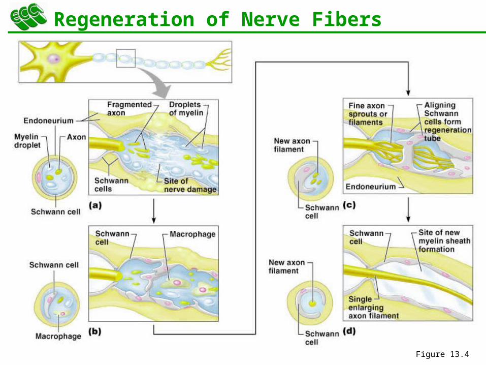
Regeneration of Nerve Fibers
Figure 13.4

33
13Peripheral Nervous System (PNS)
Cranial Nerves
34
Cranial Nerves
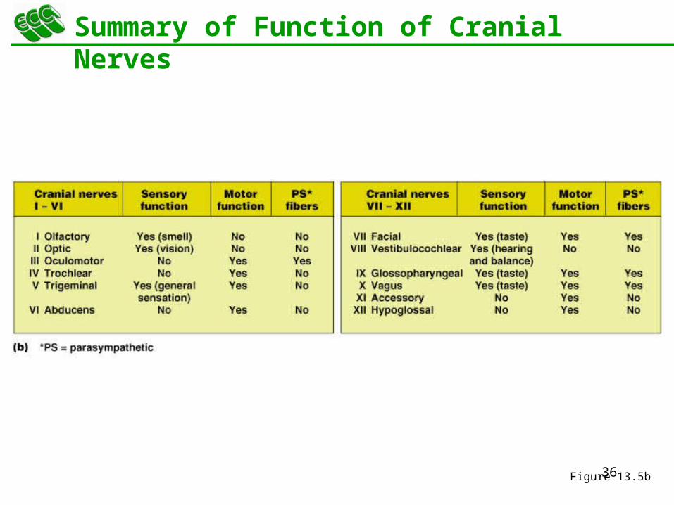
Twelve pairs of cranial nerves arise from the brain
They have sensory, motor, or both sensory and motor functions
Each nerve is identified by a number (I through XII) and a name
Four cranial nerves carry parasympathetic fibers that serve muscles and glands
35
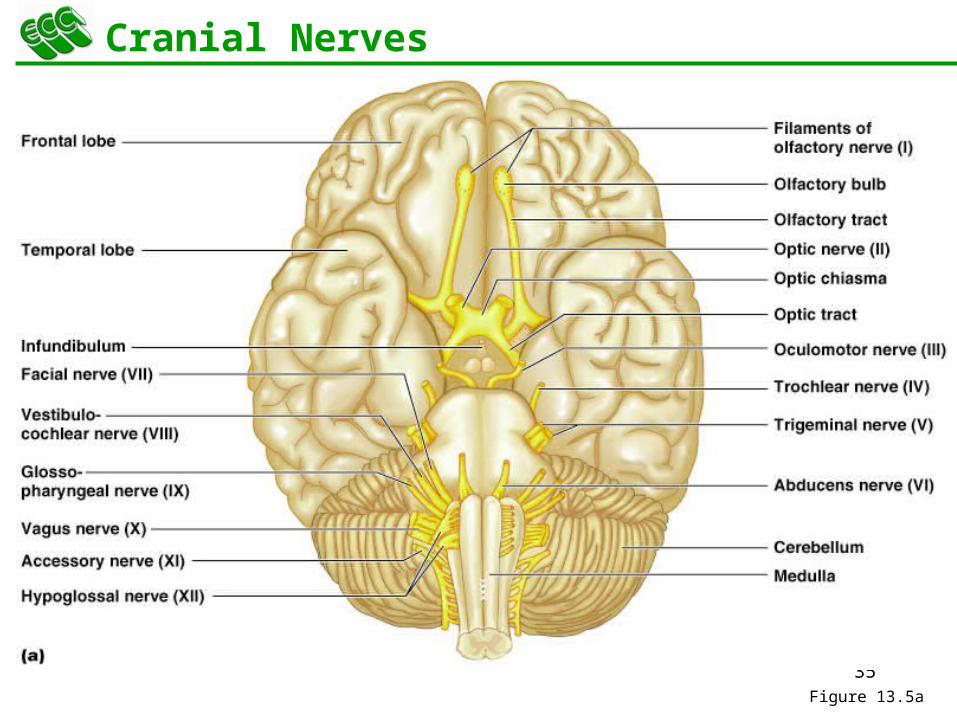
Cranial Nerves
Figure 13.5a
36
Summary of Function of Cranial Nerves
Figure 13.5b
37
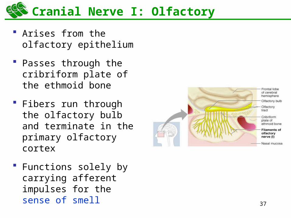
Cranial Nerve I: Olfactory
Arises from the olfactory epithelium
Passes through the cribriform plate of the ethmoid bone
Fibers run through the olfactory bulb and terminate in the primary olfactory cortex
Functions solely by carrying afferent impulses for the sense of smell
39
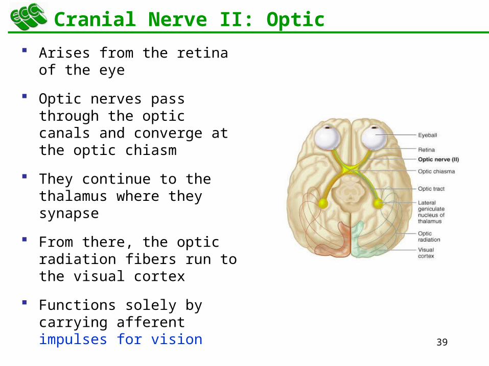
Cranial Nerve II: Optic
Arises from the retina of the eye
Optic nerves pass through the optic canals and converge at the optic chiasm
They continue to the thalamus where they synapse
From there, the optic radiation fibers run to the visual cortex
Functions solely by carrying afferent impulses for vision
41
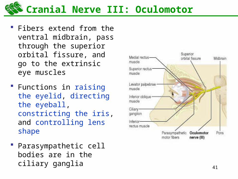
Cranial Nerve III: Oculomotor
Fibers extend from the ventral midbrain, pass through the superior orbital fissure, and go to the extrinsic eye muscles
Functions in raising the eyelid, directing the eyeball, constricting the iris, and controlling lens shape
Parasympathetic cell bodies are in the ciliary ganglia
43
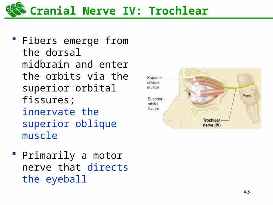
Cranial Nerve IV: Trochlear
Fibers emerge from the dorsal midbrain and enter the orbits via the superior orbital fissures; innervate the superior oblique muscle
Primarily a motor nerve that directs the eyeball
45
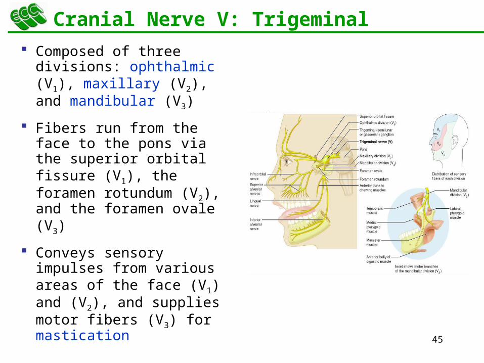
Cranial Nerve V: Trigeminal
Composed of three divisions: ophthalmic (V1), maxillary (V2), and mandibular (V3)
Fibers run from the face to the pons via the superior orbital fissure (V1), the foramen rotundum (V2), and the foramen ovale (V3)
Conveys sensory impulses from various areas of the face (V1) and (V2), and supplies motor fibers (V3) for mastication
47
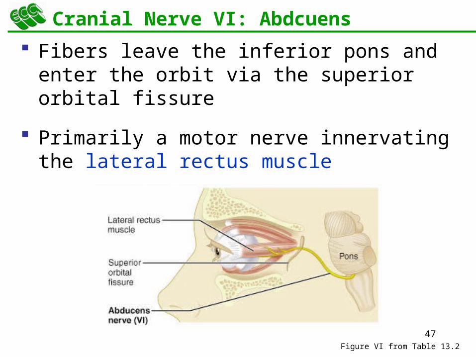
Fibers leave the inferior pons and enter the orbit via the superior orbital fissure
Primarily a motor nerve innervating the lateral rectus muscle
Figure VI from Table 13.2
Cranial Nerve VI: Abdcuens
48
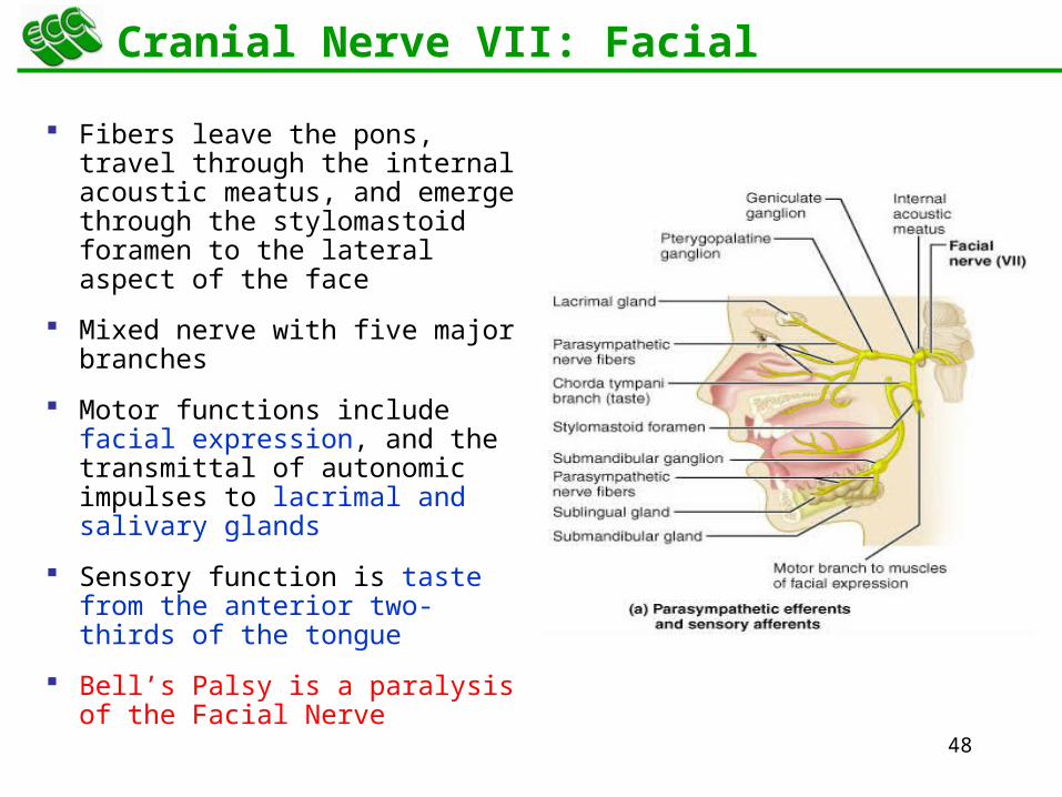
Cranial Nerve VII: Facial
Fibers leave the pons, travel through the internal acoustic meatus, and emerge through the stylomastoid foramen to the lateral aspect of the face
Mixed nerve with five major branches
Motor functions include facial expression, and the transmittal of autonomic impulses to lacrimal and salivary glands
Sensory function is taste from the anterior two-thirds of the tongue
Bell’s Palsy is a paralysis of the Facial Nerve
50
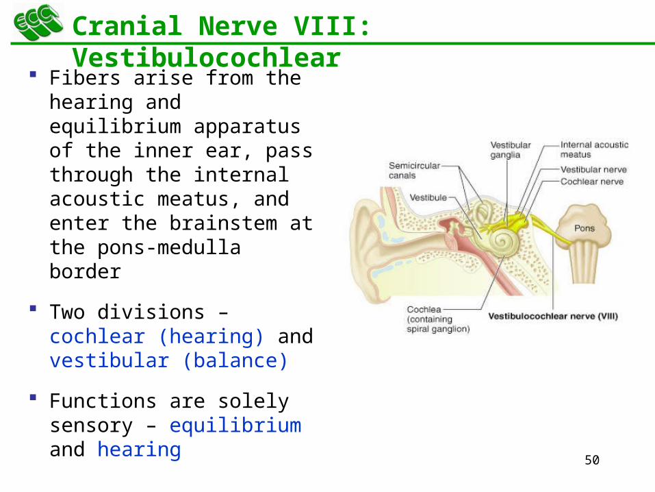
Cranial Nerve VIII: Vestibulocochlear
Fibers arise from the hearing and equilibrium apparatus of the inner ear, pass through the internal acoustic meatus, and enter the brainstem at the pons-medulla border
Two divisions – cochlear (hearing) and vestibular (balance)
Functions are solely sensory – equilibrium and hearing
52
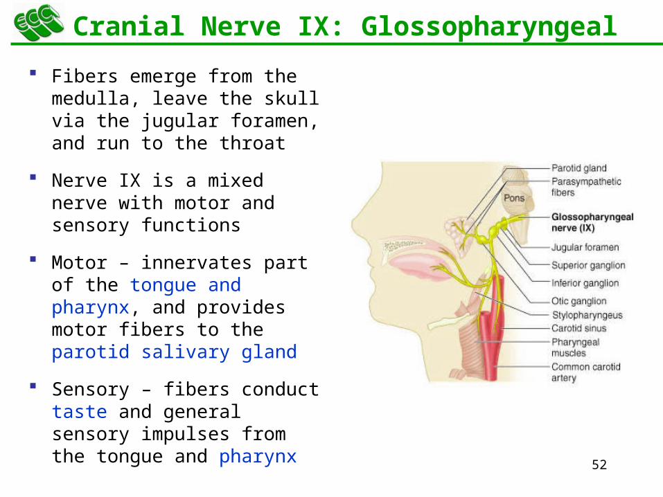
Cranial Nerve IX: Glossopharyngeal
Fibers emerge from the medulla, leave the skull via the jugular foramen, and run to the throat
Nerve IX is a mixed nerve with motor and sensory functions
Motor – innervates part of the tongue and pharynx, and provides motor fibers to the parotid salivary gland
Sensory – fibers conduct taste and general sensory impulses from the tongue and pharynx
54
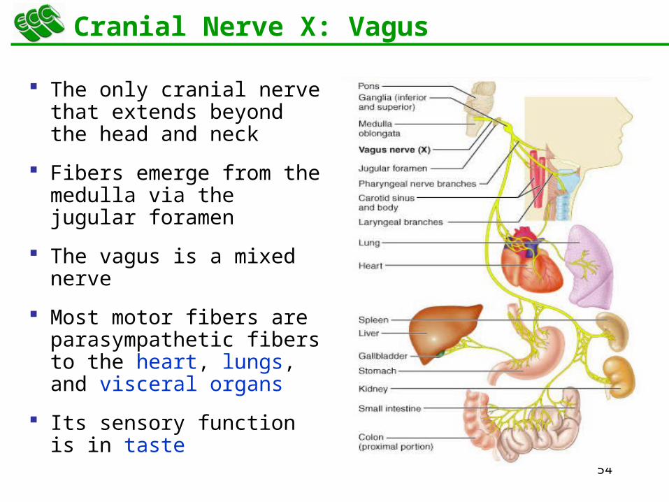
Cranial Nerve X: Vagus
The only cranial nerve that extends beyond the head and neck
Fibers emerge from the medulla via the jugular foramen
The vagus is a mixed nerve
Most motor fibers are parasympathetic fibers to the heart, lungs, and visceral organs
Its sensory function is in taste
57
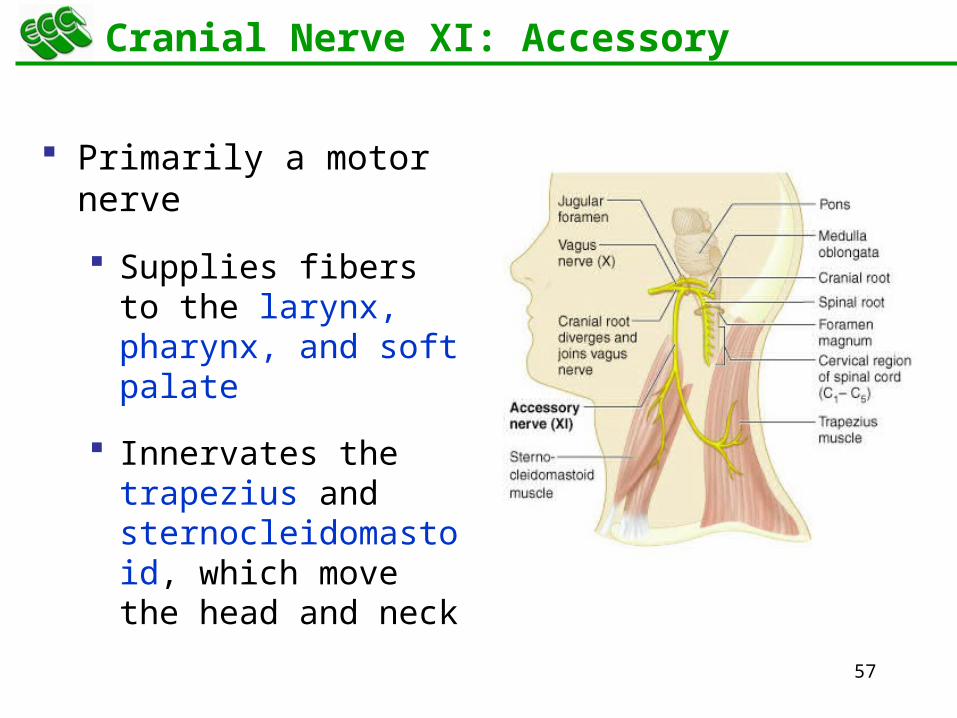
Cranial Nerve XI: Accessory
Primarily a motor nerve
Supplies fibers to the larynx, pharynx, and soft palate
Innervates the trapezius and sternocleidomastoid, which move the head and neck
59
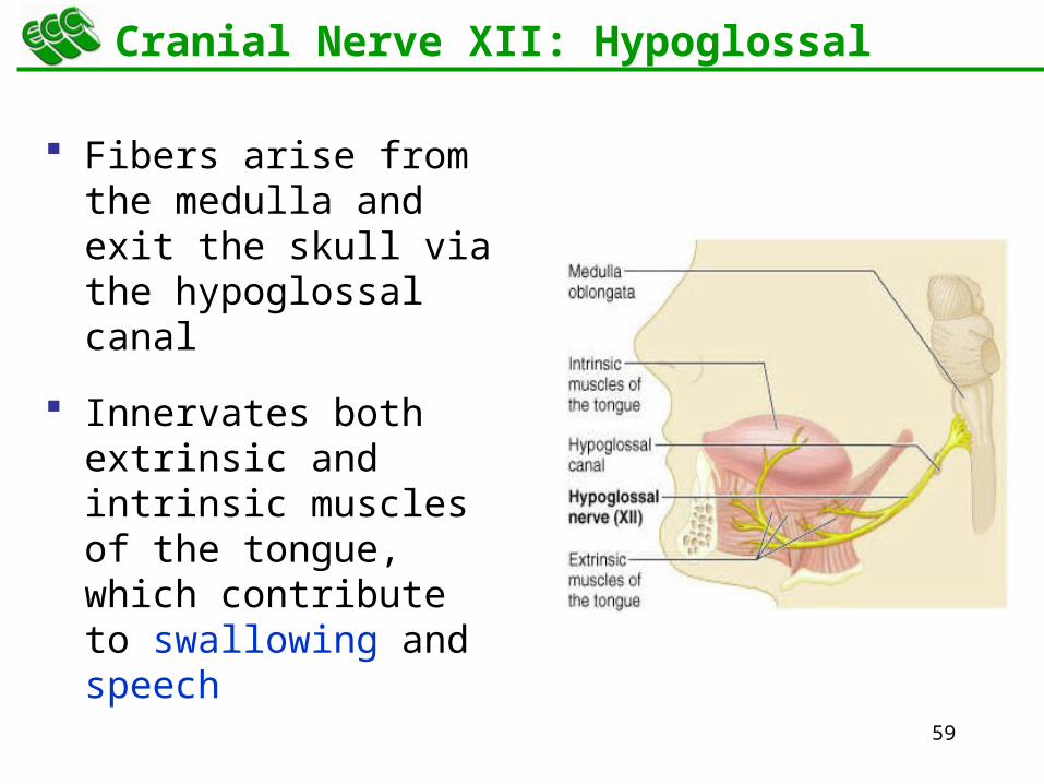
Cranial Nerve XII: Hypoglossal
Fibers arise from the medulla and exit the skull via the hypoglossal canal
Innervates both extrinsic and intrinsic muscles of the tongue, which contribute to swallowing and speech
61
13Peripheral Nervous System (PNS)
Spinal Nerves
62
Spinal Nerves
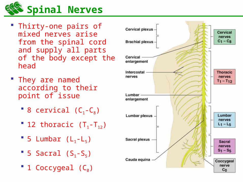
Thirty-one pairs of mixed nerves arise from the spinal cord and supply all parts of the body except the head
They are named according to their point of issue
8 cervical (C1-C8)
12 thoracic (T1-T12)
5 Lumbar (L1-L5)
5 Sacral (S1-S5)
1 Coccygeal (C0)
64
Spinal Nerves: Roots
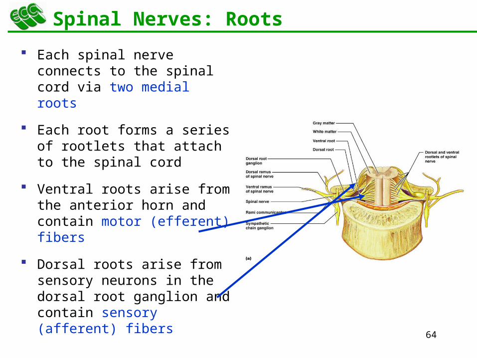
Each spinal nerve connects to the spinal cord via two medial roots
Each root forms a series of rootlets that attach to the spinal cord
Ventral roots arise from the anterior horn and contain motor (efferent) fibers
Dorsal roots arise from sensory neurons in the dorsal root ganglion and contain sensory (afferent) fibers
66
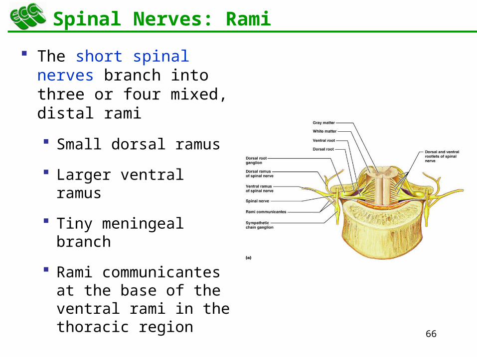
Spinal Nerves: Rami
The short spinal nerves branch into three or four mixed, distal rami
Small dorsal ramus
Larger ventral ramus
Tiny meningeal branch
Rami communicantes at the base of the ventral rami in the thoracic region
67
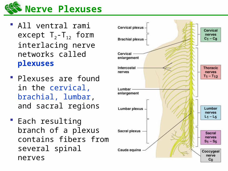
Nerve Plexuses
All ventral rami except T2-T12 form interlacing nerve networks called plexuses
Plexuses are found in the cervical, brachial, lumbar, and sacral regions
Each resulting branch of a plexus contains fibers from several spinal nerves
68
Nerve Plexuses
Fibers travel to the periphery via several different routes
Each muscle receives a nerve supply from more than one spinal nerve
Damage to one spinal segment cannot completely paralyze a muscle
69
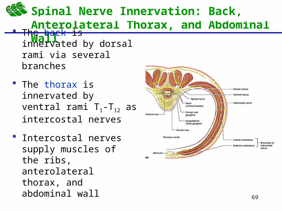
Spinal Nerve Innervation: Back, Anterolateral Thorax, and Abdominal Wall
The back is innervated by dorsal rami via several branches
The thorax is innervated by ventral rami T1-T12 as intercostal nerves
Intercostal nerves supply muscles of the ribs, anterolateral thorax, and abdominal wall

71
Cervical Plexus
The cervical plexus is formed by ventral rami of C1-C4
Most branches are cutaneous nerves of the neck, ear, back of head, and shoulders
The most important nerve of this plexus is the phrenic nerve
major motor and sensory nerve of the diaphragm
73
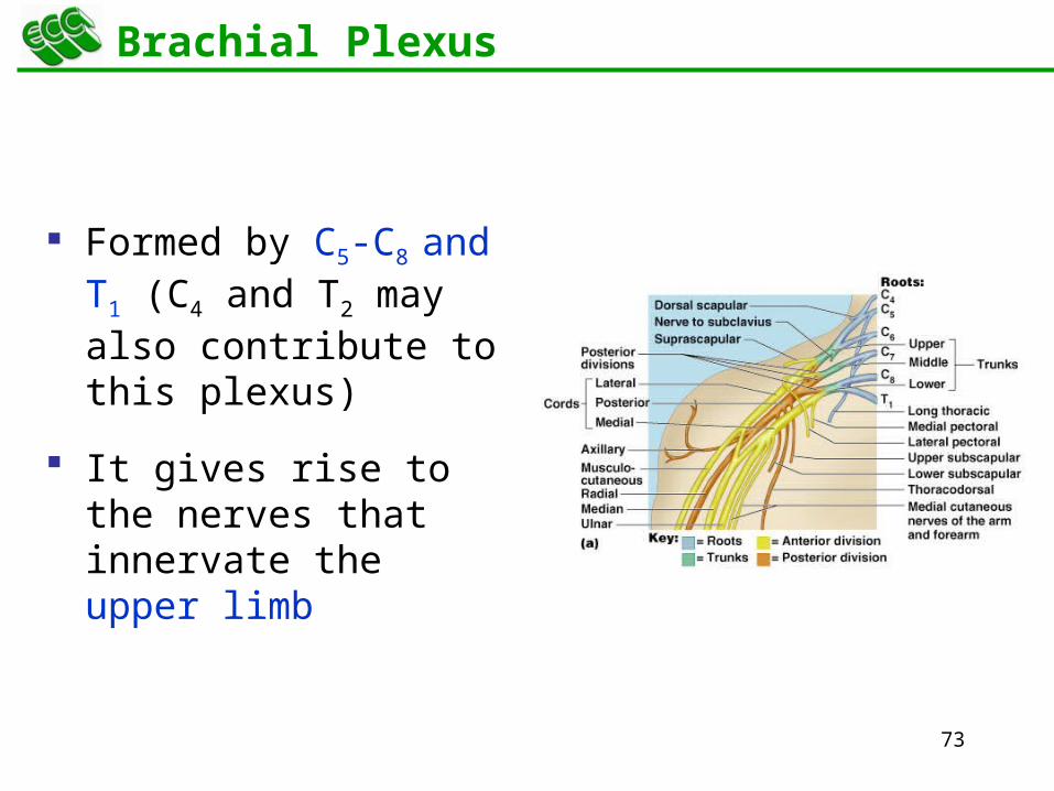
Brachial Plexus
Formed by C5-C8 and T1 (C4 and T2 may also contribute to this plexus)
It gives rise to the nerves that innervate the upper limb
74
Brachial Plexus
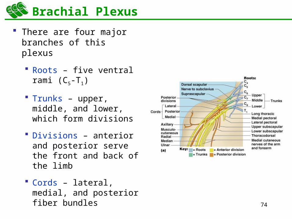
There are four major branches of this plexus
Roots – five ventral rami (C5-T1)
Trunks – upper, middle, and lower, which form divisions
Divisions – anterior and posterior serve the front and back of the limb
Cords – lateral, medial, and posterior fiber bundles
76
Brachial Plexus: Nerves
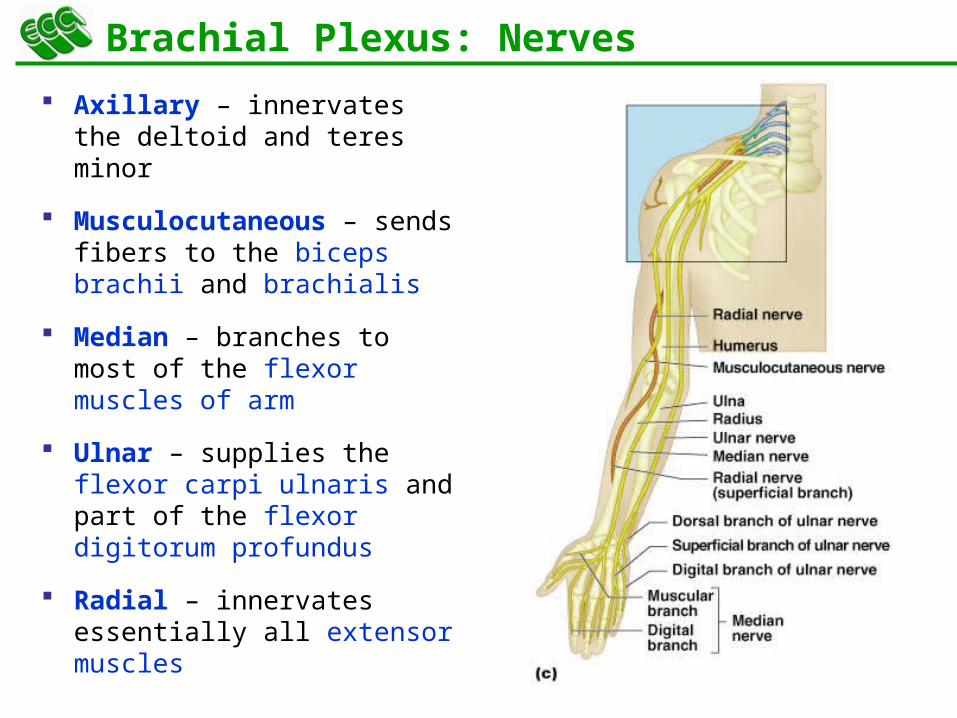
Axillary – innervates the deltoid and teres minor
Musculocutaneous – sends fibers to the biceps brachii and brachialis
Median – branches to most of the flexor muscles of arm
Ulnar – supplies the flexor carpi ulnaris and part of the flexor digitorum profundus
Radial – innervates essentially all extensor muscles
79
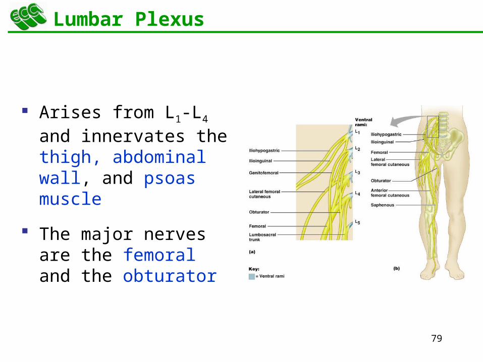
Lumbar Plexus
Arises from L1-L4 and innervates the thigh, abdominal wall, and psoas muscle
The major nerves are the femoral and the obturator

81
Sacral Plexus
Arises from L4-S4 and serves the buttock, lower limb, pelvic structures, and the perineum
The major nerve is the sciatic
the longest and thickest nerve of the body
The sciatic is actually composed of two nerves: the tibial and the common fibular (peroneal) nerves
83
Dermatomes
A dermatome is the area of skin innervated by the cutaneous branches of a single spinal nerve
All spinal nerves except C1 participate in dermatomes
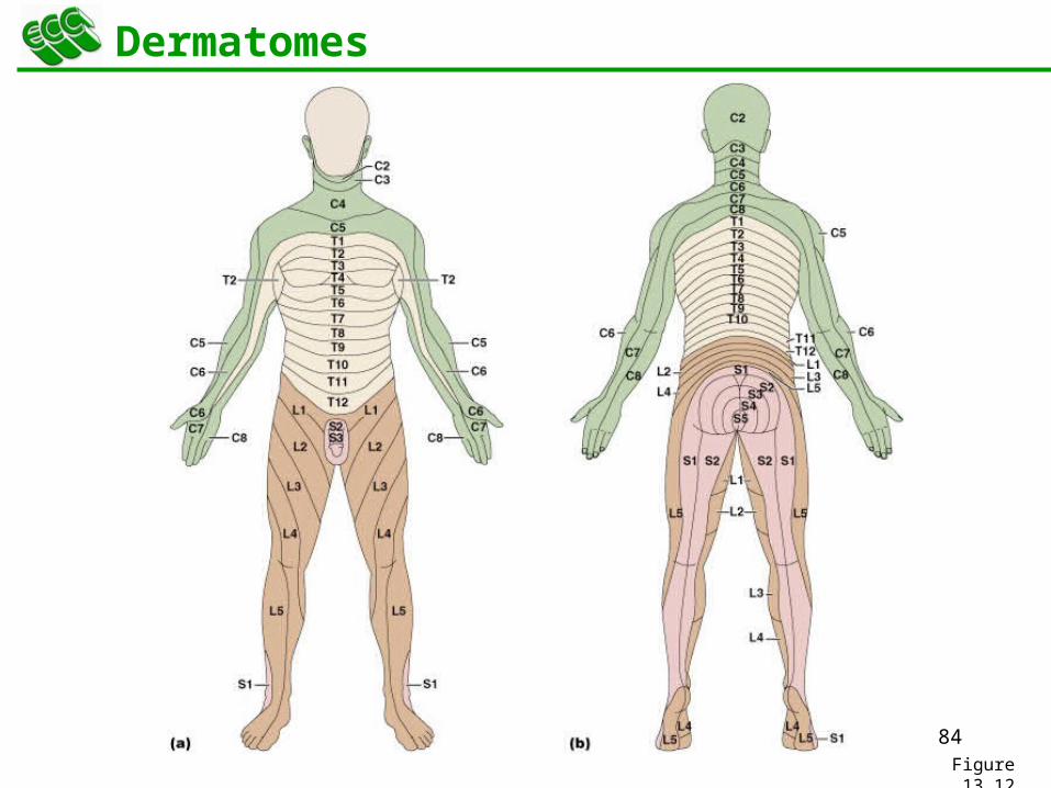
84Figure 13.12
Dermatomes
85
Innervation of Joints
Hilton’s law: any nerve serving a muscle that produces movement at a joint also innervates the joint itself and the skin over the joint
86
13Peripheral Nervous System (PNS)
Peripheral Motor Endings
87
Motor Endings
PNS elements that activate effectors by releasing neurotransmitters at:
Neuromuscular junctions
Varicosities at smooth muscle and glands
88
Innervation of Skeletal Muscle
Takes place at a neuromusclular junction
Acetylcholine is the neurotransmitter that diffuses across the synaptic cleft
ACh binds to receptors resulting in:
Movement of Na+ and K+ across the membrane
Depolarization of the interior of the muscle cell
An end-plate potential that triggers an action potential
89
Innervation of Visceral Muscle and Glands
Autonomic motor endings and visceral effectors are simpler than somatic junctions
Branches form synapses en passant via varicosities
Acetylcholine and norepinephrine are used as neurotransmitters
Visceral responses are slower than somatic responses
90
13Peripheral Nervous System (PNS)
Motor Integration: From Intention to Effect

91
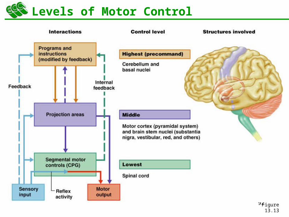
Levels of Motor Control
The three levels of motor control are
Segmental level
Projection level
Precommand level
92Figure 13.13
Levels of Motor Control
93
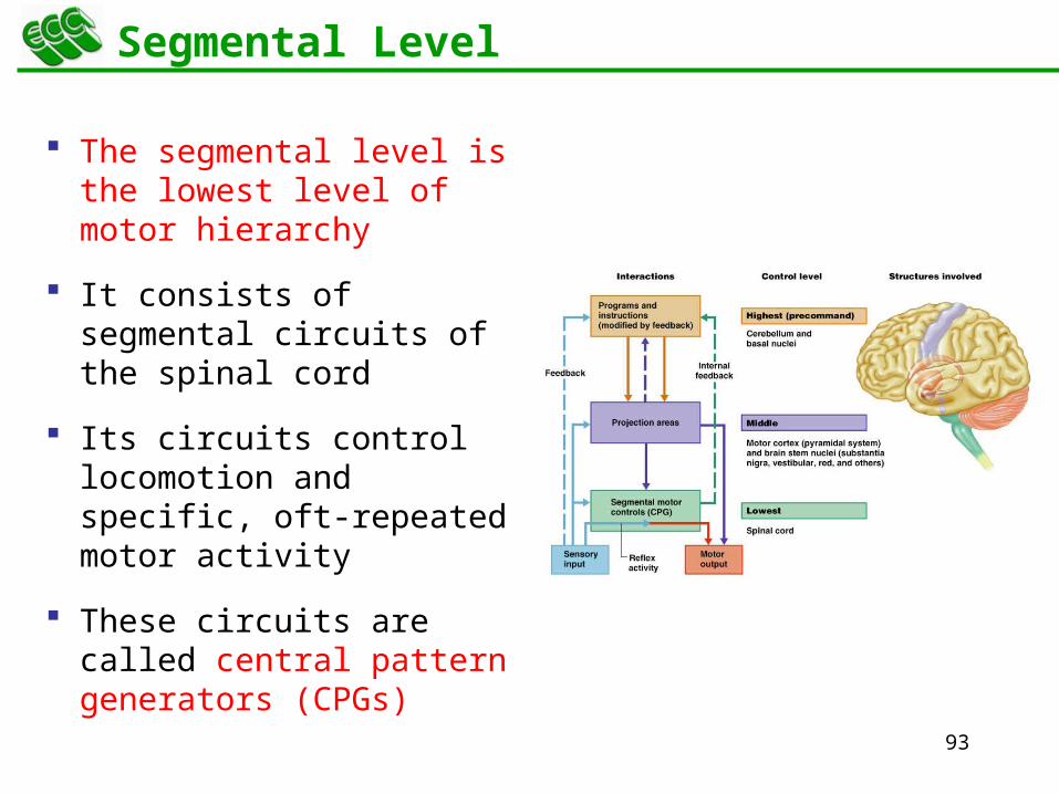
Segmental Level
The segmental level is the lowest level of motor hierarchy
It consists of segmental circuits of the spinal cord
Its circuits control locomotion and specific, oft-repeated motor activity
These circuits are called central pattern generators (CPGs)
94
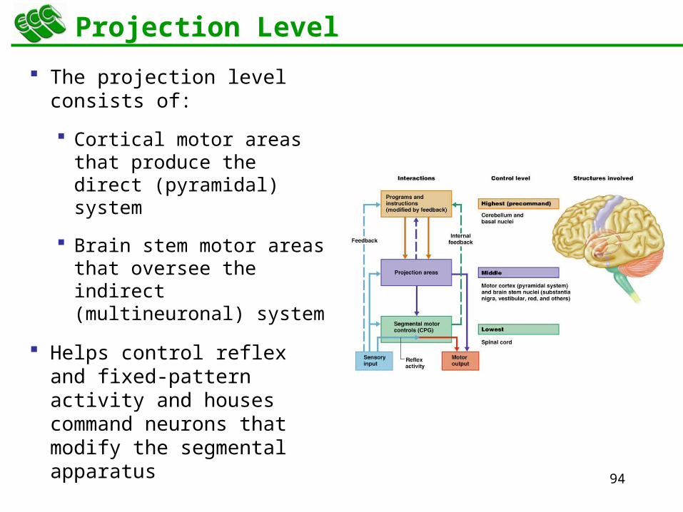
Projection Level
The projection level consists of:
Cortical motor areas that produce the direct (pyramidal) system
Brain stem motor areas that oversee the indirect (multineuronal) system
Helps control reflex and fixed-pattern activity and houses command neurons that modify the segmental apparatus
95
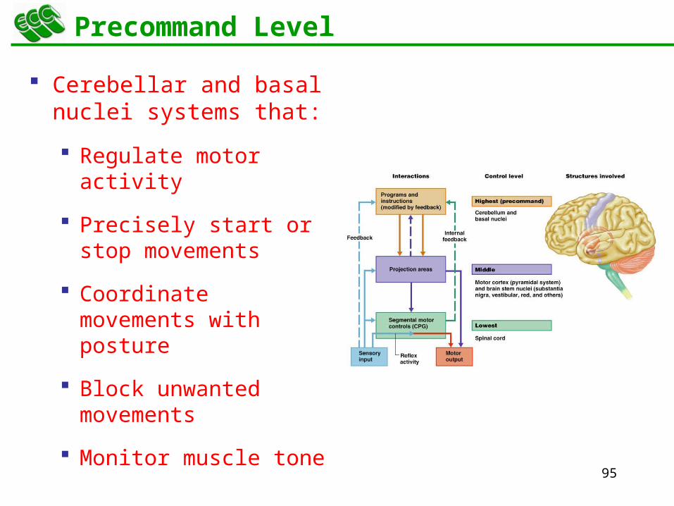
Precommand Level
Cerebellar and basal nuclei systems that:
Regulate motor activity
Precisely start or stop movements
Coordinate movements with posture
Block unwanted movements
Monitor muscle tone
96
13Peripheral Nervous System (PNS)
Reflex Arc
97
Reflexes
A reflex is a rapid, predictable motor response to a stimulus
Reflexes may:
Be inborn (intrinsic) or learned (acquired)
Involve only peripheral nerves and the spinal cord
Involve higher brain centers as well
98
Reflex Arc
There are five components of a reflex arc
Receptor – site of stimulus
Sensory neuron – transmits the afferent impulse to the CNS
Integration center – either monosynaptic or polysynaptic region within the CNS
Motor neuron – conducts efferent impulses from the integration center to an effector
Effector – muscle fiber or gland that responds to the efferent impulse
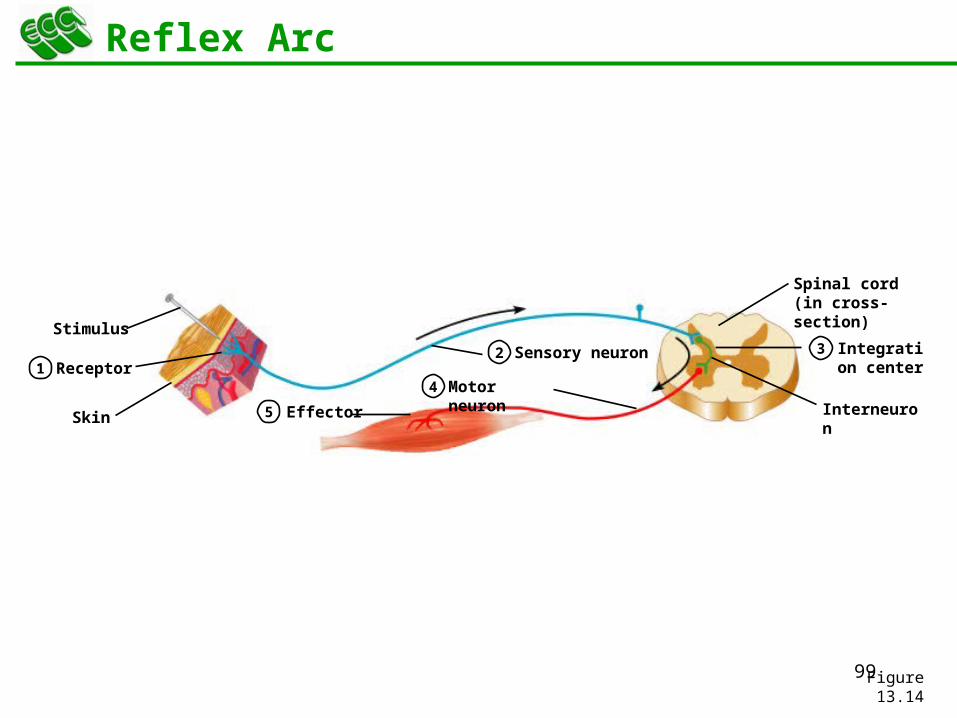
99Figure 13.14
Receptor12 3
4
Sensory neuron Integration center
5 Effector
Motor neuron
Stimulus
Skin
Spinal cord (in cross-section)
Interneuron
Reflex Arc
100
13Peripheral Nervous System (PNS)
Spinal Arc
101
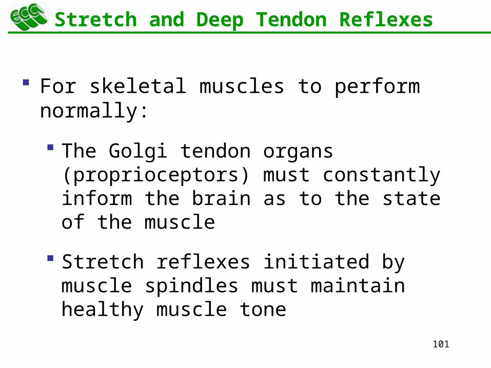
Stretch and Deep Tendon Reflexes
For skeletal muscles to perform normally:
The Golgi tendon organs (proprioceptors) must constantly inform the brain as to the state of the muscle
Stretch reflexes initiated by muscle spindles must maintain healthy muscle tone
102
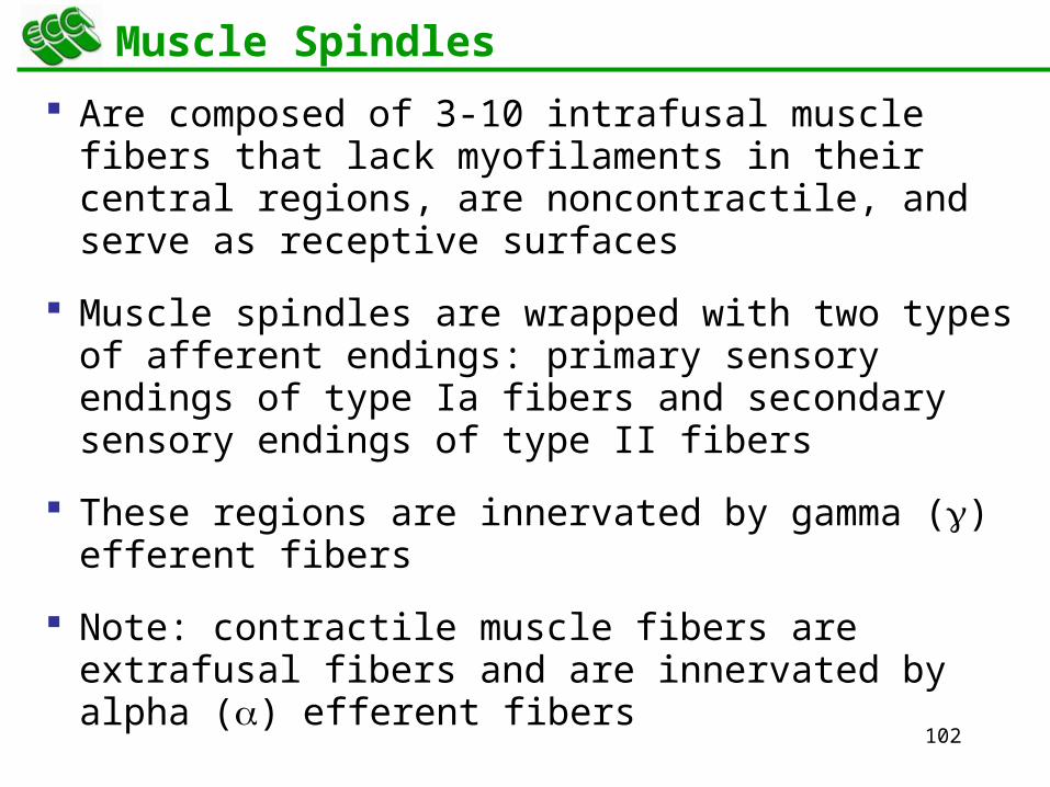
Muscle Spindles
Are composed of 3-10 intrafusal muscle fibers that lack myofilaments in their central regions, are noncontractile, and serve as receptive surfaces
Muscle spindles are wrapped with two types of afferent endings: primary sensory endings of type Ia fibers and secondary sensory endings of type II fibers
These regions are innervated by gamma () efferent fibers
Note: contractile muscle fibers are extrafusal fibers and are innervated by alpha () efferent fibers
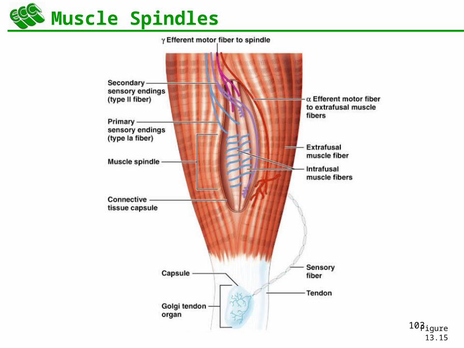
103Figure 13.15
Muscle Spindles
104
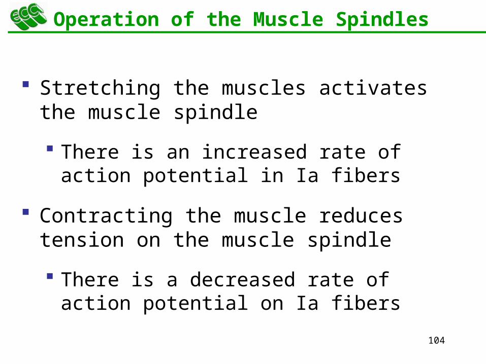
Operation of the Muscle Spindles
Stretching the muscles activates the muscle spindle
There is an increased rate of action potential in Ia fibers
Contracting the muscle reduces tension on the muscle spindle
There is a decreased rate of action potential on Ia fibers
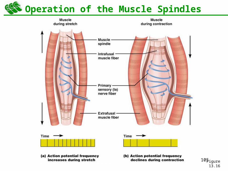
105Figure 13.16
Operation of the Muscle Spindles
106
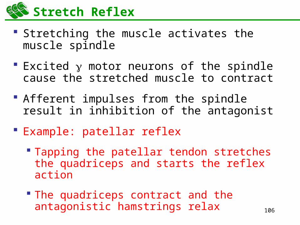
Stretch Reflex
Stretching the muscle activates the muscle spindle
Excited motor neurons of the spindle cause the stretched muscle to contract
Afferent impulses from the spindle result in inhibition of the antagonist
Example: patellar reflex
Tapping the patellar tendon stretches the quadriceps and starts the reflex action
The quadriceps contract and the antagonistic hamstrings relax
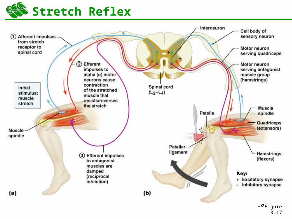
107Figure 13.17
Stretch Reflex
108
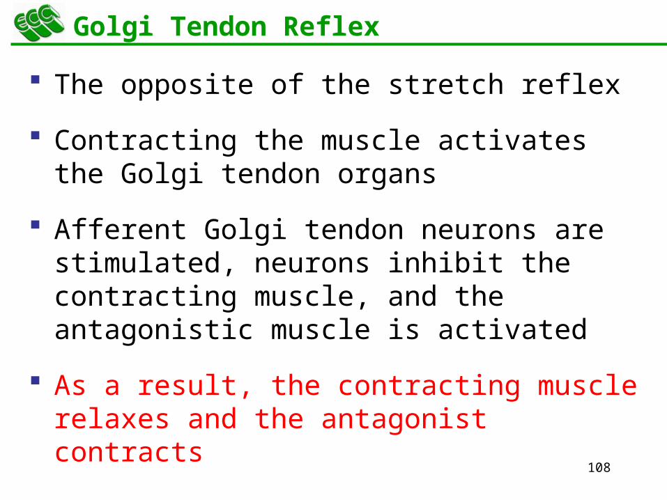
Golgi Tendon Reflex
The opposite of the stretch reflex
Contracting the muscle activates the Golgi tendon organs
Afferent Golgi tendon neurons are stimulated, neurons inhibit the contracting muscle, and the antagonistic muscle is activated
As a result, the contracting muscle relaxes and the antagonist contracts
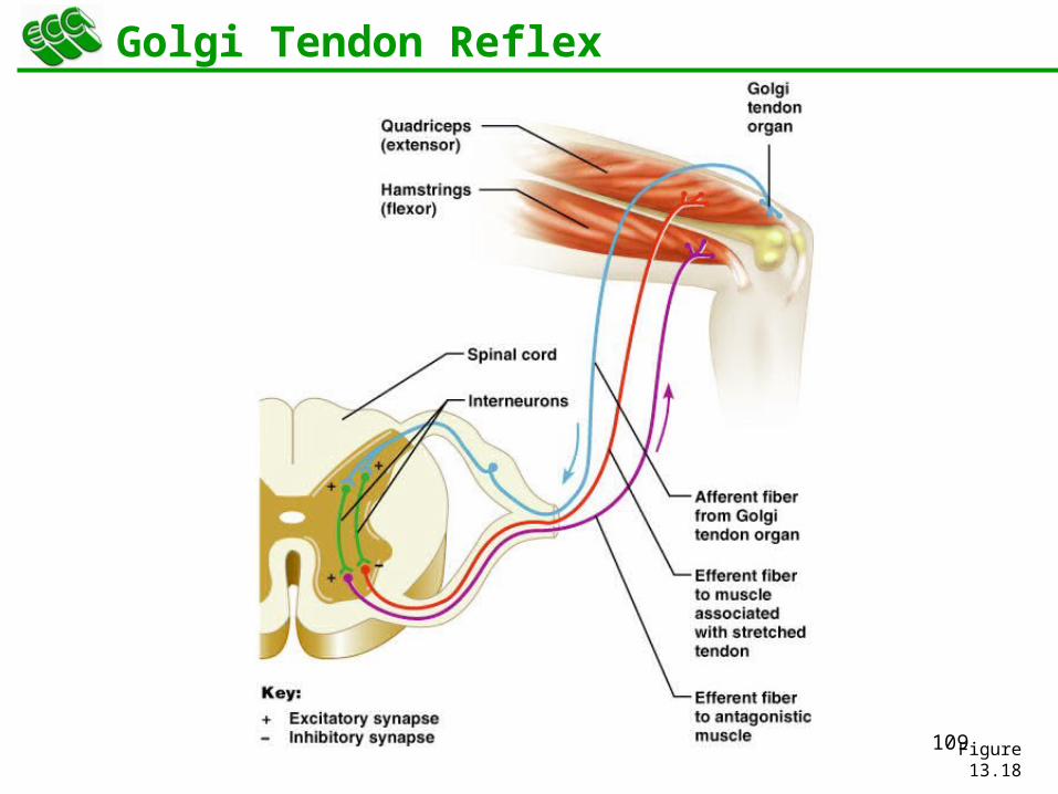
109Figure 13.18
Golgi Tendon Reflex
110
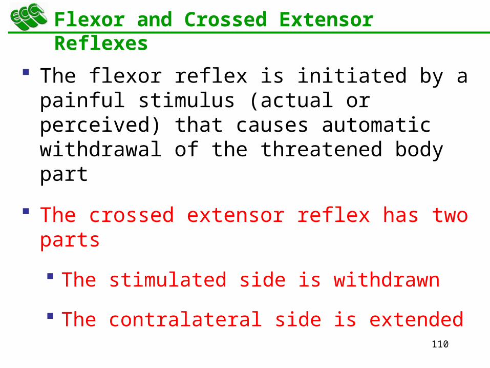
Flexor and Crossed Extensor Reflexes
The flexor reflex is initiated by a painful stimulus (actual or perceived) that causes automatic withdrawal of the threatened body part
The crossed extensor reflex has two parts
The stimulated side is withdrawn
The contralateral side is extended
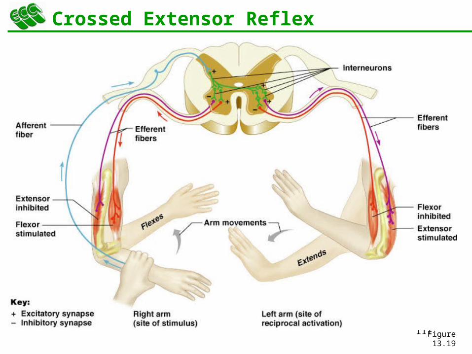
111Figure 13.19
Crossed Extensor Reflex
112
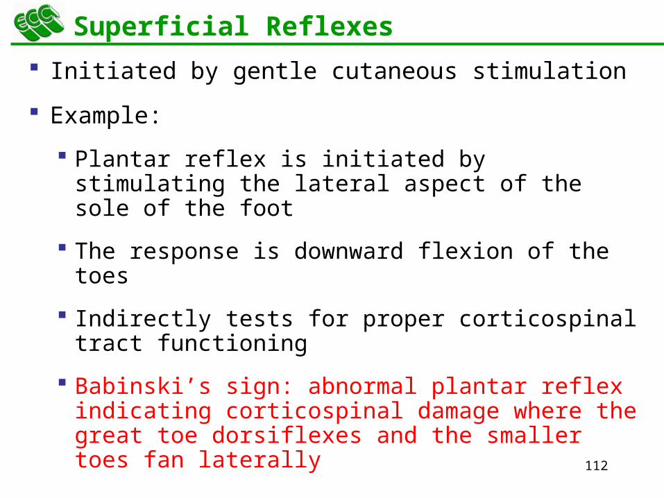
Superficial Reflexes
Initiated by gentle cutaneous stimulation
Example:
Plantar reflex is initiated by stimulating the lateral aspect of the sole of the foot
The response is downward flexion of the toes
Indirectly tests for proper corticospinal tract functioning
Babinski’s sign: abnormal plantar reflex indicating corticospinal damage where the great toe dorsiflexes and the smaller toes fan laterally
113
Developmental Aspects of the PNS
Spinal nerves branch from the developing spinal cord and neural crest cells
Supply motor and sensory function to developing muscles
Cranial nerves innervate muscles of the head
114
Developmental Aspects of the PNS
Distribution and growth of spinal nerves correlate with the segmented body plan
Sensory receptors atrophy with age and muscle tone lessens
Peripheral nerves remain viable throughout life unless subjected to trauma


![The Nervous System. Divisions of the Nervous System Central Nervous System [CNS] = Spinal Cord Brain Peripheral Nervous System [PNS]= Spinal Nerves](https://img.pdfslide.us/doc/110x75/56649d6c5503460f94a4c71d/the-nervous-system-divisions-of-the-nervous-system-central-nervous-system.jpg)