Embed Size (px)
Citation preview
PercutaneousCoronaryIntervention
ACC/AHA Pocket Guideline
Based on the ACC/AHA/SCAI 2005 Guideline Update
November 2005
Learn and Live SM
Special thanks to
Eli Lilly and Company
supported this
pocket guideline
through an
educational grant.
Eli Lilly and Company
was not involved in the
development of this
publication and in no
way influenced
its contents.
PercutaneousCoronaryInterventionNovember 2005
Writing Committee
Sidney C. Smith, Jr, MD, FACC, FAHA, Chair
Ted. E. Feldman, MD, FACC, FSCAI
John W. Hirshfeld, Jr, MD, FACC, FSCAI
Alice K. Jacobs, MD, FACC, FAHA, FSCAI
Morton J. Kern, MD, FACC, FAHA, FSCAI
Spencer B. King, III, MD, MACC, FSCAI
Douglass A. Morrison, MD, PhD, FACC, FSCAI
William W. O’Neill, MD, FACC, FSCAI
Hartzell V. Schaff, MD, FACC, FAHA
Patrick L. Whitlow, MD, FACC, FAHA
David O. Williams, MD, FACC, FAHA, FSCAI
©2005 American College
of Cardiology Foundation and
American Heart Association, Inc.
The following article was adapted from
the ACC/AHA/SCAI 2005 Guideline Update
for Percutaneous Coronary Intervention
(January 3, 2006 issue of Journal of the
American College of Cardiology, January 3,
2006 issue of Circulation, and January
2006 issue of Catheterization and
Cardiovascular Intervention).
For a copy of the full report or published
summary article, visit our Web sites at
www.acc.org, www.americanheart.org, or
www.scai.org, or call the ACC Resource
Center at 1-800-253-4636, ext. 694.
IntroductionCharacterization
Outcom
esCom
petencyClinical Presentations
Special ConsiderationsIntroduction
CharacterizationO
utcomes
Competency
Clinical PresentationsPatient M
anagement
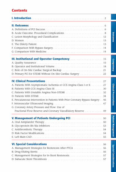
Contents
I. Introduction . . . . . . . . . . . . . . . . . . . . . . . . . . . . . . . . . . . . . . . . . . . . . 2
II. Outcomes. . . . . . . . . . . . . . . . . . . . . . . . . . . . . . . . . . . . . . . . . . . . . . . 6
A. Definitions of PCI Success . . . . . . . . . . . . . . . . . . . . . . . . . . . . . . . . . . . . 6
B. Acute Outcome: Procedural Complications . . . . . . . . . . . . . . . . . . . . . . . . . 8
C. Lesion Morphology and Classification . . . . . . . . . . . . . . . . . . . . . . . . . . . 12
D. Women. . . . . . . . . . . . . . . . . . . . . . . . . . . . . . . . . . . . . . . . . . . . . . . . . 13
E. The Elderly Patient . . . . . . . . . . . . . . . . . . . . . . . . . . . . . . . . . . . . . . . . . 13
F. Comparison With Bypass Surgery . . . . . . . . . . . . . . . . . . . . . . . . . . . . . . . 14
G. Comparison With Medicine . . . . . . . . . . . . . . . . . . . . . . . . . . . . . . . . . . . 14
III. Institutional and Operator Competency . . . . . . . . . . . . . . . . . . . . 15
A. Quality Assurance . . . . . . . . . . . . . . . . . . . . . . . . . . . . . . . . . . . . . . . . . 15
B. Operator and Institutional Volume . . . . . . . . . . . . . . . . . . . . . . . . . . . . . . 18
C. Role of On-Site Cardiac Surgical Backup . . . . . . . . . . . . . . . . . . . . . . . . . 21
D. Primary PCI for STEMI Without On-Site Cardiac Surgery . . . . . . . . . . . . . . 22
IV. Clinical Presentations . . . . . . . . . . . . . . . . . . . . . . . . . . . . . . . . . . . 27
A. Patients With Asymptomatic Ischemia or CCS Angina Class I or II. . . . . . . . 27
B. Patients With CCS Angina Class III . . . . . . . . . . . . . . . . . . . . . . . . . . . . . . 30
C. Patients With Unstable Angina/Non-STEMI . . . . . . . . . . . . . . . . . . . . . . . 32
D. Patients With STEMI. . . . . . . . . . . . . . . . . . . . . . . . . . . . . . . . . . . . . . . . 34
E. Percutaneous Intervention in Patients With Prior Coronary Bypass Surgery . . 46
F. Intravascular Ultrasound Imaging. . . . . . . . . . . . . . . . . . . . . . . . . . . . . . . 47
G. Coronary Artery Pressure and Flow: Use of
Fractional Flow Reserve and Coronary Vasodilatory Reserve . . . . . . . . . . . 49
V. Management of Patients Undergoing PCI . . . . . . . . . . . . . . . . . . . . 50
A. Oral Antiplatelet Therapy . . . . . . . . . . . . . . . . . . . . . . . . . . . . . . . . . . . . 50
B. Glycoprotein IIb/IIIa Inhibitors . . . . . . . . . . . . . . . . . . . . . . . . . . . . . . . . 53
C. Antithrombotic Therapy . . . . . . . . . . . . . . . . . . . . . . . . . . . . . . . . . . . . . 54
D. Risk Factor Modifications . . . . . . . . . . . . . . . . . . . . . . . . . . . . . . . . . . . . 54
E. Left Main CAD . . . . . . . . . . . . . . . . . . . . . . . . . . . . . . . . . . . . . . . . . . . . 55
VI. Special Considerations . . . . . . . . . . . . . . . . . . . . . . . . . . . . . . . . . . 56
A. Management Strategies for Restenosis After PTCA . . . . . . . . . . . . . . . . . . 56
B. Drug-Eluting Stents . . . . . . . . . . . . . . . . . . . . . . . . . . . . . . . . . . . . . . . . 56
C. Management Strategies for In-Stent Restenosis. . . . . . . . . . . . . . . . . . . . . 57
D. Subacute Stent Thrombosis . . . . . . . . . . . . . . . . . . . . . . . . . . . . . . . . . . 57
2
I. Introduction
The American College of Cardiology (ACC)/American
Heart Association (AHA) practice guidelines are
intended to assist healthcare providers in clinical
decision making by describing a range of generally
acceptable approaches for the diagnosis, manage-
ment, or prevention of specific diseases or condi-
tions. These percutaneous coronary intervention
(PCI) guidelines attempt to define practices that
meet the needs of most patients in most circum-
stances. The guideline recommendations reflect a
consensus of expert opinion after a thorough review
of the available, current scientific evidence and are
intended to improve patient care. If these guidelines
are used as the basis for regulatory/payer decisions,
the ultimate goal is quality of care and serving the
patient’s best interests. The ultimate judgment
regarding care of a particular patient must be made
by the healthcare provider and patient in light of
all of the circumstances presented by that patient.
Although initially limited to balloon angioplasty
and termed percutaneous transluminal coronary
angioplasty (PTCA), PCI now includes other new
techniques capable of relieving coronary narrowing.
Accordingly, in this document, implantation of
intracoronary stents and other catheter-based inter-
ventions for treating coronary atherosclerosis are
considered components of PCI. In this context,
3
PTCA will be used to refer to procedures that use only balloon
angioplasty, whereas PCI will refer to the broader group of
percutaneous techniques.
Percutaneous coronary intervention is a technique that is being
continually refined and modified; hence, continued, periodic
guideline revision is anticipated. These guidelines are to be
viewed as broad recommendations to aid in the appropriate
application of PCI. Under unique circumstances, exceptions
may exist. These guidelines are intended to complement, not
replace, sound medical judgment and knowledge. They are
intended for operators who possess the cognitive and technical
skills to perform PCI and assume that facilities and resources
required to properly perform PCI are available. As in the past,
the indications are categorized as class I, II, or III, based on a
multifactorial assessment of risk and expected efficacy viewed
in the context of current knowledge and the relative strength
of this knowledge.
The schema for classification of recommendations and level of
evidence is summarized in Table 1, which also illustrates how
the grading system provides an estimate of the size of the treat-
ment effect and an estimate of the certainty of the treatment
effect. It is recognized that the basis for recommendations graded
Level of Evidence: C is the opinion and consensus of the Writing
Group. In this setting, evidence from clinical trials can provide
important data to investigate the validity of the consensus opin-
ion and to support continuing or modifying the recommendation.
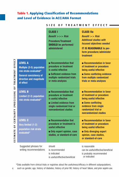
CLASS IIa
Benefit >> RiskAdditional studies withfocused objectives needed
IT IS REASONABLE to per-form procedure/administer treatment
■ Recommendation in favorof treatment or procedurebeing useful/effective
■ Some conflicting evidencefrom multiple randomized trials or meta-analyses
■ Recommendation in favorof treatment or procedurebeing useful/effective
■ Some conflicting evidence from single randomized trial or nonrandomized studies
■ Recommendation in favorof treatment or procedurebeing useful/effective
■ Only diverging expertopinion, case studies, or standard-of-care
CLASS I
Benefit >>> Risk
Procedure/TreatmentSHOULD be performed/administered
■ Recommendation that procedure or treatment is useful/effective
■ Sufficient evidence frommultiple randomized trials or meta-analyses
■ Recommendation that procedure or treatment is useful/effective
■ Limited evidence from single randomized trial ornonrandomized studies
■ Recommendation that procedure or treatment isuseful/effective
■ Only expert opinion, casestudies, or standard-of-care
shouldis recommendedis indicatedis useful/effective/beneficial
LEVEL A
Multiple (3-5) populationrisk strata evaluated*
General consistency ofdirection and magnitudeof effect
LEVEL B
Limited (2-3) populationrisk strata evaluated*
LEVEL C
Very limited (1-2)
population risk strataevaluated*
*Data available from clinical trials or registries about the usefulness/efficacy in different subpopulations,
such as gender, age, history of diabetes, history of prior MI, history of heart failure, and prior aspirin use.
Suggested phrases forwriting recommendations
is reasonablecan be useful/effective/beneficialis probably recommended
or indicated
S I Z E O F T R E A T M E N T E F F E C T
Table 1. Applying Classification of Recommendations and Level of Evidence in ACC/AHA Format
ES
TIM
AT
E O
F C
ER
TAIN
TY
(P
RE
CIS
ION
) O
F T
RE
AT
ME
NT
EF
FE
CT
4
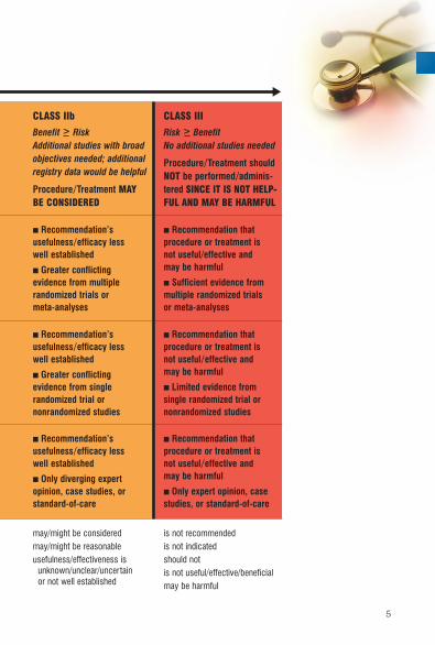
CLASS IIb
Benefit > RiskAdditional studies with broadobjectives needed; additionalregistry data would be helpful
Procedure/Treatment MAY BE CONSIDERED
■ Recommendation’s usefulness/efficacy less well established
■ Greater conflicting evidence from multiple randomized trials or meta-analyses
■ Recommendation’s usefulness/efficacy less well established
■ Greater conflicting evidence from single randomized trial or nonrandomized studies
■ Recommendation’s usefulness/efficacy less well established
■ Only diverging expert opinion, case studies, orstandard-of-care
may/might be consideredmay/might be reasonableusefulness/effectiveness is
unknown/unclear/uncertain or not well established
CLASS III
Risk > BenefitNo additional studies needed
Procedure/Treatment shouldNOT be performed/adminis-tered SINCE IT IS NOT HELP-FUL AND MAY BE HARMFUL
■ Recommendation that procedure or treatment is not useful/effective and may be harmful
■ Sufficient evidence frommultiple randomized trials or meta-analyses
■ Recommendation that procedure or treatment is not useful/effective and may be harmful
■ Limited evidence from single randomized trial ornonrandomized studies
■ Recommendation that procedure or treatment is not useful/effective and may be harmful
■ Only expert opinion, casestudies, or standard-of-care
is not recommendedis not indicatedshould notis not useful/effective/beneficialmay be harmful
▼
5
6
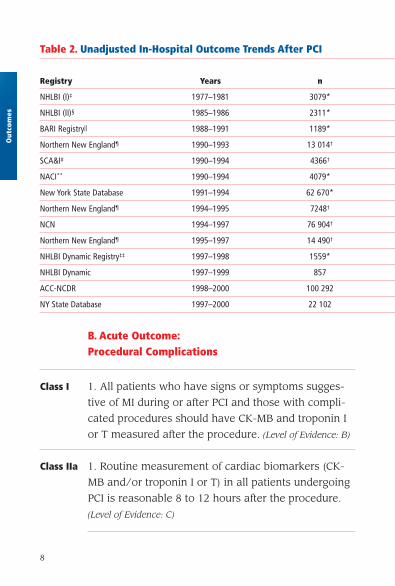
II. Outcomes
The outcomes of PCI are measured in terms of
success and complications and are related to the
mechanisms of the employed devices, as well as
the clinical and anatomic patient-related factors
(Table 2). The committee recommends the use of
such standards as the ACC-National Cardiovascular
Data Registry®whenever feasible to accommodate
the common database for the assessment of
outcomes.
A. Definitions of PCI Success
1. Angiographic Success
With the advent of advanced adjunct technology,
including coronary stents, a minimum stenosis
diameter reduction to less than 20% has been the
clinical benchmark of an optimal angiographic
result.
2. Procedural Success
Although the occurrence of emergency coronary
artery bypass surgery and death are easily identified
end points, the definition of procedure-related
myocardial infarction (MI) has been debated. The
development of Q waves in addition to a threshold
Out
com
es
7
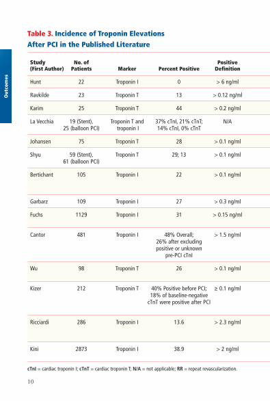
value of creatine kinase (CK) elevation has been
commonly used. The clinical significance of cardiac
biomarker elevations in the absence of Q waves
remains a subject of investigation and debate. An
increase in CK-MB greater than 5 times is associat-
ed with worsened outcome. Thus, this degree of
increase in CK-MB without Q waves is considered
by most to qualify as an associated complication
of PCI. Troponin T or I elevation occurs frequently
after PCI. Minor elevations do not appear to have
prognostic value, whereas marked (greater than
5 times) elevations are associated with worsened
1-year outcome (Table 3).
Outcom
es
8
Out
com
es
B. Acute Outcome:Procedural Complications
Class I 1. All patients who have signs or symptoms sugges-
tive of MI during or after PCI and those with compli-
cated procedures should have CK-MB and troponin I
or T measured after the procedure. (Level of Evidence: B)
Class IIa 1. Routine measurement of cardiac biomarkers (CK-
MB and/or troponin I or T) in all patients undergoing
PCI is reasonable 8 to 12 hours after the procedure.
(Level of Evidence: C)
Table 2. Unadjusted In-Hospital Outcome Trends After PCI
Registry Years n
NHLBI (I)‡ 1977–1981 3079*
NHLBI (II)§ 1985–1986 2311*
BARI Registry|| 1988–1991 1189*
Northern New England¶ 1990–1993 13 014†
SCA&I# 1990–1994 4366†
NACI** 1990–1994 4079*
New York State Database 1991–1994 62 670*
Northern New England¶ 1994–1995 7248†
NCN 1994–1997 76 904†
Northern New England¶ 1995–1997 14 490†
NHLBI Dynamic Registry‡‡ 1997–1998 1559*
NHLBI Dynamic 1997–1999 857
ACC-NCDR 1998–2000 100 292
NY State Database 1997–2000 22 102
9
Outcom
es
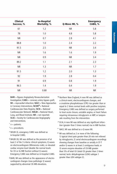
Clinical In-Hospital Emergency Success, % Mortality, % Q-Wave MI, % CABG, %
61 1.2 NR 5.8
78 1.0 4.8 5.8
NR 0.7 2.8 4.1
88.8 1.0 2.4 2.2
91.5 2.5 NR 3.4
NR 1.6 1.6 1.9
NR 0.9 NR 3.4
89.2 1.1 2.1 2.3
NR 1.3 NR 1.7
91.5 1.2 2.0 1.3
92 1.9 2.8 0.4
91 0.9 0.8 1.9
96.5 1.4 0.4 1.9
NR 0.68 NR NR
BARI = Bypass Angioplasty RevascularizationInvestigation; CABG = coronary artery bypass graft;MI = myocardial infarction; NACI = New Approachesin Coronary Interventions; NCDR®= NationalCardiovascular Data Registry; NCN = NationalCardiovascular Network; NHLBI = National Heart,Lung, and Blood Institute; NR = not reported;SCAI = Society for Cardiovascular Angiographyand Interventions.
* n = patients.† n = procedures.
‡ NHLBI (I), emergency CABG was defined as in-hospital CABG.
§ NHLBI (II), MI was defined as the presence of at least 2 of the 3 criteria: clinical symptoms, Q waves on electrocardiogram (Minnesota code), or elevatedcardiac enzyme level (double the normal levels for CK or its MB fraction without Q waves).Emergency CABG was defined as in-hospital CABG.
|| BARI, MI was defined as the appearance of electro-cardiogram changes (new pathologic Q waves) supported by abnormal CK-MB elevations.
¶ Northern New England, A new MI was defined as a clinical event, electrocardiogram changes, and a creatinine phosphokinase (CPK) rise greater than orequal to 2 times normal levels with positive isozymes.Emergency CABG was defined as surgery performed to treat acute closure, unstable angina, or heart failurerequiring intravenous nitroglycerin or ABP, or tampon-ade resulting from the intervention.
# SCAI, A new MI was defined as any significant infarc-tion (greater than 3 times normal) rise in MB fraction.
** NACI, MI was defined as a Q-wave MI.
†† MI was defined as 2 or more of the following:1) typical chest pain greater than 20 min not relieved by nitroglycerin; 2) serial electrocardiogram recordingsshowing changes from baseline or serially in ST-Tand/or Q waves in at least 2 contiguous leads; or 3) serum enzyme elevation of CK-MB greater than 5% of total CK (total CK greater than 2 timesnormal; lactic dehydrogenase (LDH) subtype 1 greater than LDH subtype 2).
10
Out
com
es
Table 3. Incidence of Troponin Elevations After PCI in the Published Literature
Study No. of Positive (First Author) Patients Marker Percent Positive Definition
Hunt 22 Troponin I 0 > 6 ng/ml
Ravkilde 23 Troponin T 13 > 0.12 ng/ml
Karim 25 Troponin T 44 > 0.2 ng/ml
La Vecchia 19 (Stent), Troponin T and 37% cTnI, 21% cTnT; N/A25 (balloon PCI) troponin I 14% cTnI, 0% cTnT
Johansen 75 Troponin T 28 > 0.1 ng/ml
Shyu 59 (Stent), Troponin T 29; 13 > 0.1 ng/ml61 (balloon PCI)
Bertichant 105 Troponin I 22 > 0.1 ng/ml
Garbarz 109 Troponin I 27 > 0.3 ng/ml
Fuchs 1129 Troponin I 31 > 0.15 ng/ml
Cantor 481 Troponin I 48% Overall; > 1.5 ng/ml26% after excluding positive or unknown
pre-PCI cTnI
Wu 98 Troponin T 26 > 0.1 ng/ml
Kizer 212 Troponin T 40% Positive before PCI; ≥ 0.1 ng/ml18% of baseline-negative
cTnT were positive after PCI
Ricciardi 286 Troponin I 13.6 > 2.3 ng/ml
Kini 2873 Troponin I 38.9 > 2 ng/ml
cTnI = cardiac troponin I; cTnT = cardiac troponin T; N/A = not applicable; RR = repeat revascularization.
11
Outcom
esSpecial Considerations
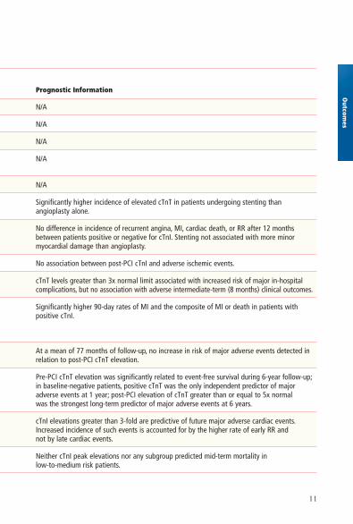
Prognostic Information
N/A
N/A
N/A
N/A
N/A
Significantly higher incidence of elevated cTnT in patients undergoing stenting than angioplasty alone.
No difference in incidence of recurrent angina, MI, cardiac death, or RR after 12 months between patients positive or negative for cTnI. Stenting not associated with more minor myocardial damage than angioplasty.
No association between post-PCI cTnI and adverse ischemic events.
cTnT levels greater than 3x normal limit associated with increased risk of major in-hospital complications, but no association with adverse intermediate-term (8 months) clinical outcomes.
Significantly higher 90-day rates of MI and the composite of MI or death in patients withpositive cTnI.
At a mean of 77 months of follow-up, no increase in risk of major adverse events detected inrelation to post-PCI cTnT elevation.
Pre-PCI cTnT elevation was significantly related to event-free survival during 6-year follow-up;in baseline-negative patients, positive cTnT was the only independent predictor of major adverse events at 1 year; post-PCI elevation of cTnT greater than or equal to 5x normal was the strongest long-term predictor of major adverse events at 6 years.
cTnI elevations greater than 3-fold are predictive of future major adverse cardiac events.Increased incidence of such events is accounted for by the higher rate of early RR and not by late cardiac events.
Neither cTnI peak elevations nor any subgroup predicted mid-term mortality in low-to-medium risk patients.
12
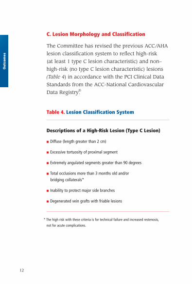
C. Lesion Morphology and Classification
The Committee has revised the previous ACC/AHA
lesion classification system to reflect high-risk
(at least 1 type C lesion characteristic) and non–
high-risk (no type C lesion characteristic) lesions
(Table 4) in accordance with the PCI Clinical Data
Standards from the ACC-National Cardiovascular
Data Registry®.
Out
com
es
Table 4. Lesion Classification System
Descriptions of a High-Risk Lesion (Type C Lesion)
■ Diffuse (length greater than 2 cm)
■ Excessive tortuosity of proximal segment
■ Extremely angulated segments greater than 90 degrees
■ Total occlusions more than 3 months old and/or
bridging collaterals*
■ Inability to protect major side branches
■ Degenerated vein grafts with friable lesions
* The high risk with these criteria is for technical failure and increased restenosis,
not for acute complications.
13
D. Women
Compared with men, women undergoing PCI are
older and have a higher incidence of hypertension,
diabetes mellitus, hypercholesterolemia, and co-
morbid disease. The hope that stents would
eliminate the difference in outcomes between
women and men has not been realized. Gender
differences in mortality have persisted for patients
treated with stents both in the setting of acute MI
and in non-acute settings.
In general, the risks and benefits of adjunctive phar-
macotherapy in women are similar to those in men,
although an increased rate of minor bleeding has
been reported in women treated with abciximab.
When glycoprotein IIb/IIIa platelet receptor antago-
nists are used with unfractionated heparin, a lower
dose of the latter should be considered to decrease
the risk of bleeding in women.
E. The Elderly Patient
With rare exception (primary PCI for cardiogenic
shock in patients greater than 75 years old), a
separate category has not been created in these
Guidelines for the elderly. However, their higher
incidence of comorbidities and risk for bleeding
complications should be taken into account when
considering the need for PCI.
Outcom
es
14
F. Comparison With Bypass Surgery
Overall, 6 trials have been published comparing PCI using stents
with coronary artery bypass grafting (CABG) in single-vessel
or multivessel disease. Both revascularization techniques
relieve angina. In aggregate, these trials have not shown a
difference between CABG and PCI in terms of mortality or pro-
cedural MI among the populations studied, which have included
mostly low-risk patients. Stents appear to have narrowed the
late repeat revascularization difference that favored CABG in
the balloon era. Some risk-adjusted registries have shown the
superiority of surgery for multivessel disease patients, especially
those with diabetes. Randomized trials, meta-analysis of trials,
and epidemiological studies have shown the superiority of drug-
eluting stents (DES) over bare-metal stents (BMS) in terms of
reducing late repeat revascularization.
G. Comparison With Medicine
Given the limited data available from randomized trials com-
paring medical therapy with PCI, it seems prudent to consider
medical therapy for the initial management of most patients
with Canadian Cardiovascular Society (CCS) classification
class I and II stable angina (Table 9, pg. 27) and reserve PCI
and CABG for those patients with more severe symptoms and
ischemia. The symptomatic patient who wishes to remain
physically active, regardless of age, will usually require PCI
or CABG to remain physically active.
Out
com
es
15
III. Institutional and Operator Competency
A. Quality Assurance
Class I 1. An institution that performs PCI should establish
an ongoing mechanism for valid peer review of
its quality and outcomes. Review should be con-
ducted both at the level of the entire program and
at the level of the individual practitioner. Quality-
assessment reviews should take risk adjustment,
statistical power, and national benchmark statistics
into consideration. Quality-assessment reviews
should include both tabulation of adverse event
rates for comparison with benchmark values and
case review of complicated procedures and some
uncomplicated procedures. (Level of Evidence: C)
2. An institution that performs PCI should partici-
pate in a recognized PCI data registry for the
purpose of benchmarking its outcomes against
current national norms. (Level of Evidence: C)
Each institution that performs PCI must establish
an ongoing mechanism for valid peer review of its
quality and outcomes. The program should provide
an opportunity for interventionalists and physicians
who do not perform angioplasty but are knowledge-
able about it to review its overall results on a
regular basis. The review process should tabulate
Competency
16
the results achieved both by individual physician
operators and by the overall program and compare
them with national benchmark standards with
appropriate risk adjustment. Valid quality assess-
ment requires that the institution maintain meticu-
lous records that include the patient demographic
and clinical characteristics necessary to assess
appropriateness and to conduct risk adjustment.
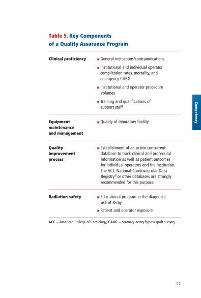
Quality assessment is a complex process that
includes more than mere tabulation of success
and complication rates. Components of quality in
coronary interventional procedures include appro-
priateness of case selection, quality of procedure
execution, proper response to intraprocedural prob-
lems, accurate assessment of procedure outcome,
and appropriateness of postprocedure management.
It is important that each of these parameters be
considered when a quality-assessment review is
conducted (Table 5).
It is recommended that an interventional cardiology
operator be certified by the American Board of
Internal Medicine in interventional cardiology.
Ideally, board certification in interventional cardiol-
ogy should be required for credentialing (Table 6).
Com
pete
ncy
17
Competency
Table 5. Key Components of a Quality Assurance Program
Clinical proficiency ■ General indications/contraindications
■ Institutional and individual operator complication rates, mortality, and emergency CABG
■ Institutional and operator procedure volumes
■ Training and qualifications of support staff
Equipment ■ Quality of laboratory facilitymaintenanceand management
Quality ■ Establishment of an active concurrent improvement database to track clinical and procedural process information as well as patient outcomes
for individual operators and the institution.The ACC-National Cardiovascular Data Registry® or other databases are strongly recommended for this purpose.
Radiation safety ■ Educational program in the diagnostic use of X-ray
■ Patient and operator exposure
ACC = American College of Cardiology; CABG = coronary artery bypass graft surgery.
18
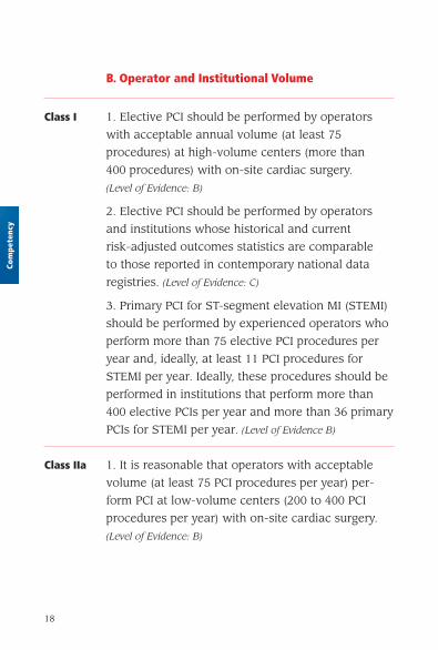
B. Operator and Institutional Volume
Class I 1. Elective PCI should be performed by operators
with acceptable annual volume (at least 75
procedures) at high-volume centers (more than
400 procedures) with on-site cardiac surgery.
(Level of Evidence: B)
2. Elective PCI should be performed by operators
and institutions whose historical and current
risk-adjusted outcomes statistics are comparable
to those reported in contemporary national data
registries. (Level of Evidence: C)
3. Primary PCI for ST-segment elevation MI (STEMI)
should be performed by experienced operators who
perform more than 75 elective PCI procedures per
year and, ideally, at least 11 PCI procedures for
STEMI per year. Ideally, these procedures should be
performed in institutions that perform more than
400 elective PCIs per year and more than 36 primary
PCIs for STEMI per year. (Level of Evidence B)
Class IIa 1. It is reasonable that operators with acceptable
volume (at least 75 PCI procedures per year) per-
form PCI at low-volume centers (200 to 400 PCI
procedures per year) with on-site cardiac surgery.
(Level of Evidence: B)
Com
pete
ncy
19
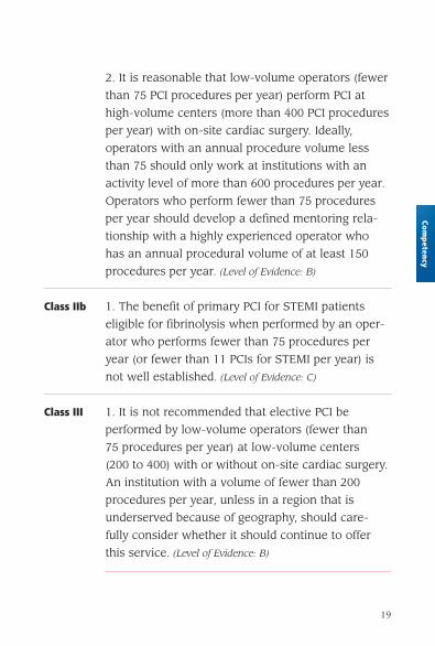
2. It is reasonable that low-volume operators (fewer
than 75 PCI procedures per year) perform PCI at
high-volume centers (more than 400 PCI procedures
per year) with on-site cardiac surgery. Ideally,
operators with an annual procedure volume less
than 75 should only work at institutions with an
activity level of more than 600 procedures per year.
Operators who perform fewer than 75 procedures
per year should develop a defined mentoring rela-
tionship with a highly experienced operator who
has an annual procedural volume of at least 150
procedures per year. (Level of Evidence: B)
Class IIb 1. The benefit of primary PCI for STEMI patients
eligible for fibrinolysis when performed by an oper-
ator who performs fewer than 75 procedures per
year (or fewer than 11 PCIs for STEMI per year) is
not well established. (Level of Evidence: C)
Class III 1. It is not recommended that elective PCI be
performed by low-volume operators (fewer than
75 procedures per year) at low-volume centers
(200 to 400) with or without on-site cardiac surgery.
An institution with a volume of fewer than 200
procedures per year, unless in a region that is
underserved because of geography, should care-
fully consider whether it should continue to offer
this service. (Level of Evidence: B)
Competency
20
Com
pete
ncy
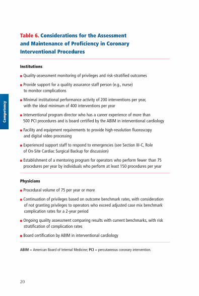
Table 6. Considerations for the Assessment and Maintenance of Proficiency in Coronary Interventional Procedures
Institutions
■ Quality-assessment monitoring of privileges and risk-stratified outcomes
■ Provide support for a quality assurance staff person (e.g., nurse) to monitor complications
■ Minimal institutional performance activity of 200 interventions per year,with the ideal minimum of 400 interventions per year
■ Interventional program director who has a career experience of more than 500 PCI procedures and is board certified by the ABIM in interventional cardiology
■ Facility and equipment requirements to provide high-resolution fluoroscopy and digital video processing
■ Experienced support staff to respond to emergencies (see Section III-C, Role of On-Site Cardiac Surgical Backup for discussion)
■ Establishment of a mentoring program for operators who perform fewer than 75 procedures per year by individuals who perform at least 150 procedures per year
Physicians
■ Procedural volume of 75 per year or more
■ Continuation of privileges based on outcome benchmark rates, with consideration of not granting privileges to operators who exceed adjusted case mix benchmark complication rates for a 2-year period
■ Ongoing quality assessment comparing results with current benchmarks, with risk stratification of complication rates
■ Board certification by ABIM in interventional cardiology
ABIM = American Board of Internal Medicine; PCI = percutaneous coronary intervention.
21
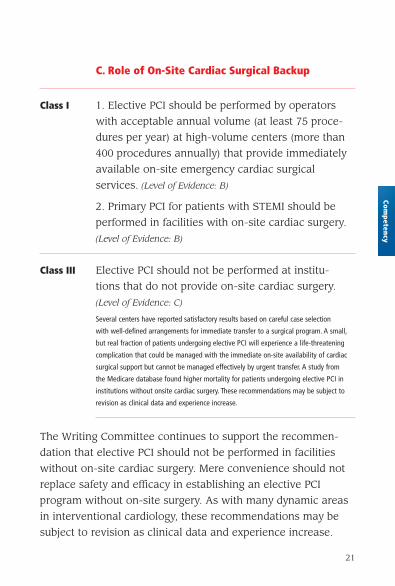
C. Role of On-Site Cardiac Surgical Backup
Class I 1. Elective PCI should be performed by operators
with acceptable annual volume (at least 75 proce-
dures per year) at high-volume centers (more than
400 procedures annually) that provide immediately
available on-site emergency cardiac surgical
services. (Level of Evidence: B)
2. Primary PCI for patients with STEMI should be
performed in facilities with on-site cardiac surgery.
(Level of Evidence: B)
Class III Elective PCI should not be performed at institu-
tions that do not provide on-site cardiac surgery.
(Level of Evidence: C)
Several centers have reported satisfactory results based on careful case selection
with well-defined arrangements for immediate transfer to a surgical program. A small,
but real fraction of patients undergoing elective PCI will experience a life-threatening
complication that could be managed with the immediate on-site availability of cardiac
surgical support but cannot be managed effectively by urgent transfer. A study from
the Medicare database found higher mortality for patients undergoing elective PCI in
institutions without onsite cardiac surgery. These recommendations may be subject to
revision as clinical data and experience increase.
The Writing Committee continues to support the recommen-
dation that elective PCI should not be performed in facilities
without on-site cardiac surgery. Mere convenience should not
replace safety and efficacy in establishing an elective PCI
program without on-site surgery. As with many dynamic areas
in interventional cardiology, these recommendations may be
subject to revision as clinical data and experience increase.
Competency

22
D. Primary PCI for STEMI Without On-Site Cardiac Surgery
Class IIb 1. Primary PCI for patients with STEMI might be con-
sidered in hospitals without on-site cardiac surgery,
provided that appropriate planning for program devel-
opment has been accomplished, including appropri-
ately experienced physician operators (more than
75 total PCIs and, ideally, at least 11 primary PCIs
per year for STEMI), an experienced catheterization
team on a 24 hours per day, 7 days per week call
schedule, and a well-equipped catheterization
laboratory with digital imaging equipment, a full
array of interventional equipment, and intra-aortic
balloon pump capability, and provided that there
is a proven plan for rapid transport to a cardiac
surgery operating room in a nearby hospital with
appropriate hemodynamic support capability for
transfer. The procedure should be limited to patients
with STEMI or MI with new or presumably new left
bundle-branch block (LBBB) on electrocardiogram
and should be performed in a timely fashion (goal
of balloon inflation within 90 minutes of presenta-
tion) by persons skilled in the procedure (at least
75 PCIs per year) and at hospitals that perform
a minimum of 36 primary PCI procedures per year.
(Level of Evidence: B)
Com
pete
ncy
23
Class III 1. Primary PCI should not be performed in hospitals
without on-site cardiac surgery and without a
proven plan for rapid transport to a cardiac surgery
operating room in a nearby hospital or without
appropriate hemodynamic support capability for
transfer. (Level of Evidence: C)
Competency
24
Com
pete
ncy
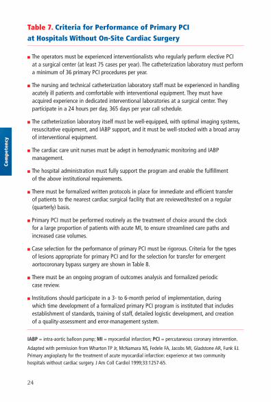
Table 7. Criteria for Performance of Primary PCI at Hospitals Without On-Site Cardiac Surgery
■ The operators must be experienced interventionalists who regularly perform elective PCI at a surgical center (at least 75 cases per year). The catheterization laboratory must perform a minimum of 36 primary PCI procedures per year.
■ The nursing and technical catheterization laboratory staff must be experienced in handling acutely ill patients and comfortable with interventional equipment. They must have acquired experience in dedicated interventional laboratories at a surgical center. They participate in a 24 hours per day, 365 days per year call schedule.
■ The catheterization laboratory itself must be well-equipped, with optimal imaging systems,resuscitative equipment, and IABP support, and it must be well-stocked with a broad array of interventional equipment.
■ The cardiac care unit nurses must be adept in hemodynamic monitoring and IABP management.
■ The hospital administration must fully support the program and enable the fulfillment of the above institutional requirements.
■ There must be formalized written protocols in place for immediate and efficient transfer of patients to the nearest cardiac surgical facility that are reviewed/tested on a regular (quarterly) basis.
■ Primary PCI must be performed routinely as the treatment of choice around the clock for a large proportion of patients with acute MI, to ensure streamlined care paths and increased case volumes.
■ Case selection for the performance of primary PCI must be rigorous. Criteria for the types of lesions appropriate for primary PCI and for the selection for transfer for emergent aortocoronary bypass surgery are shown in Table 8.
■ There must be an ongoing program of outcomes analysis and formalized periodic case review.
■ Institutions should participate in a 3- to 6-month period of implementation, during which time development of a formalized primary PCI program is instituted that includes establishment of standards, training of staff, detailed logistic development, and creation of a quality-assessment and error-management system.
IABP = intra-aortic balloon pump; MI = myocardial infarction; PCI = percutaneous coronary intervention.
Adapted with permission from Wharton TP Jr, McNamara NS, Fedele FA, Jacobs MI, Gladstone AR, Funk EJ.Primary angioplasty for the treatment of acute myocardial infarction: experience at two community hospitals without cardiac surgery. J Am Coll Cardiol 1999;33:1257-65.
25
There are important institutional considerations
in creating an effective program of primary PCI
for STEMI. An institution must commit its cathe-
terization facility to be capable of a 24 hours per
day, 7 days per week rapid response to a patient
presenting with STEMI. In addition, the institution’s
catheterization facility staff must be sufficiently
trained and experienced in the management of the
seriously ill patient with STEMI (Table 7) (Table 8).
It has been demonstrated that institutions with-
out an elective PCI program that care for a large
number of patients with STEMI can create high-
quality PCI for STEMI programs. These programs
require the 24 hours per day, 7 days per week
availability of experienced interventionalists and
an institutional commitment to invest in the
physical and cognitive resources needed to
support a high-quality program.
Competency
26
Com
pete
ncy
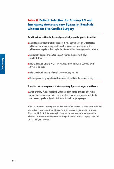
Table 8. Patient Selection for Primary PCI and Emergency Aortocoronary Bypass at Hospitals Without On-Site Cardiac Surgery
Avoid intervention in hemodynamically stable patients with:
■ Significant (greater than or equal to 60%) stenosis of an unprotected left main coronary artery upstream from an acute occlusion in the left coronary system that might be disrupted by the angioplasty catheter
■ Extremely long or angulated infarct-related lesions with TIMI grade 3 flow
■ Infarct-related lesions with TIMI grade 3 flow in stable patients with 3-vessel disease
■ Infarct-related lesions of small or secondary vessels
■ Hemodynamically significant lesions in other than the infarct artery
Transfer for emergency aortocoronary bypass surgery patients:
■ After primary PCI of occluded vessels if high-grade residual left main or multivessel coronary disease and clinical or hemodynamic instability are present, preferably with intra-aortic balloon pump support
PCI = percutaneous coronary intervention; TIMI = Thrombolysis In Myocardial Infarction.
Adapted with permission from Wharton TP Jr, McNamara NS, Fedele FA, Jacobs MI,Gladstone AR, Funk EJ. Primary angioplasty for the treatment of acute myocardial infarction: experience at two community hospitals without cardiac surgery. J Am CollCardiol 1999;33:1257–65.
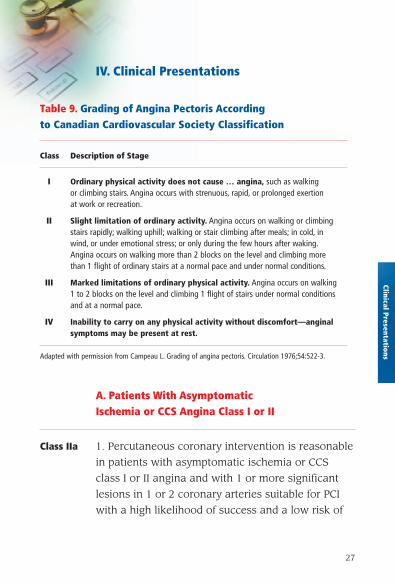
IV. Clinical Presentations
Table 9. Grading of Angina Pectoris According to Canadian Cardiovascular Society Classification
Class Description of Stage
I Ordinary physical activity does not cause … angina, such as walking or climbing stairs. Angina occurs with strenuous, rapid, or prolonged exertion at work or recreation.
II Slight limitation of ordinary activity. Angina occurs on walking or climbing stairs rapidly; walking uphill; walking or stair climbing after meals; in cold, in wind, or under emotional stress; or only during the few hours after waking.Angina occurs on walking more than 2 blocks on the level and climbing more than 1 flight of ordinary stairs at a normal pace and under normal conditions.
III Marked limitations of ordinary physical activity. Angina occurs on walking 1 to 2 blocks on the level and climbing 1 flight of stairs under normal conditions and at a normal pace.
IV Inability to carry on any physical activity without discomfort—anginal symptoms may be present at rest.
Adapted with permission from Campeau L. Grading of angina pectoris. Circulation 1976;54:522-3.
27
Clinical Presentations
A. Patients With Asymptomatic Ischemia or CCS Angina Class I or II
Class IIa 1. Percutaneous coronary intervention is reasonable
in patients with asymptomatic ischemia or CCS
class I or II angina and with 1 or more significant
lesions in 1 or 2 coronary arteries suitable for PCI
with a high likelihood of success and a low risk of
28
morbidity and mortality. The vessels to be dilated
must subtend a moderate to large area of viable
myocardium or be associated with a moderate to
severe degree of ischemia on noninvasive testing.
(Level of Evidence: B)
2. Percutaneous coronary intervention is reasonable
for patients with asymptomatic ischemia or CCS
class I or II angina, and recurrent stenosis after PCI
with a large area of viable myocardium or high-risk
criteria on noninvasive testing. (Level of Evidence: C)
3. Use of PCI is reasonable in patients with asymp-
tomatic ischemia or CCS class I or II angina with
significant left main coronary artery disease (CAD;
greater than 50% diameter stenosis) who are candi-
dates for revascularization but are not eligible for
CABG. (Level of Evidence: B)
Class IIb 1. The effectiveness of PCI for patients with asymp-
tomatic ischemia or CCS class I or II angina who
have 2- or 3-vessel disease with significant proxi-
mal left anterior descending coronary artery (LAD)
CAD who are otherwise eligible for CABG with 1
arterial conduit and who have treated diabetes or
abnormal left ventricular (LV) function is not well
established. (Level of Evidence: B)
2. Percutaneous coronary intervention might be
considered for patients with asymptomatic ischemia
Clin
ical
Pre
sent
atio
ns
29
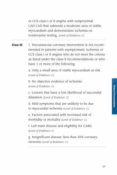
or CCS class I or II angina with nonproximal
LAD CAD that subtends a moderate area of viable
myocardium and demonstrates ischemia on
noninvasive testing. (Level of Evidence: C)
Class III 1. Percutaneous coronary intervention is not recom-
mended in patients with asymptomatic ischemia or
CCS class I or II angina who do not meet the criteria
as listed under the class II recommendations or who
have 1 or more of the following:
a. Only a small area of viable myocardium at risk
(Level of Evidence: C)
b. No objective evidence of ischemia
(Level of Evidence: C)
c. Lesions that have a low likelihood of successful
dilatation (Level of Evidence: C)
d. Mild symptoms that are unlikely to be due
to myocardial ischemia (Level of Evidence: C)
e. Factors associated with increased risk of
morbidity or mortality (Level of Evidence: C)
f. Left main disease and eligibility for CABG
(Level of Evidence: C)
g. Insignificant disease (less than 50% coronary
stenosis) (Level of Evidence: C)
Clinical Presentations
30
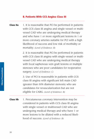
B. Patients With CCS Angina Class III
Class IIa 1. It is reasonable that PCI be performed in patients
with CCS class III angina and single-vessel or multi-
vessel CAD who are undergoing medical therapy
and who have 1 or more significant lesions in 1 or
more coronary arteries suitable for PCI with a high
likelihood of success and low risk of morbidity or
mortality. (Level of Evidence: B)
2. It is reasonable that PCI be performed in patients
with CCS class III angina with single-vessel or multi-
vessel CAD who are undergoing medical therapy
with focal saphenous vein graft lesions or multiple
stenoses who are poor candidates for reoperative
surgery. (Level of Evidence: C)
3. Use of PCI is reasonable in patients with CCS
class III angina with significant left main CAD
(greater than 50% diameter stenosis) who are
candidates for revascularization but are not
eligible for CABG. (Level of Evidence: B)
Class IIb 1. Percutaneous coronary intervention may be
considered in patients with CCS class III angina
with single-vessel or multivessel CAD who are
undergoing medical therapy and who have 1 or
more lesions to be dilated with a reduced likeli-
hood of success. (Level of Evidence: B)
Clin
ical
Pre
sent
atio
ns
31
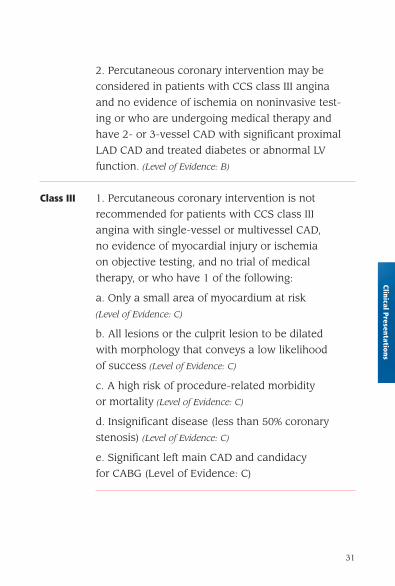
2. Percutaneous coronary intervention may be
considered in patients with CCS class III angina
and no evidence of ischemia on noninvasive test-
ing or who are undergoing medical therapy and
have 2- or 3-vessel CAD with significant proximal
LAD CAD and treated diabetes or abnormal LV
function. (Level of Evidence: B)
Class III 1. Percutaneous coronary intervention is not
recommended for patients with CCS class III
angina with single-vessel or multivessel CAD,
no evidence of myocardial injury or ischemia
on objective testing, and no trial of medical
therapy, or who have 1 of the following:
a. Only a small area of myocardium at risk
(Level of Evidence: C)
b. All lesions or the culprit lesion to be dilated
with morphology that conveys a low likelihood
of success (Level of Evidence: C)
c. A high risk of procedure-related morbidity
or mortality (Level of Evidence: C)
d. Insignificant disease (less than 50% coronary
stenosis) (Level of Evidence: C)
e. Significant left main CAD and candidacy
for CABG (Level of Evidence: C)
Clinical Presentations
32
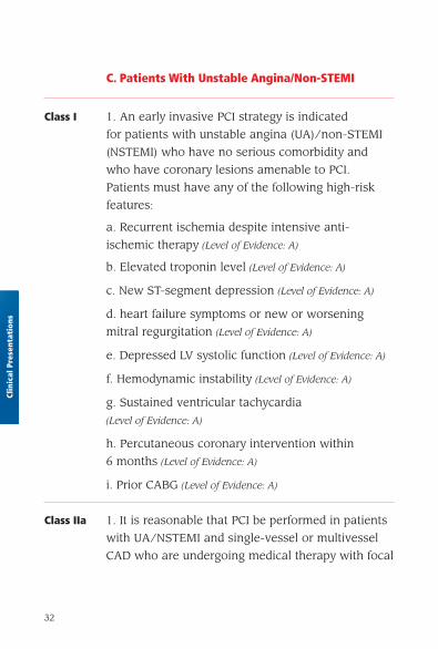
C. Patients With Unstable Angina/Non-STEMI
Class I 1. An early invasive PCI strategy is indicated
for patients with unstable angina (UA)/non-STEMI
(NSTEMI) who have no serious comorbidity and
who have coronary lesions amenable to PCI.
Patients must have any of the following high-risk
features:
a. Recurrent ischemia despite intensive anti-
ischemic therapy (Level of Evidence: A)
b. Elevated troponin level (Level of Evidence: A)
c. New ST-segment depression (Level of Evidence: A)
d. heart failure symptoms or new or worsening
mitral regurgitation (Level of Evidence: A)
e. Depressed LV systolic function (Level of Evidence: A)
f. Hemodynamic instability (Level of Evidence: A)
g. Sustained ventricular tachycardia
(Level of Evidence: A)
h. Percutaneous coronary intervention within
6 months (Level of Evidence: A)
i. Prior CABG (Level of Evidence: A)
Class IIa 1. It is reasonable that PCI be performed in patients
with UA/NSTEMI and single-vessel or multivessel
CAD who are undergoing medical therapy with focal
Clin
ical
Pre
sent
atio
ns
33
saphenous vein graft lesions or multiple stenoses
who are poor candidates for reoperative surgery.
(Level of Evidence: C)
2. In the absence of high-risk features associated
with UA/NSTEMI, it is reasonable to perform PCI
in patients with amenable lesions and no con-
traindication for PCI with either an early invasive
or early conservative strategy. See full-text
guidelines. (Level of Evidence: B)
3. Use of PCI is reasonable in patients with UA/
NSTEMI with significant left main CAD (greater
than 50% diameter stenosis) who are candidates
for revascularization but are not eligible for CABG.
(Level of Evidence: B)
Class IIb 1. In the absence of high-risk features associated
with UA/NSTEMI, PCI may be considered in patients
with single-vessel or multivessel CAD who are
undergoing medical therapy and who have 1 or
more lesions to be dilated with reduced likelihood
of success. (Level of Evidence: B)
2. Percutaneous coronary intervention may be con-
sidered in patients with UA/NSTEMI who are under-
going medical therapy who have 2- or 3-vessel
disease, significant proximal LAD CAD, and treated
diabetes or abnormal LV function. (Level of Evidence: B)
Clinical Presentations
34
Class III 1. In the absence of high-risk features associated
with UA/NSTEMI, PCI is not recommended for
patients with UA/NSTEMI with single-vessel or
multivessel CAD and no trial of medical therapy,
or who have 1 or more of the following:
a. Only a small area of myocardium at risk
(Level of Evidence: C)
b. All lesions or the culprit lesion to be dilated
with morphology that conveys a low likelihood
of success (Level of Evidence: C)
c. A high risk of procedure-related morbidity or
mortality. (Level of Evidence: C)
d. Insignificant disease (less than 50% coronary
stenosis) (Level of Evidence: C)
e. Significant left main CAD and candidacy for
CABG (Level of Evidence: B)
D. Patients With STEMI
1. General and Specific Considerations
Class I General Considerations:
1. If immediately available, primary PCI should be
performed in patients with STEMI (including true
posterior MI) or MI with new or presumably new
Clin
ical
Pre
sent
atio
ns

35
LBBB who can undergo PCI of the infarct artery
within 12 hours of symptom onset, if performed in
a timely fashion (balloon inflation goal within
90 minutes of presentation) by persons skilled in
the procedure (individuals who perform more than
75 PCI procedures per year, ideally at least 11 PCIs
per year for STEMI). The procedure should be sup-
ported by experienced personnel in an appropriate
laboratory environment (one that performs more
than 200 PCI procedures per year, of which at least
36 are primary PCI for STEMI, and that has cardiac
surgery capability). (Level of Evidence: A) Primary PCI
should be performed as quickly as possible, with a
goal of a medical contact–to-balloon or door-to-
balloon time within 90 minutes. (Level of Evidence: B)
Specific Considerations:
2. Primary PCI should be performed for patients less
than 75 years old with ST elevation or presumably
new LBBB who develop shock within 36 hours of
MI and are suitable for revascularization that can
be performed within 18 hours of shock, unless fur-
ther support is futile because of patient’s wishes or
contraindications/unsuitability for further invasive
care. (Level of Evidence: A)
Clinical Presentations
36
3. Primary PCI should be performed in patients with
severe heart failure and/or pulmonary edema (Killip
class 3) and onset of symptoms within 12 hours. The
medical contact–to-balloon or door-to-balloon time
should be as short as possible (i.e., goal within 90
minutes). (Level of Evidence: B)
Class IIa 1. Primary PCI is reasonable for selected patients
75 years or older with ST elevation or LBBB or who
develop shock within 36 hours of MI and are suitable
for revascularization that can be performed within
18 hours of shock. Patients with good prior functional
status who are suitable for revascularization and
agree to invasive care may be selected for such an
invasive strategy. (Level of Evidence: B)
2. It is reasonable to perform primary PCI for
patients with onset of symptoms within the prior
12 to 24 hours and 1 or more of the following:
a. Severe heart failure (Level of Evidence: C)
b. Hemodynamic or electrical instability
(Level of Evidence: C)
c. Evidence of persistent ischemia (Level of Evidence: C)
Clin
ical
Pre
sent
atio
ns
37
Class IIb 1. The benefit of primary PCI for STEMI patients
eligible for fibrinolysis when performed by an opera-
tor who performs fewer than 75 PCI procedures per
year (or fewer than 11 PCIs for STEMI per year)
is not well established. (Level of Evidence: C)
Class III 1. Elective PCI should not be performed in a non-
infarct-related artery at the time of primary PCI
of the infarct-related artery in patients without
hemodynamic compromise. (Level of Evidence: C)
2. Primary PCI should not be performed in asympto-
matic patients more than 12 hours after onset of
STEMI who are hemodynamically and electrically
stable. (Level of Evidence: C)
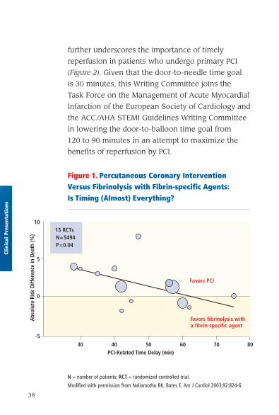
Time from symptom onset to reperfusion is an
important predictor of patient outcome. An analysis
of the randomized, controlled trials that compared
fibrinolysis with primary PCI suggests that the mor-
tality benefit with PCI exists when treatment is
delayed by no more than 60 minutes (Figure 1).
Mortality increases significantly with each 15-minute
delay in the time between arrival and restoration of
TIMI-3 flow (door-to-TIMI-3 flow time), which
Clinical Presentations
38
further underscores the importance of timely
reperfusion in patients who undergo primary PCI
(Figure 2). Given that the door-to-needle time goal
is 30 minutes, this Writing Committee joins the
Task Force on the Management of Acute Myocardial
Infarction of the European Society of Cardiology and
the ACC/AHA STEMI Guidelines Writing Committee
in lowering the door-to-balloon time goal from
120 to 90 minutes in an attempt to maximize the
benefits of reperfusion by PCI.
Clin
ical
Pre
sent
atio
ns
Figure 1. Percutaneous Coronary Intervention Versus Fibrinolysis with Fibrin-specific Agents:Is Timing (Almost) Everything?
N = number of patients; RCT = randomized controlled trial.
Modified with permission from Nallamothu BK, Bates E. Am J Cardiol 2003;92:824-6.
Abs
olut
e Ri
sk D
iffer
ence
in D
eath
(%)
30-5
0
5
10
40 50 60 70 80PCI-Related Time Delay (min)
13 RCTsN=5494P=0.04
Favors fibrinolysis witha fibrin-specific agent
Favors PCI
39
Clinical Presentations
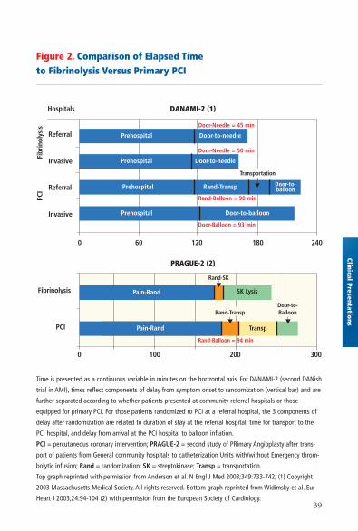
Figure 2. Comparison of Elapsed Time to Fibrinolysis Versus Primary PCI
Time is presented as a continuous variable in minutes on the horizontal axis. For DANAMI-2 (second DANish
trial in AMI), times reflect components of delay from symptom onset to randomization (vertical bar) and are
further separated according to whether patients presented at community referral hospitals or those
equipped for primary PCI. For those patients randomized to PCI at a referral hospital, the 3 components of
delay after randomization are related to duration of stay at the referral hospital, time for transport to the
PCI hospital, and delay from arrival at the PCI hospital to balloon inflation.
PCI = percutaneous coronary intervention; PRAGUE-2 = second study of PRimary Angioplasty after trans-
port of patients from General community hospitals to catheterization Units with/without Emergency throm-
bolytic infusion; Rand = randomization; SK = streptokinase; Transp = transportation.
Top graph reprinted with permission from Anderson et al. N Engl J Med 2003;349:733-742; (1) Copyright
2003 Massachusetts Medical Society. All rights reserved. Bottom graph reprinted from Widimsky et al. Eur
Heart J 2003;24:94-104 (2) with permission from the European Society of Cardiology.
DANAMI-2 (1)
PRAGUE-2 (2)
Hospitals
Fibrinolysis
PCI
Prehospital
Prehospital Door-to-balloon
Rand-Transp Door-to-balloon
Door-to-needle
Door-Needle = 45 min
Door-Needle = 50 min
Rand-Balloon = 90 min
Door-Balloon = 93 min
Rand-Balloon = 94 min
Transportation
Rand-SK
Rand-TranspDoor-to-Balloon
0 60 120
2001000 300
180 240
▼
Pain-Rand
Pain-Rand Transp▼
Fibr
inol
ysis
Referral
Invasive
Referral
Invasive
PCI
Prehospital
Prehospital
Pain-Rand SK Lysis
▼
▼
Pain-Rand
Door-to-needle
40
Primary stenting using BMS compared with primary PTCA in
9 studies showed no differences in mortality (3.0% versus 2.8%)
or reinfarction (1.8% versus 2.1%) rates. However, subsequent
target vessel revascularization rates were lower with stenting.
Preliminary reports suggest that compared with conventional
BMS, DES are not associated with increased risk when used
for primary PCI in patients with STEMI. PCI appears to have
its greatest mortality benefit in high-risk patients. In patients
with cardiogenic shock, an absolute 9% reduction in 30-day
mortality with mechanical revascularization instead of imme-
diate medical stabilization was reported in the SHOCK (SHould
we emergently revascularize Occluded Coronaries for cardio-
genic shocK?) trial.
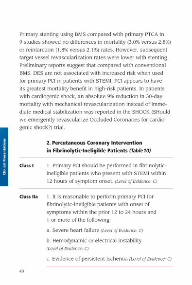
2. Percutaneous Coronary Intervention in Fibrinolytic-Ineligible Patients (Table10)
Class I 1. Primary PCI should be performed in fibrinolytic-
ineligible patients who present with STEMI within
12 hours of symptom onset. (Level of Evidence: C)
Class IIa 1. It is reasonable to perform primary PCI for
fibrinolytic-ineligible patients with onset of
symptoms within the prior 12 to 24 hours and
1 or more of the following:
a. Severe heart failure (Level of Evidence: C)
b. Hemodynamic or electrical instability
(Level of Evidence: C)
c. Evidence of persistent ischemia (Level of Evidence: C)
Clin
ical
Pre
sent
atio
ns
41
Clinical Presentations
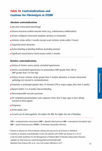
Table 10. Contraindications and Cautions for Fibrinolysis in STEMI*
Absolute contraindications
■ Any prior intracranial hemorrhage
■ Known structural cerebral vascular lesion (e.g., arteriovenous malformation)
■ Known malignant intracranial neoplasm (primary or metastatic)
■ Ischemic stroke within 3 months (except acute ischemic stroke within 3 hours)
■ Suspected aortic dissection
■ Active bleeding or bleeding diathesis (excluding menses)
■ Significant closed head or facial trauma within 3 months
Relative contraindications
■ History of chronic severe, poorly controlled hypertension
■ Severe, uncontrolled hypertension on presentation (SBP greater than 180 or DBP greater than 110 mm Hg)†
■ History of prior ischemic stroke greater than 3 months, dementia, or known intracranial pathology not covered in contraindications
■ Traumatic or prolonged (greater than 10 minutes) CPR or major surgery (less than 3 weeks)
■ Recent (within 2 to 4 weeks) internal bleeding
■ Noncompressible vascular punctures
■ For streptokinase/anistreplase: prior exposure (more than 5 days ago) or prior allergic reaction to these agents
■ Pregnancy
■ Active peptic ulcer
■ Current use of anticoagulants: the higher the INR, the higher the risk of bleeding
CPR = cardiopulmonary resuscitation; DBP = diastolic blood pressure; INR = international normalized ratio;
SBP = systolic blood pressure; STEMI = ST-elevation myocardial infarction.
.
* Viewed as advisory for clinical decision making and may not be all-inclusive or definitive.
† Could be an absolute contraindication in low-risk patients with STEMI (see Section 6.3.1.6.3.2
in the ACC/AHA Guidelines for the Management of Patients With ST-Elevation Myocardial Infarction,
available at: http://www.acc.org/clinical/guidelines/stemi/index.pdf).
Reprinted from Antman EM, et al. J Am Coll Cardiol 2004;44:e1-e211.
42
3. Percutaneous Coronary Intervention After Failed Fibrinolysis (Rescue PCI)
Class I 1. Rescue PCI should be performed in patients less
than 75 years old with ST elevation or LBBB who
develop shock within 36 hours of MI and are suit-
able for revascularization that can be performed
within 18 hours of shock, unless further support is
futile because of patient’s wishes or contraindica-
tions/unsuitability for further invasive care.
(Level of Evidence: B)
2. Rescue PCI should be performed in patients with
severe heart failure and/or pulmonary edema (Killip
class 3) and onset of symptoms within 12 hours.
(Level of Evidence: B)
Class IIa 1. Rescue PCI is reasonable for selected patients
75 years or older with ST elevation or LBBB or who
develop shock within 36 hours of MI and are suit-
able for revascularization that can be performed
within 18 hours of shock. Patients with good prior
functional status who are suitable for revasculariza-
tion and agree to invasive care may be selected for
such an invasive strategy. (Level of Evidence: B)
2. It is reasonable to perform rescue PCI for patients
with 1 or more of the following:
a. Hemodynamic or electrical instability
(Level of Evidence: C)
b. Evidence of persistent ischemia. (Level of Evidence: C)
Clin
ical
Pre
sent
atio
ns

43
Class III 1. Rescue PCI in the absence of 1 or more of the
above class I or IIa indications is not recommended.
(Level of Evidence: C)
4. Percutaneous Coronary Intervention AfterSuccessful Fibrinolysis or for Patients NotUndergoing Primary Reperfusion
Class I 1. In patients whose anatomy is suitable, PCI should
be performed when there is objective evidence of
recurrent MI. (Level of Evidence: C)
2. In patients whose anatomy is suitable, PCI should
be performed for moderate or severe spontaneous
or provocable myocardial ischemia during recovery
from STEMI. (Level of Evidence: B)
3. In patients whose anatomy is suitable, PCI should
be performed for cardiogenic shock or hemodynamic
instability. (Level of Evidence: B)
Class IIa 1. It is reasonable to perform routine PCI in patients
with LV ejection fraction less than or equal to 0.40,
heart failure, or serious ventricular arrhythmias.
(Level of Evidence: C)
2. It is reasonable to perform PCI when there is
documented clinical heart failure during the acute
episode, even though subsequent evaluation shows
preserved LV function (LV ejection fraction greater
than 0.40). (Level of Evidence: C)
Clinical Presentations

44
Class IIb 1. Percutaneous coronary intervention might be
considered as part of an invasive strategy after
fibrinolytic therapy. (Level of Evidence: C)
5. Percutaneous Coronary Intervention for Cardiogenic Shock (Figure 3)
Class I 1. Primary PCI is recommended for patients less
than 75 years old with ST elevation or LBBB who
develop shock within 36 hours of MI and are suit-
able for revascularization that can be performed
within 18 hours of shock, unless further support is
futile because of the patient’s wishes or contraindi-
cations/unsuitability for further invasive care. (Level
of Evidence: A)
Class IIa 1. Primary PCI is reasonable for selected patients 75
years or older with ST elevation or LBBB who devel-
op shock within 36 hours of MI and are suitable for
revascularization that can be performed within 18
hours of shock. Patients with good prior functional
status who are suitable for revascularization and
agree to invasive care may be selected for such an
invasive strategy. (Level of Evidence: B)
Clin
ical
Pre
sent
atio
ns
45
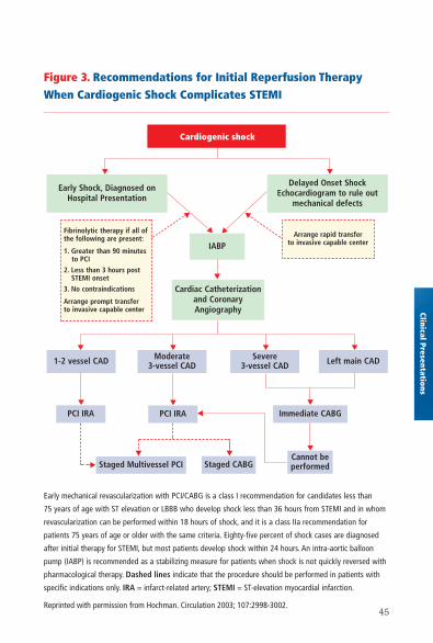
Figure 3. Recommendations for Initial Reperfusion TherapyWhen Cardiogenic Shock Complicates STEMI
Early mechanical revascularization with PCI/CABG is a class I recommendation for candidates less than
75 years of age with ST elevation or LBBB who develop shock less than 36 hours from STEMI and in whom
revascularization can be performed within 18 hours of shock, and it is a class IIa recommendation for
patients 75 years of age or older with the same criteria. Eighty-five percent of shock cases are diagnosed
after initial therapy for STEMI, but most patients develop shock within 24 hours. An intra-aortic balloon
pump (IABP) is recommended as a stabilizing measure for patients when shock is not quickly reversed with
pharmacological therapy. Dashed lines indicate that the procedure should be performed in patients with
specific indications only. IRA = infarct-related artery; STEMI = ST-elevation myocardial infarction.
Reprinted with permission from Hochman. Circulation 2003; 107:2998-3002.
Clinical Presentations
Fibrinolytic therapy if all ofthe following are present:
1. Greater than 90 minutes to PCI
2. Less than 3 hours postSTEMI onset
3. No contraindications
Arrange prompt transfer to invasive capable center
Moderate 3-vessel CAD
▼▼
Cardiogenic shock
Early Shock, Diagnosed onHospital Presentation
Delayed Onset ShockEchocardiogram to rule out
mechanical defects
Arrange rapid transfer to invasive capable center
▼
▼
Cannot be performed
PCI IRA PCI IRA Immediate CABG
Staged CABGStaged Multivessel PCI
1-2 vessel CAD Left main CADSevere 3-vessel CAD
IABP
▼▼ ▼
▼
▼
▼
▼
▼
▼
▼
▼
▼
▼▼
▼
Cardiac Catheterization and CoronaryAngiography
46
E. Percutaneous Intervention in Patients With Prior Coronary Bypass Surgery
Class I 1. When technically feasible, PCI should be per-
formed in patients with early ischemia (usually
within 30 days) after CABG. (Level of Evidence: B)
2. It is recommended that distal embolic protection
devices be used when technically feasible in
patients undergoing PCI to saphenous vein grafts.
(Level of Evidence: B)
Class IIa 1. Percutaneous coronary intervention is reasonable
in patients with ischemia that occurs 1 to 3 years
after CABG and who have preserved LV function
with discrete lesions in graft conduits. (Level of
Evidence: B)
2. Percutaneous coronary intervention is reasonable
in patients with disabling angina secondary to new
disease in a native coronary circulation after CABG.
(If angina is not typical, objective evidence of
ischemia should be obtained.) (Level of Evidence: B)
3. Percutaneous coronary intervention is reasonable
in patients with diseased vein grafts more than 3
years after CABG. (Level of Evidence: B)
4. Percutaneous coronary intervention is reasonable
when technically feasible in patients with a patent
left internal mammary artery graft who have clini-
cally significant obstructions in other vessels. (Level
of Evidence: C)
Clin
ical
Pre
sent
atio
ns
47
Class III 1. Percutaneous coronary intervention is not recom-
mended in patients with prior CABG for chronic
total vein graft occlusions. (Level of Evidence: B)
2. Percutaneous coronary intervention is not recom-
mended in patients who have multiple target lesions
with prior CABG and who have multivessel disease,
failure of multiple saphenous vein grafts, and
impaired LV function unless repeat CABG poses
excessive risk due to severe comorbid conditions.
(Level of Evidence: B)
F. Intravascular Ultrasound Imaging
Class IIa 1. Intravascular ultrasound is reasonable for
the following:
a. Assessment of the adequacy of deployment of
coronary stents, including the extent of stent appo-
sition and determination of the minimum luminal
diameter within the stent. (Level of Evidence: B)
b. Determination of the mechanism of stent
restenosis (inadequate expansion versus neointimal
proliferation) and to enable selection of appropriate
therapy (vascular brachytherapy versus repeat
balloon expansion). (Level of Evidence: B)
c. Evaluation of coronary obstruction at a location
difficult to image by angiography in a patient with a
suspected flow-limiting stenosis. (Level of Evidence: C)
Clinical Presentations
48
d. Assessment of a suboptimal angiographic result
after PCI. (Level of Evidence: C)
e. Establishment of the presence and distribution
of coronary calcium in patients for whom
adjunctive rotational atherectomy is contemplated.
(Level of Evidence: C)
f. Determination of plaque location and circumfer-
ential distribution for guidance of directional
coronary atherectomy. (Level of Evidence: B)
Class IIb 1. Intravascular ultrasound may be considered for
the following:
a. Determination of the extent of atherosclerosis in
patients with characteristic anginal symptoms and
a positive functional study with no focal stenoses
or mild CAD on angiography. (Level of Evidence: C)
b. Preinterventional assessment of lesional
characteristics and vessel dimensions as a means
to select an optimal revascularization device.
(Level of Evidence: C)
c. Diagnosis of coronary disease after cardiac
transplantation. (Level of Evidence: C)
Class III 1. Intravascular ultrasound is not recommended
when the angiographic diagnosis is clear and
no interventional treatment is planned. (Level of
Evidence: C)
Clin
ical
Pre
sent
atio
ns
49
G. Coronary Artery Pressure and Flow:Use of Fractional Flow Reserve and CoronaryVasodilatory Reserve
Class IIa 1. It is reasonable to use intracoronary physiological
measurements (Doppler ultrasound, fractional flow
reserve) in the assessment of the effects of inter-
mediate coronary stenoses (30% to 70% luminal
narrowing) in patients with anginal symptoms.
Coronary pressure or Doppler velocimetry may also
be useful as an alternative to performing noninva-
sive functional testing (e.g., when the functional
study is absent or ambiguous) to determine whether
an intervention is warranted. (Level of Evidence: B)
Class IIb 1. Intracoronary physiological measurements may
be considered for the evaluation of the success of
PCI in restoring flow reserve and to predict the risk
of restenosis. (Level of Evidence: C)
2. Intracoronary physiological measurements
may be considered for the evaluation of patients
with anginal symptoms without an apparent angio-
graphic culprit lesion. (Level of Evidence: C)
Class III 1. Routine assessment with intracoronary physio-
logical measurements such as Doppler ultrasound
or fractional flow reserve to assess the severity
of angiographic disease in patients with a positive,
unequivocal noninvasive functional study is not
recommended. (Level of Evidence: C)
Clinical Presentations
50
V. Management of Patients Undergoing PCI
A. Oral Antiplatelet Therapy
Class I 1. Patients already taking daily chronic aspirin ther-
apy should take 75 to 325 mg of aspirin before the
PCI procedure is performed. (Level of Evidence: A)
2. Patients not already taking daily chronic aspirin
therapy should be given 300 to 325 mg of aspirin at
least 2 hours and preferably 24 hours before the PCI
procedure is performed. (Level of Evidence: C)
3. After the PCI procedure, in patients with neither
aspirin resistance, allergy, nor increased risk of
bleeding, aspirin 325 mg daily should be given for
at least 1 month after BMS implantation, 3 months
after sirolimus-eluting stent implantation, and 6
months after paclitaxel-eluting stent implantation,
after which daily chronic aspirin use should be
continued indefinitely at a dose of 75 to 162 mg.
(Level of Evidence: B)
4. A loading dose of clopidogrel should be adminis-
tered before PCI is performed. (Level of Evidence: A) An
oral loading dose of 300 mg, administered at least 6
hours before the procedure, has the best established
evidence of efficacy. (Level of Evidence: B)
Pati
ent
Man
agem
ent
51
5. In patients who have undergone PCI, clopidogrel
75 mg daily should be given for at least 1 month
after BMS implantation (unless the patient is at
increased risk for bleeding; then it should be given
for a minimum of 2 weeks), 3 months after
sirolimus stent implantation, and 6 months after
paclitaxel stent implantation, and ideally up to 12
months in patients who are not at high risk of
bleeding. (Level of Evidence: B)
Class IIa 1. If clopidogrel is given at the time of procedure,
supplementation with glycoprotein (GP) IIb/IIIa
receptor antagonists can be beneficial to facilitate
earlier platelet inhibition than with clopidogrel
alone. (Level of Evidence: B)
2. For patients with an absolute contraindication to
aspirin, it is reasonable to give a 300-mg loading
dose of clopidogrel, administered at least 6 hours,
before PCI and/or GP IIb/IIIa antagonists, adminis-
tered at the time of PCI. (Level of Evidence: C)
3. When a loading dose of clopidogrel is adminis-
tered, a regimen of greater than 300 mg is reason-
able to achieve higher levels of antiplatelet activity
more rapidly, but the efficacy and safety compared
with a 300-mg loading dose are less established.
(Level of Evidence: C)
Patient Managem
ent
52
4. It is reasonable that patients undergoing
brachytherapy be given daily clopidogrel 75 mg
indefinitely and daily aspirin 75 to 325 mg indefi-
nitely unless there is significant risk for bleeding.
(Level of Evidence: C)
Class IIb 1. In patients in whom subacute thrombosis
may be catastrophic or lethal (unprotected left
main, bifurcating left main, or last patent coronary
vessel), platelet aggregation studies may be con-
sidered and the dose of clopidogrel increased
to 150 mg per day if less than 50% inhibition of
platelet aggregation is demonstrated. (Level of
Evidence: C)
Aspirin reduces the frequency of ischemic compli-
cations after PCI. A strategy of pretreatment with
clopidogrel in patients who have not already had
their coronary anatomy defined is controversial,
because patients who undergo CABG within 5 to 7
days of clopidogrel treatment have an increased
risk of bleeding.
Spec
ial C
onsi
dera
tion
sPa
tien
t M
anag
emen
t
53
B. Glycoprotein IIb/IIIa Inhibitors
Class I 1. In patients with UA/NSTEMI undergoing PCI
without clopidogrel administration, a GP IIb/IIIa
inhibitor (abciximab, eptifibatide, or tirofiban)
should be administered. (Level of Evidence: A)*
Class IIa 1. In patients with UA/NSTEMI undergoing PCI
with clopidogrel administration, it is reasonable
to administer a GP IIb/IIIa inhibitor (abciximab,
eptifibatide, or tirofiban). (Level of Evidence: B)*
2. In patients with STEMI undergoing PCI, it is
reasonable to administer abciximab as early as
possible. (Level of Evidence: B)
3. In patients undergoing elective PCI with stent
placement, it is reasonable to administer a GP
IIb/IIIa inhibitor (abciximab, eptifibatide, or
tirofiban). (Level of Evidence: B)
Class IIb 1. In patients with STEMI undergoing PCI, treatment
with eptifibatide or tirofiban may be considered.
(Level of Evidence: C)
*It is acceptable to administer the GP IIb/IIIa inhibitor before performance of the
diagnostic angiogram (“upstream treatment”) or just before PCI (“in-lab treatment”).
Patient Managem
ent
54
C. Antithrombotic Therapy
1. Unfractionated Heparin,Low-Molecular-Weight Heparin, and Bivalirudin
Class I 1. Unfractionated heparin should be administered
to patients undergoing PCI. (Level of Evidence: C)
2. For patients with heparin-induced thrombocy-
topenia, it is recommended that bivalirudin
or argatroban be used to replace heparin.
(Level of Evidence: B)
Class IIa 1. It is reasonable to use bivalirudin as an alterna-
tive to unfractionated heparin and GP IIb/IIIa
antagonists in low-risk patients undergoing elective
PCI. (Level of Evidence: B)
2. Low-molecular-weight heparin is a reasonable
alternative to unfractionated heparin in patients
with UA/NSTEMI undergoing PCI. (Level of Evidence: B)
Class IIb 1. Low-molecular-weight heparin may be
considered as an alternative to unfractionated
heparin in patients with STEMI undergoing PCI.
(Level of Evidence: B)
D. Risk Factor Modifications
All patients should be instructed about necessary behavior
and risk factor modification, and the appropriate medical
therapies should be initiated for the secondary prevention
Pati
ent
Man
agem
ent
55
of atherosclerosis before the patient leaves the hospital.
The interventional cardiologist should emphasize the impor-
tance of these measures directly to the patient, because
failure to do so may suggest that secondary prevention
therapies are not necessary. The interventional cardiologist
should interact with the primary care physician to ensure
that the necessary secondary prevention therapies initiated
during hospitalization are maintained by patients after dis-
charge from the hospital. Secondary prevention measures
are an essential part of long-term therapy because they
can reduce future morbidity and mortality associated with
the atherosclerotic process.
E. Left Main CAD
Class IIa 1. It is reasonable that patients undergoing PCI
to unprotected left main coronary obstructions
be followed up with coronary angiography between
2 and 6 months after PCI. (Level of Evidence: C)
Percutaneous coronary intervention is not recommended
in patients with left main disease who are eligible for CABG.
(See Section IV. Clinical Presentations.) In those patients
with significant left main CAD (greater than 50% diameter
stenosis) who are not eligible for CABG in whom PCI is
performed, careful postprocedure surveillance, including
coronary angiography, is needed to prevent fatal MI or
sudden death that may be associated with in-stent restenosis
with a large area of myocardium in jeopardy.
Patient Managem
ent
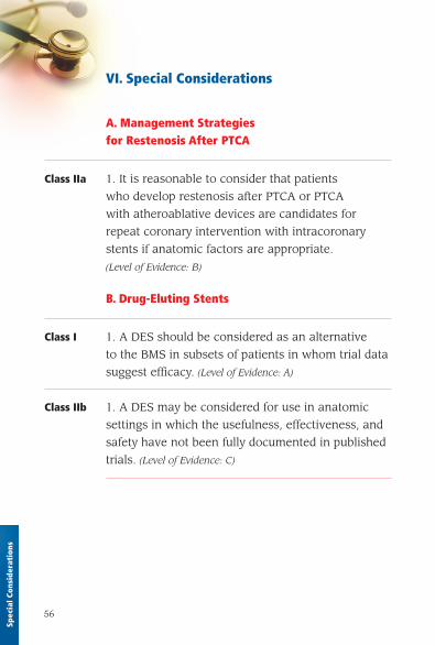
56
VI. Special Considerations
A. Management Strategies for Restenosis After PTCA
Class IIa 1. It is reasonable to consider that patients
who develop restenosis after PTCA or PTCA
with atheroablative devices are candidates for
repeat coronary intervention with intracoronary
stents if anatomic factors are appropriate.
(Level of Evidence: B)
B. Drug-Eluting Stents
Class I 1. A DES should be considered as an alternative
to the BMS in subsets of patients in whom trial data
suggest efficacy. (Level of Evidence: A)
Class IIb 1. A DES may be considered for use in anatomic
settings in which the usefulness, effectiveness, and
safety have not been fully documented in published
trials. (Level of Evidence: C)
Spec
ial C
onsi
dera
tion
s
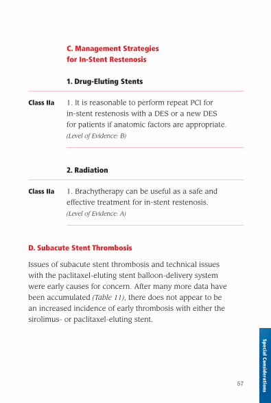
57
C. Management Strategies for In-Stent Restenosis
1. Drug-Eluting Stents
Class IIa 1. It is reasonable to perform repeat PCI for
in-stent restenosis with a DES or a new DES
for patients if anatomic factors are appropriate.
(Level of Evidence: B)
2. Radiation
Class IIa 1. Brachytherapy can be useful as a safe and
effective treatment for in-stent restenosis.
(Level of Evidence: A)
D. Subacute Stent Thrombosis
Issues of subacute stent thrombosis and technical issues
with the paclitaxel-eluting stent balloon-delivery system
were early causes for concern. After many more data have
been accumulated (Table 11), there does not appear to be
an increased incidence of early thrombosis with either the
sirolimus- or paclitaxel-eluting stent.
Special Considerations
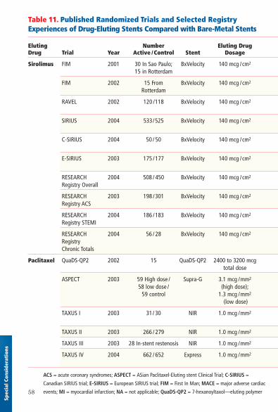
58
Spec
ial C
onsi
dera
tion
s
Table 11. Published Randomized Trials and Selected Registry Experiences of Drug-Eluting Stents Compared with Bare-Metal Stents
Eluting Number Eluting Drug Drug Trial Year Active /Control Stent Dosage
Sirolimus FIM 2001 30 In Sao Paulo; BxVelocity 140 mcg /cm2
15 in Rotterdam
FIM 2002 15 From BxVelocity 140 mcg /cm2
Rotterdam
RAVEL 2002 120 /118 BxVelocity 140 mcg /cm2
SIRIUS 2004 533/525 BxVelocity 140 mcg /cm2
C-SIRIUS 2004 50 / 50 BxVelocity 140 mcg /cm2
E-SIRIUS 2003 175 /177 BxVelocity 140 mcg /cm2
RESEARCH 2004 508 / 450 BxVelocity 140 mcg /cm2
Registry Overall
RESEARCH 2003 198 /301 BxVelocity 140 mcg /cm2
Registry ACS
RESEARCH 2004 186 /183 BxVelocity 140 mcg /cm2
Registry STEMI
RESEARCH 2004 56 / 28 BxVelocity 140 mcg /cm2
RegistryChronic Totals
Paclitaxel QuaDS-QP2 2002 15 QuaDS-QP2 2400 to 3200 mcg total dose
ASPECT 2003 59 High dose / Supra-G 3.1 mcg /mm2
58 low dose / (high dose);59 control 1.3 mcg /mm2
(low dose)
TAXUS I 2003 31/ 30 NIR 1.0 mcg /mm2
TAXUS II 2003 266 / 279 NIR 1.0 mcg /mm2
TAXUS III 2003 28 In-stent restenosis NIR 1.0 mcg /mm2
TAXUS IV 2004 662 / 652 Express 1.0 mcg /mm2
ACS = acute coronary syndromes; ASPECT = ASian Paclitaxel-Eluting stent Clinical Trial; C-SIRIUS =
Canadian SIRIUS trial; E-SIRIUS = European SIRIUS trial; FIM = First In Man; MACE = major adverse cardiac
events; MI = myocardial infarction; NA = not applicable; QuaDS-QP2 = 7-hexanoyltaxol—eluting polymer
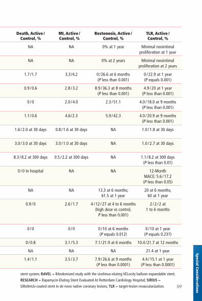
59
Special Considerations
Death, Active / MI, Active / Restenosis, Active / TLR, Active /Control, % Control, % Control, % Control, %
NA NA 0% at 1 year Minimal neointimal proliferation at 1 year
NA NA 0% at 2 years Minimal neointimal proliferation at 2 years
1.7 /1.7 3.3/4.2 0 /26.6 at 6 months 0 / 22.9 at 1 year (P less than 0.001) (P equals 0.001)
0.9 / 0.6 2.8 / 3.2 8.9 / 36.3 at 8 months 4.9 / 20 at 1 year (P less than 0.001) (P less than 0.001)
0 / 0 2.0 / 4.0 2.3 / 51.1 4.0 / 18.0 at 9 months (P less than 0.001)
1.1/ 0.6 4.6/2.3 5.9 / 42.3 4.0 / 20.9 at 9 months (P less than 0.001)
1.6 / 2.0 at 30 days 0.8 / 1.6 at 30 days NA 1.0 / 1.8 at 30 days
3.0 / 3.0 at 30 days 3.0 / 1.0 at 30 days NA 1.0 / 2.7 at 30 days
8.3 / 8.2 at 300 days 0.5 / 2.2 at 300 days NA 1.1/ 8.2 at 300 days (P less than 0.01)
0 / 0 In hospital NA NA 12-MonthMACE: 5.6 / 17.2(P less than 0.05)
NA NA 13.3 at 6 months; 20 at 6 months;61.5 at 1 year 60 at 1 year
0.9 / 0 2.6 / 1.7 4 / 12 / 27 at 4 to 6 months 2 / 2 / 2 at (high dose vs control, 1 to 6 months
P less than 0.001)
0 / 0 0 / 0 0 /10 at 6 months 0 /10 at 1 year (P equals 0.012) (P equals 0.237)
0 / 0.8 3.1 / 5.3 7.1/ 21.9 at 6 months 10.4 /21.7 at 12 months
NA NA NA 21.4 at 1 year
1.4 / 1.1 3.5 / 3.7 7.9 / 26.6 at 9 months 4.4 / 15.1 at 1 year (P less than 0.0001) (P less than 0.0001)
stent system; RAVEL = RAndomized study with the sirolimus-eluting VELocity balloon expandable stent;
RESEARCH = Rapamycin-Eluting Stent Evaluated At Rotterdam Cardiology Hospital; SIRIUS =
SIRolImUs-coated stent in de novo native coronary lesions; TLR = target-lesion revascularization.