Embed Size (px)
Citation preview
University of Groningen
Outcome after percutaneous coronary interventionFokkema, Marieke
IMPORTANT NOTE: You are advised to consult the publisher's version (publisher's PDF) if you wish to cite fromit. Please check the document version below.
Document VersionPublisher's PDF, also known as Version of record
Publication date:2013
Link to publication in University of Groningen/UMCG research database
Citation for published version (APA):Fokkema, M. L. (2013). Outcome after percutaneous coronary intervention Groningen: s.n.
CopyrightOther than for strictly personal use, it is not permitted to download or to forward/distribute the text or part of it without the consent of theauthor(s) and/or copyright holder(s), unless the work is under an open content license (like Creative Commons).
Take-down policyIf you believe that this document breaches copyright please contact us providing details, and we will remove access to the work immediatelyand investigate your claim.
Downloaded from the University of Groningen/UMCG research database (Pure): http://www.rug.nl/research/portal. For technical reasons thenumber of authors shown on this cover page is limited to 10 maximum.
Download date: 26-03-2018
4
The golden hours of primary PCI
Quantitative analysis of the impact of total ischemic time on
myocardial perfusion and clinical outcome in patients with
ST- elevation myocardial infarction
Marieke L. FokkemaWouter G. Wieringa
Iwan C. van der HorstEric BoersmaFelix Zijlstra
Bart J. de Smet
Am J Cardiol. 2011;108:1536-1541
CHAPTER 4
ABSTRACT Early reperfusion of the infarct related coronary artery is an important issue in improvement of outcome after STEMI. We evaluated the clinical significance of total ischemic time on myocardial reperfusion and clinical outcome in patients with ST-elevation myocardial infarction (STEMI) treated with primary percutaneous coronary intervention (PCI) and thrombus aspiration and additional triple anti-platelet therapy. Total ischemic time was defined as time from symptom onset to first intracoronary therapy (first balloon inflation or thrombus aspiration). We selected all STEMI patients treated with primary PCI with total ischemic time of at least 30 minutes and up to 24 hours in our center between 2005 and 2008. Ischemic time was available in 1383 patients, of which 18.4% presented with total ischemic time ≤2h, 31.2% >2-3h, 26.8% >3-5h and 23.5% >5h. Increased ischemic time was associated with age, female gender, hypertension and diabetes. Patients with total ischemic time <5h more often had myocardial blush grade 3 (40%-45% vs. 22%; p<0.001) and complete ST- segment resolution (55%-60% vs. 42%; p = 0.002) than their counterparts with total ischemic time >5h. In addition, patients with total ischemic time ≤5h had a lower 30-day mortality (1.5% vs. 4.0%; p = 0.032) than patients with total ischemic time >5h. In conclusion, in this contemporary cohort of STEMI patients treated with primary PCI, triple anti-platelet therapy and thrombus aspiration, short ischemic time is associated with a better myocardial reperfusion and a decreased mortality. After a 5-hour period in which outcome remains relatively stable, myocardial reperfusion becomes suboptimal and mortality increases.
54
The golden hours of primary PCI
INTRODUCTIONEarly reperfusion of the infarct related coronary artery is an important issue in the improvement of outcome after ST-elevation myocardial infarction (STEMI). Although the myocardium is damaged during ischemia, it is viable in part early after symptom onset and may be salvaged by rapid reperfusion.1,2 The presence of microvascular obstruction increases with longer ischemic times, resulting in an increased infarct size.2,3
In previous studies the best clinical results of reperfusion by primary PCI have been observed in patients treated within 90-120 minutes after symptom onset.2-5 Pretreatment with aspirin, heparin and clopidogrel before hospital admission, and the administration of a glycoprotein IIb/IIIa inhibitor during primary PCI is associated with an improvement of myocardial reperfusion and clinical outcome.6-8 Furthermore, it has been demonstrated that thrombus aspiration results in an additional improvement of myocardial reperfusion.9,10 The application of these innovative pharmacologic and intracoronary treatment strategies could influence the time window to obtain optimal reperfusion and clinical outcome by primary PCI in patients with STEMI. The aim of this study was to evaluate the impact of total ischemic time on myocardial reperfusion and clinical outcome in a large contemporary cohort of STEMI patients treated with primary PCI, with thrombus aspiration and triple anti-platelet therapy.
METHODSWe performed an analysis of ischemic time data from consecutive STEMI patients presenting to the University Medical Center of Groningen from January 2005 to July 2008. Inclusion criteria were symptoms of chest pain suggestive for acute myocardial infarction lasting at least 30 minutes and up to 24 hours before hospital admission, an electrocardiogram (ECG) showing ST-segment elevation of more than 0.1 mV in 2 or more leads, and the performance of a primary PCI procedure. The exclusion criteria were presence of cardiogenic shock, existence of a life threatening disease with a life expectancy of less than six months. Patients treated with acute coronary artery bypass grafting after primary PCI were not enrolled. The University Medical Center of Groningen provides 24-hours emergency cardiac care 7 days a week. It is situated in a region with 750 000 inhabitants and has 7 referral hospitals. When an acute coronary syndrome is suspected, a 12 lead ECG is performed and interpreted by the ambulance physician, aided by a computer algorithm and feed-back after fax transmission from our coronary care unit. After confirmation of STEMI, the STEMI treatment protocol is initiated. This includes that the coronary care unit of our center is contacted and informed about the arriving patient, and direct activation of the cardiac catheterisation team. The patient is directly transported to the catheterisation laboratory, thereby bypassing other regional hospitals. In our region, ambulance transfer times vary until a maximum of 30 minutes. The STEMI protocol has been initiated in January 2004 and has remained unchanged during the period.
All patients were treated with aspirin (500 mg), heparin (5000 IU) and clopidogrel (600 mg) after confirmation of ST-segment elevation on the first ECG, usually performed in the ambulance before hospital admission. During primary PCI patients received the glycoprotein IIb/IIIa inhibitor abciximab (0.25 mg/kg intravenously) if not
55
CHAPTER 4
contraindicated. Additional heparin was administered during procedure guided by the activated clotting time. As the initial step during primary PCI, manual thrombus aspiration was performed in about half of the patients until 2006. After 2006, thrombus aspiration was performed in all patients whenever possible. After restoration of flow through the infarct related lesion, a stent was implanted. Balloon pre- and postdilatation were used when necessary to achieve visualisation of the infarct related lesion before stent placement or optimal stent deployment. After primary PCI, patients received aspirin, clopidogrel (>1 month), β blocker, lipid-lowering agent, and angiotensin converting enzyme inhibitors or angiotensin II receptor blockers.11
Total ischemic time was defined as the time from symptom onset to the first intracoronary therapy (first balloon inflation or thrombus aspiration). Information on the time of symptom onset was systematically collected by asking the patient or his relatives about initiation of continuous chest pain before hospital admission.
Angiographic records before and after primary PCI were evaluated by 2 experienced observers blinded for clinical data. On the initial angiogram and on the final angiogram, Thrombolysis In Myocardial Infarction (TIMI) flow grade, angiographic evidence of thrombus in the infarct-related lesion and distal embolization were assessed.12-14 In addition, Myocardial Blush Grade (MBG) was assessed on the angiogram after stenting.15 The 12 lead ECG’s made at presentation and at 30-60 minutes after primary PCI were evaluated by 2 experienced observers blinded for angiographic and clinical data. ST-segment elevation resolution and the presence of Q waves was assessed.16,17
Aspirated material was collected and analysed for patients from 2005 to 2006. Thrombus aspiration was defined as effective when atherothrombotic material was present in the aspirated samples.
Follow-up data at 30 days after primary PCI were collected from hospital records, written questionnaires and telephone interviews. We report all cause mortality. Reinfarction was defined as the onset of recurrent symptoms of ischemia combined with new ST-segment elevations and/or a second increase of serum CK or CK-MB to at least twice the upper limit of the normal range. Target vessel revascularization (TVR) was defined as PCI or bypass grafting of the infarct-related coronary artery.
The primary end point of our study was optimal myocardial reperfusion, defined by MBG 3 and/or ST-segment resolution >70%. Secondary end points were the presence of new Q waves on the ECG after primary PCI, enzymatic infarct size as assessed by the maximum CK-MB level, and mortality, reinfarction and TVR at 30 days after primary PCI.
Patients were classified in 4 categories according to total ischemic time. Time categories of whole hours were chosen as they approach a distribution in quartiles. Categorical variables are presented as frequency values and proportions, and differences between ischemic time categories were evaluated by the chi square test or Fisher’s exact test. Continuous variables with a normal distribution are presented as mean values ± one standard deviation, whereas variables with a non-normal distribution are presented as median values with interquartile range (IQR). Differences in continuous variables between ischemic time categories were evaluated with one way analysis of variance or
56
The golden hours of primary PCI
the Kruskal-Wallis nonparametric test, as appropriate. The cumulative incidence of clinical endpoints was evaluated by the method of Kaplan-Meier, and differences in cumulative event rates according to ischemic time were evaluated by using the log-rank test. Univariable and multivariable logistic regression analyses were applied to study the relation between ischemic time and the primary endpoint myocardial reperfusion, assessed by MBG 3 and ST-segment resolution >70%. In multivariable analysis, we adjusted for potential confounders associated with the endpoints in univariable analysis. We report crude and adjusted odds ratios (ORs) together with the corresponding 95% confidence interval (CI). For all analyses, p-values of less than 0.05 2-sided were defined as significant. Statistical analysis was performed using the Statistical Package for Social Sciences (SPSS Inc, Chicago, IL) version 16.0.
57
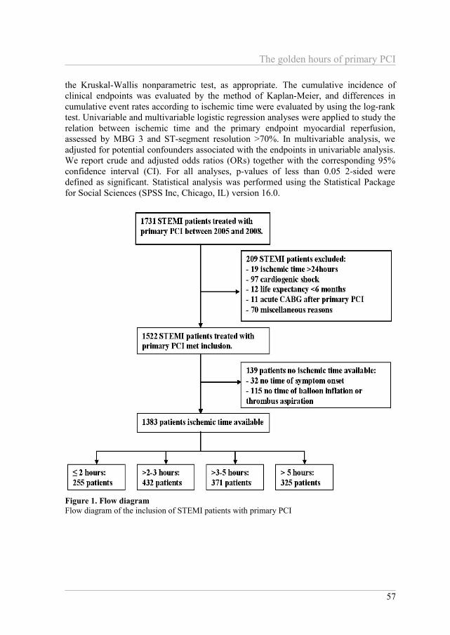
Figure 1. Flow diagramFlow diagram of the inclusion of STEMI patients with primary PCI
CHAPTER 4
Table 1. Baseline characteristics
Myocardial Ischemic Time (hours)
Variable ≤2N = 255
>2-3N = 432
>3-5 N = 371
> 5 N = 325 p
Ischemic time, hours (IQR)
1.83 (1.50-1.92)
2.50 (2.25-2.75)
3.75 (3.33-4.25)
7.25 (5.87-10.58)
<0.001
Age (years) (±SD) 61.6 (12.0) 61.9 (12.2) 62.5 (13.0) 65.2 (12.6) 0.001
Men 193/255 (75.7%) 321/432 (74.3%) 266/371 (71.7%) 215/325 (66.2%) 0.006
Hypertension 84/249 (33.7%) 143/411 (34.8%) 118/364 (32.4%) 139/316 (44.0%) 0.022
Hypercholesterolemia 66/217 (30.4%) 98/357 (27.5%) 88/315 (27.9%) 80/280 (28.6%) 0.763
Diabetes mellitus 18/254 (7.1%) 44/426 (10.3%) 34/368 (9.2%) 50/323 (15.5%) 0.003
Myocardial infarction 18/254 (7.1%) 42/426 (9.9%) 36/368 (9.8%) 31/323 (9.6%) 0.392
Previous PCI 18/254 (7.1%) 28/427 (6.6%) 25/366 (6.8%) 20/322 (6.2%) 0.735
Previous CABG 4/255 (1.6%) 9/427 (2.1%) 15/368 (4.1%) 10/323 (3.1%) 0.115
Current smoker 135/231 (58.4%) 199/397 (50.1%) 175/337 (51.9%) 133/293 (45.4%) 0.011
SD = standard deviation, CABG = coronary artery bypass grafting
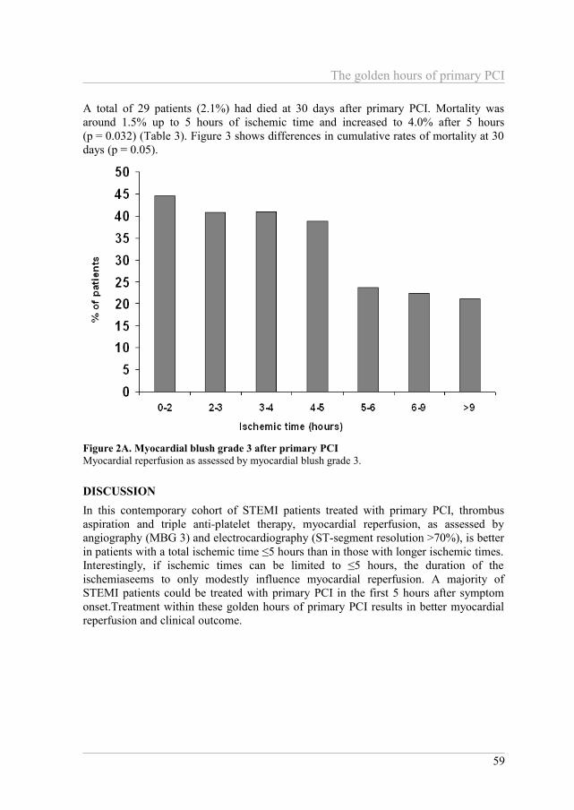
RESULTSBetween January 2005 and July 2008 1731 consecutive STEMI patients were treated with primary PCI in our hospital (Figure 1). Ischemic time was available in 1383 included patients, 79.9% of all 1731 STEMI patients. Of these, 255 patients (18.4%) had an ischemic time ≤2 hours, 432 patients (31.2%) >2-3 hours, 371 patients (26.8%) >3-5 hours and 325 patients (23.5%) >5 hours. The median ischemic time was 3.1 hours (IQR 2.3- 4.8 hours). As shown in table 1, prolonged ischemic time was associated with age, female gender, hypertension, diabetes and smoking status. Angiographic and procedural characteristics are shown in table 2. Ischemic time was associated with multivessel disease, the presence of collaterals, the use of GPIIb/IIIa inhibitor, balloon dilatation and stent implantation. The incidence of TIMI flow 3 decreased from 93.3% to 79.9% after 5 hours (p<0.001).
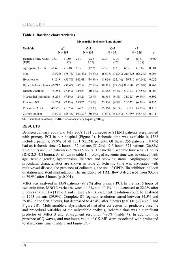
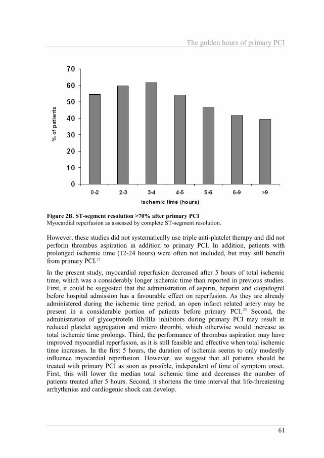
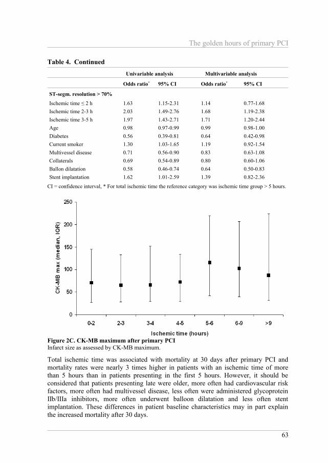
MBG was analysed in 1358 patients (98.2%) after primary PCI. In the first 5 hours of ischemic time, MBG 3 varied between 44.6% and 40.1%, but decreased to 22.3% after 5 hours (p<0.001)) (Table 3 and Figure 2A). ST-segment resolution could be analysed in 1243 patients (89.9%). Complete ST-segment resolution varied between 54.5% and 59.8% in the first 5 hours, but decreased to 42.4% after 5 hours (p<0.001) (Table 3 and Figure 2B). Multivariable analysis showed that after correction for predictive baseline and procedural variables of the univariable analysis, ischemic time was a significant predictor of MBG 3 and ST-segment resolution >70% (Table 4). In addition, the presence of Q waves, and maximum value of CK-MB were associated with prolonged total ischemic time (Table 3 and Figure 2C).
58
The golden hours of primary PCI
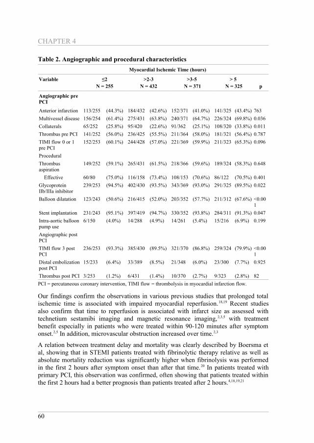
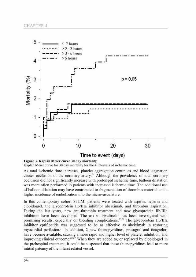
A total of 29 patients (2.1%) had died at 30 days after primary PCI. Mortality was around 1.5% up to 5 hours of ischemic time and increased to 4.0% after 5 hours (p = 0.032) (Table 3). Figure 3 shows differences in cumulative rates of mortality at 30 days (p = 0.05).
Figure 2A. Myocardial blush grade 3 after primary PCIMyocardial reperfusion as assessed by myocardial blush grade 3.
DISCUSSIONIn this contemporary cohort of STEMI patients treated with primary PCI, thrombus aspiration and triple anti-platelet therapy, myocardial reperfusion, as assessed by angiography (MBG 3) and electrocardiography (ST-segment resolution >70%), is better in patients with a total ischemic time ≤5 hours than in those with longer ischemic times. Interestingly, if ischemic times can be limited to ≤5 hours, the duration of the ischemiaseems to only modestly influence myocardial reperfusion. A majority of STEMI patients could be treated with primary PCI in the first 5 hours after symptom onset.Treatment within these golden hours of primary PCI results in better myocardial reperfusion and clinical outcome.
59
CHAPTER 4
Table 2. Angiographic and procedural characteristicsMyocardial Ischemic Time (hours)
Variable ≤2N = 255
>2-3N = 432
>3-5 N = 371
> 5 N = 325 p
Angiographic pre PCI
Anterior infarction 113/255 (44.3%) 184/432 (42.6%) 152/371 (41.0%) 141/325 (43.4%) 763Multivessel disease 156/254 (61.4%) 275/431 (63.8%) 240/371 (64.7%) 226/324 (69.8%) 0.036Collaterals 65/252 (25.8%) 95/420 (22.6%) 91/362 (25.1%) 108/320 (33.8%) 0.011Thrombus pre PCI 141/252 (56.0%) 236/425 (55.5%) 211/364 (58.0%) 181/321 (56.4%) 0.787TIMI flow 0 or 1 pre PCI
152/253 (60.1%) 244/428 (57.0%) 221/369 (59.9%) 211/323 (65.3%) 0.096
ProceduralThrombus aspiration
149/252 (59.1%) 265/431 (61.5%) 218/366 (59.6%) 189/324 (58.3%) 0.648
Effective 60/80 (75.0%) 116/158 (73.4%) 108/153 (70.6%) 86/122 (70.5%) 0.401Glycoprotein IIb/IIIa inhibitor
239/253 (94.5%) 402/430 (93.5%) 343/369 (93.0%) 291/325 (89.5%) 0.022
Balloon dilatation 123/243 (50.6%) 216/415 (52.0%) 203/352 (57.7%) 211/312 (67.6%) <0.001
Stent implantation 231/243 (95.1%) 397/419 (94.7%) 330/352 (93.8%) 284/311 (91.3%) 0.047Intra-aortic balloon pump use
6/150 (4.0%) 14/288 (4.9%) 14/261 (5.4%) 15/216 (6.9%) 0.199
Angiographic post PCITIMI flow 3 post PCI
236/253 (93.3%) 385/430 (89.5%) 321/370 (86.8%) 259/324 (79.9%) <0.001
Distal embolization post PCI
15/233 (6.4%) 33/389 (8.5%) 21/348 (6.0%) 23/300 (7.7%) 0.925
Thrombus post PCI 3/253 (1.2%) 6/431 (1.4%) 10/370 (2.7%) 9/323 (2.8%) 82PCI = percutaneous coronary intervention, TIMI flow = thrombolysis in myocardial infarction flow.
Our findings confirm the observations in various previous studies that prolonged total ischemic time is associated with impaired myocardial reperfusion.18,19 Recent studies also confirm that time to reperfusion is associated with infarct size as assessed with technetium sestamibi imaging and magnetic resonance imaging,2,3,5 with treatment benefit especially in patients who were treated within 90-120 minutes after symptom onset.2,5 In addition, microvascular obstruction increased over time.2,3
A relation between treatment delay and mortality was clearly described by Boersma et al, showing that in STEMI patients treated with fibrinolytic therapy relative as well as absolute mortality reduction was significantly higher when fibrinolysis was performed in the first 2 hours after symptom onset than after that time.20 In patients treated with primary PCI, this observation was confirmed, often showing that patients treated within the first 2 hours had a better prognosis than patients treated after 2 hours.4,18,19,21
60
The golden hours of primary PCI
However, these studies did not systematically use triple anti-platelet therapy and did not perform thrombus aspiration in addition to primary PCI. In addition, patients with prolonged ischemic time (12-24 hours) were often not included, but may still benefit from primary PCI.22
In the present study, myocardial reperfusion decreased after 5 hours of total ischemic time, which was a considerably longer ischemic time than reported in previous studies. First, it could be suggested that the administration of aspirin, heparin and clopidogrel before hospital admission has a favourable effect on reperfusion. As they are already administered during the ischemic time period, an open infarct related artery may be present in a considerable portion of patients before primary PCI.23 Second, the administration of glycoptroteïn IIb/IIIa inhibitors during primary PCI may result in reduced platelet aggregation and micro thrombi, which otherwise would increase as total ischemic time prolongs. Third, the performance of thrombus aspiration may have improved myocardial reperfusion, as it is still feasible and effective when total ischemic time increases. In the first 5 hours, the duration of ischemia seems to only modestly influence myocardial reperfusion. However, we suggest that all patients should be treated with primary PCI as soon as possible, independent of time of symptom onset. First, this will lower the median total ischemic time and decreases the number of patients treated after 5 hours. Second, it shortens the time interval that life-threatening arrhythmias and cardiogenic shock can develop.
61
Figure 2B. ST-segment resolution >70% after primary PCI Myocardial reperfusion as assessed by complete ST-segment resolution.
CHAPTER 4
Table 3. Outcome characteristicsMyocardial Ischemic Time (hours)
Variable ≤2N = 255
>2-3N = 432
>3-5 N = 371
> 5 N = 325 p
Myocardial reperfusion
Myocardial blush grade 3
112/251 (44.6%) 173/425 (40.7%) 146/364 (40.1%) 71/318 (22.3%) <0.001
ST-segm. resolution > 70%
126/231 (54.5%) 234/391 (59.8%) 197/333 (59.2%) 122/288 (42.4%) 0.002
Measures of infarct sizeQ- waves 186/238 (78.2%) 315/403 (78.2%) 268/343 (78.1%) 251/295 (85.1%) 0.047CK-MB maximum (U/L) (IQR)
71.0 (26.9-145.0)
65.5 (28.0-132.8)
68.9 (29.1-147.7)
98.2 (36.0-222.3)
<0.001
Clinical outcome at 30 days
Mortality 4/255 (1.6%) 6/430 (1.4%) 6/371 (1.6%) 13/325 (4.0%) 0.032Reinfarction 4/255 (1.6%) 3/430 (0.7%) 6/371 (1.6%) 5/325 (1.5%) 0.647Target vessel revascularization
11/255 (4.3%) 14/430 (3.3%) 22/371 (5.9%) 19/325 (5.8%) 0.134
CK-MB = myocardial band of creatine kinase, IQR = interquartile range, U/L = units per liter.
Table 4. Univariable and multivariable analysis between total ischemic time and myocardial reperfusion, assessed by myocardial blush grade 3 and ST-segment resolution >70%.
Univariable analysis Multivariable analysis
Odds ratio* 95% CI Odds ratio* 95% CI
Myocardial blush grade 3
Ischemic time ≤ 2 h 2.80 1.95-4.03 2.66 1.76-4.03
Ischemic time 2-3 h 2.39 1.72-3.31 2.33 1.60-3.38
Ischemic time 3-5 h 2.33 1.66-3.26 2.31 1.57-3.39
Age 0.97 0.96-0.98 0.98 0.97-0.99
Current smoker 1.35 1.07-1.70 1.10 0.85-1.43
Multivessel disease 0.79 0.63-0.99 1.10 0.85-1.44
Collaterals 0.66 0.493-0.89 0.80 0.60- 1.08
Glycoprotein IIb/IIIa inhibitor 1.65 1.04-2.60 1.08 0.62- 1.87
Balloon dilatation 0.41 0.33-0.52 0.48 0.37-0.61
62
The golden hours of primary PCI
Table 4. Continued
Univariable analysis Multivariable analysis
Odds ratio* 95% CI Odds ratio* 95% CI
ST-segm. resolution > 70%
Ischemic time ≤ 2 h 1.63 1.15-2.31 1.14 0.77-1.68Ischemic time 2-3 h 2.03 1.49-2.76 1.68 1.19-2.38Ischemic time 3-5 h 1.97 1.43-2.71 1.71 1.20-2.44Age 0.98 0.97-0.99 0.99 0.98-1.00Diabetes 0.56 0.39-0.81 0.64 0.42-0.98Current smoker 1.30 1.03-1.65 1.19 0.92-1.54Multivessel disease 0.71 0.56-0.90 0.83 0.63-1.08Collaterals 0.69 0.54-0.89 0.80 0.60-1.06Ballon dilatation 0.58 0.46-0.74 0.64 0.50-0.83Stent implantation 1.62 1.01-2.59 1.39 0.82-2.36
CI = confidence interval, * For total ischemic time the reference category was ischemic time group > 5 hours.
Total ischemic time was associated with mortality at 30 days after primary PCI and mortality rates were nearly 3 times higher in patients with an ischemic time of more than 5 hours than in patients presenting in the first 5 hours. However, it should be considered that patients presenting late were older, more often had cardiovascular risk factors, more often had multivessel disease, less often were administered glycoprotein IIb/IIIa inhibitors, more often underwent balloon dilatation and less often stent implantation. These differences in patient baseline characteristics may in part explain the increased mortality after 30 days.
63
Figure 2C. CK-MB maximum after primary PCI Infarct size as assessed by CK-MB maximum.
CHAPTER 4
As total ischemic time increases, platelet aggregation continues and blood stagnation causes occlusion of the coronary artery.24 Although the prevalence of total coronary occlusion did not significantly increase with prolonged ischemic time, balloon dilatation was more often performed in patients with increased ischemic time. The additional use of balloon dilatation may have contributed to fragmentation of thrombus material and a higher incidence of embolization into the microvasculature.
In this contemporary cohort STEMI patients were treated with aspirin, heparin and clopidogrel, the glycoprotein IIb/IIIa inhibitor abciximab, and thrombus aspiration. During the last years, new anti-thrombin treatment and new glycoprotein IIb/IIIa inhibitors have been developed. The use of bivalirudin has been investigated with promising results, especially on bleeding complications.25,26 The glycoprotein IIb/IIIa inhibitor eptifibatide was suggested to be as effective as abciximab in restoring myocardial perfusion.27 In addition, 2 new thienopyridines, prasugrel and ticagrelor, have become available, causing a more rapid and higher level of platelet inhibition, and improving clinical outcome.28,29 When they are added to, or replaced by clopidogrel in the prehospital treatment, it could be suspected that these thienopyridines lead to more intitial patency of the infarct related vessel.
64
Figure 3. Kaplan Meier curve 30 day mortalityKaplan Meier curve for 30 day mortality for the 4 intervals of ischemic time.
The golden hours of primary PCI
Several limitations should be taken into consideration. This is a single center study, and therefore data can not automatically be extrapolated to other PCI centers, although with an inclusion of 80% of all STEMI patients, this patient cohort does reflect a real world clinical practice. However, it should be mentioned that patients presenting with cardiogenic shock were excluded in our analysis, which may have influenced our results. In patients with cardiogenic shock it was often not possible to assess the onset of ischemic time. Ischemic time data were not available for all patients because of a missing times of symptom onset, thrombus aspiration or balloon inflation. The exclusion of these patients may have influenced our results, although the baseline characteristics of the included and excluded patients were similar. Furthermore, thrombus aspiration was performed in only 60% of patients. However, the incidence of thrombus aspiration did not differ between the ischemic time groups. In addition, ST-segment resolution could only be analysed in 90% of patients, because the ECG at presentation or the ECG after primary PCI was not available, or because of the occurrence of an intraventricular conduction delay. Furthermore, maximum CK-MB levels were only measured during the stay in our hospital. As some patients were transferred to a regional hospital 1 day after primary PCI, CK-MB maximum may be measured too low.
REFERENCES1. Reimer KA, Lowe JE, Rasmussen MM, Jennings RB. The wavefront phenomenon of
ischemic cell death. 1. Myocardial infarct size vs duration of coronary occlusion in dogs. Circulation 1977;56:786-794.
2. Francone M, Bucciarelli-Ducci C, Carbone I, Canali E, Scardala R, Calabrese FA, Sardella G, Mancone M, Catalano C, Fedele F, Passariello R, Bogaert J, Agati L. Impact of primary coronary angioplasty delay on myocardial salvage, infarct size, and microvascular damage in patients with ST-segment elevation myocardial infarction: insight from cardiovascular magnetic resonance. J Am Coll Cardiol 2009;54:2145-2153.
3. Tarantini G, Cacciavillani L, Corbetti F, Ramondo A, Marra MP, Bacchiega E Napodano M, Bilato C, Razzolini R, Iliceto S. Duration of ischemia is a major determinant of transmurality and severe microvascular obstruction after primary angioplasty: a study performed with contrast-enhanced magnetic resonance. J Am Coll Cardiol 2005;46:1229-1235.
4. Brodie BR, Stuckey TD, Wall TC, Kissling G, Hansen CJ, Muncy DB Weintraub RA, Kelly TA. Importance of time to reperfusion for 30-day and late survival and recovery of left ventricular function after primary angioplasty for acute myocardial infarction. J Am Coll Cardiol 1998;32:1312-1319.
5. Brodie BR, Webb J, Cox DA, Qureshi M, Kalynych A, Turco M, Schultheiss HP, Dulas D, Rutherford B, Antoniucci D, Stuckey T, Krucoff M, Gibbons R, Lansky A, Na Y, Mehran R, W Stone GW. Impact of time to treatment on myocardial reperfusion and infarct size with primary percutaneous coronary intervention for acute myocardial infarction (from the EMERALD Trial). Am J Cardiol 2007;99:1680-1686.
6. De Luca G, Suryapranata H, Stone GW, Antoniucci D, Tcheng JE, Neumann FJ, Van de Werf F, Antman EM, Topol EJ.. Abciximab as adjunctive therapy to reperfusion in acute ST-segment elevation myocardial infarction: a meta-analysis of randomized trials. JAMA 2005;293:1759-1765.
7. Montalescot G, Barragan P, Wittenberg O, Ecollan P, Elhadad S, Villain P, Boulenc JM, Morice MC, Maillard L, Pansiéri M, Choussat R, Pinton P. Platelet glycoprotein IIb/IIIa
65
CHAPTER 4
inhibition with coronary stenting for acute myocardial infarction. N Engl J Med 2001;344:1895-1903.
8. Antoniucci D, Rodriguez A, Hempel A, Valenti R, Migliorini A, Vigo F, Parodi G, Fernandez-Pereira C, Moschi G, Bartorelli A, Santoro GM, Bolognese L, Colombo A. A randomized trial comparing primary infarct artery stenting with or without abciximab in acute myocardial infarction. J Am Coll Cardiol 2003;42:1879-1885.
9. Svilaas T, Vlaar PJ, van der Horst IC, Diercks GF, de Smet BJ, van den Heuvel AF, Anthonio RL, Jessurun GA, Tan ES, Suurmeijer AJ, Zijlstra F. Thrombus aspiration during primary percutaneous coronary intervention. N Engl J Med 2008;358:557-567.
10. De Luca G, Dudek D, Sardella G, Marino P, Chevalier B, Zijlstra F. Adjunctive manual thrombectomy improves myocardial perfusion and mortality in patients undergoing primary percutaneous coronary intervention for ST-elevation myocardial infarction: a meta-analysis of randomized trials. Eur Heart J 2008;29:3002-3010.
11. Silber S, Albertsson P, Avilés FF, Camici PG, Colombo A, Hamm C, Jørgensen E, Marco J, Nordrehaug JE, Ruzyllo W, Urban P, Stone GW, Wijns W. Guidelines for percutaneous coronary interventions. The Task Force for Percutaneous Coronary Interventions of the European Society of Cardiology. Eur Heart J 2005;26:804-847.
12. The Thrombolysis in Myocardial Infarction (TIMI) trial. Phase I findings. TIMI Study Group. N Engl J Med 1985;312:932-936.
13. Mabin TA, Holmes DR Jr, Smith HC, Vlietstra RE, Bove AA, Reeder GS, Chesebro JH, Bresnahan JF, Orszulak TA. Intracoronary thrombus: role in coronary occlusion complicating percutaneous transluminal coronary angioplasty. J Am Coll Cardiol 1985;5:198-202.
14. Henriques JP, Zijlstra F, Ottervanger JP, de Boer MJ, van 't Hof AW, Hoorntje JC, Suryapranata H. Incidence and clinical significance of distal embolization during primary angioplasty for acute myocardial infarction. Eur Heart J 2002;23:1112-1117.
15. van 't Hof AW, Liem A, Suryapranata H, Hoorntje JC, de Boer MJ, Zijlstra F. Angiographic assessment of myocardial reperfusion in patients treated with primary angioplasty for acute myocardial infarction: myocardial blush grade. Zwolle Myocardial Infarction Study Group. Circulation 1998;97:2302-2306.
16. van 't Hof AW, Liem A, de Boer MJ, Zijlstra F. Clinical value of 12-lead electrocardiogram after successful reperfusion therapy for acute myocardial infarction. Zwolle Myocardial infarction Study Group. Lancet 1997;350:615-619.
17. Thygesen K, Alpert JS, White HD. Universal definition of myocardial infarction. Eur Heart J 2007;28:2525-2538.
18. De Luca G, Suryapranata H, Zijlstra F, van 't Hof AW, Hoorntje JC, Gosselink AT, Dambrink JH, de Boer MJ. Symptom-onset-to-balloon time and mortality in patients with acute myocardial infarction treated by primary angioplasty. J Am Coll Cardiol 2003;42:991-997.
19. De Luca G, van 't Hof AW, de Boer MJ, Ottervanger JP, Hoorntje JC, Gosselink AT, Dambrink JH, Zijlstra F, Suryapranata H. Time-to-treatment significantly affects the extent of ST-segment resolution and myocardial blush in patients with acute myocardial infarction treated by primary angioplasty. Eur Heart J 2004;25:1009-1013.
20. Boersma E, Maas AC, Deckers JW, Simoons ML. Early thrombolytic treatment in acute myocardial infarction: reappraisal of the golden hour. Lancet 1996;348:771-775.
21. Boersma E. Does time matter? A pooled analysis of randomized clinical trials comparing primary percutaneous coronary intervention and in-hospital fibrinolysis in acute myocardial infarction patients. Eur Heart J 2006;27:779-788.
22. Gierlotka M, Gasior M, Wilczek K, Hawranek M, Szkodzinski J, Paczek P, Lekston A, Kalarus Z, Zembala M, Polonski L. Reperfusion by primary percutaneous coronary intervention in patients with ST-segment elevation myocardial infarction within 12 to 24
66
The golden hours of primary PCI
hours of the onset of symptoms (from a prospective national observational study [PL-ACS]). Am J Cardiol 2011;107:501-508.
23. Vlaar PJ, Svilaas T, Damman K, de Smet BJ, Tijssen JG, Hillege HL, Zijlstra F. Impact of pretreatment with clopidogrel on initial patency and outcome in patients treated with primary percutaneous coronary intervention for ST-segment elevation myocardial infarction: a systematic review. Circulation 2008;118:1828-1836.
24. Falk E, Thuesen L. Pathology of coronary microembolisation and no reflow. Heart 2003;89:983-985.
25. Stone GW, Witzenbichler B, Guagliumi G, Peruga JZ, Brodie BR, Dudek D, Kornowski R, Hartmann F, Gersh BJ, Pocock SJ, Dangas G, Wong SC, Kirtane AJ, Parise H, Mehran R; HORIZONS-AMI Trial Investigators. Bivalirudin during primary PCI in acute myocardial infarction. N Engl J Med 2008;358:2218-2230.
26. Sejersten M, Nielsen SL, Engstrøm T, Jørgensen E, Clemmensen P. Feasibility and safety of prehospital administration of bivalirudin in patients with ST-elevation myocardial infarction. Am J Cardiol 2009;103:1635-1640.
27. Zeymer U, Margenet A, Haude M, Bode C, Lablanche JM, Heuer H, Schröder R, Kropff S, Bourkaib R, Banik N, Zahn R, Teiger E. Randomized comparison of eptifibatide versus abciximab in primary percutaneous coronary intervention in patients with acute ST-segment elevation myocardial infarction: results of the EVA-AMI Trial. J Am Coll Cardiol 2010;56:463-469.
28. Montalescot G, Wiviott SD, Braunwald E, Murphy SA, Gibson CM, McCabe CH, Antman EM; TRITON-TIMI 38 investigators. Prasugrel compared with clopidogrel in patients undergoing percutaneous coronary intervention for ST-elevation myocardial infarction (TRITON-TIMI 38): double-blind, randomised controlled trial. Lancet 2009;373:723-731.
29. Wallentin L, Becker RC, Budaj A, Cannon CP, Emanuelsson H, Held C, Horrow J, Husted S, James S, Katus H, Mahaffey KW, Scirica BM, Skene A, Steg PG, Storey RF, Harrington RA; PLATO Investigators, Freij A, Thorsén M. Ticagrelor versus clopidogrel in patients with acute coronary syndromes. N Engl J Med 2009;361:1045-1057.
67