Embed Size (px)
Citation preview
ORIGINAL RESEARCH—PEYRONIE’S DISEASE
Penile Duplex Ultrasonography in Men with Peyronie’s Disease:Is it Veno-Occlusive Dysfunction or Poor Cavernosal ArterialInflow that Contributes to Erectile Dysfunction?jsm_2501 3446..3451
Eric Chung, MD, FRACS,* Ling De Young, MD, MS,† and Gerald B. Brock, MD, FACS†
*Department of Urology, Princess Alexandra Hospital, Brisbane, QLD, Australia; †Division of Urology, St Joseph HealthCare, London, ON, Canada
DOI: 10.1111/j.1743-6109.2011.02501.x
A B S T R A C T
Introduction. At least 20% of men with Peyronie’s disease (PD) suffer from erectile dysfunction (ED). Thefundamental mechanism is thought to arise from the progression of penile fibrosis, which was initially limited to thePD plaque within the tunica albuginea. However, recent studies have highlighted the possibility of fibrosis ofthe cavernosal vessel media wall leading to impairment of arterial inflow.Aim. To evaluate the penile duplex ultrasonographic findings in PD of impotent men and to determine whetherearly features of PD might predict clinical progression.Main Outcome Measures. Patient demographic, comorbidities, International Index of Erectile Function-5 scores,surgical intervention, and physical findings were documented. Penile curvature, plaque size, and peak systolicvelocity (PSV) and end-diastolic velocity (EDV) on color duplex ultrasonography (CDU) were recorded.Methods. We performed a retrospective review of all men presenting with penile curvature and length loss whounderwent penile CDU between January 2001 and January 2010.Results. A total of 1,120 men underwent penile CDU during the 10-year period. Complete information wasobtained in 810 men; 250 men complained of decreased penile rigidity, while 150 men were unable to sustainerection. Comorbidities were similar between men with PD with and without ED. Tunical thickening (65%) was themost common CDU feature, and mean cumulative calcifications was 24.2 mm2 (1–360 mm2, standard deviation 76).The PSV and EDV on the right cavernosal artery were 14.2 cm/second and 3.5 cm/second, while the left cavernosalartery measurements were 15.1 cm/second and 3.2 cm/second. Multivariate logistic regression model showed strongcorrelation between plaque size and development of ED. Both veno-occlusive dysfunction and impaired cavernosalarterial inflow were associated with ED.Conclusions. Veno-occlusive dysfunction and impaired cavernosal arterial inflow contributed to the development ofED, and larger plaque size is a strong predictor of surgical intervention. Chung E, De Young L, and Brock GB.Penile duplex ultrasonography in men with Peyronie’s disease: Is it veno-occlusive dysfunction or poorcavernosal arterial inflow that contributes to erectile dysfunction? J Sex Med 2011;8:3446–3451.
Key Words. Duplex Ultrasonography; Peyronie’s Disease; Erectile Dysfunction; Corporal Veno-Occlusive Dys-function; Cavernosal Arterial Insufficiency
Introduction
P eyronie’s disease (PD) is a localized inflamma-tory process within the bilaminar tunica
albuginea layers and is associated with penile pain,curvature and/or deformity, and, ultimately, sexualdysfunction [1]. Although PD was first describedmore than 250 years ago, very little is known aboutthe exact molecular and cellular pathophysiologyof this condition [2]. Recent epidemiologicalSource of funding: None.
3446
J Sex Med 2011;8:3446–3451 © 2011 International Society for Sexual Medicine
studies reported that the prevalence of PD wasapproaching 9%; this number is higher in prostatecancer-screened population [3]. While this condi-tion might not be life-threatening, many men whosuffer from PD reported high rates of psychosocialdistress [4]. The natural history of PD remainsuncertain, with spontaneous resolution of the scarand curvature only occurring in a minority of cases.
In contrast, erectile dysfunction (ED), defined asthe persistent inability to attain and maintain anerection sufficient to permit satisfactory sexual per-formance [5], is often the result of alterations in thetone and compliance of corporal smooth musclefunction. Epidemiologic studies such as the Massa-chusetts Male Aging Study reported the prevalenceof ED in men aged 40–70 years to be around 50%[6]. Among the cohorts of men with PD, at least20% of men suffer from ED, which may be causedby PD itself or by a variety of etiologies that includepsychogenic factors. The fundamental mechanismof PD causing ED is thought to arise from theprogression of penile fibrosis into the corporalsmooth muscle, which was initially limited withinthe tunica albuginea. Profibrotic factors such astransforming growth factor (TGF) b-1 and reactiveoxygen species have been implicated in tissue fibro-sis and scarring. The ability of TGF b1 to induce itsown production propagates the fibrotic cycle andresults in a chronic progressive condition thateradicates tissue architecture [7]. Furthermore,these profibrotic factors intensify the expression ofmyofibroblasts, causing failure of myofibroblasts toundergo apoptosis and lead to an inappropriate andpersistent cellular contraction [2,8]. The conse-quence of extension of fibrosis into the corporalsmooth muscle is corporal veno-occlusive dysfunc-tion (CVOD), hence the development of ED in thelater stage of PD.
Over the last few years, it has become evidentthat in addition to corporal smooth muscle fibro-sis, this fibrotic condition also involves the mediaof penile arteries with loss of smooth musclecells, causing a vasculogenic ED [9]. While it isnot uncommon for men with PD to suffer fromED, many men with significant penile curvaturedo not report ED, and yet some men with non-palpable Peyronie’s plaque have ED despite thelack of cardiovascular risk factors. Furthermore,the impact of both conditions together on vascu-lar impairments has not been well addressed. Thepresent study evaluates whether CVOD and/orimpairment in cavernosal arterial inflow is themajor cause of ED in men with PD. To ourknowledge, this is the largest and most compre-
hensive study that examines the associationamong Peyronie’s plaque size, cavernosal bloodflow, and risk of ED.
Patients and Methods
Study Population and End PointsFollowing Institutional Medical Ethics ReviewBoard approval, all patients who underwent high-resolution penile color Doppler ultrasonography(CDU) between January 2001 and January 2010were recruited. A retrospective review of patientclinical charts was undertaken, and men with com-plaints of penile curvature and length loss wereincluded in this study. Men with congenital curva-ture were excluded from this study.
Clinical data such as patient demographics, pastand/or current comorbidities, health-relatedbehaviors, history of penile trauma, and Interna-tional Index of Erectile Function-5 scores werecompiled. Time interval from symptom onset toclinical assessment, previous therapies and thera-pies following diagnosis were also documented.
Physical examination was conducted to charac-terize any palpable plaque and penile CDU withpharmacologically induced erections using5–15 mg prostaglandin E1 was performed to docu-ment the degree of penile curvature, presence ofpenile calcifications, septal fibrosis, tunical thick-ening (tunica thickness > 2 mm), and intracavern-osal fibrosis. These PD characteristics on CDUhave been previously described in the literature[10,11]. After intracavernosal injection of 5 mgprostaglandin E1, patients were allowed 5 minutesfor manual self-stimulation, and if the erection wasdeemed as suboptimal, a further 5–10 mg of pros-taglandin E1 would be given. In all cases, prostag-landin E1 dose was chosen to achieve completesmooth muscle relaxation with self-stimulation,and redosing was used if inadequate penile erec-tion was achieved. Less than 10% of patientsrequired additional doses. Penile CDU was per-formed with gray-scale ultrasonography using a7.5–12 MHz small parts linear array probe. Colorand pulse wave Doppler were used to determinethe cavernosal vascular status such as the peak sys-tolic velocity (PSV) and end-diastolic velocity(EDV). The ultrasound transducer was placed atthe dorsolateral aspect on the proximal, one thirdof the penis base. If necessary, the transducer wasrun along the shaft of the penis in order to obtainthe best CDU images and record any changes inPSV value proximal, at, or distal to the plaque.
Penile Doppler Study in Men with Peyronie’s Disease and ED 3447
J Sex Med 2011;8:3446–3451
The values used for different vascular statusdefinitions were PSV less than 25 cm per second(cm/second) for pure arterial insufficiency andEDV greater than 5 cm/second for CVOD. ThePeyronie’s plaque size was measured using theradiological caliper available on CDU images.The sonographic appearance of PD characteristicswas not mutually exclusive, and some patients hadmultiple sonographic characteristics. Measure-ment of calcified plaque length and width weremade in the longitudinal and transverse axes tocalculate the total area of calcification. For menwho had more than one plaque, the sum of eachcalcified area was added to calculate the cumulativeburden of the plaque size. The presence or absenceof CDU findings and measurement of the plaquesize on the CDU images were reviewed by one ofthe authors (EC), and any questionable findingsbetween the sonographic images and reportedCDU findings in the medical records were dis-cussed and adjusted, and a consensus was reachedby both authors (GBB and EC).StatisticsCategorical variables were tested using a chi-squared and nonpaired student t-tests. Bivariateanalysis and logistic regression were utilized toassess the association between penile CDU PDfindings and clinical characteristics. All statisticalanalyses were conducted using STATA®10 (Stata-Corp, College Station, TX, USA); a significancelevel of P < 0.05 was utilized, and all tests weretwo-sided.
Results
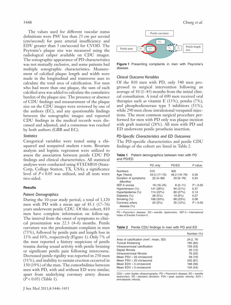
Patient DemographicsDuring the 10-year study period, a total of 1,120men with PD with a mean age of 45.1 (17–76)years underwent penile CDU. Of this cohort, 810men have complete information on follow-up.The interval from the onset of symptoms to clini-cal presentation was 22.3 (4–6) months. Penilecurvature was the predominant complaint in men(75%), followed by penile pain and length loss in15% and 10%, respectively (Figure 1). Only 7% ofthe men reported a history suspicious of peniletrauma during sexual activity with penile bruisingor significant penile pain following intercourse.Decreased penile rigidity was reported in 250 men(31%), and inability to sustain erection occurred in150 (19%) of the men. The comorbidities betweenmen with PD, with and without ED were similar,apart from underlying coronary artery disease(P < 0.05) (Table 1).
Clinical Outcome VariablesOf the 810 men with PD, only 540 men pro-gressed to surgical intervention following anaverage of 10 (1–45) months from the initial clini-cal consultation. A total of 690 men received oraltherapies such as vitamin E (13%), potaba (7%),and phosphodiesterase type 5 inhibitors (51%),while 290 men chose intralesional verapamil injec-tions. The most common surgical procedure per-formed for men with PD only was plaque incisionwith graft material (28%). All men with PD andED underwent penile prosthesis insertion.
PD-Specific Characteristics and ED OutcomesThe PD-specific characteristics and penile CDUfindings of the cohort are listed in Table 2.
Penile curvature
Penile length loss Penile pain
75%
7% 3% 11% 4%
Figure 1 Presenting complaints in men with Peyronie’sdisease.
Table 1 Patient demographics between men with PDand PD/ED
PD only PD/ED P value
Numbers 510 300Age (Years) 43.3 (17–72) 46.2 (19–76) 0.35Duration of symptoms
(Months)22 (4–68) 29 (6–76) 0.24
IIEF-5 scores 18 (16–25) 8 (0–11) P < 0.05Hypertension (%) 141 (28%) 94 (31%) 0.37Hyperlipidemia (%) 114 (22%) 80 (27%) 0.14Diabetes (%) 28 (5%) 19 (6%) 0.27Smoking (%) 168 (33%) 68 (23%) 0.08Coronary artery
disease (%)25 (5%) 35 (12%) P < 0.05
PD = Peyronie’s disease; ED = erectile dysfunction; IIEF-5 = InternationalIndex of Erectile Function-5.
Table 2 Penile CDU findings in men with PD and ED
Number (%)
Area of calcification (mm2, mean, SD) 24.2, 76Tunical thickening 195 (65)Intracavernosal calcification 159 (53)Septal fibrosis 35 (12)Intracavernosal fibrosis 76 (25)Mean PSV > 25 cm/second 58 (19)Mean PSV < 25 cm/second 242 (81)Mean EDV > 5 cm/second 168 (56)Mean EDV < 5 cm/second 134 (44)
CDU = color duplex ultrasonography; PD = Peyronie’s disease; ED = erectiledysfunction; SD = standard deviation; PSV = peak systolic velocity; EDV =end-diastolic velocity.
3448 Chung et al.
J Sex Med 2011;8:3446–3451

Penile CDU evaluation of tunical thickening,calcifications, septal fibrosis, and intracavernosalfibrosis were observed in 65%, 53%, 12%, and25% of the patients, respectively. The most fre-quent combination penile CDU finding was thepresence of tunical thickening and calcification(17%). Among men with Peyronie’s plaque calci-fications, the mean cumulative calcification was24.2 mm2 (1–360 mm2, standard deviation 76).The mean PSV and EDV on the right cavernosalartery were 14.2 cm/second and 3.5 cm/second,while the left cavernosal artery measurementswere 15.1 cm/second and 3.2 cm/second.
The presence of intracavernosal fibrosis wasstrongly associated with lower PSV (P < 0.05) andhigher EDV (P < 0.05). Both CVOD and impairedcavernosal arterial inflow were associated with ED(P < 0.05). A linear regression model multivariateanalysis was performed to predict the significanteffect of patient’s medical comorbidities, history ofpresenting complaint, penile PD CDU character-istics, and the size of Peyronie’s plaque, as predic-tors of ED. There was a strong correlationbetween Peyronie’s plaque size and developmentof ED (r2 = 0.76, P < 0.05 with df = 2). History ofpresenting complaint and other penile CDU char-acteristics were not significant predictors for thedevelopment of ED.
Discussion
The present study explores the relationshipbetween penile CDU findings in men with PDand ED. Experimental rat models in aging, dia-betes, cavernous injury, and castration showedloss of smooth muscle cells and decrease insmooth muscle/collagen ratio, proving the asso-ciation between corporal fibrosis and ED [12–16].These histological alterations underlie the patho-physiology for the development of CVOD [17].In contrast, many studies identified excessivetunical fibrosis as responsible for CVOD and thedevelopment of ED based on earlier knowledgeon the pathophysiology of PD. CVOD related toPD is thought to be secondary to a structuralalteration in the fibroelastic components of thetrabeculae against the tunica albuginea and sub-sequently the inability to compress the subtunicalvenules and activate the veno-occlusive mecha-nism [18]. However, a recent concept describingthe mechanism of penile fibrosis [2,9] has high-lighted the development of fibrosis within thecavernosal smooth muscle and media of penilearteries as important factors in the development
of vasculogenic ED in men with PD. The recur-rent penile trauma sustained in men with PDduring sexual activities not only resulted infurther delamination of the tunical layers, it alsoled to hemorrhage of the underlying cavernosalsmooth muscle [19]. The excessive stimulation ofmyofibroblasts following the inciting event andthe exposure to repetitive trauma sustainedduring sexual activities, resulted in the extensionand development of fibrotic process within thecorporal cavernosa smooth muscle and media ofcavernosal arteries.
Penile CDU is a useful investigative tool ini-tially described in the mid 1980s [11], to localizePeyronie’s plaques, follow progression of plaquesize, and evaluate arterial and vascular parameters.While these specialized high-resolution ultra-sound machines are not readily available in manyurological practices, it is relatively inexpensive,noninvasive, and effective in identifying penileabnormalities such as septal hematoma or vascularanomalies. Recent publications from Lue andassociates [20,21] have demonstrated strong corre-lation between penile CDU findings and clinicaloutcomes. The presence of intracavernosal fibrosisand tunical thickening were associated withdecreased ability to have intercourse [20], and afteradjustment for PD-specific characteristics, base-line calcifications correlated strongly with the pro-gression to surgery [21].
Several studies have supported the adverse roleof calcification in clinical outcomes. Experimentalstudies using cell cultures media derived from PDtissues demonstrate cytogenetic instability andtumorigenic potential within the myofibroblast[22,23], and that these osteogenic progenitor cellscan undergo calcification and/or ossification, acondition observed in advanced cases of PD [23].Indeed, the most frequent combination penileCDU finding in our study was the presence oftunical thickening and calcification (17%).Andresen [24] reported negative associationbetween plaque calcification and successful out-comes following intralesional injection therapy.Similar conclusions were drawn by Bekos [25]who found that larger plaque calcification corre-lated strongly with decreased rates of spontane-ous resolution and the ability to have intercourse.Both articles concluded that calcified lesionsignals a more chronic form of PD [24,25].While we did not detect any significant differ-ence between interval from the onset of symp-toms to clinical presentation, and size of plaquecalcification, men with larger plaque calcification
Penile Doppler Study in Men with Peyronie’s Disease and ED 3449
J Sex Med 2011;8:3446–3451
tend to fail conservative treatment and requirepenile prosthesis implant.
While the presence of intracavernosal fibrosisand plaque calcification correlated strongly withhigher ED rates i.e., CVOD, lower PSV were alsoidentified in men with PD with ED complaints. Ofthe men with PD who complained of decreasedpenile rigidity and inability to sustain erection, wefound that significant numbers of these men haveboth lower PSV and higher EDV values. None-theless it is often difficult to correlate specific flowrates and hemodynamic values with the clinicalsymptoms, as an adequate erection for one manmay be viewed as completely inadequate foranother. Expectations of rigidity and performancevary widely from individual to individual and alsoacross age strata. While decreased penile rigidity isusually demonstrated by a low PSV, some menmay complain of similar symptoms despite normalor “high” PSV. Similarly, not every man with ahigh EDV will have difficulty sustaining an erec-tion, owing to a compensated arterial inflow andshort-lived time requirements for his sexual act.
This study has demonstrated that both CVODand impaired cavernosal arterial inflow contributeto the development of ED in men with PD, andthe presence of one condition is not mutuallyexclusive of the other. In fact, the presence of int-racavernosal fibrosis was strongly associated witharterial insufficiency and CVOD (P < 0.05). Wealso found that larger Peyronie’s plaque size cor-related strongly with the development of CVODand risk of ED, and the need for subsequent sur-gical intervention.
We acknowledged several limitations in ourstudy. The study cohort is derived from patientspresenting to our tertiary center and may not berepresentative of general PD population due toselection bias from referral pattern. Secondly, thiscross-sectional study may not fully address thecausal relationship and the changes to the penileCDU findings and ED in men with PD in the longterm that is provided by longitudinal observationstudy. While an ultrasonograph is a user-dependant modality with a possibility of underre-porting the prevalence of tunical thickening, allpenile CDUs were performed by an experiencedoperator (GBB), and all penile CDU images werereviewed independently by two of the authors (ECand GBB) to confirm the thickness of tunicalcalcified plaque, and rule out any discrepancies.
The demonstration of early blood flow and per-sistence of flow in the dorsal vein on cavernosog-raphy and cavernosometry is considered the gold
standard of diagnosing venous leak or CVOD.However, this test is not without its own draw-backs because of its invasive nature, includingincreased sympathetic tone induction, risk of con-trast anaphylaxis, and time-consuming procedure.Furthermore, this study did not incorporate otherpenile CDU parameters such as retrograde caver-nosal flow and resistance index to diagnosis venousleak. Lastly, some would argue that phentolamineshould be utilized rather than prostaglandins E1injection as the erectogenic agent among youngermen, given phentolamine has an alpha-adrenergicresponse and therefore lower EDV due to inhibi-tion of sympathetic neuronal activity.
To our knowledge, this is the largest and mostcomprehensive analysis of penile CDU findingsand clinical outcomes in men with PD and ED.The large sample size and abundance of clinicalvariables allow for multivariate and regressionmodel analysis to provide comprehensive evalua-tion of the penile CDU findings and clinical out-comes. The coexistence of CVOD and poorcavernosal arterial flow suggested that the devel-opment of ED in men with PD is a result of penilefibrosis/fibrotic changes that occur across all tissuecomponents. In addition, penile CDU findingsdemonstrated that PD is not localized to thetunical layers alone as evidenced by the presence ofintracavernosal fibrosis and calcification.
Nonetheless, further studies are required andshould be designed to focus on the casual relation-ship between penile CDU findings and functionaloutcomes from a longitudinal perspective. Theability of penile CDU to identify men who willrespond to treatment or not is exciting and willplay a significant factor in directing clinicians toact decisively to alleviate the burden of the diseasein men with PD.
Conclusions
Penile CDU provides a valuable tool to documentpertinent features of PD and investigate underly-ing vascular anomalies. CVOD and impairedcavernosal arterial inflow contribute to the devel-opment of ED, and larger Peyronie’s plaque size isa strong predictor of the need for surgical inter-vention. This information, enabling the treatingphysician to understand the underlying vascularhealth of the patient, is essential to optimize sur-gical outcomes.
Corresponding Author: Eric Chung, MD, Depart-ment of Urology, Princess Alexandra Hospital, Brisbane,
3450 Chung et al.
J Sex Med 2011;8:3446–3451

Queensland, 4102, Australia. Tel: +617-33242468; Fax:+617-33242546; E-mail: [email protected]
Conflict of Interest and Disclosure: None.
Statement of Authorship
Category 1(a) Conception and Design
Eric Chung; Gerald B. Brock(b) Acquisition of Data
Eric Chung; Gerald B. Brock(c) Analysis and Interpretation of Data
Eric Chung; Ling De Young; Gerald B. Brock
Category 2(a) Drafting the Article
Eric Chung(b) Revising It for Intellectual Content
Gerald B. Brock
Category 3(a) Final Approval of the Completed Article
Eric Chung; Ling De Young; Gerald B. Brock
References
1 Ralph D, Gonzalez-Cadavid N, Mirone V, Perovic S, Sohn M,Usta M, Levine L. The management of Peyronie’s disease:Evidence-based 2010 guidelines. J Sex Med 2010;7:2359–74.
2 Gonzalez-Cadavid NF, Rajfer J. Mechanisms of disease: Newinsights into the cellular and molecular pathology of Peyro-nie’s disease. Nat Clin Pract Urol 2005;2:291–7.
3 Mulhall JP, Creech SD, Boorjian SA, Ghaly S, Kim ED, MotyA, Davis R, Hellstrom W. Subjective and objective analysis ofthe prevalence of Peyronie’s disease in a population of menpresenting for prostate cancer screening. J Urol 2004;171:2350–3.
4 Rosen R, Catania J, Lue T, Althof S, Henne J, Hellstrom W,Levine L. Impact of Peyronie’s disease on sexual and psycho-social functioning: Qualitative findings in patients and con-trols. J Sex Med 2008;5:1977–84.
5 Montorsi F, Adaikan G, Becher E, Giuliano F, Khoury S, LueTF, Sharlip I, Althof SE, Andersson KE, Brock G, BroderickG, Burnett A, Buvat J, Dean J, Donatucci C, Eardley I, Fugl-Meyer KS, Goldstein I, Hackett G, Hatzichristou D, Hell-strom W, Incrocci L, Jackson G, Kadioglu A, Levine L, LewisRW, Maggi M, McCabe M, McMahon CG, Montague D,Montorsi P, Mulhall J, Pfaus J, Porst H, Ralph D, Rosen R,Rowland D, Sadeghi-Nejad H, Shabsigh R, Stief C, Vardi Y,Wallen K, Wasserman M. Summary of the recommendationson sexual dysfunctions in men. J Sex Med 2010;7:3572–88.
6 Johannes CB, Araujo AB, Feldman HA, Derby CA, KleinmanKP, McKinlay JB. Incidence of erectile dysfunction in men 40to 69 years old: Longitudinal results from the MassachusettsMale Aging Study. J Urol 2000;163:460–3.
7 El-Sakka AI, Hassoba HM, Pillaritsetty RJ, Dahiya R, Lue TF.Peyronie’s disease is associated with an increase in
transforming growth factor-beta protein expression. J Urol1997;158:1391–4.
8 Tomasek JJ, Gabbiani G, Hinz B, Chapponier C, Brown RA.Myofibroblasts and mechano-regulation of connective tissueremodelling. Nat Rev Mol Cell Biol 2002;3:349–63.
9 Gonzalez-Cadavid NF. Mechanism of penile fibrosis. J SexMed 2009;6:353–62.
10 Wilkins CJ, Sriprasad S, Sidhu PS. Colour Doppler ultrasoundof penis. Clin Radiol 2003;58:514–23.
11 Lopez JA, Jarow JP. Duplex ultrasound findings in men withPeyronie’s disease. Urol Radiol 1991;12:199–202.
12 Pryor J, Akkus E, Alter G, Jordan G, Lebret T, Levine L,Mulhall J, Perovic S, Ralph D, Stackl W. Peyronie’s disease. JSex Med 2004;1:110–5.
13 Ferrini M, Magee TR, Vernet D, Rajfer J, Gonzalez-CadavidNF. Aging-related expression of inducible nitric oxide syn-thase (iNOS) and markers of tissue damage in the rat penis.Biol Reprod 2001;64:974–82.
14 Traish AM, Munarriz R, O’Connell L, Choi S, Kim SW, KimNN, Huang YH, Goldstein I. Effects of medical or surgicalcastration on erectile function in an animal model. J Androl2003;24:381–7.
15 Kovanecz I, Rambhatla A, Ferrini MG, Verdet D, Sanchez S,Rajfer J, Gonzalez-Cadavid NF. Chronic daily tadalafil pre-vents the corporal fibrosis and veno-occlusive dysfunction thatoccurs after cavernosal nerve resection. BJU Int 2008;101:203–10.
16 Ferrini MG, Kovanecz I, Sanchez S, Vernet D, Umeh C,Rajfer J, Gonzalez-Cadavid NF. Fibrosis and loss of smoothmuscle in the corpora cavernosa precede corporal veno-occlusive dysfunction (CCVOD) induced by experimentalcavernosal nerve damage in the rat. J Sex Med 2009;6:415–28.
17 Davila HH, Rajfer J, Gonzalez-Cadavid NF. Corporal veno-occlusive dysfunction in the aging rat. Evaluation by caverno-sometry and cavernosography. Urology 2004;64:1261–6.
18 Brock G, Hsu GL, Nunes L, von Heyden B, Lue TF. Theanatomy of the tunica albuginea in the normal penis and Pey-ronie’s disease. J Urol 1997;157:276–81.
19 Devine CJ Jr, Somers KD, Jordan SG, Schlossberg SM. Pro-posal trauma as the cause of the Peyronie’s lesion. J Urol1997;157:285–90.
20 Smith JF, Brant WO, Fradet V, Shindel AW, Vittinghoff E,Chi T, Huang YC, Davis CB, Conti S, Lue TF. Penile sono-graphic and clinical characteristic in men with Peyronie’sdisease. J Sex Med 2006;6:2858–67.
21 Breyer BN, Shindel AW, Huang YC, Eisenberg ML, WeissDA, Lue TF, Smith JF. Are sonographic characteristics asso-ciated with progression to surgery in men with Peyronie’sdisease? J Urol 2010;183:1484–8.
22 Mulhall JP, Nicholson B, Pierpaoli S, Lubrano T, ShankeyTV. Chromosomal instability is demontrstaed by fibroblastsderived from tunica of men with Peyronie’s disease. Int JImpot Res 2004;16:288–93.
23 Vernet D, Nolazco G, Cantini L, Magee TR, Qian A, Rajfer J,Gonzalez-Cadavid NF. Evidence that osteogenic progenitorcells in the human tunica albuginea may originate from stemcells: Implications for Peyronie disease. Biol Reprod 2005;73:1199–210.
24 Andresen R, Wegner HE, Banzer D, Miller K. Ultrasound andsoft-tissue radiography to monitor local interferon-alpha 2Btreatment in Peyronie’s disease. Acta Radiol 1996;37:352–6.
25 Bekos A, Arvaniti M, Hatzimouratidis K, Moysidis K, TzortzisV, Hatzichristou D. The natural history of Peyronie’s disease:An ultrasonography-based study. Eur Urol 2008;53:644–50.
Penile Doppler Study in Men with Peyronie’s Disease and ED 3451
J Sex Med 2011;8:3446–3451