Embed Size (px)
Citation preview
www.elsevier.com/locate/braindev
Brain & Development 29 (2007) 69–78
Pediatric intractable epilepsy syndromes: Reason for earlysurgical intervention
Shiyong Liu a,1, Ning An a,1, Hui Yang a,*, Meihua Yang a, Zhi Hou a,Lihong Liu b, Yong Liu c
a Department of Neurosurgery, Xinqiao Hospital, The Third Military Medical University, Chongqing, 400037, PR Chinab Department of Pediatrics, Xinqiao Hospital, The Third Military Medical University, Chongqing, 400037, PR Chinac Department of Neurology, Xinqiao Hospital, The Third Military Medical University, Chongqing, 400037, PR China
Received 5 December 2005; received in revised form 28 June 2006; accepted 30 June 2006
Abstract
Drug-resistance in several childhood epilepsy syndromes is common, and these patients may tolerate epilepsy surgery. In thisstudy, the surgical outcomes of 24 pediatric patients with various intractable epilepsy syndromes and three patients with tuberoussclerosis were examined at Xinqiao hospital between 1997 and 2004. The study included nine cases of Lennox–Gastaut syndrome,two cases of Rasmussen‘s syndrome, one case of Sturge–Weber syndrome, three cases of West syndrome, three cases of tuberoussclerosis and nine cases of mesial temporal lobe epilepsy syndrome. In each case, different surgical procedures were performedaccording to preoperative evaluation and ECoG. At an average of 4.5 years after surgery, 14 out of 27 patients (51.9%) had an EngelClass I outcome after surgery, and an additional eight patients (29.6%) had rare seizure (Engel ClassII). Three patients showed asignificant decrease in seizure frequency (Engel Class III). The mean IQ increased from 61.4 ± 12.2 to 75.0 ± 11.0, and greaterIQ increase was seen in patients with shorter seizure history and drug-resistance. Temporary complications were observed in fourpatients and there were no deaths. In conclusion, early surgical intervention in pediatric intractable epilepsy syndromes may resultsin a favorable outcome in a high percentage of cases and may provides an important opportunity to prevent irreversible decline inintelligence and disability.� 2006 Elsevier B.V. All rights reserved.
Keywords: Intractable epilepsy; Epilepsy syndromes; Epilepsy surgery; Multilobe resection; Pediatric patients
1. Introduction
Drug-resistance in some childhood-onset epilepsysyndromes such as Lennox–Gastaut syndrome, Rasmus-sen syndrome, Sturge–Weber syndrome, West syn-drome, and mesial temporal lobe epilepsy syndrome iscommon, and is also predictable in a certain extent[1–3]. Although each of these syndromes manifests in
0387-7604/$ - see front matter � 2006 Elsevier B.V. All rights reserved.
doi:10.1016/j.braindev.2006.06.009
* Corresponding author. Tel.: +86 23 68755610; fax: +86 2365218204.
E-mail address: [email protected] (H. Yang).1 Both authors have contributed equally to this work.
an age-specific manner and is defined by distinct electro-clinical features, they are all refractory to antiepilepticdrugs and are invariably associated with psychomotordeficits [1–5]. In the most severe cases, these pediatric epi-lepsy syndromes can lead to either epileptic encephalop-athy or progressive neurodegeneration. Due to the poorresponse to antiepileptic drugs (AEDs), early surgicalinterventions represents an important part of a compre-hensive treatment plan for this group of children, espe-cially in regard to the developmental delays and mentalretardation that are the hallmarks of these syndromes[5–7]. This study reports the surgical outcome of 27 pedi-atric patients with intractable epileptic syndromes.
70 S. Liu et al. / Brain & Development 29 (2007) 69–78
2. Methods
The study population consisted of 27 pediatric caseswhich accounted for the 13.9% of all pediatric surgicalpatients at same term (194 cases), including nine casesof Lennox–Gastaut syndrome, two cases of Rasmussensyndrome, one case of Sturge–Weber syndrome, threecases of West syndrome, and nine cases of mesial tempo-ral lobe epilepsy syndrome. Three cases of tuberous scle-rosis also included due to their some similarities withpediatric syndromes, such as the child-onset epilepsy,drug-resistance epilepsy and mental retardation in child-hood. Tuberous sclerosis patients in this group did notmanifest as above pediatric syndromes. The syndromicdiagnoses in this study have been described by manyauthors and ILAE (international league against epilep-sy, ILAE). Briefly, West syndrome is characterized byinfantile spasms, hypsarrhythmia and mental retarda-tion [2]. Lennox–Gastaut syndrome is defined by the tri-ad of (1) multiple seizure types including tonic, atonicand myoclonic seizures, and atypical absences, (2) slowspike-and-wave EEG disturbance, and (3) mental defi-ciency [1,4]. Rasmussen syndrome patients demonstrateepilepsia partialis continua, slowly progressive intellec-tual deterioration, and hemiparesis, with radiologic evi-dence of nonspecific hemispheric brain atrophy [8].Sturge–Weber syndrome is characterized by a facial portwine stain, contralateral hemiparesis, mental retarda-tion, and seizures as a result of leptomeningeal angioma-tosis. CNS disorder and lesion of tuberous sclerosisinclude cortically located tubers and/or subependymalnodules, refractory epilepsy and mental retardation[10]. Mesial temporal lobe epilepsy syndrome involvesseizures in the mesial temporal regions at the onset.The EEG of such patients shows temporal focal dis-charge, and magnetic resonance imaging (MRI) is sug-gestive of hippocampal sclerosis [11].
Criteria for epilepsy surgery: (1) frequent or severeseizures interfering with patient‘s life; (2) drug-resistancefor at least one year (at least two AEDs failure of first-and second-line antiepileptics to control seizures); (3)seizure has been demonstrated to originated from a sin-gle focus or multiple foci at same hemisphere; (4) lesionor lobe resection should be surgical accessible and with-out severe deficit to patients; and (5) parents understandthe risks and benefits of procedures.
All patients in the study underwent a comprehensiveevaluation including detailed history and neurologicalexamination, routine and ambulatory EEG, long-termvideo EEG monitoring, and magnetic resonance imag-ing (MRI). Nineteen patients underwent single-photonemission computed tomography (SPECT) scan. Preop-erative and postoperative neuropsychological testingwas assessed by a psychologist. Three patients requiredinvasive intracranial monitoring with subdural platesand depth electrodes to further delineate the epileptic
zone. The patients were discussed at a multidisciplinaryEpilepsy Conference and then those that were deemedsuitable candidates were subjected to surgery. Surgicalincisions were designed according to the preoperativeevaluation of each patient, and potential epileptogenicfoci should be involved in exposed area. Single loberesection or lesionectomy, and/or multiple subpial tran-section (MST) was performed when focal epileptic dis-charge was indicated according to preoperativeevaluation and ECoG, but multilobe resection withMST and/or corpus callosotomy were chosen to dealwith hemispheric multiple epileptogenic foci. Antiepilep-tic drugs were continued for at least two years and thengradually tapered. The patients were postoperativelyassessed at 6 months, 1 year, and then annually. Theduration of follow-up was between 1 and 8 years witha mean of 4.5 years. Seizure outcome was assessed usingEngel’s criteria. All subjects were the patients underwentsurgery but had follow-up at least one year, and thosewho had follow-up less than one year were excluded.Statistical analysis was performed using independent-samples t-test and paired-samples t-test on SPSS soft-ware, P < 0.05 was taken as statistically significant.
3. Results
3.1. Patient information
Sixteen patients underwent single lobe resection orlesionectomy, and eleven patients received multiloberesection. Among the 11 patients with multilobe resec-tion, four patients received partial corpus callosotomy.The age of the Patients at surgery ranged from 3 to 15years. All patients showed drug-resistance for at leasttwo AEDs (failure of first- and second-line antiepilepticsto control seizures) ranging from 1 to 10 years. TheAED-resistant periods were for 1–2 years in eightpatients, 2–4 years in 10 patients and over 4 years in ninepatients (see Table 1). The lag between onset and sur-gery of epilepsy ranged 1–11 years.
3.2. Electroencephalograms
Routine EEG results were abnormal in 25 of 27 cases,indicating epileptic discharge in 19 patients, and general-ized slowing in six patients. Ambulatory EEG monitor-ing demonstrated unilateral epileptic discharge in 23patients. Long-term video electroencephalographicmonitoring localized the epileptic zone in 24 cases.Three patients required invasive recording with intracra-nial electrodes to delineate the epileptic zone. The epi-leptic foci were determined by ictal EEG and interictalEEG finding. Bemegride was used to induce seizurewhen ambulatory EEG and video EEG observed no sei-zure, and use of this drug was limited to children older
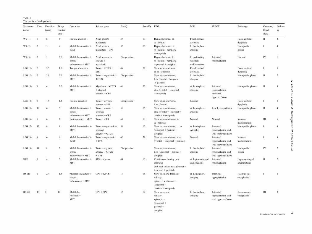
Table 1
The profile of each patients
Syndrome
name
Year Duration
(year)
Drug-
resistant
time
Operation Seizure types Pre-IQ Post-IQ EEG MRI SPECT Pathology Outcome/
Engel
class
Follow-
up
WS (1) 7 6 6 Frontal excision Axial spasms
in clusters
47 60 Hypsarrhythmia, rt.
sz (frontal)
Focal cortical
dysplasia
Focal cortical
dysplasia
II 2
WS (2) 5 5 4 Multilobe resection +
MST
Axial spasms
in clusters + CPS
52 66 Hypsarrhythmial, lt.
sz (frontal + temporal
+ occipital)
lt. hemisphere
atrophy
Nonspecific
gliosis
I 4
WS (3) 3 3 2.6 Multilobe resection +
corpus
callosotomy + MST
Axial spasms in
clusters +
myoclonic
Disoperative Hypsarrhythmia, lt.
sz (frontal + temporal
+ parietal + occipital)
lt. perforating
ventricle
malformation
Interictal
hypoperfusion
Normal IV 1
LGS (1) 6 2.9 1.8 Temporal excision Tonic + GTCS +
SPS
46 72 Slow spike-and-wave,
rt. sz (temporal)
Focal cortical
dysplasia
Focal cortical
dysplasia
I 5
LGS (2) 7 2.8 2.6 Multilobe resection +
MST
Tonic + myoclonic +
GTCS
Disoperative Slow spike-and-wave,
lt.sz (frontal + temporal
+ parietal)
lt. hemisphere
atrophy
Nonspecific gliosis II 7
LGS (3) 9 4 2.5 Multilobe resection +
MST
Myoclonic + GTCS
+ atypical
absence + CPS
61 73 Slow spike-and-wave,
rt.sz (frontal + temporal
+ occipital)
rt. hemisphere
atrophy
Interictal
hypoperfusion
and ictal
hyperperfusion
Nonspecific gliosis II 3
LGS (4) 6 1.9 1.4 Frontal resection Tonic + atypical
absence + SPS
Disoperative Slow spike-and-wave,
lt.sz (frontal)
Normal Focal cortical
dysplasia
I 8
LGS (5) 10 6 5 Multilobe resection +
corpus
callosotomy + MST
Tonic + atonic +
atypical
absence + CPS
51 63 Slow spike-and-wave,
rt.sz (frontal + temporal +
parietal + occipital)
rt. hemisphere
atrophy
Ictal hyperperfusion Nonspecific gliosis II 4
LGS (6) 9 6 6 Lesionectomy + MST Tonic + CPS 65 68 Slow spike-and-wave, lt.
sz (parietal)
Normal Normal Vascular
malformation
III 6
LGS (7) 13 9 9 Multilobe resection +
MST
Tonic + myoclonic +
atypical
absence + GTCS
58 65 Slow spike-and-wave, rt. sz
(temporal + parietal +
occipital)
rt. hemisphere
Atrophy
Interictal
hypoperfusion and
ictal hyperperfusion
Nonspecific gliosis I 4
LGS (8) 9 6 6 Multilobe resection +
MST
Tonic + myoclonic
+ CPS
62 78 Slow spike-and-wave, lt.sz
(frontal + temporal + parietal)
Normal Interictal
hypoperfusion and
ictal hyperperfusion
Vascular
malformation
I 3
LGS (9) 11 8 7 Multilobe resection +
corpus
callosotomy + MST
Tonic + atypical
absence + GTCS
+ CPS
Disoperative Slow spike-and-wave,
lt.sz (temporal + parietal +
occipital)
lt. hemisphere
atrophy
Interictal
hypoperfusion and
ictal hyperperfusion
Nonspecific
gliosis
IV 7
SWS 9 4 3 Multilobe resection +
MST
SPS + absence 44 66 Continuous slowing, and
interictal
and ictal spikes, rt.sz (frontal +
temporal + parietal)
rt. leptomeningeal
angiomatosis
Interictal
hypoperfusion
Leptomeningeal
angiomatosis
II 2
RS (1) 6 2.4 1.8 Multilobe resection +
corpus
callosotomy + MST
CPS + GTCS 53 68 Slow wave and frequent
solitary
spikes, rt.sz (frontal +
temporal +
parietal + occipital)
rt. hemisphere
atrophy
Interictal
hypoperfusion
Rasmussen’s
encephalitis
I 8
RS (2) 13 11 10 Multilobe
resection +
MST
CPS + SPS 57 67 Slow wave and
solitary
spikes,lt. sz
(temporal +
parietal +
occipital)
lt. hemisphere
atrophy
Interictal
hypoperfusion and
ictal hyperperfusion
Rasmussen’s
encephalitis
III 3
(continued on next page)
S.
Liu
eta
l./
Bra
in&
Develo
pm
ent
29
(2
00
7)
69
–7
871
Table 1 (continued)
Syndrome
name
Year Duration
(year)
Drug-
resistant
time
Operation Seizure
types
Pre-IQ Post-IQ EEG MRI SPECT Pathology Outcome/
Engel
class
Follow-
up
TSS (1) 8 2.5 1.6 Lesionectomy SPS + GTCS 65 90 lt.sz (frontal) Cortex tubers
and a
subependymal
nodule
Ictal hyperperfusion Tuberous
sclerosis
I 1
TSS (2) 9 5 2.9 lesionectomy +
MST
SPS + absence 67 72 rt.sz (parietal) A cortex tuber
and subependymal
nodules
Interictal
hypoperfusion and
ictal hyperperfusion
Tuberous
sclerosis
II 2
TSS (3) 7 2.9 2.6 Temporal
lobectomy
CPS 61 78 rt. sz (temporal) Subependymal
noduletex tubers
Interictal
hypoperfusion
Tuberous
sclerosis
I 4
MTLE (1) 12 2.2 1.7 Temporal
lobectomy
SPS + GTCS 59 73 rt. sz (temporal) HS Ictal hyperperfusion HS I 8
MTLE (2) 9 1.9 1.3 Temporal
lobectomy
CPS 73 93 rt. sz (temporal) HS HS I 3
MTLE (3) 12 2.6 1.6 Temporal
lobectomy
CPS + GTCS 67 86 rt. sz (temporal) HS Interictal
hypoperfusion and
ictal hyperperfusion
HS I 6
MTLE (4) 15 2.4 1.8 Temporal
lobectomy
SPS + GTCS 86 95 lt. sz (temporal) HS Normal HS II 2
MTLE (5) 13 4 4 Temporal
lobectomy
SPS + CPS 53 64 rt. sz (temporal) HS Interictal
hypoperfusion
HS I 8
MTLE (6) 11 5 4 Temporal
lobectomy
CPS 75 79 lt. sz (temporal) HS HS II 1
MTLE (7) 12 4 2.8 Temporal
lobectomy
CPS 49 67 rt. sz (temporal) HS HS I 7
MTLE (8) 14 8 8 Temporal
lobectomy
CPS + GTCS 91 96 lt. sz (temporal) HS Normal HS III 5
MTLE (9) 11 7 7 Temporal
lobectomy
CPS 72 85 lt. sz (temporal) HS Interictal
hypoperfusion
HS I 7
Abbreviations: CPS, complex partial seizure; EEG, electroencephalogram; HS, hippocampal sclerosis; LGS, Lennox–Gastaut syndrome; lt., Left; MRI, magnetic resonance imaging; MTLE, Mesial temporal lobe epilepsy; RS, Rasussen’s syndrome; rt., Right; SPS,
simple partial seizure; SWS, Sturge–Weber syndrome; sz., seizure; TSS, tuberous sclerosis syndrome; WS, West syndrome.
72S
.L
iuet
al.
/B
rain
&D
evelop
men
t2
9(
20
07
)6
9–
78
S. Liu et al. / Brain & Development 29 (2007) 69–78 73
than 9 years and at low dose because it may be risky forthe immature and developing brain.
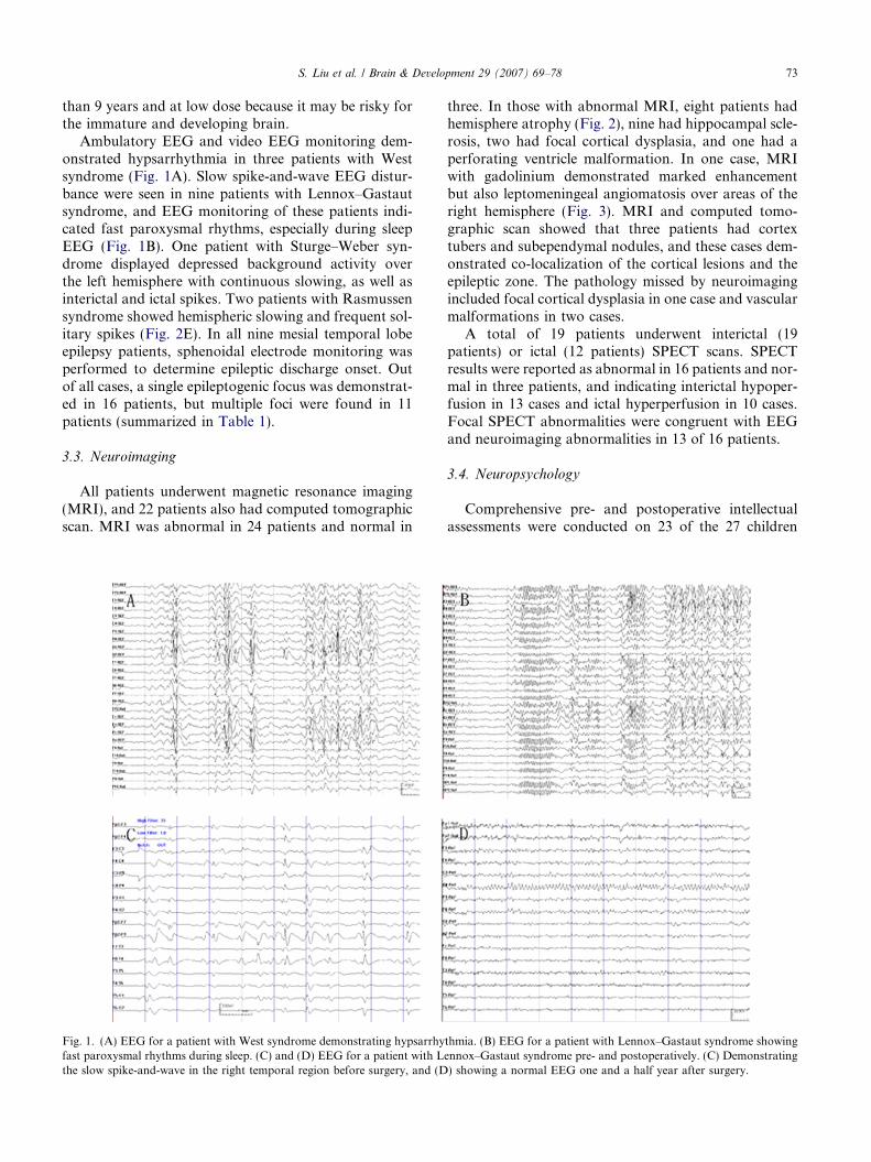
Ambulatory EEG and video EEG monitoring dem-onstrated hypsarrhythmia in three patients with Westsyndrome (Fig. 1A). Slow spike-and-wave EEG distur-bance were seen in nine patients with Lennox–Gastautsyndrome, and EEG monitoring of these patients indi-cated fast paroxysmal rhythms, especially during sleepEEG (Fig. 1B). One patient with Sturge–Weber syn-drome displayed depressed background activity overthe left hemisphere with continuous slowing, as well asinterictal and ictal spikes. Two patients with Rasmussensyndrome showed hemispheric slowing and frequent sol-itary spikes (Fig. 2E). In all nine mesial temporal lobeepilepsy patients, sphenoidal electrode monitoring wasperformed to determine epileptic discharge onset. Outof all cases, a single epileptogenic focus was demonstrat-ed in 16 patients, but multiple foci were found in 11patients (summarized in Table 1).
3.3. Neuroimaging
All patients underwent magnetic resonance imaging(MRI), and 22 patients also had computed tomographicscan. MRI was abnormal in 24 patients and normal in
Fig. 1. (A) EEG for a patient with West syndrome demonstrating hypsarrhyfast paroxysmal rhythms during sleep. (C) and (D) EEG for a patient with Lthe slow spike-and-wave in the right temporal region before surgery, and (D
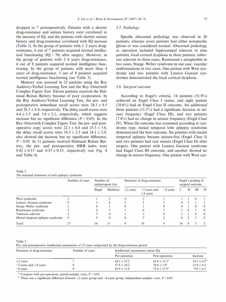
three. In those with abnormal MRI, eight patients hadhemisphere atrophy (Fig. 2), nine had hippocampal scle-rosis, two had focal cortical dysplasia, and one had aperforating ventricle malformation. In one case, MRIwith gadolinium demonstrated marked enhancementbut also leptomeningeal angiomatosis over areas of theright hemisphere (Fig. 3). MRI and computed tomo-graphic scan showed that three patients had cortextubers and subependymal nodules, and these cases dem-onstrated co-localization of the cortical lesions and theepileptic zone. The pathology missed by neuroimagingincluded focal cortical dysplasia in one case and vascularmalformations in two cases.
A total of 19 patients underwent interictal (19patients) or ictal (12 patients) SPECT scans. SPECTresults were reported as abnormal in 16 patients and nor-mal in three patients, and indicating interictal hypoper-fusion in 13 cases and ictal hyperperfusion in 10 cases.Focal SPECT abnormalities were congruent with EEGand neuroimaging abnormalities in 13 of 16 patients.
3.4. Neuropsychology
Comprehensive pre- and postoperative intellectualassessments were conducted on 23 of the 27 children
thmia. (B) EEG for a patient with Lennox–Gastaut syndrome showingennox–Gastaut syndrome pre- and postoperatively. (C) Demonstrating) showing a normal EEG one and a half year after surgery.
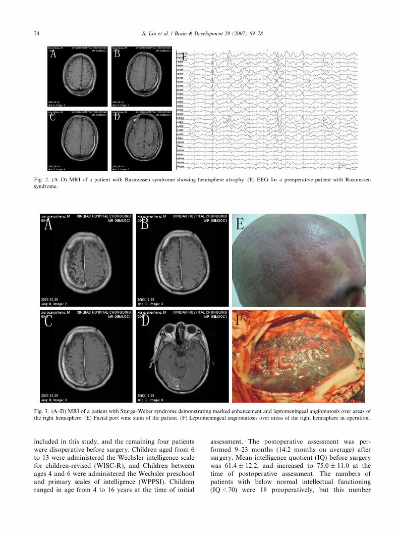
Fig. 2. (A–D) MRI of a patient with Rasmussen syndrome showing hemisphere atrophy. (E) EEG for a preoperative patient with Rasmussensyndrome.
Fig. 3. (A–D) MRI of a patient with Sturge–Weber syndrome demonstrating marked enhancement and leptomeningeal angiomatosis over areas ofthe right hemisphere. (E) Facial port wine stain of the patient. (F) Leptomeningeal angiomatosis over areas of the right hemisphere in operation.
74 S. Liu et al. / Brain & Development 29 (2007) 69–78
included in this study, and the remaining four patientswere disoperative before surgery. Children aged from 6to 13 were administered the Wechsler intelligence scalefor children-revised (WISC-R), and Children betweenages 4 and 6 were administered the Wechsler preschooland primary scales of intelligence (WPPSI). Childrenranged in age from 4 to 16 years at the time of initial
assessment. The postoperative assessment was per-formed 9–23 months (14.2 months on average) aftersurgery. Mean intelligence quotient (IQ) before surgerywas 61.4 ± 12.2, and increased to 75.0 ± 11.0 at thetime of postoperative assessment. The numbers ofpatients with below normal intellectual functioning(IQ < 70) were 18 preoperatively, but this number
S. Liu et al. / Brain & Development 29 (2007) 69–78 75
dropped to 7 postoperatively. Patients with a shorterdrug-resistance and seizure history were correlated tothe increase of IQ, and the patients with shorter seizurehistory and drug-resistance correlated with IQ increase(Table 2). In the group of patients with 1–2 years drug-resistance, 6 out of 7 patients acquired normal intellec-tual functioning (IQ > 70) after surgery. However, inthe group of patients with 2–4 years drug-resistance,4 out of 8 patients acquired normal intelligence func-tioning. In the group of patients with more than 4years of drug-resistance, 3 out of 8 patients acquirednormal intelligence functioning (see Table 3).
Memory was assessed in 22 patients using the ReyAuditory-Verbal Learning Test and the Rey–OsterriethComplex Figure Test. Eleven patients received the Hal-stead–Reitan Battery because of poor cooperation. Inthe Rey Auditory-Verbal Learning Test, the pre- andpostoperative immediate recall scores were 24.1 ± 9.5and 28.3 ± 8.4, respectively. The delay recall scores were4.4 ± 1.5 and 5.8 ± 2.1, respectively, which suggestsincrease but no significant difference (P > 0.05). In theRey–Osterrieth Complex Figure Test, the pre- and post-operative copy scores were 22.1 ± 4.4 and 25.3 ± 3.8,the delay recall scores were 16.3 ± 2.5 and 18.1 ± 2.9,also showed the increase but no significant difference,P > 0.05. In 11 patients received Halstead–Reitan Bat-tery, the pre- and postoperative HRB index were0.42 ± 0.15 and 0.33 ± 0.15, respectively (see Fig. 4and Table 4).
Table 2The itemized statement of each epilepsy syndrome
Number of cases Number ofepileptogenic foc
Single Multif
West syndrome 3 1 2Lennox–Gastaut syndrome 9 3 6Sturge–Weber syndrome 1 0 1Rasmussen syndrome 2 0 2Tuberous sclerosis 3 3 0Mesial temporal epilepsy syndrome 9 9 0
Total 27 16 11
Table 3Pre- and postoperative intellectual assessments of 23 cases categorized by th
Duration of drug-resistance Number of cases I
P
62 years 7 6>2 years and 64 years 8 5>4 years 8 6
a Compare with pre-operation, paired-samples t-test, P < 0.05.b There was a significant difference between 62 years group and >4 years
3.5. Pathology
Specific abnormal pathology was observed in 20patients, whereas seven patients had either nonspecificgliosis or was considered normal. Abnormal pathologyat operation included hippocampal sclerosis in ninepatients, focal cortical dysplasia in three patients, tuber-ous sclerosis in three cases, Rasmussen’s encephalitis intwo cases, Sturge–Weber syndrome in one case, vascularmalformations in two cases. One patient with West syn-drome and two patients with Lennox–Gastaut syn-dromes demonstrated the focal cortical dysplasia.
3.6. Surgical outcome
According to Engel‘s criteria, 14 patients (51.9%)achieved an Engel Class I status, and eight patient(29.6%) had an Engel Class II outcome. An additionalthree patients (11.1%) had a significant decrease in sei-zure frequency (Engel Class III), and two patients(7.4%) had no change in seizure frequency (Engel ClassIV). When the outcome was examined according to syn-drome type, mesial temporal lobe epilepsy syndromedemonstrated the best outcome. Six patients with mesialtemporal epilepsy became seizure-free (Engel Class I)and two patients had rare seizure (Engel Class II) aftersurgery. One patient with Lennox–Gastaut syndromehad Engel Class III outcome, and another showed nochange in seizure frequency. One patient with West syn-
iDuration of drug-resistance Engel’s grading of
surgical outcome
oci 62 years >2 years and64 years
>4 years I II III IV
0 2 1 1 1 0 12 2 5 4 3 1 10 1 0 0 1 0 01 0 1 1 0 1 01 2 0 2 1 0 04 3 2 6 2 1 0
8 10 9 14 8 3 2
e drug-resistance period
ntellectual assessments (mean IQ)
re-operation Post-operation Increase
4.1 ± 13.2 82.4 ± 11.1a 18.3 ± 6.1b
7.8 ± 10.2 70.6 ± 5.8a 12.9 ± 6.22.9 ± 13.8 72.8 ± 12.5a 9.9 ± 4.5
group, independent-samples t-test, P < 0.05.
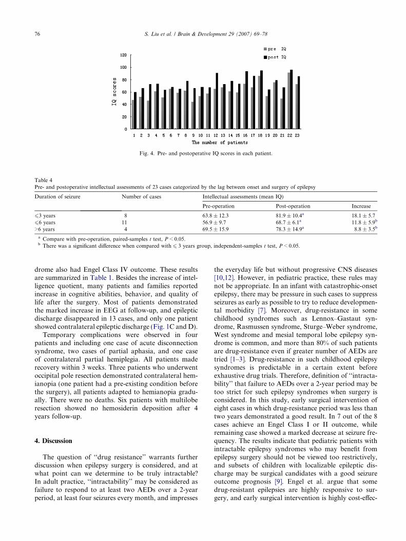
Fig. 4. Pre- and postoperative IQ scores in each patient.
Table 4Pre- and postoperative intellectual assessments of 23 cases categorized by the lag between onset and surgery of epilepsy
Duration of seizure Number of cases Intellectual assessments (mean IQ)
Pre-operation Post-operation Increase
63 years 8 63.8 ± 12.3 81.9 ± 10.4a 18.1 ± 5.766 years 11 56.9 ± 9.7 68.7 ± 6.1a 11.8 ± 5.9b
>6 years 4 69.5 ± 15.9 78.3 ± 14.9a 8.8 ± 3.5b
a Compare with pre-operation, paired-samples t test, P < 0.05.b There was a significant difference when compared with 6 3 years group, independent-samples t test, P < 0.05.
76 S. Liu et al. / Brain & Development 29 (2007) 69–78
drome also had Engel Class IV outcome. These resultsare summarized in Table 1. Besides the increase of intel-ligence quotient, many patients and families reportedincrease in cognitive abilities, behavior, and quality oflife after the surgery. Most of patients demonstratedthe marked increase in EEG at follow-up, and epilepticdischarge disappeared in 13 cases, and only one patientshowed contralateral epileptic discharge (Fig. 1C and D).
Temporary complications were observed in fourpatients and including one case of acute disconnectionsyndrome, two cases of partial aphasia, and one caseof contralateral partial hemiplegia. All patients maderecovery within 3 weeks. Three patients who underwentoccipital pole resection demonstrated contralateral hem-ianopia (one patient had a pre-existing condition beforethe surgery), all patients adapted to hemianopia gradu-ally. There were no deaths. Six patients with multiloberesection showed no hemosiderin deposition after 4years follow-up.
4. Discussion
The question of ‘‘drug resistance’’ warrants furtherdiscussion when epilepsy surgery is considered, and atwhat point can we determine to be truly intractable?In adult practice, ‘‘intractability’’ may be considered asfailure to respond to at least two AEDs over a 2-yearperiod, at least four seizures every month, and impresses
the everyday life but without progressive CNS diseases[10,12]. However, in pediatric practice, these rules maynot be appropriate. In an infant with catastrophic-onsetepilepsy, there may be pressure in such cases to suppressseizures as early as possible to try to reduce developmen-tal morbidity [7]. Moreover, drug-resistance in somechildhood syndromes such as Lennox–Gastaut syn-drome, Rasmussen syndrome, Sturge–Weber syndrome,West syndrome and mesial temporal lobe epilepsy syn-drome is common, and more than 80% of such patientsare drug-resistance even if greater number of AEDs aretried [1–3]. Drug-resistance in such childhood epilepsysyndromes is predictable in a certain extent beforeexhaustive drug trials. Therefore, definition of ‘‘intracta-bility’’ that failure to AEDs over a 2-year period may betoo strict for such epilepsy syndromes when surgery isconsidered. In this study, early surgical intervention ofeight cases in which drug-resistance period was less thantwo years demonstrated a good result. In 7 out of the 8cases achieve an Engel Class I or II outcome, whileremaining case showed a marked decrease at seizure fre-quency. The results indicate that pediatric patients withintractable epilepsy syndromes who may benefit fromepilepsy surgery should not be viewed too restrictively,and subsets of children with localizable epileptic dis-charge may be surgical candidates with a good seizureoutcome prognosis [9]. Engel et al. argue that somedrug-resistant epilepsies are highly responsive to sur-gery, and early surgical intervention is highly cost-effec-
S. Liu et al. / Brain & Development 29 (2007) 69–78 77
tive for patients with surgically remediable syndromes,so there is no need to pursue exhaustive drug trials withthese patients [5,13,14].
Some childhood epilepsies halt cognitive and socialdevelopment with permanent long-term effects [1]. Morethan one third of patients with childhood epilepsy haveintellectual and cognitive problem. This is particularlytrue in some severe epilepsy, such as Lennox–Gastautsyndrome, in which only few children (<10%) are intel-lectually normal [1,7]. Although some authors claim thatintellectual performance and memory remains eitherunchanged or further impaired after surgery, a growingbody of evidence suggests that early seizure controlmight have a positive effect on cognitive developmentand social adjustment. Thus, surgical intervention forintractable epilepsy may provides a good opportunityto prevent irreversible decline of intelligence and cogni-tive function [1,15–17]. In this study, pre- and postoper-ative intellectual assessments demonstrate pediatricpatients with intractable epilepsy syndromes benefitfrom surgery, and mean IQ increases. Memory testsand the Halstead–Reitan Battery also indicated increase,and many patients and families reported increase in cog-nitive abilities, behavior, and quality of life after the sur-gery. Moreover, the results here demonstrate that thepatients with shorter seizure history and drug-resistancetime correlate with higher IQ, and this maybe the reasonthat we have more patients who had IQ gains of P15 IQthan Freitag‘s group, because the duration of seizure in8 cases is no more than 3 years in this study (6/8 patientshad IQ gains of P15 IQ) [14]. This suggests that earlysurgical intervention in pediatric patients with intracta-ble epilepsy syndromes means not only good seizurecontrol, but also better intelligence. Nine patients hadIQ gains of P15 IQ in this group after surgery, andmost patients demonstrated the postsurgical IQincrease, but these IQ increase need to be further reex-amined, because other factors, such as test–retest effect,practice effect, disease severity and maturation will affectthe postsurgical IQ [18].
A subset of pediatric patients with intractable epilep-sy syndromes demonstrate focal epileptic discharge, asin mesial temporal lobe epilepsy syndrome [11]. Len-nox–Gastaut syndrome and West syndrome are general-ly accepted as generalized seizure, but some patientswith such syndromes are localizable and manifest multi-lobar or hemispheric diffuse epileptic discharge, especial-ly emerges in some symptomatic seizure [1,19–21].Traditional anatomic or functional hemispherectomyhas been advocated for children with hemispheric-dom-inant multilobar or diffuse epileptic discharge. However,the majority of these candidates have preexistent hemi-plegia associated with a structural abnormality of thecontralateral hemisphere and seizures proven to arisefrom that hemisphere [9,21,22]. The strict indicationsfor hemispherectomy may lead to exclusion of some
patients without preexistent hemiplegia, and challengeare also exist in such patients with hemispheric multilo-bar epileptic discharge [17,22,23]. The results of thisstudy indicates that multilobe resection with MST or/and corpus callosotomy is a reasonable alternative tohemispherectomy. In 11 cases, 8 patients achieved anEngel Class I or II outcome. These outcomes are similarto the hemispherectomy results of Kossoff et al., butshow lesser complication [22,23]. Our results also showthat surgical patients have better seizure control andcognitive function in contrast to single corpus callosoto-my, MST or vagus nerve stimulation alone [24]. Theresults of this study highlight the advantages of multi-lobe resection combined with MST and/or corpuscallosotomy in severe intractable hemispheric epilepsy[21].
Using Engel‘s criteria, approximately 80% of patientsachieve an Engel Class I or Class II outcome after sur-gery. When the results are examined by syndrome type,patients with mesial temporal lobe epilepsy syndromedemonstrated the best outcome compared to otherpatients with intractable epilepsy. Pediatric patients withother intractable epilepsy syndromes also had goodresults, with 60% achieving the Engel Class I or ClassII outcome at least. These results suggest that aggressivesurgical intervention for such patients is considerable formore practice [13,25].
5. Conclusion
Epilepsy surgery in children with intractable epilepsysyndrome is effective and safe. The results here suggestthat pediatric patients with intractable epilepsy syn-dromes that may benefit from epilepsy surgery shouldnot be viewed too restrictively, and subsets of such pedi-atric patients with localizable epileptic discharge may begood surgical candidates. Early surgical intervention inpediatric intractable epilepsy syndromes may results ina favorable outcome in a high percentage of cases andmay provides an important opportunity to prevent irre-versible decline in intelligence and disability.
References
[1] Camfield P, Camfield C. Epileptic syndromes in childhood:clinical features, outcomes, and treatment. Epilepsia 2002;43(Suppl. 3):27–32.
[2] Rho JM. Basic Science Behind the catastrophic epilepsies.Epilepsia 2004;45(suppl. 5):5–11.
[3] Nordli DR. Infantile seizures and epilepsy syndromes. Epilepsia2002;43(Suppl. 3):11–6.
[4] Shields WD. Diagnosis of infantile spasms, Lennox–Gastautsyndrome, and progressive myoclonic epilepsy. Epilepsia2004;45(Suppl. 5):2–4.
[5] Wheless JW. Nonpharmacologic treatment of the catastrophicepilepsies of childhood. Epilepsia 2004;45(Suppl. 5):17–22.
78 S. Liu et al. / Brain & Development 29 (2007) 69–78
[6] Asano E, Chugani DC, Juhasz C, Muzik O, Chugani HT.Surgical treatment of West syndrome. Brain Dev 2001;23:668–76.
[7] Cross JH. Epilepsy surgery in childhood. Epilepsia 2002;43(Suppl. 3):65–70.
[8] Marjanovic BD, Stojanov LM, Zdravkovic DS, Kravljanac RM,Djordjevic MS. Rasmussen syndrome and long-term response tothalidomide. Pediatr Neurol 2003;29:151–6.
[9] Tuxhorn IEB, Pannek HW. Epilepsy surgery in bilateral Sturge–Weber syndrome. Pediatric Neurol 2002;26:394–7.
[10] Romanelli P, Verdecchia M, Rodas R, Seri S, Curatolo P.Epilepsy surgery for tuberous sclerosis. Pediatr Neurol2004;31:239–47.
[11] Bartolomei F, Wendling F, Regis J, Gavaret M, Guye M,Chauvel P. Pre-ictal synchronicity in limbic networks of mesialtemporal lobe epilepsy. Epilepsy Research 2004;61:89–104.
[12] Loyning Y, Hauglie-Hanssen E. When should surgery be consid-ered? In: Johannesen SI, editor. Intractable Epilepsy. Peters-field: Wrightson Biomedical Publishing; 1995. p. 211–9.
[13] Engel J. Early versus late surgery for intractable seizures. AdvExp Med Biol 2002;497:99–105.
[14] Freitag H, Tuxhorn I. Cognitive function in preschool childrenafter epilepsy surgery: rationale for early intervention. Epilepsia2005;46:561–7.
[15] Sugimoto T, Otsubo H, Hwang PA, Hoffman HJ, Jay V, SneadOC. Outcome of epilepsy surgery in the first three years of life.Epilepsia 1999;40:560–5.
[16] Smith ML, Elliott IM, Lach L. Cognitive skills in children withintractable epilepsy: comparison of surgical and nonsurgicalcandidates. Epilepsia 2002;43:631–7.
[17] Huf RL, Mamelak A, Kneedy-Cayem K. Vagus nerve stimulationtherapy: 2-year prospective open-label study of 40 subjects withrefractory epilepsy and low IQ who are living in long-term carefacilities. Epilepsy Behav 2005;6:417–23.
[18] Sherman E, Slick DJ, Connolly MB, Steinbok P, Martin R,Strauss E, et al. Reexamining the effects of epilepsy surgery on IQin children: use of regression-based change scores. J Int Neuro-psychol Soc 2003;9:879–86.
[19] Ohtahara S, Ohtsuka Y, Kobayashi K. Lennox–Gastaut syn-drome: a new vista. Psychiat Clin Neurosci 1995;49:S179–83.
[20] Fisher RS, Niedermeyer E. Depth EEG studies in the Lennox–Gastaut syndrome. Clin Electroencephalogr 1987;18:191–200.
[21] Patil AA, Andrews RV, Torkekon R. Surgical treatment ofintractable seizures with multilobar or bihemispheric seizure foci.Surg. Neurol. 1997;47:72–8.
[22] Kossoff EH, Buck C, Freeman JM. Outcomes of 32 hemispher-ectomies for Sturge–Weber syndrome worldwide. Neurology2002;59:1735–8.
[23] Kossoff EH, Vining EP, Pillas DJ, Pyzik PL, Avellino AM,Carson BS, et al. Hemispherectomy for intractable unihemi-spheric epilepsy Etiology vs outcome. Neurology 2003;61:887–90.
[24] Valencia I, Holder DL, Helmers SL, Madsen JR, Riviello JJ.Vagus nerve stimulation in pediatric epilepsy: a review. Pediatr.Neurol. 2001;25:368–76.
[25] Sinclair DB, Aronyk KE, Snyder TJ, Wheatley BM, McKean JD,Bhargava R, et al. Pediatric epilepsy surgery at the University ofAlberta: 1988–2000. Pediatr. Neurol. 2003;29:302–11.


![Personalized translational epilepsy research - novel ... · focal (mostly lesional) epilepsy syndromes who are candidates for epilepsy surgery [6]. The ... characterized by hypo-,](https://img.pdfslide.us/doc/110x75/5f2c017e847cd27046085bd0/personalized-translational-epilepsy-research-novel-focal-mostly-lesional.jpg)