Embed Size (px)
Citation preview
THE FINE STRUCTURE OFVON EBNER'S GLAND OF THE RAT
ARTHUR R . HAND
340
INTRODUCTION
The surface of the mammalian tongue is well sup-plied with glandular elements, predominately of amucous nature . Located beneath the large circum-vallate papillae on the dorsum, and the foliatepapillae on the sides of the tongue, are a smallgroup of branching tubuloalveolar glands (3, 10,43) known as von Ebner's gland . They are classi-cally described as serous in nature (3, 11, 29) andtheir ducts open into the trough at the base of thepapillae.
The function of von Ebner's gland has oftenbeen described as one of washing out taste sub-stances from the trough and readying the taste re-ceptors in the walls of the papilla for a new stimu-lus (5, 11, 36) . Their secretions have been impliedto be of importance in the sense of taste because oftheir enzyme content (2) . The present study wasundertaken to determine the ultrastructural char-
From the Laboratory of Biological Structure, National Institute of Dental Research, NationalInstitutes of Health, Bethesda, Maryland 90014
ABSTRACT
The fine structure of von Ebner's gland was studied in untreated rats and rats stimulated tosecrete by fasting-refeeding or injection of pilocarpine . Cytological features were similar tothose reported for pancreas and parotid gland . Abundant granular endoplasmic reticulumfilled the basal portion of the cell, a well-developed Golgi complex was located in the vicinityof the nucleus, and the apical portion of the cell was filled with dense secretory granules .Dense heterogeneous bodies resembling lysosomes were closely associated with the Golgicomplex . Coated vesicles were seen in the Golgi region and also in continuity with the cellmembrane. Granule discharge occurred by fusion of the granule membrane with the cellmembrane at the secretory surface . Successive fusion of adjacent granules to the previouslyfused granule formed a connected string of granules in the apical cytoplasm . Myoepithelialcells were present within the basement membrane, and nerve processes were seen adjacent toacinar and myoepithelial cells. Duct cells resembled the intercalated duct cells of the majorsalivary glands.
acteristics of these glands and their mode of secre-tion .
MATERIALS AND METHODS
Von Ebner's glands were obtained from the tonguesof 5- to 8-week-old male Sprague-Dawley rats, weigh-ing 170-270 g, maintained on laboratory chow andwater ad libitum . Specimens from animals in whichsecretion was stimulated were also included . Food waswithheld from some animals for 24-48 hr, then theywere refed and sacrificed at times from 5 min to 15 hrafter feeding . Other animals received intraperitonealinjections of pilocarpine (160 mg/kg) and weresacrificed at times from 5 min to 4 hr after injection .
A variety of fixation procedures was used . The mostreliable fixation was obtained by vascular perfusionthrough the heart of an anesthetized animal (50 mg/kg sodium pentobarbital, intraperitoneally) whichwas artificially respired with a mixture of 95% oxygen
on May 10, 2018jcb.rupress.org Downloaded from http://doi.org/10.1083/jcb.44.2.340Published Online: 1 February, 1970 | Supp Info:
and 5% carbon dioxide . Other animals were killed bya blow on the head . In either instance, the tissue wasrapidly excised, placed in cold fixative, and cut intoI mm cubes. Fixatives employed were phosphate-buffered 2 .5 0/0 glutaraldehyde (28, 50), phosphate-buffered 1 % osmium tetroxide (28), or cacodylate-buffered 4% formaldehyde (made from paraform-aldehyde powder) or formaldehyde-glutaraldehydemixtures (4), all at pH 7.3 . Aldehyde fixation wascarried out for 2-6 hr, followed by postfixation in l %
osmium tetroxide in phosphate or cacodylate bufferfor 2-3 hr . Some tissues were stained in block for 2 hrwith 0.5 or 2.0% uranyl acetate (24) prior to dehydra-tion . They were dehydrated in ethanol and embeddedin Araldite (27) .
For light microscopy, 6 u cryostat sections werefixed in calcium acetate-formalin (26) and stained
with hmatoxylin and eosin, and 0 .5 µ sections ofAraldite-embedded tissues were stained with tolui-dine blue . For electron microscopy, thin sections werecut on a Porter-Blum microtome (Ivan Sorvall Inc .,Norwalk, Conn .), stained with uranyl acetate and/orlead citrate (49), and examined in a SiemensElmiskop I at 80 kv . Micrographs were obtained withinitial magnifications of 1800 to 33,000 .
RESULTS
Light Microscopy
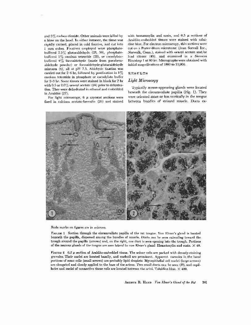
Typically serous-appearing glands were locatedbeneath the circumvallate papilla (Fig . 1) . Theywere oriented more or less vertically in the tonguebetween bundles of striated muscle. Ducts ex-
Scale marks on figures are in microns .
FIGURE 1 Section through the circumvallate papilla of the rat tongue . Von Ebner's gland is locatedbeneath the papilla, dispersed among the bundles of muscle . Ducts can be seen extending toward thetrough around the papilla (arrows) and, on the right, one duct is seen opening into the trough . Portionsof the mucous glands of the tongue are seen lateral to von Ebner's gland . Hematoxylin and eosin . X 40.
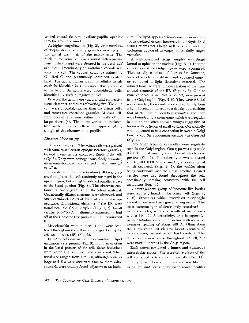
FIGURE 2 0 .5 .s section of Araldite-embedded tissue. The acinar cells are packed with densely-staininggranules. Their nuclei are located basally, and nucleoli are prominent. Apparent vacuoles in the basalportions of some cells (small arrows) are probably lipid droplets . Myoepithelial cell nuclei (large arrows)are elongated and closely applied to the base of the acinus . Two small ducts can be seen (D), and capil-laries and nuclei of connective tissue cells are located between the acini . Toluidine blue . X 400.
ARTHUR R. HAND Von Ebner's Gland of the Rat 341
tended toward the circumvallate papilla, openinginto the trough around it .
At higher magnification (Fig. 2), large numbersof deeply stained secretory granules were seen inthe apical two-thirds of the acinar cells . Thenuclei of the acinar cells were round with a promi-nent nucleolus and were situated in the basal halfof the cell . Occasionally an unstained vacuole wasseen in a cell . The droplet could be stained byOil Red 0 and presumably contained neutrallipid . The acinar lumen and intercellular canalscould be identified in some cases . Closely appliedto the base of the acinus were myoepithelial cells,identified by their elongated nuclei .
Between the acini were vascular and connectivetissue elements, and ducts of varying size. The ductcells were cuboidal, smaller than the acinar cells,and sometimes contained granules. Mucous cellswere occasionally seen within the walls of thelarger ducts (3) . The ducts varied in thicknessfrom one to four or five cells as they approached thetrough of the circumvallate papilla .
Electron Microscopy
ACINAR CELLS : The acinar cells were packedwith numerous electron-opaque secretory granules,located mainly in the apical two-thirds of the cells(Fig. 3) . They were homogeneous, finely granular,membrane-bounded, and ranged in size from 0 .5to 1 .7 y .
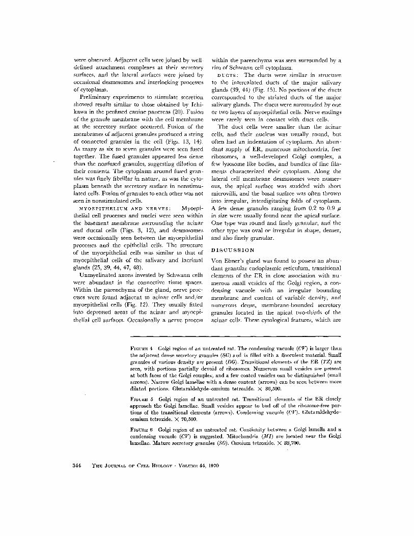
Granular endoplasmic reticulum (ER) was pres-ent throughout the cell, randomly arranged in theapical region, but in highly ordered parallel arraysin the basal portion (Fig. 3) . The cisternae con-tained a finely granular or flocculent material .Occasionally dilated cisternae were observed, andoften certain elements of ER had a vesicular ap-pearance . Transitional elements of the ER werefound near the Golgi complex (Figs . 4, 5) . Smallvesicles 400-700 A in diameter appeared to budoff of the ribosome-free portions of the transitionalER .
Mitochondria were numerous and were scat-tered throughout the cell or were aligned along thecell membranes (39) (Fig. 3) .
In many cells one or more electron-lucent lipidinclusions were present (Fig . 3), found most oftenin the basal portion of the cell. Some inclusionswere membrane bounded ; others were not . Theirusual size ranged from 1 to 3 ,u, although some aslarge as 5-6 u were observed . One or more mito-chondria were usually found adjacent to an inclu-
342 THE JOURNAL OF CELL BIOLOGY • VOLUME 44, 1970
Bion. The lipid appeared homogeneous in osmiumtetroxide-fixed tissues ; however, in aldehyde-fixedtissues, it was not always well preserved and theinclusions appeared as empty or partially emptyvacuoles .A well-developed Golgi complex was found
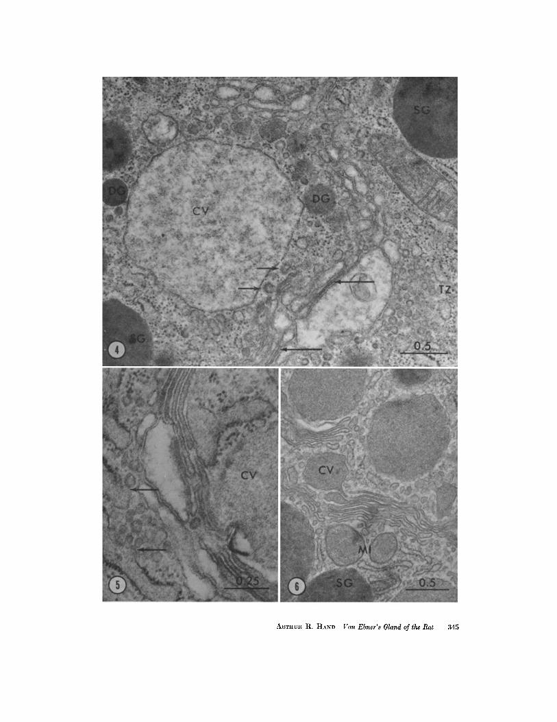
lateral or apical to the nucleus (Figs . 3-6) . In somecells two or three Golgi regions were recognized .They usually consisted of four to five lamellae,some of which were dilated and appeared emptyor contained a light flocculent material . Thedilated lamellae were in close relation to the tran-sitional elements of the ER (Figs . 4, 5) . One ormore condensing vacuoles (7, 22, 23) were presentin the Golgi region (Figs . 4-6) . They were 0.8-2 .0µ in diameter, their content varied in density froma light flocculent material to a density approachingthat of the mature secretory granules, and theywere bounded by a membrane which was irregularin outline and often showed images suggestive offusion with or fission of small vesicles . Occasionallywhat appeared to be a connection between a Golgilamella and the condensing vacuole was observed(Fig . 6) .Two other types of organelles were regularly
seen in the Golgi region . One type was a granule0.2-0.4 s in diameter, a number of which werepresent (Fig. 4). The other type was a coatedvesicle, 500-1800 A in diameter, a population ofwhich occurred, (Figs . 4, 7), the vesicles oftenbeing continuous with the Golgi lamellae . Coatedvesicles were also found throughout the cell,occasionally showing continuity with the cellmembrane (Fig. 11) .
A heterogeneous group of lysosome-like bodieswere regularly found in the acinar cells (Figs . 3,7-10) . Structures which resembled autophagicvacuoles contained recognizable organelles . Themost common type of dense body contained nu-merous vesicles, whorls or scrolls of membraneswith a 120-160 A periodicity, or a hexagonally-packed tubular-crystalline structure with a center-to-center spacing of about 200 A . Often thesestructures contained electron-lucent vacuoles ofvarious sizes, suggestive of lipid content. Thedense bodies were found throughout the cell, butwere most numerous in the Golgi region .
Each acinus contained a lumen and numerousintercellular canals. The secretory surfaces of thecell contained a few small microvilli (Fig. 11) .The cytoplasm beneath the surface was fibrillarin nature, and occasionally microtubular profiles
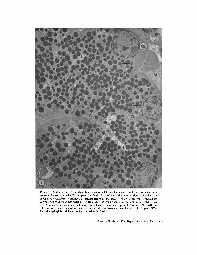
FIGURE 3 Major portion of an acinus from a rat fasted for 24 hr ; parts of at least nine acinar cellsare seen . Secretory granules fill the apical two-thirds of the cells, and the nuclei are located basally. Theendoplasmic reticulum is arranged in parallel arrays in the basal portions of the cells . Intercellularcanals and part of the acinar lumen are evident (L) . Condensing vacuoles are located in the Golgi regions(G) . Numerous heterogeneous bodies and autophagic vascuoles are present (arrows) . Myoepithelialcell process (M) are located peripherally but within the basement membrane . Lipid droplets (FD) .Formaldehyde-glutaraldehyde-osmium tetroxide. X 5500 .
ARTHUR R. HAND Von Rlmer's Gland of the Rat
343
were observed . Adjacent cells were joined by well-defined attachment complexes at their secretorysurfaces, and the lateral surfaces were joined byoccasional desmosomes and interlocking processesof cytoplasm .
Preliminary experiments to stimulate secretionshowed results similar to those obtained by Ichi-kawa in the perfused canine pancreas (20) . Fusionof the granule membrane with the cell membraneat the secretory surface occurred . Fusion of themembranes of adjacent granules produced a stringof connected granules in the cell (Figs . 13, 14) .As many as six to seven granules were seen fusedtogether . The fused granules appeared less densethan the nonfused granules, suggesting dilution oftheir contents . The cytoplasm around fused gran-ules was finely fibrillar in nature, as was the cyto-plasm beneath the secretory surface in nonstimu-lated cells . Fusion of granules to each other was notseen in nonstimulated cells .MYOEPITHELIUM AND NERVES :
Myoepi-thelial cell processes and nuclei were seen withinthe basement membrane surrounding the acinarand ductal cells (Figs . 3, 12), and desmosomeswere occasionally seen between the myoepithelialprocesses and the epithelial cells . The structureof the myoepithelial cells was similar to that ofmyoepithelial cells of the salivary and lacrimalglands (25, 39, 44, 47, 48) .
Unmyelinated axons invested by Schwann cellswere abundant in the connective tissue spaces .Within the parenchyma of the gland, nerve proc-esses were found adjacent to acinar cells and/ormyoepithelial cells (Fig . 12) . They usually fittedinto depressed areas of the acinar and myoepi-thelial cell surfaces . Occasionally a nerve process
344
FIGURE 4 Golgi region of an untreated rat . The condensing vacuole (CV) is larger thanthe adjacent dense secretory granules (SG) and is filled with a flocculent material . Smallgranules of various density are present (DG). Transitional elements of the ER (TZ) areseen, with portions partially devoid of ribosomes. Numerous small vesicles are presentat both faces of the Golgi complex, and a few coated vesicles can be distinguished (smallarrows). Narrow Golgi lamellae with a dense content (arrows) can be seen between moredilated portions . Glutaraldehyde-osmium tetroxide . X 36,500 .
FIGURE 5 Golgi region of an untreated rat . Transitional elements of the ER closelyapproach the Golgi lamellae . Small vesicles appear to bud off of the ribosome-free por-tions of the transitional elements (arrows) . Condensing vacuole (CV) . Glutaraldehyde-osmium tetroxide. X 70,500.
FIGURE 6 Golgi region of an untreated rat. Continuity between a Golgi lamella and acondensing vacuole (CV) is suggested. Mitochondria (MI) are located near the Golgilamellae. Mature secretory granules (SG) . Osmium tetroxide . X 33,700 .
THE JOURNAL OF CELL BIOLOGY • VOLUME 44, 1970
within the parenchyma was seen surrounded by arim of Schwann cell cytoplasm .DUCTS : The ducts were similar in structure
to the intercalated ducts of the major salivaryglands (39, 44) (Fig . 15) . No portions of the ductscorresponded to the striated ducts of the majorsalivary glands. The ducts were surrounded by oneor two layers of myoepithelial cells . Nerve endingswere rarely seen in contact with duct cells .
The duct cells were smaller than the acinarcells, and their nucleus was usually round, butoften had an indentation of cytoplasm . An abun-dant supply of ER, numerous mitochondria, freeribosomes, a well-developed Golgi complex, afew lysosome-like bodies, and bundles of fine fila-ments characterized their cytoplasm . Along thelateral cell membrane desmosomes were numer-ous, the apical surface was studded with shortmicrovilli, and the basal surface was often throwninto irregular, interdigitating folds of cytoplasm .A few dense granules ranging from 0.2 to 0 .9 .sin size were usually found near the apical surface .One type was round and finely granular, and theother type was oval or irregular in shape, denser,and also finely granular.
DISCUSSION
Von Ebner's gland was found to possess an abun-dant granular endoplasmic reticulum, transitionalelements of the ER in close association with nu-merous small vesicles of the Golgi region, a con-densing vacuole with an irregular boundingmembrane and content of variable density, andnumerous dense, membrane-bounded secretorygranules located in the apical two-thirds of theacinar cells . These cytological features, which are
ARTHUR R. HAND Von Elmer's Gland of the Rat
345
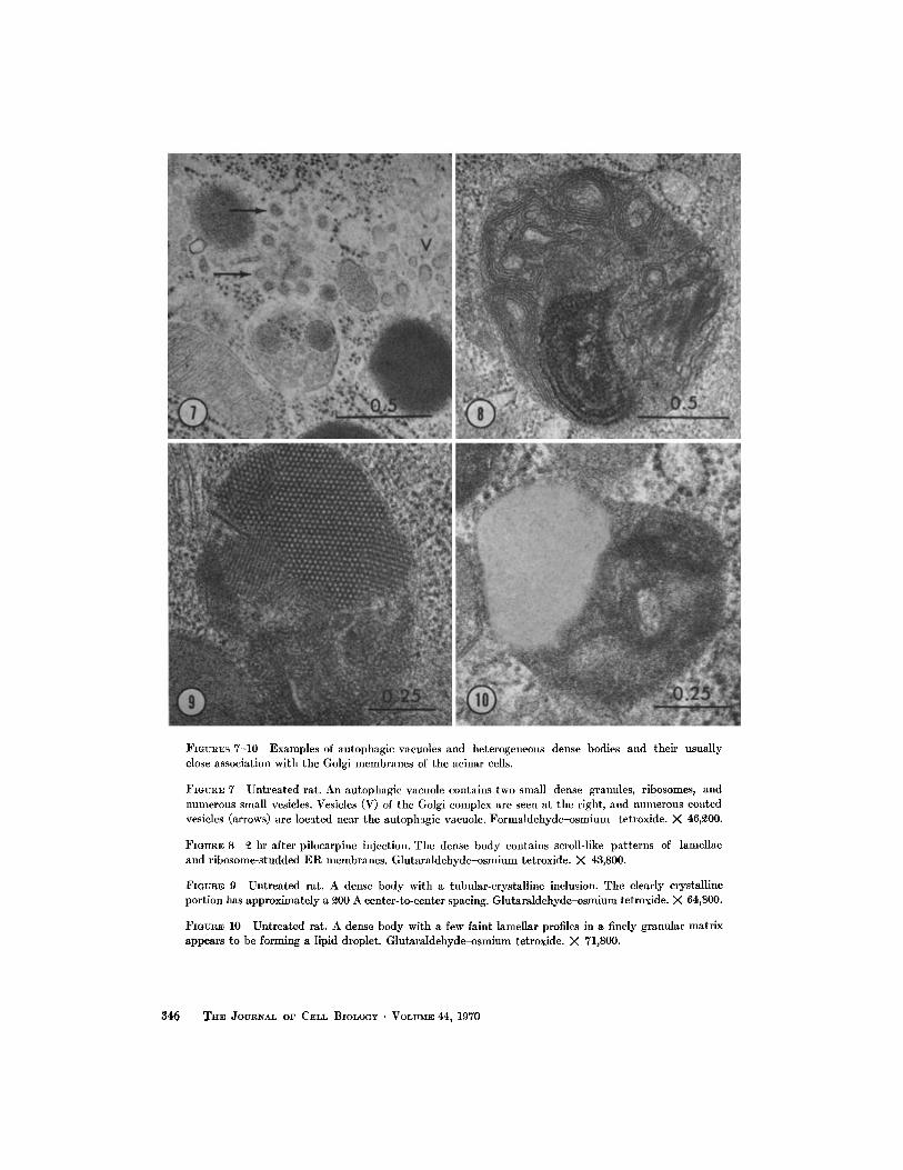
FIGURES 7-10 Examples of autophagic vacuoles and heterogeneous dense bodies and their usuallyclose association with the Golgi membranes of the acinar cells .
FIGURE 7 Untreated rat . An autophagic vacuole contains two small dense granules, ribosomes, andnumerous small vesicles . Vesicles (V) of the Golgi complex are seen at the right, and numerous coatedvesicles (arrows) are located near the autophagic vacuole . Formaldehyde-osmium tetroxide . X 46,200 .
FIGURE 8 2 hr after pilocarpine injection . The dense body contains scroll-like patterns of lamellaeand ribosome-studded ER membranes. Glutaraldehyde-osmium tetroxide. X 43,800 .
FIGURE 9 Untreated rat. A dense body with a tubular-crystalline inclusion . The clearly crystallineportion has approximately a 200 A center-to-center spacing . Glutaraldehyde-osmium tetroxide . X 64,800 .
FIGURE 10 Untreated rat . A dense body with a few faint lamellar profiles in a finely granular matrixappears to be forming a lipid droplet. Glutaraldehyde-osmium tetroxide . X 71,800 .
346 THE JOURNAL OF CELL BIOLOGY . VOLUME 44, 1970
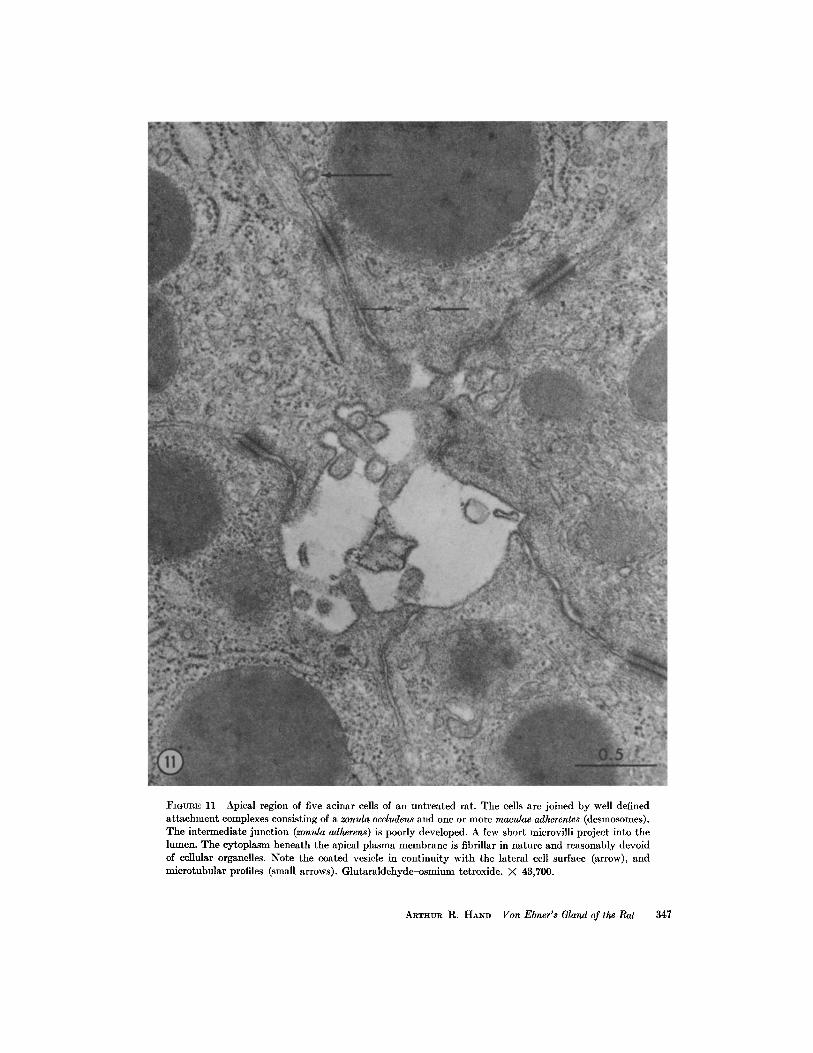
FIGURE 11 Apical region of five acinar cells of an untreated rat. The cells are joined by well definedattachment complexes consisting of a zonula occludens and one or more maculae adherentes (desmosomes) .The intermediate junction (zonula adherens) is poorly developed . A few short microvilli project into thelumen. The cytoplasm beneath the apical plasma membrane is fibrillar in nature and reasonably devoidof cellular organelles . Note the coated vesicle in continuity with the lateral cell surface (arrow), andmicrotubular profiles (small arrows) . Glutaraldehyde-osmium tetroxide. X 43,700 .
ARmmun R . HAND Von Ebner's Gland of the Rat
347
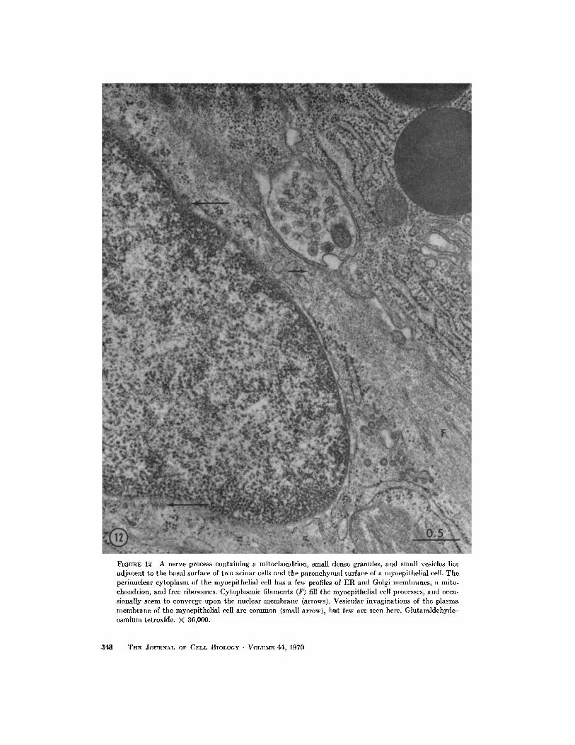
FIGURE 12 A nerve process containing a mitochondrion, small dense granules, and small vesicles liesadjacent to the basal surface of two acinar cells and the parenchymal surface of a myoepithelial cell . Theperinuclear cytoplasm of the myoepithelial cell has a few profiles of ER and Golgi membranes, a mito-chondrion, and free ribosomes . Cytoplasmic filaments (F) fill the myoepithelial cell processes, and occa-sionally seem to converge upon the nuclear membrane (arrows) . Vesicular invaginations of the plasmamembrane of the myoepithelial cell are common (small arrow), but few are seen here . Glutaraldehyde-osmium tetroxide. X 36,000 .
348
THE JOURNAL OF CELL BIOLOGY • VOLUME 44, 1970
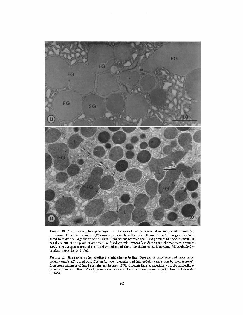
FIGURE 13 5 min after pilocarpine injection. Portions of two cells around an intercellular canal (L)are shown. Four fused granules (FG) can be seen in the cell on the left, and three to four granules havefused to make the large figure on the right . Connections between the fused granules and the intercellularcanal are out of the plane of section . The fused granules appear less dense than the nonfused granules(SG) . The cytoplasm around the fused granules and the intercellular canal is fibrillar . Glutaraldehyde-osmium tetroxide . X 21,900 .
FIGURE 14 Rat fasted 48 hr, sacrificed 5 min after refeeding . Portions of three cells and three inter-cellular canals (L) are shown. Fusion between granules and intercellular canals can be seen (arrows) .Numerous examples of fused granules can be seen (FG), although their connections with the intercellularcanals are not visualized . Fused granules are less dense than nonfused granules (SG) . Osmium tetroxide .X 9000.
349
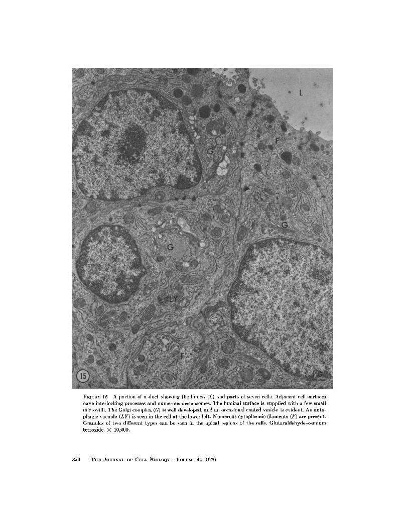
FIGURE 15 A portion of a duct showing the lumen (L) and parts of seven cells . Adjacent cell surfaceshave interlocking processes and numerous desmosomes . The luminal surface is supplied with a few smallmicrovilli . The Golgi complex (G) is well developed, and an occasional coated vesicle is evident . An auto-phagic vacuole (LY) is seen in the cell at the lower left . Numerous cytoplasmic filaments (F) are present.Granules of two different types can be seen in the apical regions of the cells . Glutaraldehyde-osmiumtetroxide. X 10,200.
3 50
THE JOURNAL OF CELL BIOLOGY . VOLUME 44, 1970
essentially similar to those of other exocrine secre-tory glands (7, 23, 38, 39, 44, 45), suggest thatthe secretory product of von Ebner's gland is pro-teinaceous, and that its intracellular synthesisand transport is similar to that postulated for thepancreas (7, 22, 23, 38, 40) .
In the pancreas, protein synthesis presumablyoccurs on the ribosomes of the ER ; the protein istransferred to the cisternae of the ER (40), andis then transported to the condensing vacuole viathe ER cisternae, the transitional elements, andthe peripheral vesicles of the Golgi region, whichsupposedly act as "shuttle carriers" between theER and the condensing vacuole (22, 23) . TheGolgi lamellae do not appear to be involved inthe transport of protein, but may instead add asecond component to the condensing vacuole . Inother cells, the Golgi region has been shown to bethe site of incorporation of carbohydrate moietiesinto the secretory product (31, 32) . The acidhydrolytic activity associated with newly formedsecretory granules in some glands (33, 46) maybe contributed by the Golgi apparatus . Anotherfunction attributed to the Golgi complex is thatof supplying membranes to surround the secretorygranules . Occasionally, apparent connectionswere observed between the Golgi lamellae and thecondensing vacuole of von Ebner's gland . Thesignificance of these connections is being exploredfurther.
The Golgi complex has also been implicatedin the formation of lysosomes in other cells (9,12, 15, 34) and may play a similar role in vonEbner's gland. The dense heterogeneous lysosome-like bodies with a content of vesicular and lamellarprofiles showed a consistent topographical rela-tionship with the Golgi region . Lysosomes insecretory cells such as von Ebner's gland mayparticipate in removal or degradation of excessiveor exhausted membranous material . Fowler andde Duve (14) have shown that extracts of lyso-somes isolated from liver are enzymatically ca-pable of degrading the membranes of mitochondriaand microsomes. Hicks (17, 18) attributed mem-brane breakdown to dense heterogeneous bodiesin transitional epithelium of the rat ureter andbladder . She was able to demonstrate acid phos-phatase activity in some of these bodies, therebyclassifying them as part of the lysosomal system .Acid phosphatase and nonspecific esterase havepreviously been demonstrated in von Ebner'sgland by light microscopy (2, 21, 30), and Bogart
(6) has recently shown acid phosphatase activityassociated with lipofuscin granules in the ratsubmandibular gland, some of which show simi-larities to the dense bodies described in thepresent study .
Stimulation of secretion by fasting-refeeding orpilocarpine administration showed that granuledischarge occurred by fusion of the granule mem-brane with the cell membrane at the secretorysurface . Once fusion had occurred between onegranule and the cell membrane, another granulewas able to fuse with the first granule . Repetitionof this process apparently leads to a string of con-nected granules in the cell . The density of fusedgranules was always less than that of granules inthe same cell which were not fused . The cytoplasmimmediately surrounding the fused granules hadthe characteristics of the apical cytoplasm innonstimulated cells, i.e . finely fibrillar and devoidof organelles . These findings indicate, as suggestedby other workers (1, 20), that the membrane ofthe fused granule takes on the properties of thecell membrane, a transformation which enablesother granules to fuse with it .
Once the content of the granules has beenreleased, the fused granule membranes appear tobe left as an invagination of the now enlargedlumen into the apical cytoplasm of the cell . Thefate of this membrane as the cell resumes its formershape is unknown . Fawcett (13) has suggested thatan amount of membrane equivalent to thatwhich was added during secretion is withdrawnfrom the cell membrane at a molecular level andreassembled into visible membrane in the Golgiregion . Palade (37) has suggested that in pan-creatic acinar cells small "empty" vesicles seenafter secretion of zymogen granules might repre-sent return of membrane to a "membrane depot"in the Golgi region. In the parotid gland stimu-lated to secrete with isoproterenol, Amsterdamet al. (1) have demonstrated numerous vesiclesin the apical region of the cell during the reductionof the size of the lumen. Comparable vesicles werenot observed in the present study ; however, amassive secretion as caused by isoproterenol in theparotid gland was not achieved in von Ebner'sgland .
Coated vesicles have been described in manyother cells (8, 16, 19, 35, 41, 42) where they aremainly involved in protein uptake from the ex-tracellular space . They have also been observed
ARTHUR R. HAND Von Ebner's Gland of the Rat
351
to originate from the Golgi complex and to con-tain hydrolytic enzymes (16) . The function ofcoated vesicles in von Ebner's gland is unknown,but their association with the Golgi complex sug-gests a possible role in secretory granule or lyso-some formation .
REFERENCES
1. AMSTERDAM, A ., I. OHAD, and M. SCHRAMM .1969 . Dynamic changes in the ultrastructure ofthe acinar cell of the rat parotid gland duringthe secretory cycle . J. Cell Biol. 41 :753 .
2. BARADI, A . F., and G. H. BOURNE . 1953. Gusta-tory and olfactory epithelia . Intern . Rev . Cytol.2 :289 .
3. BAUMGARTNER, E . A. 1917. The development ofthe serous glands (von Ebner's) of the vallatepapillae in man . Amer. J. Anat. 22 :365 .
4. BERKOWITZ, L., O. FIORELLO, L . KRUGER, andD. S. MAXWELL . 1968 . Selective staining ofnervous tissue for light microscopy followingpreparation for electron microscopy . J. Histo-chem . Cytochem. 16:808 .
5. BLOOM, W., and D. W. FAWCETT. 1962 . A Text-book of Histology . W . B. Saunders Co., Philadel-phia . 8th edition. 405 .
6. BOGART, B. I . 1968 . The fine structural localiza-tion of alkaline and acid phosphatase activityin the rat submandibular gland . J. Histochem .Cytochem . 16 :572 .
7. CARO, L. G., and G . E. PALADE . 1964 . Proteinsynthesis, storage, and discharge in the pan-creatic exocrine cell . An autoradiographicstudy . J. Cell Biol . 20 :473 .
8. CUNNINGHAM, W . P., D . J . MOORE, and H . H .MOLLENHAUER . 1966. Structure of isolatedplant Golgi apparatus revealed by negativestaining . J . Cell Biol. 28:169.
9 . DE DUVE, C ., and R. WATTIAUX . 1966 . Functionsof lysosomes . Annu. Rev . Physiol. 28:435.
10. EBNER, V . VON . 1873 . Die acinosen Drusen derZunge and ihre Beziehungen zu den Gesch-macksorganen . Leuschner and Lubensky, Graz,Austria .
11 . ELLIS, R . A. 1959. Circulatory patterns in thepapillae of the mammalian tongue . Anat. Rec.133 :579 .
12. ERICSSON, J . L. E., and B. F. TRUMP. 1966 . Elec-tron microscopic studies of the epithelium ofthe proximal tubule of rat kidney . III. Micro-bodies, multivesicular bodies, and Golgi ap-paratus . Lab . Invest. 15:16 10 .
13. FAWCETT, D. W. 1962 . Significant specializationsof the cell surface . Circulation . 26 :1105 .
14. FOWLER, S., and C . DE DUVE . 1969 . Digestiveactivity of lysosomes. III . The digestion of
352
THE JOURNAL OF CELL BIOLOGY . VOLUME 44, 1970
The author is indebted to Dr. Marie U. Nylen forinvaluable advice and assistance, and to Drs. StephenGobel and John F . Goggins for critically reviewingthe manuscript .
Received for publication 25 July 1969, and in revised form 1October 1969.
lipids by extracts of rat liver lysosomes. J. Biol .Chem . 244 :471 .
15. FRIEND, D. S. 1969. Cytochemical staining ofmultivesicular body and Golgi vesicles. J. CellBiol. 41 :269.
16. FRIEND, D . S ., and M . G. FARQUHAR. 1967 . Func-tions of coated vesicles during protein absorp-tion in the rat vas deferens. J. Cell Biol. 35 :357 .
17. HICKS, R. M. 1965 . The fine structure of thetransitional epithelium of rat ureter . J . CellBiol . 26 :25.
18. Hlcxs, R . M. 1966 . The function of the Golgicomplex in transitional epithelium . Synthesisof the thick cell membrane . J . Cell Biol . 30 :623 .
19. HOLTZMAN, E., and R . DOMINITZ . 1968 . Cyto-chemical studies of lysosomes, Golgi apparatus,and endoplasmic reticulum in secretion andprotein uptake by adrenal medulla cells of therat . J . Histochem. Cytochem. 16 :320 .
20. ICHIKAWA, A . 1965 . Fine structural changes inresponse to hormonal stimulation of the per-fused canine pancreas . J. Cell Biol . 24:369 .
21. IWAYAMA, T ., and O . NADA . 1967 . Histochemicalobservation on the phosphatases of the tongue,with special reference to taste buds . Arch . Histol.Jap. 28 :151 .
22 . JAMIESON, J . D., and G. E. PALADE . 1967 . Intra-cellular transport of secretory proteins in thepancreatic exocrine cell . I . Role of the periph-eral elements of the Golgi complex . J. Cell Biol.34:577 .
23 . JAMIESON, J . D., and G. E. PALADE . 1967 . Intra-cellular transport of secretory proteins in thepancreatic exocrine cell . II . Transport to con-densing vacuoles and zymogen granules. J .Cell Biol . 34 :597.
24. KARNOVSKY, M . J . 1967. The ultrastructuralbasis of capillary permeability studied withperoxidase as a tracer . J. Cell Biol. 35 :213 .
25. LEESON, C . R. 1960. The electron microscopy ofthe myoepithelium in the rat exorbital lacrimalgland . Ant. Rec. 137 :45 .
26. LILLIE, R. D. 1965 . Histopathologic Technic andPractical Histochemistry . McGraw-Hill BookCompany, New York., 3rd edition . 37 .
27 . LUFT, J . H. 1961. Improvements in epoxy resinembedding methods. J . Biophys . Biochem . Cytol .9 :409 .
28 . MILLONIG, G . 1962 . Further observations on aphosphate buffer for osmium solutions in fixa-tion . Proc . Intern . Conf. Electron Microscopy, 5th,Philadelphia, 1962. 2 :8.
29. MIRA, E . 1963 . Contributo alla conoscenza isto-chimica delle ghiandole di von Ebner dei mam-miferi . Arch . Ital . Otol . 74:570.
30. MIRA, E . 1965 . Cytochemical localization of oxi-dative and hydrolytic enzymes in von Ebner'sglands . Acta oto-laryngol . 59 :88 .
31 . NEUTRA, M ., and C . P. LEBLOND . 1966 . Synthesisof the carbohydrate of mucus in the Golgicomplex as shown by electron microscoperadioautography of goblet cells from rats in-jected with glucose-H 3. J. Cell Biol. 30 :119.
32. NEUTRA, M ., and C . P. LEBLOND. 1966. Radio-autographic comparison of the uptake of galac-tose-H 3 and glucose-H 3 in the Golgi region ofvarious cells secreting glycoproteins or muco-polysaccharides . J. Cell Biol . 30:137 .
33. NovIKOFF, A . B. 1962. Cytochemical stainingmethods for enzyme activities : their applica-tion to the rat parotid gland . Jewish MemorialHospital Bulletin 7 :70 .
34, NOVIKOFF, A. B ., E. ESSNER, and N . QUINTANA .1964. Golgi apparatus and lysosomes . Fed .Proc . 23:1010 .
35, NOVIKOFF, A. B., P . S. ROHEIM, and N . QUIN-TANA. 1966 . Changes in rat liver cells inducedby orotic acid feeding . Lab. Invest. 15 :27 .
36. ORBAN, B . J ., and H. SICHER . 1962 . Oral mucousmembrane . In Orban's Oral Histology and Em-bryology . H. Sicher, editor . The C . V. MosbyCompany, St. Louis, 5th edition . 265.
37. PALADE, G. E. 1959 . Functional changes in thestructure of cell components . In SubcellularParticles. T. Hayashi, editor . The RonaldPress Company, New York. 64 .
38. PALADE, G. E . 1961 . The secretory process of thepancreatic exocrine cell . In Electron Microscopyin Anatomy . J . D. Boyd, F, R. Johnson, andJ. D. Lever, editors . The Williams & WilkinsCompany, Baltimore .
39. PARKS, H . F . 1961 . On the fine structure of theparotid gland of mouse and rat . Amer . J. Anat .108 :303 .
40. REDMAN, C. M ., P . SIEKEVITZ, and G . E. PALADE .1966. Synthesis and transfer of amylase inpigeon pancreatic microsomes. J. Biol. Chem .241 :1150 .
41 . ROSENBLUTH, J ., and S. L. WISSIG . 1964. Thedistribution of exogenous ferritin in toad spinalganglia and the mechanism of its uptake byneurons . J. Cell Biol . 23 :307 .
42. ROTH, T . F., and K . R. PORTER . 1962 . Special-ized sites on the cell surface for protein uptake .Proc . Intern . Conf. Electron Microscopy, 5th, Phila-delphia, 1962. 2:LL4 .
43. SCHWALBE, G . 1868. Uber die Geschmachsorganeder Saugethiere and des Menchen. Arch . mikr .Anat. Entwmech . 4 :154 .
44. SCOTT, B. L., and D . C. PEASE. 1959 . Electronmicroscopy of the salivary and lacrimal glandsof the rat . Amer. J. Anat . 104 :115 .
45. SJ6STRAND, F . S . 1961 . The fine structure of theexocrine pancreas cells. In : Ciba FoundationSymposium The Exocrine Pancreas . A. V . S . deReuck and M. P. Cameron, editors . Little,Brown, & Co . Inc ., Boston .
46. SMITH, R. E., and M . G. FARQUHAR . 1966 . Lyso-some function in the regulation of the secretoryprocess in cells of the anterior pituitary gland .J. Cell Biol. 31 :319 .
47. TAMARIN, A . 1966 . Myoepithelium of the rat sub-maxillary gland . J. Ultrastruct . Res . 16 :320 .
48. TANDLER, B . 1965 . Ultrastructure of the humansubmaxillary gland. III . Myoepithelium. Z .Zellforsch . Mikroskop . Anat . 68 :852 .
49. VENABLE, J . H., and R . COGGESHALL . 1965. Asimplified lead citrate stain for use in electronmicroscopy. J. Cell Biol . 25:407 .
50. WARSHAWSKY, H., and G. MooRE. 1967. Atechnique for the fixation and decalcification ofrat incisors for electron microscopy. J . Histo-chem . Cytochem . 15 :542 .
ARTHUR R. HAND Von Ebner's Gland of the Rat
353