Embed Size (px)
Citation preview
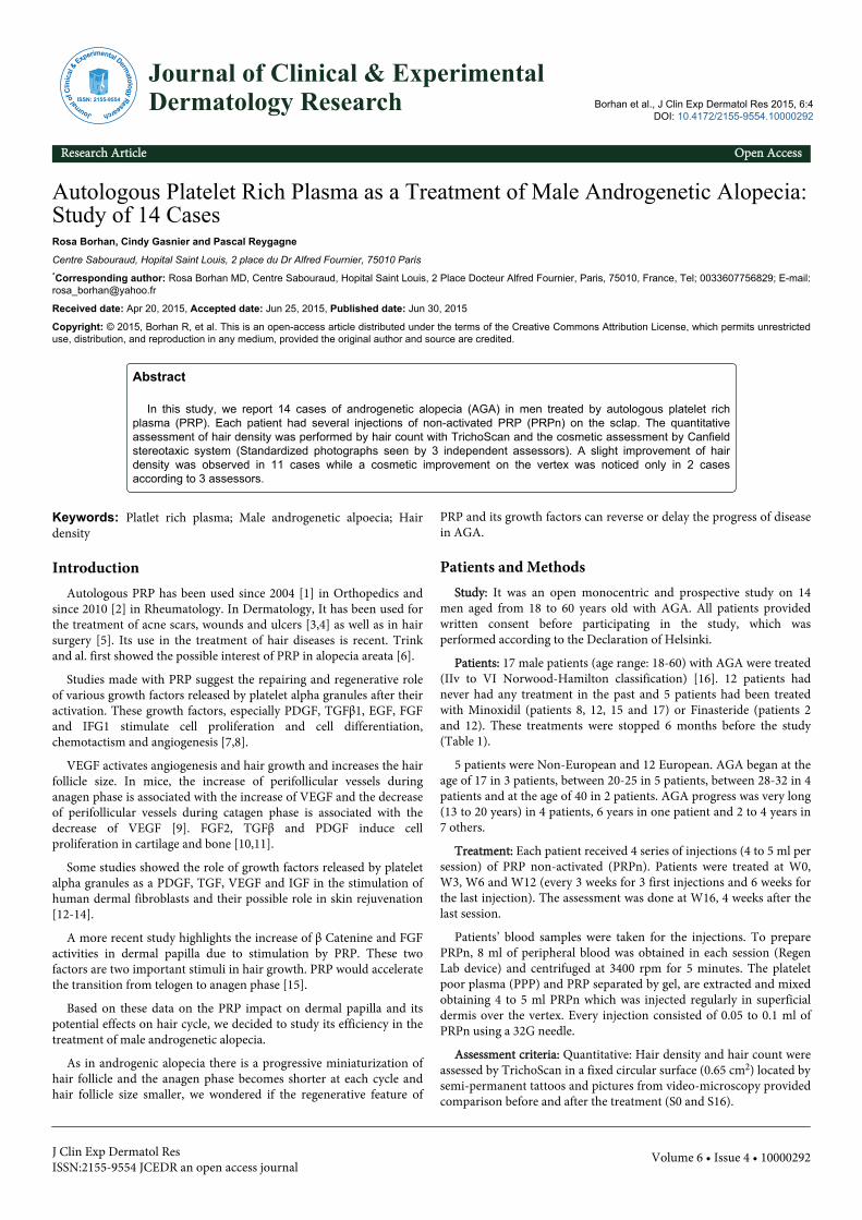
Autologous Platelet Rich Plasma as a Treatment of Male Androgenetic Alopecia:Study of 14 CasesRosa Borhan, Cindy Gasnier and Pascal Reygagne
Centre Sabouraud, Hopital Saint Louis, 2 place du Dr Alfred Fournier, 75010 Paris*Corresponding author: Rosa Borhan MD, Centre Sabouraud, Hopital Saint Louis, 2 Place Docteur Alfred Fournier, Paris, 75010, France, Tel; 0033607756829; E-mail:[email protected]
Received date: Apr 20, 2015, Accepted date: Jun 25, 2015, Published date: Jun 30, 2015
Copyright: © 2015, Borhan R, et al. This is an open-access article distributed under the terms of the Creative Commons Attribution License, which permits unrestricteduse, distribution, and reproduction in any medium, provided the original author and source are credited.
Abstract
In this study, we report 14 cases of androgenetic alopecia (AGA) in men treated by autologous platelet richplasma (PRP). Each patient had several injections of non-activated PRP (PRPn) on the sclap. The quantitativeassessment of hair density was performed by hair count with TrichoScan and the cosmetic assessment by Canfieldstereotaxic system (Standardized photographs seen by 3 independent assessors). A slight improvement of hairdensity was observed in 11 cases while a cosmetic improvement on the vertex was noticed only in 2 casesaccording to 3 assessors.
Keywords: Platlet rich plasma; Male androgenetic alpoecia; Hairdensity
IntroductionAutologous PRP has been used since 2004 [1] in Orthopedics and
since 2010 [2] in Rheumatology. In Dermatology, It has been used forthe treatment of acne scars, wounds and ulcers [3,4] as well as in hairsurgery [5]. Its use in the treatment of hair diseases is recent. Trinkand al. first showed the possible interest of PRP in alopecia areata [6].
Studies made with PRP suggest the repairing and regenerative roleof various growth factors released by platelet alpha granules after theiractivation. These growth factors, especially PDGF, TGFβ1, EGF, FGFand IFG1 stimulate cell proliferation and cell differentiation,chemotactism and angiogenesis [7,8].
VEGF activates angiogenesis and hair growth and increases the hairfollicle size. In mice, the increase of perifollicular vessels duringanagen phase is associated with the increase of VEGF and the decreaseof perifollicular vessels during catagen phase is associated with thedecrease of VEGF [9]. FGF2, TGFβ and PDGF induce cellproliferation in cartilage and bone [10,11].
Some studies showed the role of growth factors released by plateletalpha granules as a PDGF, TGF, VEGF and IGF in the stimulation ofhuman dermal fibroblasts and their possible role in skin rejuvenation[12-14].
A more recent study highlights the increase of β Catenine and FGFactivities in dermal papilla due to stimulation by PRP. These twofactors are two important stimuli in hair growth. PRP would acceleratethe transition from telogen to anagen phase [15].
Based on these data on the PRP impact on dermal papilla and itspotential effects on hair cycle, we decided to study its efficiency in thetreatment of male androgenetic alopecia.
As in androgenic alopecia there is a progressive miniaturization ofhair follicle and the anagen phase becomes shorter at each cycle andhair follicle size smaller, we wondered if the regenerative feature of
PRP and its growth factors can reverse or delay the progress of diseasein AGA.
Patients and MethodsStudy: It was an open monocentric and prospective study on 14
men aged from 18 to 60 years old with AGA. All patients providedwritten consent before participating in the study, which wasperformed according to the Declaration of Helsinki.
Patients: 17 male patients (age range: 18-60) with AGA were treated(IIv to VI Norwood-Hamilton classification) [16]. 12 patients hadnever had any treatment in the past and 5 patients had been treatedwith Minoxidil (patients 8, 12, 15 and 17) or Finasteride (patients 2and 12). These treatments were stopped 6 months before the study(Table 1).
5 patients were Non-European and 12 European. AGA began at theage of 17 in 3 patients, between 20-25 in 5 patients, between 28-32 in 4patients and at the age of 40 in 2 patients. AGA progress was very long(13 to 20 years) in 4 patients, 6 years in one patient and 2 to 4 years in7 others.
Treatment: Each patient received 4 series of injections (4 to 5 ml persession) of PRP non-activated (PRPn). Patients were treated at W0,W3, W6 and W12 (every 3 weeks for 3 first injections and 6 weeks forthe last injection). The assessment was done at W16, 4 weeks after thelast session.
Patients’ blood samples were taken for the injections. To preparePRPn, 8 ml of peripheral blood was obtained in each session (RegenLab device) and centrifuged at 3400 rpm for 5 minutes. The plateletpoor plasma (PPP) and PRP separated by gel, are extracted and mixedobtaining 4 to 5 ml PRPn which was injected regularly in superficialdermis over the vertex. Every injection consisted of 0.05 to 0.1 ml ofPRPn using a 32G needle.
Assessment criteria: Quantitative: Hair density and hair count wereassessed by TrichoScan in a fixed circular surface (0.65 cm2) located bysemi-permanent tattoos and pictures from video-microscopy providedcomparison before and after the treatment (S0 and S16).
Borhan et al., J Clin Exp Dermatol Res 2015, 6:4 DOI: 10.4172/2155-9554.10000292
Research Article Open Access
J Clin Exp Dermatol ResISSN:2155-9554 JCEDR an open access journal
Volume 6 • Issue 4 • 10000292
Journal of Clinical & ExperimentalDermatology ResearchJourna
l of C
linic
al &
Experimental Dermatology Research
ISSN: 2155-9554
Cosmetic: The assessment was done comparing standardized digitalphotographs taken before and after the treatment (S0 and S16). Thesephotographs were made by Canfield stereotaxic system [17]. One angleof incidence of the frontal anterior zone and the other, the vertex.Three assessors (2 independent) noted the qualitative change of hairusing a scale of 7 scores.*
*Scale based on 7 scores (-3: highly decreased, -2: moderatelydecreased, -1: slightly decreased, 0: unchanged, 1: slightly increased, 2:moderately increased, 3: highly increased.)
Self-assessment: a self-assessment questionnaire based on 5 criteriawas filled by each patient: change in hair texture (quality), change involume, shedding decrease, pigmentation and grow back. Also anotherquestionnaire regarding the quality of life (DLQI) before and aftertreatment (S0 and S16) was filled by patients.
Patient
Noorwid-HamiltonClassification scale
Age Treatmentprior tostudy
Platelets(peripheralblood)
Disease-onset(year-old)
Ethnicgroup
1 V 31 NO 200 G/l 17 European
3 IIv 20 NO 259 G/l 17 European
4 IIv 24 NO 233 G/l 20 European
5 Femalepattern
23 NO 231 G/l 17 Asian
6 III Vertex 33 NO 210 G/l 28 European
7 VI 59 NO 190 G/l 40 European
8 IV 43 Minoxidil 223 G/l 30 European
9 Femalepattern
28 NO 240 G/l 25 European
11 IV 58 NO 220 G/l 40 European
12 IV 25 Minoxidil+Finasteride
220 G/l 22 European
13 Femalepattern
36 NO 169 G/l 32 Arabian
14 III 31 NO 253 G/l 28 Pakistani
15 III 36 Minoxidil 190 G/l 32 European
17 III 28 Minoxidil 211 G/l 23 European
2 III 25 Finasteride 250 G/l 22 European
10 III 24 NO 219 G/l 22 African
16 Femalepattern
32 NO 311 G/l 29 Asian
Table 1: Patients’ characteristics at baseline.
Results2 of 17 patients included interrupted the study for personal reasons
after 2 and 3 injections (travelling…). One patient was retreated fromthe follow-up after the treatment. Finally 14 patients enrolled into thestudy, completed the treatment and were assessed at W0 and W16.
Quantitative assessmentMean hair density varies from 128.7 (at W0) to131.9 (at W16). The
mean increase of density is 3.1/0.65 cm2 or 2.9℅ (Table 2).
TrichoScan assessment showed an increase in hair density in 11patients (+0.50℅ to +10℅). Gain in hair number was more than 7℅ in3 patients. We observed a decrease of hair density in 3 patients (-1.44℅ to -1.72℅). Globally the number of new hair follicles was 33 and thenumber of vellus hairs was 16 in our study.
Hair Count (W0; W16)*
Patients Hair Gain Hair density (W0) Hair density(W16)
W0-W16
1 1.57 190 193 3
3 2.17 138 141 3
4 0.50 198 199 1
5 0.85 117 118 1
6 2.65 151 155 4
7 3.38 59 61 2
8 -1.44 69 68 -1
9 -1.72 116 114 -2
11 10 80 88 8
12 0.94 212 214 2
13 7.22 83 89 6
14 7.43 121 130 9
15 8.26. 150 160 10
17 -1.68 119 117 -2
MEAN 2.9 128.8 131.9 3.1
Standard Deviation 47.9 48 3.9
2 154 Studyinterrupted
10 119 Studyinterrupted
16 114 Studyinterrupted
*Surface of measured hair density: 0.65 cm2
Table 2: Hair density measured before and after treatment byTrichoScanTM.
Citation: Borhan R, Gasnier C, Reygagne P (2015) Autologous Platelet Rich Plasma as a Treatment of Male Androgenetic Alopecia: Study of 14Cases. J Clin Exp Dermatol Res 6: 292. doi:10.4172/2155-9554.10000292
Page 2 of 6
J Clin Exp Dermatol ResISSN:2155-9554 JCEDR an open access journal
Volume 6 • Issue 4 • 10000292
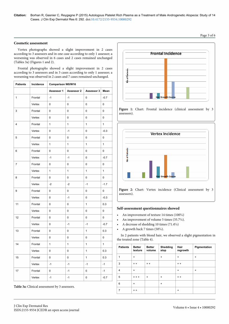
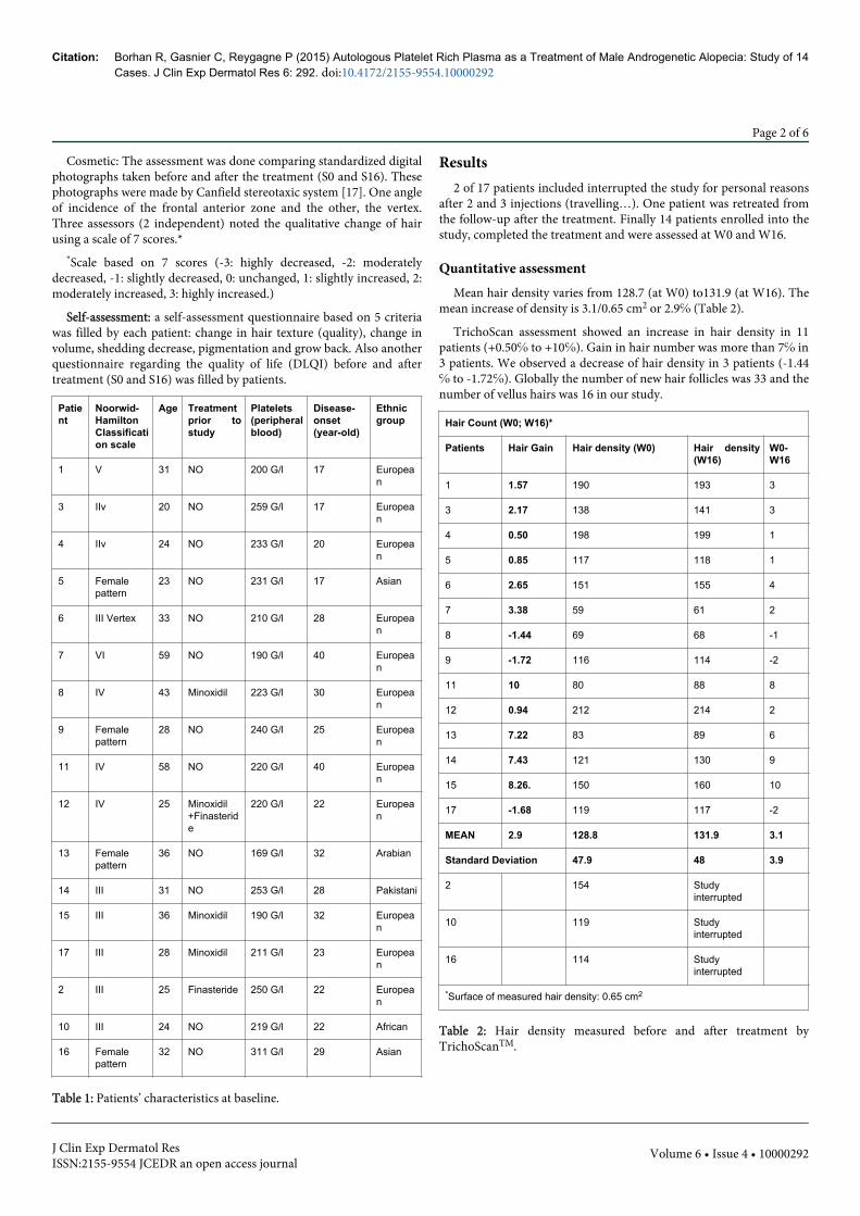
Cosmetic assessmentVertex photographs showed a slight improvement in 2 cases
according to 3 assessors and in one case according to only 1 assessor; aworsening was observed in 6 cases and 2 cases remained unchanged(Tables 3a) (Figures 1 and 2).
Frontal photographs showed a slight improvement in 2 casesaccording to 3 assessors and in 3 cases according to only 1 assessor; aworsening was observed in 2 cases and 7 cases remained unchanged.
Patients Incidence Comparison W0/W16
Assessor 1 Assessor 2 Assessor 3 Mean
1 Frontal -1 -1 0 -0.7
Vertex 0 0 0 0
3 Frontal 0 0 0 0
Vertex 0 0 0 0
4 Frontal 1 1 1 1
Vertex 0 -1 0 -0.3
5 Frontal 0 0 0 0
Vertex 1 1 1 1
6 Frontal 0 0 0 0
Vertex -1 -1 0 -0.7
7 Frontal 0 0 0 0
Vertex 1 1 1 1
8 Frontal 0 0 0 0
Vertex -2 -2 -1 -1.7
9 Frontal 0 0 0 0
Vertex 0 -1 0 -0.3
11 Frontal 0 0 1 0.3
Vertex 0 0 0 0
12 Frontal 0 0 0 0
Vertex 0 -1 -1 -0.7
13 Frontal 0 0 1 0.3
Vertex 0 0 0 0
14 Frontal 1 1 1 1
Vertex 0 0 1 0.3
15 Frontal 0 0 1 0.3
Vertex -1 -1 -1 -1
17 Frontal 0 -1 0 -1
Vertex -1 -1 0 -0.7
Table 3a: Clinical assessment by 3 assessors.
Figure 1: Chart: Frontal incidence (clinical assessment by 3assessors).
Figure 2: Chart: Vertex incidence (Clinical assessment by 3assessors).
Self-assessment questionnaires showed• An improvement of texture 14 times (100℅)• An improvement of volume 5 times (35.7℅).• A decrease of shedding 10 times (71.4℅)• A growth back 7 times (50℅).
In 2 patients with blond hair, we observed a slight pigmentation inthe treated zone (Table 4).
Patients Bettertexture
Bettervolume
Sheddingstop
Hairregrowth
Pigmentation
1 + + + +
3 + + + + + +
4 + + +
5 + + + + + + +
6 + +
7 + + +
Citation: Borhan R, Gasnier C, Reygagne P (2015) Autologous Platelet Rich Plasma as a Treatment of Male Androgenetic Alopecia: Study of 14Cases. J Clin Exp Dermatol Res 6: 292. doi:10.4172/2155-9554.10000292
Page 3 of 6
J Clin Exp Dermatol ResISSN:2155-9554 JCEDR an open access journal
Volume 6 • Issue 4 • 10000292
8 + + +
9 +
11 + + + + + + +
12 + + + + +
13 + +
14 + + + +
15 + + +
17 + + +
+ : slight change++ : moderate change +++ : significant change
Table 4: Patients self-assessments.
Questionnaires of quality of life before and after treatmentshow a slight improvement of quality of life in 8 patients
Patients 1 3 4 5 6 7 8 9 11 12 13 14 15 17
Before
treatment
0 0 0 0 0 0 0 4 0 1 5 0 0 0
After
Treatment
1 0 0 2 0 0 2 6 1 5 10 2 0 2
Table 5: Dermatology life quality index before and after treatment(DLQI).
DiscussionEven though our study was conducted without a control group, it
does not totally confirm the interest for the use of PRP in AGA alreadymentioned in two previous studies [18,19].
Our study showed an increase in hair density in 11 out of 14patients who completed the treatment. This change is slight though(2.9℅). In 3 cases of 14, it is higher than 7℅ and could be compared toresults obtained by reference treatments of AGA like Minoxidil andFinasteride [20,21]. Topical Minoxidil 5℅ (foam) induced a meanincrease of hair count of 13.4℅ in 16 weeks and Finasteride (oral) 1mg/day induced a mean increase of hair density of 11℅ in 12 months[21].
In the specific conditions of this study our results are less positivethan the ones of these two reference treatments of AGA. We observedthat 3 of 4 best responders were 31 to 36 -year-old and the bestresponse was seen in AGA stage III to IV. Among the best responders,3 patients had a very recent history of AGA (less than 4 years).
The hair density measurement was done only in vertex in the samezone in each patient (equal distance between two ears). We did notmeasure the hair density in frontal area and we do not know if the PRPacts differently in this zone.
The cosmetic assessment results with global photographs aredisappointing, and in this study the hair density did not allow toobtain cosmetically attractive results. Nevertheless, the clinical self-
assessment of patients showed a better hair texture in all cases and abetter growth back for half of them, as well as the interruption ofshedding in more than half of patients.
In this study, we used a regular injection technique covering all thevertex and the frontal area( 4 to 5ml of PRPn) in contrast with otherstudies where only a limited area was treated so that , in these studiesthe biologically active product in this limited zone is concentrated andprobably increases the effectiveness of the treatment [22].
Cervelli and al. have treated a group of male patients with AGA andhave obtained a mean gain of 27.7 hair/cm 2 or 19.41℅ [22]. Anotherrecently published study used a large volume of peripheral blood(60ml) and obtained high platelet concentrations (6 or 7 fold morethan peripheral blood) leading to better results [18]. This last studyshowed an improvement of hair volume in early AGA in men andwomen but the PRP was rich in leucocytes and the assessment wasdone through photographs without hair counting, hence it is subject tocriticism [18].
The use of PRP and the assessment of its effects raise issues ofdefinition and exact determination of its content. Regarding its nature,in this study we used non activated PRP (without adding CalciumGluconate or Calcium Citrate) considering that the centrifugation,also intradermal injections (micro blood vessels injuries), and thecontact with dermal fibroblasts would be sufficient to activate plateletsand to release growth factors [12,14,23]. The activated PRP ( PRPa) isobtained by adding Calcium Gluconate to PRPn which triggers therelease of growth factors by platelet α-granules. Therefore PRPa couldbe associated with increased production of growth factors which inturn would show more efficacy.Two studies [6,22] using PRPa in twodifferent indications had interesting results: Cervelli and al. usingPRPa in AGA obtained almost.
19℅ of gain in mean hair density while Trink and al. using PRPa inalopecia areata noticed better results compared to patients treated with2.5mg/ml of Triamcinolone acetonide (60℅ of complete remissioncompared to 27℅). This suggests that PRPa could be more efficientthan PRPn. Consequently it would be interesting to perform a studycomparing these two types of PRP.
Regarding its content, it is essential to measure after centrifugationand before injection in each session, platelet concentration and ideallygrowth factors to assess both the quality and the quantity of what toinject. We know that there are a wide range of devices on the market( 16 various systems without counting blood banks and privatelaboratories ) and the heterogeneity of these systems leads to a largevariability of platelet and growth factors concentrations, thus makingany comparison impossible. A recent study compared this variabilitybetween 5 systems and the authors propose to use “platelet dose” as aterminology to standardize the technique and determine plateletconcentration of PRP. In fact, the quantity of injected cells is the onlyparameter correlated to clinical efficacy as shown with bone marrowtransplant in hematology [25]. They propose the followingformulas:Injected platelet dose in PRP (X 10)=volume of PRP obtained(ml) × Platelet concentration in PRP (G/ml)/1000
Growth factor dose (pg)=volume of PRP obtained (ml) × Growthfactors concentration in PRP (pg/ml)
We know there is a positive correlation between platelet dose andgrowth factor dose for all growth factors (VEGF, PDGF, EGF, TGFβ1)[24]. Another interesting notion is “platelet capture efficacy” thatcorrelates obtained platelet concentration in the sample with platelet
Citation: Borhan R, Gasnier C, Reygagne P (2015) Autologous Platelet Rich Plasma as a Treatment of Male Androgenetic Alopecia: Study of 14Cases. J Clin Exp Dermatol Res 6: 292. doi:10.4172/2155-9554.10000292
Page 4 of 6
J Clin Exp Dermatol ResISSN:2155-9554 JCEDR an open access journal
Volume 6 • Issue 4 • 10000292
concentration in peripheral blood. These notions allow to biologicallycharacterize the obtained PRP.
Once PRP biologically characterized, we have to determine theoptimal platelet concentration for each indication. The optimalconcentration is different for each tissue. For example for Achillestendinopathy or bone surgery (anterior cruciate ligament) the optimalconcentration is 1 to 6 fold more than peripheral blood concentration[26,27]. In studies done in dermatology, the platelet concentration was1.6 fold more than in peripheral blood. Once we have determined theoptimal platelet dose, we can resolve the problem of volume to inject,because the volume depends on platelet dose contained in one ml ofPRP.
Another aspect consists of taking into consideration the half-life ofplatelets (7 to 10 days) and to perform more frequent sessions tomaintain long lasting stimulation by growth factors in dermis,especially when we use a small amount of PRPn.
Nevertheless, It is important to determine the frequency and thenumber of sessions not only based on half-life of platelets but also upto patient background. The role of age, AGA staging and probablycorrelation with androgenic activity may be considered. These factorscould interfere with PRP efficiency. For example, in a young subject,the regenerative capacity is high but androgenic activity is equallyhigh. These two factors can thwart variously and alter PRP efficacy.One the other hand AGA staging and disease progress are important.PRP seems to be less effective in late AGA and/or rapidly progressingAGA.
In this study we observed a few side effects such as erythema orheadaches. Two patients with seborrheic dermatitis noticed animprovement with PRP (probably anti-inflammatory effect?) and weobserved a darkening of treated hair in two patients with blond hair.
We did not realize biopsies as Cervelli and al. did. In their study,scalp biopsies showed the increase of hair follicle number and theincrease of epidermis thickness confirming clinical data anddermoscopic measurements [22]. The angiogenic role of PRP inendothelial cells is known at high concentration (1500000 platele/μl)[28]. It would be interesting to realize further studies to measuremolecular and cellular effect of PRP in scalp and skin.
Kang et al. obtained better results in AGA in male and femalepatients by adding bone marrow derived CD34+ cells to activated PRPbut this study does not permit to assess the efficiency of PRP solely.This technique deserves further studies [29].
Unlike the conventional treatments of AGA, our approach does notrequire long term therapy, potential side effects and/or high degree ofcompliance.
Patients' compliance is not always very high with Minoxidil,because of cosmetic effects and long term use. Also Finasteride hasraised concerns for some patients regarding its sexual side effects(decreased libido, hypofertility). PRP as an autologous and non-chemical treatment is better accepted by patients who wish to avoidthese disadvantages.
Consequently for non-responders to these therapies, PRP can be analternative. Also as the mechanism of action of PRP is different fromthe two other treatments, we may have an additional positive effect byusing PRP in responders to Minoxidil and Finasteride. In terms ofcosts, PRP may be more cost effective in long term than conventionaltherapies.
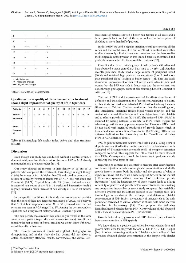
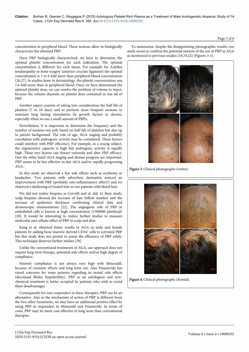
To summarize, despite the disappointing photographic results, ourstudy seems to confirm the potential interest of the use of PRP in AGAas mentioned in previous studies [18,19,22] (Figures 3-5).
Figure 3: Clinical photographs (vertex).
Figure 4: Clinical photographs (frontal).
Citation: Borhan R, Gasnier C, Reygagne P (2015) Autologous Platelet Rich Plasma as a Treatment of Male Androgenetic Alopecia: Study of 14Cases. J Clin Exp Dermatol Res 6: 292. doi:10.4172/2155-9554.10000292
Page 5 of 6
J Clin Exp Dermatol ResISSN:2155-9554 JCEDR an open access journal
Volume 6 • Issue 4 • 10000292
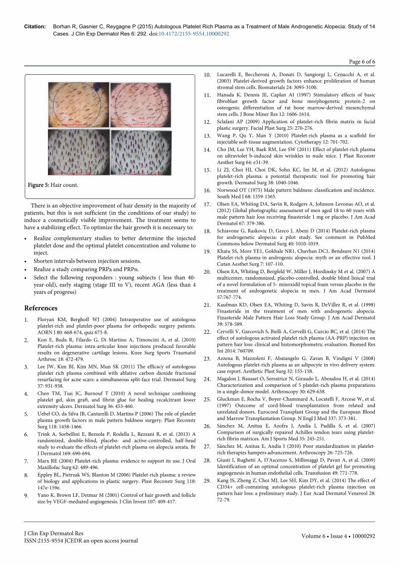
Figure 5: Hair count.
There is an objective improvement of hair density in the majority ofpatients, but this is not sufficient (in the conditions of our study) toinduce a cosmetically visible improvement. The treatment seems tohave a stabilizing effect. To optimize the hair growth it is necessary to:
• Realize complementary studies to better determine the injectedplatelet dose and the optimal platelet concentration and volume toinject.
• Shorten intervals between injection sessions.• Realize a study comparing PRPa and PRPn.• Select the following responders : young subjects ( less than 40-
year-old), early staging (stage III to V), recent AGA (less than 4years of progress)
References1. Floryan KM, Berghoff WJ (2004) Intraoperative use of autologous
platelet-rich and platelet-poor plasma for orthopedic surgery patients.AORN J 80: 668-674, quiz 675-8.
2. Kon E, Buda R, Filardo G, Di Martino A, Timoncini A, et al. (2010)Platelet-rich plasma: intra-articular knee injections produced favorableresults on degenerative cartilage lesions. Knee Surg Sports TraumatolArthrosc 18: 472-479.
3. Lee JW, Kim BJ, Kim MN, Mun SK (2011) The efficacy of autologousplatelet rich plasma combined with ablative carbon dioxide fractionalresurfacing for acne scars: a simultaneous split-face trial. Dermatol Surg37: 931-938.
4. Chen TM, Tsai JC, Burnouf T (2010) A novel technique combiningplatelet gel, skin graft, and fibrin glue for healing recalcitrant lowerextremity ulcers. Dermatol Surg 36: 453-460.
5. Uebel CO, da Silva JB, Cantarelli D, Martins P (2006) The role of plateletplasma growth factors in male pattern baldness surgery. Plast ReconstrSurg 118: 1458-1466.
6. Trink A, Sorbellini E, Bezzola P, Rodella L, Rezzani R, et al. (2013) Arandomized, double-blind, placebo- and active-controlled, half-headstudy to evaluate the effects of platelet-rich plasma on alopecia areata. BrJ Dermatol 169: 690-694.
7. Marx RE (2004) Platelet-rich plasma: evidence to support its use. J OralMaxillofac Surg 62: 489-496.
8. Eppley BL, Pietrzak WS, Blanton M (2006) Platelet-rich plasma: a reviewof biology and applications in plastic surgery. Plast Reconstr Surg 118:147e-159e.
9. Yano K, Brown LF, Detmar M (2001) Control of hair growth and folliclesize by VEGF-mediated angiogenesis. J Clin Invest 107: 409-417.
10. Lucarelli E, Beccheroni A, Donati D, Sangiorgi L, Cenacchi A, et al.(2003) Platelet-derived growth factors enhance proliferation of humanstromal stem cells. Biomaterials 24: 3095-3100.
11. Hanada K, Dennis JE, Caplan AI (1997) Stimulatory effects of basicfibroblast growth factor and bone morphogenetic protein-2 onosteogenic differentiation of rat bone marrow-derived mesenchymalstem cells. J Bone Miner Res 12: 1606-1614.
12. Sclafani AP (2009) Application of platelet-rich fibrin matrix in facialplastic surgery. Facial Plast Surg 25: 270-276.
13. Wang P, Qu Y, Man Y (2010) Platelet-rich plasma as a scaffold forinjectable soft-tissue augmentation. Cytotherapy 12: 701-702.
14. Cho JM, Lee YH, Baek RM, Lee SW (2011) Effect of platelet-rich plasmaon ultraviolet b-induced skin wrinkles in nude mice. J Plast ReconstrAesthet Surg 64: e31-39.
15. Li ZJ, Choi HI, Choi DK, Sohn KC, Im M, et al. (2012) Autologousplatelet-rich plasma: a potential therapeutic tool for promoting hairgrowth. Dermatol Surg 38: 1040-1046.
16. Norwood OT (1975) Male pattern baldness: classification and incidence.South Med J 68: 1359-1365.
17. Olsen EA, Whiting DA, Savin R, Rodgers A, Johnson-Levonas AO, et al.(2012) Global photographic assessment of men aged 18 to 60 years withmale pattern hair loss receiving finasteride 1 mg or placebo. J Am AcadDermatol 67: 379-386.
18. Schiavone G, Raskovic D, Greco J, Abeni D (2014) Platelet-rich plasmafor androgenetic alopecia: a pilot study. See comment in PubMedCommons below Dermatol Surg 40: 1010-1019.
19. Khatu SS, More YE1, Gokhale NR1, Chavhan DC1, Bendsure N1 (2014)Platelet-rich plasma in androgenic alopecia: myth or an effective tool. JCutan Aesthet Surg 7: 107-110.
20. Olsen EA, Whiting D, Bergfeld W, Miller J, Hordinsky M et al. (2007) Amulticenter, randomnized, placebo-controlled, double blind linical trialof a novel formulation of 5- minoxidil topical foam versus placebo in thetreatment of androgenetic alopecia in men. J Am Acad Dermatol57:767-774.
21. Kaufman KD, Olsen EA, Whiting D, Savin R, DeVillez R, et al. (1998)Finasteride in the treatment of men with androgenetic alopecia.Finasteride Male Pattern Hair Loss Study Group. J Am Acad Dermatol39: 578-589.
22. Cervelli V, Garcovich S, Bielli A, Cervelli G, Curcio BC, et al. (2014) Theeffect of autologous activated platelet rich plasma (AA-PRP) injection onpattern hair loss: clinical and histomorphometric evaluation. Biomed ResInt 2014: 760709.
23. Azzena B, Mazzoleni F, Abatangelo G, Zavan B, Vindigni V (2008)Autologous platelet-rich plasma as an adipocyte in vivo delivery system:case report. Aesthetic Plast Surg 32: 155-158.
24. Magalon J, Bausset O, Serratrice N, Giraudo L, Aboudou H, et al. (2014)Characterization and comparison of 5 platelet-rich plasma preparationsin a single-donor model. Arthroscopy 30: 629-638.
25. Gluckman E, Rocha V, Boyer-Chammard A, Locatelli F, Arcese W, et al.(1997) Outcome of cord-blood transplantation from related andunrelated donors. Eurocord Transplant Group and the European Bloodand Marrow Transplantation Group. N Engl J Med 337: 373-381.
26. Sánchez M, Anitua E, Azofra J, Andía I, Padilla S, et al. (2007)Comparison of surgically repaired Achilles tendon tears using platelet-rich fibrin matrices. Am J Sports Med 35: 245-251.
27. Sánchez M, Anitua E, Andia I (2010) Poor standardization in platelet-rich therapies hampers advancement. Arthroscopy 26: 725-726.
28. Giusti I, Rughetti A, D'Ascenzo S, Millimaggi D, Pavan A, et al. (2009)Identification of an optimal concentration of platelet gel for promotingangiogenesis in human endothelial cells. Transfusion 49: 771-778.
29. Kang JS, Zheng Z, Choi MJ, Lee SH, Kim DY, et al. (2014) The effect ofCD34+ cell-containing autologous platelet-rich plasma injection onpattern hair loss: a preliminary study. J Eur Acad Dermatol Venereol 28:72-79.
Citation: Borhan R, Gasnier C, Reygagne P (2015) Autologous Platelet Rich Plasma as a Treatment of Male Androgenetic Alopecia: Study of 14Cases. J Clin Exp Dermatol Res 6: 292. doi:10.4172/2155-9554.10000292
Page 6 of 6
J Clin Exp Dermatol ResISSN:2155-9554 JCEDR an open access journal
Volume 6 • Issue 4 • 10000292