Embed Size (px)
Citation preview
At
Sa
b
a
ARRAA
KASHB
1
aSo
2
prniortcosar(u
h2c
CASE REPORT – OPEN ACCESSInternational Journal of Surgery Case Reports 28 (2016) 71–73
Contents lists available at ScienceDirect
International Journal of Surgery Case Reports
journa l h omepage: www.caserepor ts .com
ngiomyxolipoma of the right sub-brow: Case report with review ofhe literature
ami Al-Bdairat (M.D.)a,∗, Isam Sh. Alrawia, Mohammed A. Obaida, Yahia F. Dajanib
Eye Specialty Hospital, Amman, JordanGrand Medical Laboratories, Amman, Jordan
r t i c l e i n f o
rticle history:eceived 27 June 2016eceived in revised form 4 September 2016ccepted 5 September 2016vailable online 22 September 2016
eywords:
a b s t r a c t
INTRODUCTION: Angiomyxolipoma (AML) is a rare variant of benign lipoma with characteristichistopathological and immuno-histochemical features. It consists of fatty tissue admixed with myxoidstroma and blood vessels. It was first described by Mai et al. in 1996 [1], with a total number of 19 casesreported since.PRESENTATION: This is the first report of an AML in subcutaneous tissue of the face, presenting as a 4-month old cystic lesion in a 78-year old lady. Diagnosis was based on radiological and histopathological
ngiomyxolipomaub-browistopathologyenign
with cytochemical findings.DISCUSSION: It is important to distinguish this lesion as distinct from malignant subcutaneous lesions offatty tissue, especially with short history as seen in our case.CONCLUSION: Precise diagnosis of angiomyxolipoma is important to avoid unnecessary investigations,stress and misdiagnosis of myxoid liposarcoma.
© 2016 The Authors. Published by Elsevier Ltd on behalf of IJS Publishing Group Ltd. This is an openhe CC
access article under t. Introduction
Lipoma is the most common benign mesenchymal tumor indults, which may occur singly or multiple in any part of the body.everal variants of lipoma have been described and angiomyx-lipoma is one rare subtype [2].
. Case report
A 78-year-old lady attended the Department of Orbit & Oculo-lasty in our hospital, complaining of a painless swelling on theight side of her forehead, involving the right sub-brow region. Sheoticed the lesion four months earlier with progressive increase
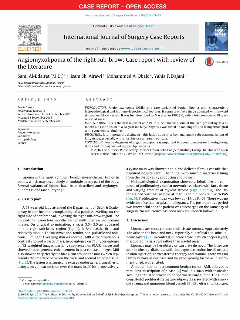
n size. On physical examination, a mass 3.0 × 3.5 cm appearedn the right sub-brow region (Fig. 1). It felt elastic, firm andelatively mobile. The mass was non-tender, non-pulsatile and non-ransilluminant. Overlying skin was normal. MRI with intra-venousontrast showed a cystic mass, hypo-intense on T1, hyper-intensen T2-weighted images, partially suppressed on FLAIR images andhowed heterogeneous enhancement in post contrast images, MRIlso showed very clearly the black-rim around the mass which rep-
esents the interface between the mass and normal adipose tissue,Fig. 2). The lesion was surgically excised under general anesthesia,sing a curvilinear incision over the mass itself. Intra-operatively,∗ Corresponding author.E-mail address: [email protected] (S. Al-Bdairat).
ttp://dx.doi.org/10.1016/j.ijscr.2016.09.024210-2612/© 2016 The Authors. Published by Elsevier Ltd on behalf of IJS Publishing Greativecommons.org/licenses/by-nc-nd/4.0/).
BY-NC-ND license (http://creativecommons.org/licenses/by-nc-nd/4.0/).
a cystic mass was showed a thin and delicate fibrous capsule thatruptured despite careful handling, with mucoid material issuingfrom the cystic cavity producing a foul smell.
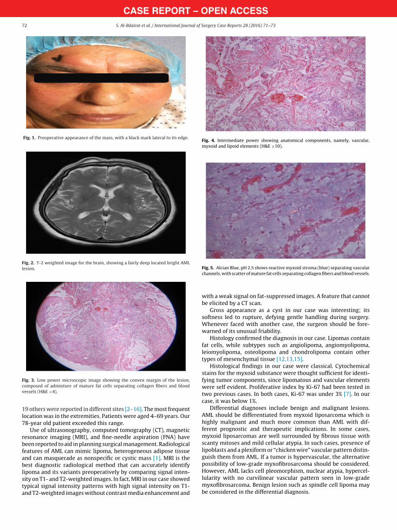
Histopathological examination showed a lobular lesion com-posed of proliferating vascular network associated with fatty tissueand varying amount of myxoid stroma (Figs. 3 and 4). The lat-ter stained with Alcian blue at pH2.5 and did not stain with PAS(Fig. 5). Proliferative index was low at <1% by Ki-67. There was noevidence of cellular atypia or malignancy. The postoperative periodwas uneventful and the patient was discharged on the same day ofsurgery. No recurrence has been seen at 6-month follow up.
3. Discussion
Lipomas are most common soft tissue tumors. Approximately15% arise in the head and neck, especially superficial and subcuta-neous layers [17]. In contrast, our case arose in much deeper layers,masquerading as a cyst rather than a solid mass.
Lipomas may be hereditary or can arise de novo. The latter areseen in obesity, diabetes, radiation exposure, endocrine disorders,insulin injection, corticosteroid therapy and trauma. There was nofamily history in our case and no predisposing factor as in afore-mentioned, was elicited.
Although lipoma is a common benign tumor, AML subtype is
rare. First description of a case [2] was in a man with testicularswelling that later proved to be spermatic cord tumor. The tumorconsisted of proliferating mature adipocytes associated with a myx-oid stroma and numerous blood vessels [2–15]. After this first case,roup Ltd. This is an open access article under the CC BY-NC-ND license (http://
CASE REPORT – OPEN ACCESS72 S. Al-Bdairat et al. / International Journal of Surgery Case Reports 28 (2016) 71–73
Fig. 1. Preoperative appearance of the mass, with a black mark lateral to its edge.
Fig. 2. T-2 weighted image for the brain, showing a fairly deep located bright AMLlesion.
Fcv
1l7
rbfablsta
Fig. 4. Intermediate power showing anatomical components, namely, vascular,myxoid and lipoid elements (H&E ×10).
ig. 3. Low power microscopic image showing the convex margin of the lesion,omposed of admixture of mature fat cells separating collagen fibers and bloodessels (H&E ×4).
9 others were reported in different sites [2–16]. The most frequentocation was in the extremities. Patients were aged 4–69 years. Our8-year old patient exceeded this range.
Use of ultrasonography, computed tomography (CT), magneticesonance imaging (MRI), and fine-needle aspiration (FNA) haveeen reported to aid in planning surgical management. Radiologicaleatures of AML can mimic lipoma, heterogeneous adipose tissuend can masquerade as nonspecific or cystic mass [1]. MRI is theest diagnostic radiological method that can accurately identify
ipoma and its variants preoperatively by comparing signal inten-
ity on T1- and T2-weighted images. In fact, MRI in our case showedypical signal intensity patterns with high signal intensity on T1-nd T2-weighted images without contrast media enhancement andFig. 5. Alcian Blue, pH 2.5 shows reactive myxoid stroma (blue) separating vascularchannels, with scatter of mature fat cells separating collagen fibers and blood vessels.
with a weak signal on fat-suppressed images. A feature that cannotbe elicited by a CT scan.
Gross appearance as a cyst in our case was interesting; itssoftness led to rupture, defying gentle handling during surgery.Whenever faced with another case, the surgeon should be fore-warned of its unusual friability.
Histology confirmed the diagnosis in our case. Lipomas containfat cells, while subtypes such as angiolipoma, angiomyolipoma,leiomyolipoma, osteolipoma and chondrolipoma contain othertypes of mesenchymal tissue [12,13,15].
Histological findings in our case were classical. Cytochemicalstains for the myxoid substance were thought sufficient for identi-fying tumor components, since lipomatous and vascular elementswere self evident. Proliferative index by Ki-67 had been tested intwo previous cases. In both cases, Ki-67 was under 3% [7]. In ourcase, it was below 1%.
Differential diagnoses include benign and malignant lesions.AML should be differentiated from myxoid liposarcoma which ishighly malignant and much more common than AML with dif-ferent prognostic and therapeutic implications. In some cases,myxoid liposarcomas are well surrounded by fibrous tissue withscanty mitoses and mild cellular atypia. In such cases, presence oflipoblasts and a plexiform or “chicken wire” vascular pattern distin-guish them from AML. If a tumor is hypervascular, the alternativepossibility of low-grade myxofibrosarcoma should be considered.However, AML lacks cell pleomorphism, nuclear atypia, hypercel-lularity with no curvilinear vascular pattern seen in low-grade
myxofibrosarcoma. Benign lesion such as spindle cell lipoma maybe considered in the differential diagnosis.
– Oal of S
4
ftmrrn
C
F
E
C
A
R
G
[
[
[
[
[
[
[
[
OTpc
CASE REPORTS. Al-Bdairat et al. / International Journ
. Conclusion
Angiomyxolipoma is a rare variant of lipoma. Cases were mostrequently reported in extremities. Precise diagnosis is importanto avoid unnecessary investigations, stress and misdiagnosis of
yxoid liposarcoma. Our case, being the oldest (78 year-old) evereported, is the first to have facial location and myxoid contenteacting to connective tissue mucin stain. Awareness of the diag-osis should be stressed.
onflicts of interest
None.
unding
None.
thical approval
Consent form, which was signed by the patient.
onsent
Informed consent was signed by the patient.
uthor contribution
Sami AL-Bdairat: study concept ,data analysis.Isam Alrawi : Data analysis and interpretation.Mohammad Obaid : Manuscript preparation and editing.Yahia dajani: data collection, quality control.
egistration of research studies
This is case report not a clinical study.
uarantor
Sami AL-Bdairat.
pen Accesshis article is published Open Access at sciencedirect.com. It is distribermits unrestricted non commercial use, distribution, and reproductredited.
PEN ACCESSurgery Case Reports 28 (2016) 71–73 73
References
[1] K.T. Mai, H.M. Yazdi, J.P. Collins, Vascular myxolipoma (angiomyxolipoma) ofthe spermatic cord, Am. J. Surg. Pathol. 20 (1996) 1145–1148.
[2] S.C. Debnath, A. Saikia, Lipoma of the parotid gland extending from thesuperficial to the deep lobe: a rarity, Br. J. Oral Maxillofac. Surg. 48 (2010)203–204.
[3] M. Zamecnik, Vascular myxolipoma (angiomyxolipoma) of subcutaneoustissue, Histopathology 34 (1999) 180–181.
[4] O. Okafor, A. Panizo, J. Pardo-Mindan, Angiomyxolipoma: a variant of lipoma,Rev. Esp. Patol. 33 (2000) 41–45.
[5] R. Sciot, M. Debiec-Rychter, I. De Wever, A. Hagemeijer, Angiomyxolipomashares cytogenetic changes with lipoma, spindle cell/pleomorphic lipoma andmyxoma, Virchows Arch. 438 (2001) 66–69.
[6] J.C. Tardio, L.M. Martin-Fragueiro, Angiomyxolipoma (vascular myxolipoma)of subcutaneous tissue, Am. J. Dermatopathol. 26 (2004) 222–224.
[7] H.W. Lee, D.K. Lee, M.W. Lee, et al., Two cases of angiomyxolipoma (vascularmyxolipoma) of subcutaneous tissue, J. Cutan. Pathol. 32 (2005) 379–382.
[8] Al Shraim, M. Hasan, A. Hawan, K. Radad, R. Eid, Plantar angiomyxolipoma ina child, BMJ Case Rep. (2011) 8.
[9] Y.S. Kang, W.S. Choi, U.H. Lee, et al., A case of multiple angiomyxolipoma,Korean J. Dermatol. 46 (2008) 1090–1095.
10] M. Song, S. Seo, D. Jung, et al., Angiomyxolipoma (vascular myxolipoma) ofsubcutaneous tissue, Ann. Dermatol. 21 (2009) 189–192.
11] P. Sanchez Sambucety, T.A. Alonso, P.G. Agapito, et al., Subungualangiomyxolipoma, Dermatol. Surg. 33 (2007) 508–509.
12] P.F. Bergin, C. Milchteim, G.P. Beaulieu, K.A. Brindle, A.M. Schwartz, C.R.Faulks, Intra-articular knee mass in a 51-year-old woman, Orthopedics 34 (3)(2011) 224–231.
13] H.J. Kim, I. Yang, A.Y. Jung, J.H. Hwang, M.K. Shin, Angiomyxolipoma (vascularmyxolipoma) of the knee in a 9-year-old boy, Pediatr. Radiol. 40 (1) (2010)30–33.
14] G. Martínez-Mata, M.F. Rocío, L.E. Juan, A.O. Paes, M.T. Adalberto,Angiomyxolipoma (vascular myxolipoma) of the oral cavity. Report of a caseand review of the literature, Head Neck Pathol. 5 (2) (2011) 184–187.
15] M. Pukar, Angiomyxolipoma of transverse colon – a case report, Turk. J.Gastroenterol. 23 (2) (2012) 156–159.
16] S. Hantous-Zannad, H. Neji, A. Zidi, E. Braham, I. Baccouche, M.S. Boudaya, K.Ben Miled-M’rad, Posterior mediastinal angiomyxolipoma with spinal canalextension, Tunis. Med. 90 (11) (2012) 816–818.
17] F. Dispenza, A. De Stefano, G. Romano, A. Mazzoni, Post-Traumatic lipoma ofthe parotid gland: case report, Acta Otorhinolaryngol. Ital. 28 (2008) 87–88.
uted under the IJSCR Supplemental terms and conditions, whichion in any medium, provided the original authors and source are