Embed Size (px)
Citation preview
陨灶贼 允 韵责澡贼澡葬造皂燥造熏 灾燥造援 5熏 晕燥援 2熏 Apr.18, 圆园12 www. IJO. cn栽藻造押8629原愿圆圆源缘员苑圆 8629-83085628 耘皂葬蚤造押ijopress岳员远猿援糟燥皂
Implantation of Iakymenko keratoprosthesis inpatients with severe ocular injury
窑Clinical Research窑
Foundation items: Supported by National Natural ScienceFoundation of China (No.81000368); Medical ScientificResearch Foundation of Guangdong Province, China(B2008091, A2011327)1Department of Ophthalmology, the First Affiliated Hospitalof Jinan University, Guangzhou 510632, GuangdongProvince, China2The Filatov Institute of Eye Disease and Tissue Therapy,Odessa, Ukraine3Institute of Ophthalmology, Medical College, JinanUniversity, Guangzhou 510632, Guangdong Province, China4Key Laboratory for Regenerative Medicine, Ministry ofEducation, Jinan University, Guangzhou 510632, GuangdongProvince, China5Department of Ophthalmology, the Third AffiliatedHospital of Jinan University, Zhuhai 519000, GuangdongProvince, China6Henan Eye Institute, Zhengzhou 450032, Henan Province,ChinaCorrespondence to: Jin-Tang Xu. Department of Ophthalmo-logy, The First Affiliated Hospital of Jinan University,Guangzhou 510632, Guangdong Province, China. [email protected]: 2011-12-19 Accepted: 2012-03-20
Abstract·AIM: To present the results of implantation of Iakymenko
keratoprosthesis in five patients with vascularized cornealleukoma caused by severe ocular injury.
·METHODS: Iakymenko keratoprosthesis was implanted into
5 eyes of 5 patients: 4 patients were suffered from chemicalburns and 1 patient from explosive injury. The preoperative
visual acuity ranged from light perception to hand motion.The implantation surgery was composed of two-stageprocedures. The follow-up period was from 9 months to 11years. The outcome measures were visual acuity, retention,and complications of the keratoprosthesis.
· RESULTS: Vision improvements were achieved in most
patients. All keratoprosthesis were retained within thefollow-up period. Corneal melting occurred in one patient and
fibrous closure in another patient, both of which were
successfully treated. Retinal detachment occurred in one
patient after surgery.
·CONCLUSION: Iakymenko keratoprosthesis seems to be a
promising alternative for the patients with severe corneal
injury, but further investigation is needed to evaluate the role
of Iakymenko keratoprosthesis.
·KEYWORDS:Iakymenko keratoprosthesis;visual rehabilitation;
complications
DOI:10.3980/j.issn.2222-3959.2012.02.10
Pan HW, Iakymenko S, Xu JT, Hou GH, Sun BJ, Zheng AN.
Implantation of Iakymenko keratoprosthesis in patients with severe
ocular injury. 2012;5(2):167-171
INTRODUCTION
C orneal disease is one of the most important causes ofblindness. For many corneal diseases such as corneal
scar, keratoconus and endothelial failure, penetratingkeratoplasty yields acceptable results. However, somecomplicated corneal lesions with limbal stem celldeficiency, severe neovascularization or repeated cornealgraft failure, are poor candidates for penetratingkeratoplasty. Implantation of a synthetic cornealreplacement, namely keratoprosthesis, has been developedfor treatment of such diseases. Now many types ofkeratoprostheses have been designed and applied in theclinical practice, from the totally synthetic Bostonkeratoprothesis, to the combined synthetic optic andbiological frame of the osteo-odonto keratoprothesis(OOKP) and the entirely biological tissue engineeredcornea. The indications for this procedure have beenexpanded for more corneal diseases including Stevens Johnson syndrome, chemical burns, disorders with limbalstem cell deficiency, ocular cicatricial pemphigoid, andsevere corneal vascularization.The Iakymenko keratoprosthesis (I-KPro) was developed inFilatov Institute of Eye Disease & Tissue Therapy, Ukraine[1, 2].We learned the surgical skill from Dr. Iakymenko and
167
introduced it into China. From 1999, we performed I-KProimplantation in 5 patients with vascularized cornealleukoma caused by severe chemical injury or explosiveinjury. We now present the result of I-KPro implantationwith follow-up of up to 11 years.MATERIALS AND METHODSMaterials Inclusion criteria for this study were severecorneal diseases with poor prognosis for keratoplastyincluding chemical injury, allograft failure and deep cornealvascularization, visual acuity not better than 20/500,potential for significant visual improvement, blindness in thecontralateral eye and age older than 18 years. Patients withactive inflammation, uncontrolled raised intraocularpressure, cicatrizing ocular diseases such as ocularcicatricial pemphigoid or Stevens-Johnson syndrome, andultrasound evidence of significant posterior segmentpathology were excluded. Ethics board committee approvalwas obtained for the study. All patients provided writteninformed consent. Five patients with severe vascularizedcorneal leucoma were included in this study. Thekeratoprothesis implantation was performed from 1999 andfollow-up duration ranged from 9 months to 11 years.MethodsSurgical technique The I-KPro was composed of a centralrigid polymethyl methacrylate (PMMA) optical cylinder anda titanium-tantalum frame. The optic cylinder had adiameter of 3.5mm in the anterior part and 2.6mm in theposterior part (Figure 1). The I-KPro implantation surgerywas composed of two stage procedures, both of which wereperformed under local anesthesia. In stage I procedure, afterexposing the ocular surface with lid retraction suture, asuperior, 270 degree of conjunctival peritomy and recessionwere performed to make a conjunctival flap. Flieringafixation ring was fixed with 6-0 black silk, then a superior270° scleral incision at approximately 1.0mm posterior tothe limbus was performed and extended into the cornea todivide the cornea into two layers at maximal possible depth,superficial and deep, using a lamellar dissecting blade. Thedissection was then extended into the inferior cornea so thata central corneal pocket was created. For preparation of theauricular cartilage, the ear skin was incised to expose theunderlying cartilage after infiltration anesthesia, and then acartilage graft was obtained using a 9mm trephine followedby clearance of the superficial connective tissues. A trephinewith similar diameter of the optic cylinder of I-KPro,3.3mm, was used to make a hole in the center of thecartilage graft. After that, the optic cylinder of Iakymenkokeratoprosthesis was fixed in the hole of auricular cartilage,and placed in gentamycin-containing saline for use. The
superficial corneal flap was then reflected inferiorly toexpose the deep cornea, to allow trephination of the centralposterior lamella with a 2.5mm trephine to enter the anteriorchamber. The piece of posterior lamella was removed.Cataract extraction (2 patients) was performed through thetrephination hole in central posterior cornea after anteriorcapsulorhexis (1 case), in the 2nd case cataract wasremoved in the capsule through additional 180° limbalincision of posterior corneal layers. Then the I-KPro withauricular cartilage was placed between the two layers withinthe pocket, the optic cylinder in the posterior lamellaropening. The superficial corneal flap was then replaced andsutured at the limbus with pre-placed absorbable suture, andthe conjunctival flap was placed over the cornea.Stage II procedure was performed at about 6 months afterthe stage I procedure. Care was taken to determine theposition of optic cylinder. Trephination of the tissuessuperficial to I-KPro including conjunctiva and anteriorcorneal lamella using a 3.5mm trephine was performed toexpose the optic cylinder.To examine the biocompatibility of auricular cartilage withcornea, we previously performed animal experiments. Theautologous auricular cartilage was obtained from the rabbitear and then implanted into its corneal stroma. After 4weeks, tissue section was prepared and HE staining wasperformed to observe the biocompatibility of auricularcartilage. The results showed that the auricular cartilage wascompletely integrated into cornea without any space betweenthe two different tissues, indicating a satisfactory compatibilityof auricular cartilage with corneal stroma (Figure 2).Postoperative medication After stage I procedure,systemic antibiotics (cefradine 2g once daily) and systemicsteroid (Dexamethasone 15mg daily) was administeredintravenously for a week. Multiple vitamins were takenorally. Topical Ofloxacin and dexamethasone eyedrops wereadministered three times daily. 1% atropine eye ointmentand tetracycline ointment were administered once daily.Two weeks after the stage I implantation, basic fibroblastgrowth factor (bFGF) eyedrops was given to promote theepithelial growth. After stage II procedure, Ofloxacin and
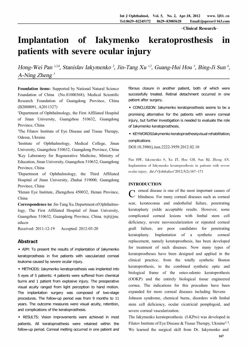
Figure 1 Photograph of Iakymenko keratoprosthesis.
Implantation of Iakymenko keratoprosthesis
168

陨灶贼 允 韵责澡贼澡葬造皂燥造熏 灾燥造援 5熏 晕燥援 2熏 Apr.18, 圆园12 www. IJO. cn栽藻造押8629原愿圆圆源缘员苑圆 8629-83085628 耘皂葬蚤造押ijopress岳员远猿援糟燥皂
Dexamethasone eyedrops were administered three timesdaily, tetracycline ointment once daily. Dexamethasoneeyedrops administration was stopped one week later.Artificial tears were applied for patients with dry eye.RESULTSPreoperative Characteristics A total of five I-KPros wereimplanted in 5 eyes of 5 patients. All the patients weremale, with a mean age of 35 years (range, 27-41 years). Allthe five patients suffered from severe vascularized cornealleucoma, of which 4 patients were secondary to chemicalburns and one patient caused by explosive injury. The timeafter injury ranged from 1.4 to 4 years. Allograftkeratoplasty was performed in 3 patients previously butfailed to improve visual acuity due to graft rejection.Cataract extraction was performed in 3 patients beforekeratoprosthesis. The preoperative best spectacle-correctedvisual acuity ranged from counting fingers to light perception;preoperatively the contralateral eye had no light perception.Visual Acuity The keratoprosthesis dramatically improvedthe vision in most patients. At 6 month afterkeratoprosthesis, the best uncorrected visual acuity (BUVA)of 20/200 or better was achieved in four patients, and onlyone patient had no significant improvement in BUVA whowas found to have retinal detachment in the operated eye.The BUVA remained almost unchanged within the follow-upperiod, except an improvement from 10/200 to 20/200 inone patient. The visual outcomes are summarized Figure 3.Retention In all the five cases, the prosthesis was retainedwithout dislocation or extrusion within the follow-upduration. A longest retention was obtained in one patientwith a follow-up of 11 years (Figure 4). The retention durationfor some patients was not updated for lost of follow-up.Complications Corneal melting was observed in one
patient 2.5 months after stage I procedure, without obviousaqueous humor leakage. After administration of bFGFeyedrops four times daily, the I-KPro was covered byepithelium and the underlying connective tissues one monthlater (Figure 5). Fibrous reclosure of the anterior openingoccurred in one case. Excision of the fibrous membrane and
Figure 2 Microphotograph showing the good compatibility ofauricular cartilage with corneal stroma 4 weeks after theimplantation of auricular cartilage into corneal stroma. Thetissue section was stained with haematoxylin and eosin. Noobvious border between auricular cartilage and cornealstroma was found, and the collagen layers was arranged inorder without infiltration of inflammatory cells (伊100).
Figure 5 A: Corneal melting occurred 2.5 months after stageI procedure; B: After treatment with bFGF eyedrops for 3weeks, connective tissues grew on the surface ofkeratoprosthesis, with a pinhole-like defect in the center.
Figure 3 Graph showing the preoperative and postoperativevisual acuity of the 5 patients over the follow-up period. Foreach patient, the preoperative visual acuity is showed at 0 oftime axis, and the posterative corrected visual acuity is showedat the point of each visit HM: Handmovement; LP:Lightperception.
Figure 4 Anterior segment photograph obtained 11 yearsafter Iakymenko keratoprosthesis implantation.
169
cauterization of the peripheral tissue was performed toregain a clear visual axis. Retinal detachment was found inone patient 7 months after surgery. Other complicationsreported in other studies such as surface infection, endo-phthalmitis, intractable glaucoma were not found in our study.DISCUSSIONAs the last resort of ophthalmologists for many refractorycorneal diseases, keratoprosthesis has achieved greatprogresses in device design and postoperative management,which in turn increase the number of implantation andexpand the indications. Moreover, the keratoprothesis isconsidered to be highly cost-effective compared withcorneal transplantation [3]. However, keratoprosthesis did notdevelop so rapidly in China as in those developed countries.Few studies on keratoprosthesis from China have beenpublished over the past decades [4,5]. We performed I-KProimplantation on several patients and the follow-up periodranged from 9 months to 11 years.Presently several types of keratoprostheses are applied inclinical use. Boston KPro is the most commonly usedkeratoprosthesis, with more than 3,000 being implantedworldwide [6]. Many clinical trials have demonstratedsatisfactory retention rate and visual improvement, with themost common complications of retroprosthetic membraneformation and raised intraocular pressure [7-9]. Theosteo-odonto-keratoprosthesis (OOKP) uses the patient'sown tooth root and surrounding alveolar bone to support acentrally cemented optical cylinder and it requires atwo-stage procedure. It is theoretically reasonable that thebiological material might increase the tissue integration andreduce extrusion rate. OOKP has wider applicationcompared with other KPros and it can even be used forpatients with sever dry eye caused by Stevens-Johnsonsyndrome, ocular cicatricial pemphigoid, . The results ofseveral studies on OOKP proved it to be well retained andthe complications rate were generally low [10-12]. Alphacorkeratoprosthesis is made from a single biocompatiblepolymer and involves a two-stage procedure. However, theresults of clinical studies showed a relatively low retentionrate due to corneal stromal melting and a high incidence ofcomplications in cases with a history of herpetic eyedisease [13-16]. Additionally, many other types of Kpro weredeveloped but the clinical studies have not extended enoughto draw a definite conclusion [17-19]. The I-KPro is differentfrom the above mentioned keratoprosthesis, but it sharesmuch common characteristics. The optic cylinder is made ofPMMA, which is also used in Boston keratoprosthesis.Auricular cartilage is used for increasing tissue integrationof I-KPro, just as the role of bone and tooth used in OOKP.
The surgical procedure of I-KPro implantation is similar tothat of AlphaCor, both composed of two stages andcharacterized by placing the device in the pocket betweenanterior and posterior corneal lamella. More than 1000I-KPro implantations were performed in the Filatov Institutesince 1966[20].Appropriate patients selection is of great importance for theprognosis of the surgery. The indications for different typesof keratoprostheses vary. As reported in previous studies,Boston keratoprosthesis and AlphaCor keratoprosthesis arenot suitable for patients with abnormal lid function or severedry eye, while OOKP has much wider application. Ourpreliminary results suggest chemical injury and corneal graftfailure might be indications for I-KPro. However, our studyis limited by the number of patients included, and it may beexpanded to more corneal diseases by future clinicalresearches.It is difficult to compare Iakymenko keratoprosthesis withother types of keratoprostheses because the patients'situation and surgeons' skill vary in different studies. Poortissue integration is a major problem for keratoprosthesis,which leads to failure of retention. In our study, no deviceextrusion was found within the follow-up period. We thinkmany factors contributed to this result. First, the use ofautologous cartilage significantly enforced the mechanicalstrength and biological compatibility of the I-KPro. Second,postoperative administration of tetracycline reduced theactivity of collagenase and inhibited tissue degradation.Third, the application of bFGF successfully promoted tissuegrowth and prevented corneal melting. Retinal detachmentoccurred in one patient, but we did not definitely know thereason. We supposed it might be related with the fragileretina of the patient and the impact of cataract extraction.In conclusion, despite the small sample size in our study, thepreliminary result of I-KPro was acceptable. All of the fivepatients achieved a middle-term to long-term visualimprovement and implant stability. No severe complicationwas found in these patients except retinal detachement inone patient. So I-KPro implantation seems to be a promisingalternative for the patients with severe corneal scar oropacity, which is no suitable for keratoplasty. Our experiencewith I-KPro might be helpful for consideration of impro-vement in the keratoprosthesis design and surgical skills.REFERENCES1 Iakymenko SA. Optic penetrating keratoprosthesis using new models of
corneal prostheses. 1981;36(2):102-104
2 Iakymenko SA. Results of PMMA/Titanium keratoprosthesis in 502 eyes.
1993; 9:197-198
3 Ament JD, Stryjewski TP, Ciolino JB, Todani A, Chodosh J, Dohlman
CH. Cost-effectiveness of the Boston keratoprosthesis.
Implantation of Iakymenko keratoprosthesis
170
陨灶贼 允 韵责澡贼澡葬造皂燥造熏 灾燥造援 5熏 晕燥援 2熏 Apr.18, 圆园12 www. IJO. cn栽藻造押8629原愿圆圆源缘员苑圆 8629-83085628 耘皂葬蚤造押ijopress岳员远猿援糟燥皂
149(2):221-8 e2
4 Yu J, Huang Y. Keratoprosthesis in China. 150(2):291
5 Huang Y, Yu J, Liu L, Du G, Song J, Guo H. Moscow Eye Microsurgery
Complex in Russia Keratoprosthesis in Beijing. 2011; 118
(1):41-46
6 Gomaa A, Comyn O, Liu C. Keratoprostheses in clinical practice - a
review. 2010; 38(2):211-224
7 Aquavella JV, Qian Y, McCormick GJ, Palakuru JR. Keratoprosthesis:
the Dohlman-Doane device. 2005; 140(6):1032-1038
8 Zerbe BL, Belin MW, Ciolino JB. Results from the multicenter Boston
Type 1 Keratoprosthesis Study. 2006; 113(10):1779 e1-7
9 Aldave AJ, Kamal KM, Vo RC, Yu F. The Boston type I keratoprosthesis:
improving outcomes and expanding indications. 2009; 116
(4):640-651
10 Falcinelli G, Falsini B, Taloni M, Colliardo P. Modified osteo-
odonto-keratoprosthesis for treatment of corneal blindness: long-term
anatomical and functional outcomes in 181 cases. 2005;
123(10):1319-1329
11 Liu C, Okera S, Tandon R, Herold J, Hull C, Thorp S. Visual
rehabilitation in end-stage inflammatory ocular surface disease with the
osteo-odonto-keratoprosthesis: results from the UK.
2008; 92(9):1211-1217
12 Tan DT, Tay AB, Theng JT, Lye KW, Parthasarathy A, Por YM, et al.
Keratoprosthesis surgery for end-stage corneal blindness in asian eyes.
2008; 115(3):503-10 e3
13 Hicks CR, Crawford GJ, Lou X, Tan DT, Snibson GR, Sutton G,
Downie N, Werner L, Chirila TV, Constable IJ. Corneal replacement using
a synthetic hydrogel cornea, AlphaCor: device, preliminary outcomes and
complications. 2003; 17(3):385-392
14 Hicks CR, Crawford GJ, Dart JK, Grabner G, Holland EJ, Stulting RD,
Tan DT, Bulsara M. AlphaCor: Clinical outcomes. 2006; 25 (9):
1034-1042
15 Crawford GJ, Hicks CR, Lou X, Vijayasekaran S, Tan D, Mulholland B,
Chirila TV, Constable IJ. The Chirila Keratoprosthesis: phase I human
clinical trial. 2002; 109(5):883-889
16 Holak SA, Holak HM, Bleckmann H. AlphaCor keratoprosthesis:
postoperative development of six patients.
2009; 247(4):535-539
17 Kim MK, Lee SM, Lee JL, Chung TY, Kim YH, Wee WR, Lee JH.
Long-term outcome in ocular intractable surface disease with Seoul-type
keratoprosthesis. 2007; 26(5):546-551
18 Ghaffariyeh A, Honarpisheh N, Karkhaneh A, Abudi R, Moroz ZI,
Peyman A, Faramarzi A, Abasov F. Fyodorov-Zuev keratoprosthesis
implantation: long-term results in patients with multiple failed corneal
grafts. 2011; 249(1):93-101
19 Hollick EJ, Watson SL, Dart JK, Luthert PJ, Allan BD. Legeais
BioKpro III keratoprosthesis implantation: long term results in seven
patients. 2006; 90(9):1146-1151
20 Iakymenko SA. Keratoprosthesis - 40-years experience of study and
application in the Filatov Institute (analysis of 1000 operations).
2010; 2010:p. 41
171


![Draft 43c PH title page main body - Heidelberg University€¦ · Ocdn m`n`\m^c _d_ ijo m`^`dq` \it nk`^dad^ bm\io amjh api_dib \b`i^d`n di oc` kp]gd^' ^jhh`m^d\g' jm ijo(ajm(kmjado](https://img.pdfslide.us/doc/110x75/60ad97ffeb8878035b2c097b/draft-43c-ph-title-page-main-body-heidelberg-university-ocdn-mnmc-d-ijo.jpg)


![D]rt]In]o b]Ijo Cezo - · PDF fileD]rt]In]o b]Ijo Cezo 3 1. s]om]]B]]w am]eirk]m]]\ g]om]t]Ip¶r jev]] n]]n]kz] g]]m]m]]\T]I s]om]]B]]wn]o B]]rt] p]rN]Ino amoirk] jw v]sy]o ht]o](https://img.pdfslide.us/doc/110x75/5a7309777f8b9aac538e3262/drtino-bijo-cezo-a-drtino-bijo-cezo-3-1-sombw-ameirkm.jpg)