Embed Size (px)
Citation preview

©1987-2002 Romero-Pilu-Jeanty-Ghidini-Hobbins
The Lungs Fetal Lung Congenital Anomalies/ Lung Sequestration / 202
195 Bronchogenic Cysts / 205 Chylothorax / 195 Congenital Cystic Adenomatoid Malformation of the Lung / 198
Fetal Lung Congenital Anomalies During fetal life, the lungs are not inflated and are visualized as solid structures that fill the space between the heart and the rib cage. Interest in these organs has increased in an attempt to recognize sonographic changes related to pulmonic maturity.
Fetal lungs appear as two paracardiac structures whose uniform echogenicity varies in comparison to that of fetal liver along gestation. Some authors have proposed that these differences may be helpful in the assessment of fetal lung maturity. They have reported that the lung becomes more reflective than the liver with advancing gestational age.3 Other investigators have found that these changes do not correlate with the L:S ratio and phosphatylglycerol measurements.2 On the other hand, preliminary reports suggest that lung compressibility-assessed subjectively from the passive movements of the lung adjacent to the cardiac ventricular walls during diastole--varies throughout gestation and correlates with lung maturity. In a report of 54 infants delivered within 36 hours of ultrasound dynamic lung studies, a fetal
lung stiff pattern was associated with respiratory distress syndrome (RDS), whereas fetuses with compliant lungs had no RDS.2 These observations need confirmation.
This chapter focuses on the four most common thoracic anomalies diagnosed with ultrasound: chylothorax, cystic adenomatoid tumor, lung sequestration, and bronchogenic cysts. A nomogram for thoracic dimensions is displayed on p. 330. It is helpful in the assessment of pulmonary hypoplasia.
REFERENCES
1. Birnholz JC, Farrell EE: Fetal lung development:
Compressibility as a measure of maturity. Radiology 157:495, 1985.
2. Fried AM, Loh FK, Umer MA, et al.: Echogenicity of fetal lung: Relation to fetal age and maturity. AJR 145:591, 1985.
3. Reeves GS, Garrett WJ, Warren PS, Fisher CC: Observations of fetal lung reflectivity using real-time ultrasound. Aust NZ J Obstet Gynaecol 24:91, 1984.
Chylothorax Definition Chylothorax is an accumulation of chyle in the pleural cavity Incidence Chylothorax is a common cause of pleural effusion during the first days of neonatal life.5 Its prevalence
has been estimated to be 1:10,000 deliveries.15 The male to female ratio is 2:1.4,25 Etiology Accumulation of lymph within the pleural cavity can result from overproduction or impaired reabsorption of lymph. The latter could be due to either an obstruction
195

©1987-2002 Romero-Pilu-Jeanty-Ghidini-Hobbins
196 THE LUNGS
Figure 5-1. Transverse scan of a patient with congenital chylothorax. A predominantly left chylothorax (*) deviates the heart (H) toward the right. L, Lung; Sp, spine. to pulmonary lymph drainage or abnormal lymphatic vessels. Pathology Pediatric series indicate that chylothorax occurs as a unilateral pleural effusion involving the right side of the lung in most instances. In rare cases, pleural effusions can be bilateral.4,8 Chylothorax may lead to lung compression and the development of pulmonary hypoplasia. Unilateral pleural effusion can also shift the mediastinum, impair venous return, and lead to congestive heart failure and hydrops. There is a paucity of information about the histopathology of congenital chylothorax. In one autopsy series, dilated distal lymphatics that apparently failed to communicate with the more proximal lymphatic vessels were demonstrated, but this may be a nonspecific finding. 19 Associated Anomalies Chylothorax may be associated with trisomy 21.6,24,26 Anomalies reported in association with chylothorax include congenital pulmonary lymphangiectasis,13 tracheoesophageal fistula,11 extralobar lung sequestration,3,9,12 and a multiple malformation complex (anemia, tracheoesophageal fistula, Klippel-Feil deformity).18 Diagnosis Chylothorax should be suspected in the presence of a pleural effusion (Figs. 5-1, 5-2, 5-3). A specific diagnosis of chylothorax is not possible on the basis of the
Figure 5-2. Longitudinal scan of the fetus shown in Figure 5-1. The arrow points to the presence of ascites. *, chylothorax; LVR, liver. gross appearance of the fluid. Indeed, lymph looks serous at birth and becomes lactescent only after oral feedings.8,10,15-17,20,21,24,25 It has been suggested that identification of abundant lymphocytes (>60 percent) in pleural fluid indicates chylothorax, since these are the predominant cells in lymph.2,24 The prenatal diagnosis of chylothorax has been reported in several instances.7,10,14,17,21-23 However, documentation of these cases is generally poor. A definitive diagnosis can be made by demonstrating a change in the character of the pleural fluid after feedings. The value of lymphocyte counts in the differential diagnosis of intrauterine-diagnosed pleural effusions has not been established. In severe cases, this condition may be associated with nonimmune hydrops (Fig. 5-2).1,7,14,22,24 Polyhydramnios has been noted in all cases prenatally diagnosed and registered in many neonatal series.1,8,11,15,16,25,26 It may be the result of esophageal compression by the pleural effusion. The differential diagnosis of congenital chylothorax is problematic. The condition can appear as isolated pleural effusions or as nonimmune hydrops. It is not known if biochemical or cytologic examination of the pleural fluid can permit a differential diagnosis between the effusion seen in congenital chylothorax and that seen in other causes of nonimmune hydrops. Prognosis It is extremely difficult to provide prognostic figures for congenital chylothorax diagnosed in utero because of the limited experience with this condition and the uncertainty surrounding its diagnosis. Of all cases
©1987-2002 Romero-Pilu-Jeanty-Ghidini-Hobbins
CHYLOTHORAX 197
claimed to have been prenatally diagnosed, 5 of 11 died perinatally; 2 were stillbirths and 3 were neonatal deaths. Hydrops was present in 3 of the dead infants and in only 1 of the survivors. The survivor was treated in utero with thoracentesis. 22 In pediatric series in which the diagnosis is made postnatally, symptoms of respiratory distress are present at birth in 40 percent of infants and become manifest within 24 hours in 64 percent and within the first week in 79 percent.4 The mortality rate is 15 percent.4 Weight loss is a sign of wastage and can lead to death. Treatment consists of pleural drainage (repeated thoracentesis or continuous thoracic drainage), careful replacement of the nutritional losses, and reduction of the volume of lymph flow. A diet composed of medium-chain triglycerides, which are absorbed into the portal system rather than through the lymphatic system, has been used.6,24 In most cases, chylothorax resolves spontaneously with time. If control of the chylous effusion is not achieved within 3 to 4 weeks, surgical ligature of the thoracic duct is performed.1 At an average follow-up of 15 months, 19 of 20 patients were normal.4 Obstetrical Management When a diagnosis is made before viability, the option of pregnancy termination can be offered. Karyotyping is indicated, since this condition has been associated with chromosomal anomalies, such as trisomy 21. After viability, the management depends on the gestational age and the development of signs of hydrops or mediastinal shift. In term infants, a thoracentesis should be considered before delivery to avoid respiratory failure due to a large pleural effusion. Decompression may also prevent rupture or inversion of the diaphragm secondary to pressure on the fetal thorax as it passes through the birth canal.17 The management of preterm infants depends on the degree of mediastinal shift present. If the pleural effusion is small and no mediastinal shift is demonstrated, intervention is not justified. Serial scans are required to monitor the evolution of the pleural effusion. If mediastinal shift and signs of hydrops develop, we would consider intervening to prevent pulmonary hypoplasia. It must be emphasized that fluid reaccumulation occurs rapidly, and repeated thoracentesis will probably be required. Two cases successfully managed with repeated thoracenteses have been reported.2,21 The uncontrolled nature of these observations prevents firm conclusions about the benefit of this type of intervention. In one fetus, weekly thoracenteses were initiated at the 19th week and carried out until the 23d week of intrauterine life, at which time the effusion spontaneously disappeared.2 The procedure may have prevented the development of pulmonary hypoplasia. There is little
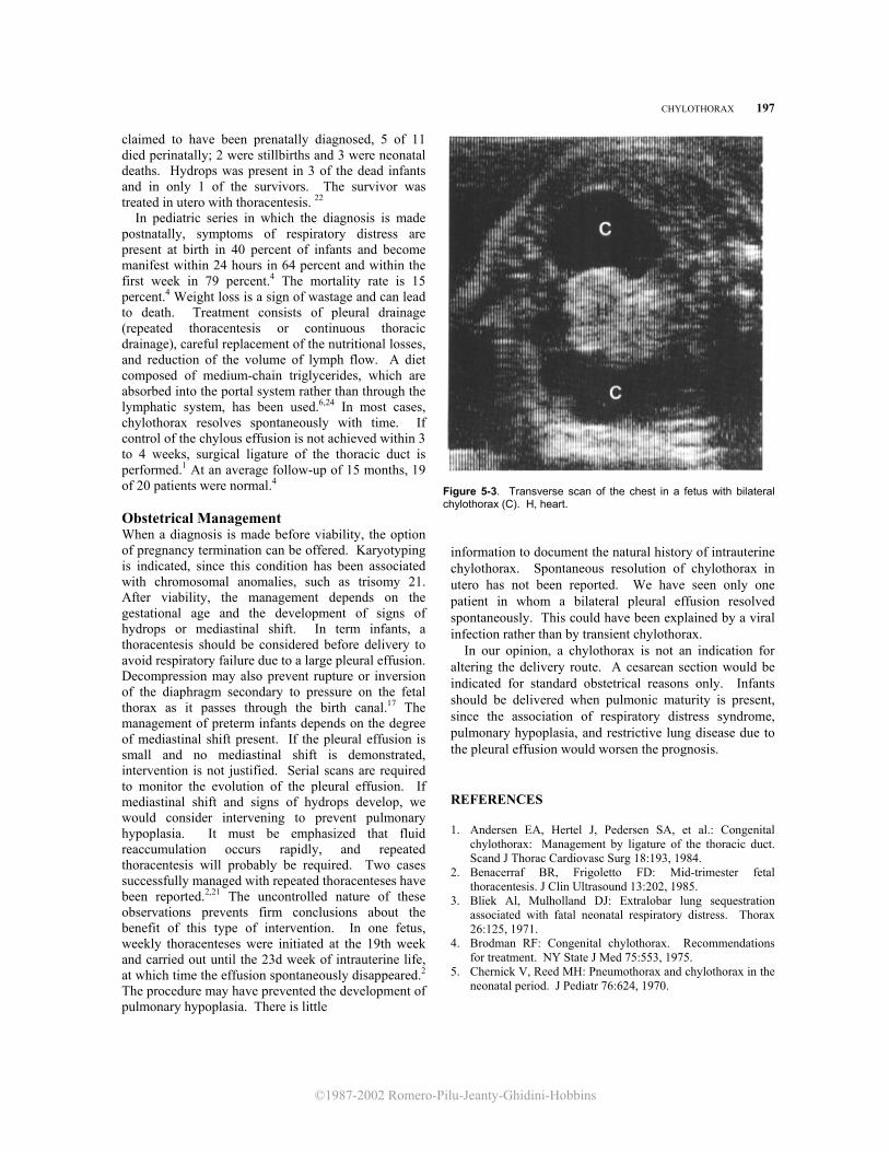
Figure 5-3. Transverse scan of the chest in a fetus with bilateral chylothorax (C). H, heart.
information to document the natural history of intrauterine chylothorax. Spontaneous resolution of chylothorax in utero has not been reported. We have seen only one patient in whom a bilateral pleural effusion resolved spontaneously. This could have been explained by a viral infection rather than by transient chylothorax. In our opinion, a chylothorax is not an indication for altering the delivery route. A cesarean section would be indicated for standard obstetrical reasons only. Infants should be delivered when pulmonic maturity is present, since the association of respiratory distress syndrome, pulmonary hypoplasia, and restrictive lung disease due to the pleural effusion would worsen the prognosis.
REFERENCES
1. Andersen EA, Hertel J, Pedersen SA, et al.: Congenital chylothorax: Management by ligature of the thoracic duct. Scand J Thorac Cardiovasc Surg 18:193, 1984.
2. Benacerraf BR, Frigoletto FD: Mid-trimester fetal thoracentesis. J Clin Ultrasound 13:202, 1985.
3. Bliek Al, Mulholland DJ: Extralobar lung sequestration associated with fatal neonatal respiratory distress. Thorax 26:125, 1971.
4. Brodman RF: Congenital chylothorax. Recommendations for treatment. NY State J Med 75:553, 1975.
5. Chernick V, Reed MH: Pneumothorax and chylothorax in the neonatal period. J Pediatr 76:624, 1970.
©1987-2002 Romero-Pilu-Jeanty-Ghidini-Hobbins
198 THE LUNGS 6. Craenen JM, Williams Jr. TE, Kilman JW: Simplified
management of chylothorax in neonates and infants. Ann Thoracic Surg 24:275, 1977.
7. Defoort P, Thiery M: Antenatal diagnosis of congenital chylothorax by gray scale sonography. J Clin Ultrasound 6:47, 1978.-
8. Doolittle WM, Ohmart D, Egan EA: Congenital bilateral pleural effusions: A cause for respiratory failure in the newborn. Am J Dis Child 125:435, 1973.
9. Dresler S: Massive pleural effusion and hypoplasia of the lung accompanying extralobar pulmonary sequestration. Hum Pathol 12:862, 1981.
10. Dubos JP, Bouchez MC, Kacet N, et al.: Anasarque non immunologique et chylothorax congenital. Arch Fr Pediatr 42:537, 1985.
11. Harvey JG, Houlsby W, Sherman K, et al.: Congenital chylothorax: Report of unique case associated with"H"-type tracheo-oesophageal fistula. Br J Surg 66:485, 1979.
12. Horowitz RN: Extralobar sequestration of lung in a newborn infant. Am J Dis Child 110:195, 1965.
13. Hunter WS, Becroft DMQ: Congenital pulmonarylymphangiectasis associated with pleural effusions. Arch Dis Child 59:278, 1984.
14. Jaffa Al, Barak S, Kaysar N, et al.: Case report. Antenatal diagnosis of bilateral congenital chylothorax with pericardial effusion. Acta Obstet Gynecol Scand 64:455, 1985.
15. John E: Pleural effusion in the newborn. Med J Aust 1:102,1974
16. Koffler H, Papile LA, Burstein RL: Congenital chylo-
thorax: Two cases associated with maternal polyhydramnios. Am J Dis Child 132:638, 1978.
17. Lange IR, Manning FA: Antenatal diagnosis of congenital pleural effusions. Am J Obstet Gynecol 140:839, 1981.
18. Lazarus KH, McCurdy FA: Multiple congenital anomalies in a patient with Diamond-Blackfan syndrome. Clin Pediatr 23:520, 1984.
19. McKendry JBJ, Lindsay WK, Gerstein MC: Congenital defects of the lymphatics in infancy. Pediatrics 19:21, 1957.
20. Perry RE, Hodgman J, Cass AB: Pleural effusion in the neonatal period. J Pediaty 62:838, 1963.
21. Petres RE, Redwine FO, Cruikshank DP: Congenitalbilateral chylothorax. Antepartum diagnosis and successful intrauterine surgical management. JAMA248:1360 ' 1982.
22. Schmidt W, Harms E, Wolf D: Successful prenatal of nonimmune hydrops fetalis due to congenital chylothorax.
Case report. Br J Obstet Gynecol 92:685, 1985. 23. Thomas DB, Anderson JC: Antenatal detection of fetal effusions and neonatal management. Med J Aust 2:435, 1979. 24. Van Aerde J, Campbell AN, Smyth JA, et al.: Sponta chylothorax in newborns. Am J Dis Child 138:961, 1984. 25. Yancy WS, Spock A: Spontaneous neonatal pleural effusion. J
Pediatr Surg 2:313, 1967. 1:102, 1974. 26. Yoss BS, Lipsitz PJ: Chylothorax in two mongoloid infants.
Clin Genet 12:357, 1977.
Congenital Cystic Adenomatoid Malformation of the Lung Synonym Adenomatoid hamartoma. Definition Congenital cystic adenomatoid malformation of the lung (CCAML) is a hamartoma of the lung characterized by overgrowth of terminal bronchioles (adenomatoid) at the expense of saccular spaces. The term "hamartoma" refers to a benign tumorlike malformation which reproduces, in a disorderly manner, the mature structure of the organ from which it is derived. Embryology The respiratory tract includes the conducting airways and the respiratory component. They have different embryologic origins. The conducting airways are derived from the foregut (endoderm), whereas the respiratory component arises from mesenchyma that
concentrate around the tips of the growing bronchi. The embryonic problem responsible for CCAML is thought to be an arrest of the connection of the two systems and subsequent overgrowth of the terminal bronchioles.21,24 The insult seems to occur before the 5th week of conceptional age. Incidence CCAML is a rare malformation of the lung, and about 200 cases have been reported in the literature to date. There is no sex predilection. Pathology The lesion is almost always unilateral with no preference of right or left lung.18 Only a few cases of bilateral involvement have been reported.21 The disease generally affects one lobe, and the tumor appears as a mass of variable size that deforms the lung. It is often large enough to cause a shift of the
©1987-2002 Romero-Pilu-Jeanty-Ghidini-Hobbins
CONGENITAL CYSTIC ADENOMATOID MALFORMATION OF THE LUNG 199
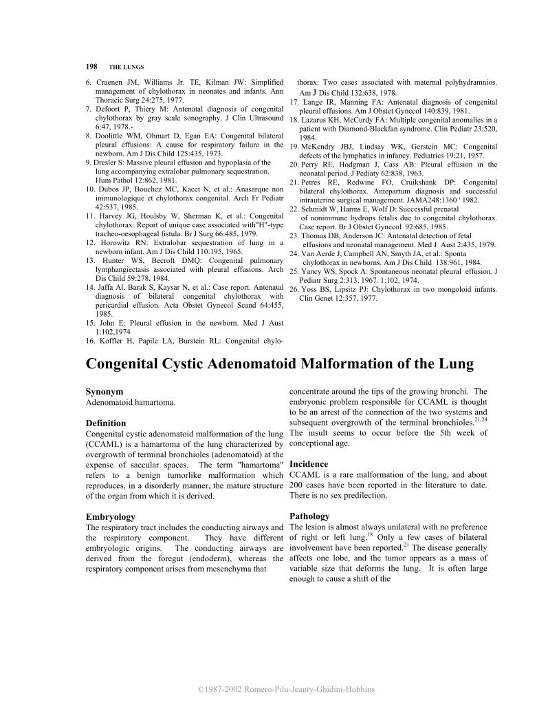
Figure 5-4. Classification of congenital cystic adenomatoid malformation. Type I is composed of a small number of large cysts with thick smooth muscle and elastic tissue walls. Relatively normal alveoli are seen between and adjacent to these cysts. Mucous glands may be present. Type II contains numerous smaller cysts (< 1 cm in diameter), with a thin muscular coat beneath the ciliated columnar epithelium. The area between the cysts is occupied by large alveoluslike structures. The lesion blends with the normal parenchyma. Type III occupies the entire lobe or lobes and is composed of regularly spaced bronchiolelike structures separated by masses of cuboidal epithelium-lined alveoluslike structures. (Reproduced with permission from Stocker et al.: Hum Pathol 8:155, 1977.)
mediastinal structures and to compress the contralat-eral lung. Stocker et al. proposed a classification of CCAML into three subtypes according to the size of the cysts24: type I has large cysts, type II has multiple small cysts of less than 1.2 cm in diameter, and type III consists of a noncystic lesion producing mediastinal shift (Fig. 5-4). The worst prognosis is seen in type III lesions. Associated anomalies are frequently present in type II. Another classification suggests that lesions are separated into two groups: macrocystic tumors with cysts of at least 5 mm in
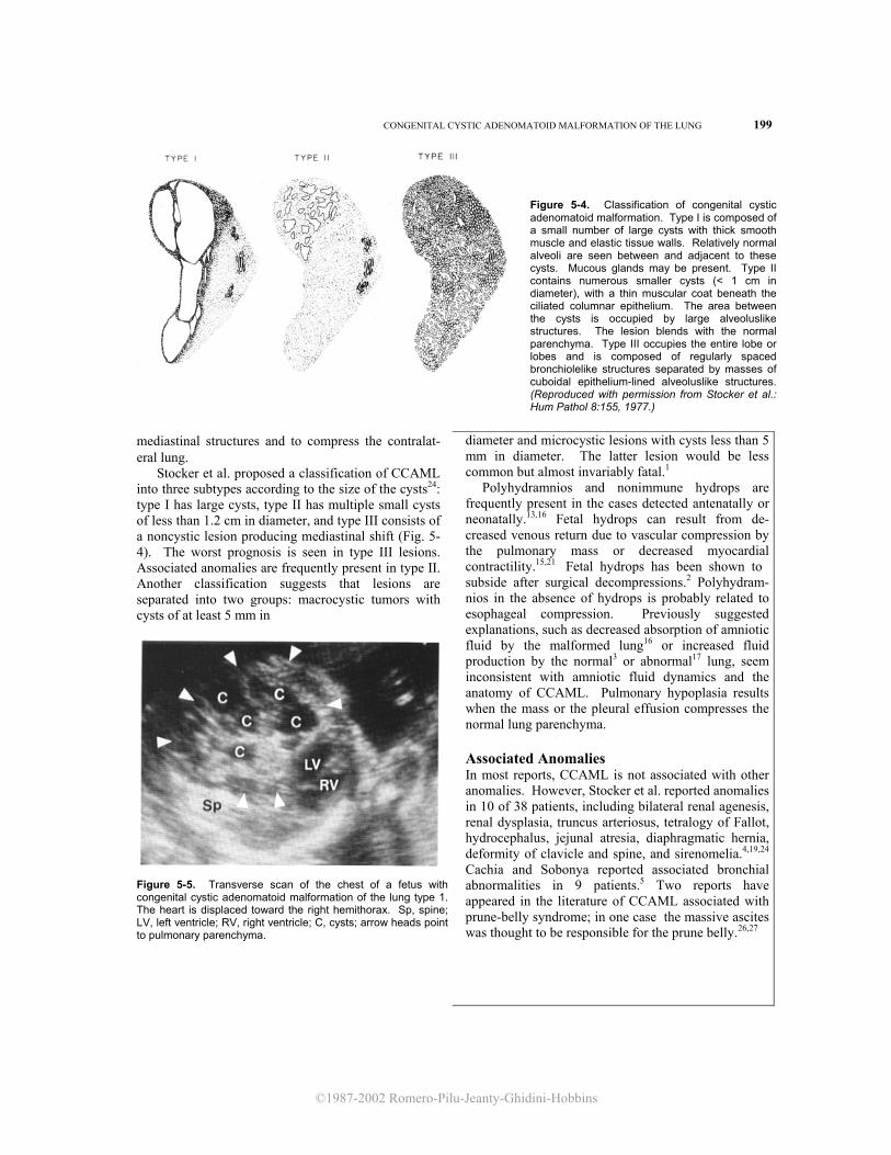
Figure 5-5. Transverse scan of the chest of a fetus with congenital cystic adenomatoid malformation of the lung type 1. The heart is displaced toward the right hemithorax. Sp, spine; LV, left ventricle; RV, right ventricle; C, cysts; arrow heads point to pulmonary parenchyma.
diameter and microcystic lesions with cysts less than 5 mm in diameter. The latter lesion would be less common but almost invariably fatal.1 Polyhydramnios and nonimmune hydrops are frequently present in the cases detected antenatally or neonatally.13,16 Fetal hydrops can result from de- creased venous return due to vascular compression by the pulmonary mass or decreased myocardial contractility.15,21 Fetal hydrops has been shown to subside after surgical decompressions.2 Polyhydram- nios in the absence of hydrops is probably related to esophageal compression. Previously suggested explanations, such as decreased absorption of amniotic fluid by the malformed lung16 or increased fluid production by the normal3 or abnormal17 lung, seem inconsistent with amniotic fluid dynamics and the anatomy of CCAML. Pulmonary hypoplasia results when the mass or the pleural effusion compresses the normal lung parenchyma. Associated Anomalies In most reports, CCAML is not associated with other anomalies. However, Stocker et al. reported anomalies in 10 of 38 patients, including bilateral renal agenesis, renal dysplasia, truncus arteriosus, tetralogy of Fallot, hydrocephalus, jejunal atresia, diaphragmatic hernia, deformity of clavicle and spine, and sirenomelia.4,19,24 Cachia and Sobonya reported associated bronchial abnormalities in 9 patients.5 Two reports have appeared in the literature of CCAML associated with prune-belly syndrome; in one case the massive ascites was thought to be responsible for the prune belly.26,27
©1987-2002 Romero-Pilu-Jeanty-Ghidini-Hobbins
200 THE LUNGS
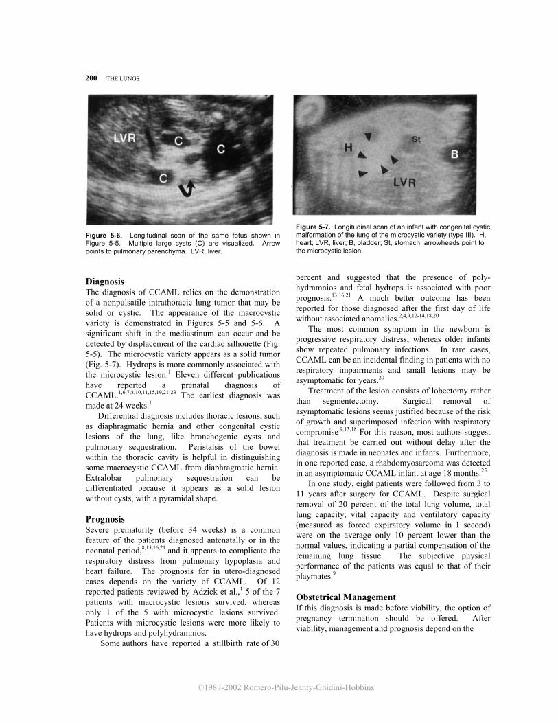
Figure 5-6. Longitudinal scan of the same fetus shown in Figure 5-5. Multiple large cysts (C) are visualized. Arrow points to pulmonary parenchyma. LVR, liver. Diagnosis The diagnosis of CCAML relies on the demonstration of a nonpulsatile intrathoracic lung tumor that may be solid or cystic. The appearance of the macrocystic variety is demonstrated in Figures 5-5 and 5-6. A significant shift in the mediastinum can occur and be detected by displacement of the cardiac silhouette (Fig. 5-5). The microcystic variety appears as a solid tumor (Fig. 5-7). Hydrops is more commonly associated with the microcystic lesion.1 Eleven different publications have reported a prenatal diagnosis of CCAML.1,6,7,8,10,11,15,19,21-23 The earliest diagnosis was made at 24 weeks.1
Differential diagnosis includes thoracic lesions, such as diaphragmatic hernia and other congenital cystic lesions of the lung, like bronchogenic cysts and pulmonary sequestration. Peristalsis of the bowel within the thoracic cavity is helpful in distinguishing some macrocystic CCAML from diaphragmatic hernia. Extralobar pulmonary sequestration can be differentiated because it appears as a solid lesion without cysts, with a pyramidal shape. Prognosis Severe prematurity (before 34 weeks) is a common feature of the patients diagnosed antenatally or in the neonatal period,8,15,16,21 and it appears to complicate the respiratory distress from pulmonary hypoplasia and heart failure. The prognosis for in utero-diagnosed cases depends on the variety of CCAML. Of 12 reported patients reviewed by Adzick et al.,1 5 of the 7 patients with macrocystic lesions survived, whereas only 1 of the 5 with microcystic lesions survived. Patients with microcystic lesions were more likely to have hydrops and polyhydramnios. Some authors have reported a stillbirth rate of 30
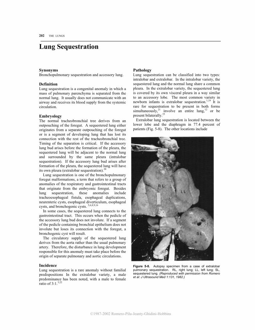
Figure 5-7. Longitudinal scan of an infant with congenital cystic malformation of the lung of the microcystic variety (type III). H, heart; LVR, liver; B, bladder; St, stomach; arrowheads point to the microcystic lesion. percent and suggested that the presence of poly-hydramnios and fetal hydrops is associated with poor prognosis.13,16,21 A much better outcome has been reported for those diagnosed after the first day of life without associated anomalies.2,4,9,12-14,18,20
The most common symptom in the newborn is progressive respiratory distress, whereas older infants show repeated pulmonary infections. In rare cases, CCAML can be an incidental finding in patients with no respiratory impairments and small lesions may be asymptomatic for years.20
Treatment of the lesion consists of lobectomy rather than segmentectomy. Surgical removal of asymptomatic lesions seems justified because of the risk of growth and superimposed infection with respiratory compromise.9,13,18 For this reason, most authors suggest that treatment be carried out without delay after the diagnosis is made in neonates and infants. Furthermore, in one reported case, a rhabdomyosarcoma was detected in an asymptomatic CCAML infant at age 18 months.25
In one study, eight patients were followed from 3 to 11 years after surgery for CCAML. Despite surgical removal of 20 percent of the total lung volume, total lung capacity, vital capacity and ventilatory capacity (measured as forced expiratory volume in I second) were on the average only 10 percent lower than the normal values, indicating a partial compensation of the remaining lung tissue. The subjective physical performance of the patients was equal to that of their playmates.9 Obstetrical Management If this diagnosis is made before viability, the option of pregnancy termination should be offered. After viability, management and prognosis depend on the

©1987-2002 Romero-Pilu-Jeanty-Ghidini-Hobbins
CONGENITAL CYSTIC ADENOMATOID MALFORMATION OF THE LUNG 201
presence of associated hydrops. In patients without hydrops, delivery can wait until fetal and pulmonic maturity is reached. Serial scans are recommended to monitor the growth of the lesion and to look for early signs of hydrops. To date, fetuses with hydrops have had a 100 percent mortality rate. Nonaggressive management may be offered to the parents in these cases. Repeated thoracenteses do not seem to guarantee long-lasting decompression of the chest. In utero surgery for fetuses with hydrops and large lesions in early pregnancy has been suggested but has not been carried out at the time of this writing.1 Antenatal diagnosis of CCAML mandates delivery in a tertiary care center, where immediate thoracic surgery can be performed.
REFERENCES 1. Adzick NS, Harrison MR, Glick PL, et al.: Fetal cystic
adenomatoid malformation: Prenatal diagnosis and natural history. J Pediatr Surg 20:483, 1985.
2. Aslam PA, Korones SB, Richardson RL, et al.: Congenital cystic adenomatoid malformation with anasarca. JAMA 212:622, 1970.
3. Bates HR Jr: Fetal pulmonary hypoplasia and hydramnios. Am J Obstet Gynecol 91:295, 1965.
4. Birdsell DC, Wentworth P, Reilly BJ, et al.: Congenital cystic adenomatoid malformation of the lung: A report of eight cases. Can J Surg 9:350, 1966.
5. Cachia R, Sobonya RE: Congenital cystic adenomatoid malformation of the lung with bronchial atresia. Hum Pathol 12:947, 1981.
6. Dibbins AW, Curci MR, McCrann DJ: Prenatal diagnosis of congenital anomalies requiring surgical correction. Am J Surg 149:528, 1985.
7. Diwan RV, Brennan JN, Philipson EH, et al.: Ultrasonic prenatal diagnosis of type III congenital cystic adenomatoid malformation of lung. J Clin Ultrasound 11:218, 1983.
8. Donn SM, Martin JN Jr, White SJ: Antenatal ultrasound findings in cystic adenomatoid malformation. Pediatr Radiol 10:180, 1981.
9. Frenckner B, Freyschuss U: Pulmonary function after lobectomy for congenital lobar emphysema and congenital cystic adenomatoid malformation: A follow-up study. Scand J Thorac Cardiovasc Surg 16:293, 1982.
10. Garrett WJ, Kossoff G, Lawrence R: Gray scale echography in the diagnosis of hydrops due to fetal lung tumor. J Clin Ultrasound 3:45, 1975.
11. Graham D, Winn K, Dex W, et al.: Prenatal diagnosis of cystic adenomatoid malformation of the lung. J Ultrasound Med 1:9, 1982.
12. Haller JA, Golladay ES, Pickard LR, et al.: Surgical management of lung bud anomalies: Lobar emphysema, bronchogenic cyst, cystic adenomatoid malformation, and intralobar pulmonary sequestration. Ann Thorac Surg 28:33, 1979.
13. Halloran LG, Silverberg SG, Salzberg AM: Congenital cystic adenomatoid malformation of the lung. Arch Surg 104:715, 1972.
14. Hartenberg MA, Brewer WH: Cystic adenomatoid malformation of the lung: Identification by sonography. AJR 140:693, 1983.
15. Johnson JA, Rumack CM, Johnson ML, et al.: Cystic adenomatoid malformation: Antenatal demonstration. AJR 142:483, 1984.
16. Kohler HG, Rymer BA: Congenital cystic malformation of the lung and its relation to hydramnios. J Obstet Gynaecol Br Commonw 80:130, 1973.
17. Krous HF, Harper PE, Perlman M: Congenital cystic adenomatoid malformation in bilateral renal agenesis: Its mitigation of Potter's syndrome. Arch Pathol Lab Med 104:368, 1980.
18. Madewell JE, Stocker JT, Korsower JM: Cystic adenomatoid malformation of the lung. AJR 124:436, 1975.
19. Mayden KL, Tortora M, Chervenak FA, et al.: The antenatal monographic detection of lung masses. Am J Obstet Gynecol 148:349, 1984.
20. Moncrieff MW, Cameron AH, Astley R, et al.: Congenital cystic adenomatoid malformation of the lung. Thorax 24:476, 1969.
21. Oestoer AG, Fortune DW: Congenital cystic adenomatoid malformation of the lung. Am J Clin Pathol 70:595, 1978.
22. Pezzuti RT, Isler RJ: Antenatal ultrasound detection of cystic adenomatoid malformation of lung: Report of a case and review of the recent literature. J Clin Ultrasound 11:342, 1983.
23. Stauffer UG, Savoldelli G, Mieth D: Antenatal ultrasound diagnosis in cystic adenomatoid malformation of the lung. Case report. J Pediatr Surg 19:141, 1984.
24. Stocker JT, Madewell JE, Drake RM: Congenital cystic adenomatoid malformation of the lung. Classification and morphologic spectrum. Hum Pathol 8:155, 1977.
25. Ueda K, Gruppo R, Unger F, et al.: Rhabdomyosarcoma of lung arising in congenital cystic adenomatoid malformation. Cancer 40:383, 1977.
26. Weber ML, Rivard G, Perreault G: Prune-belly syndrome associated with congenital cystic adenomatoid malformation of the lung. Am J Dis Child 132:316, 1978.
27. Wilson SK, Moore GW, Hutchins GM: Congenital cystic adenomatoid malformation of the lung associated with abdominal musculature deficiency (prune belly). Pediatrics 62:421, 1978.
©1987-2002 Romero-Pilu-Jeanty-Ghidini-Hobbins
202 THE LUNGS
Lung Sequestration Synonyms Bronchopulmonary sequestration and accessory lung. Definition Lung sequestration is a congenital anomaly in which a mass of pulmonary parenchyma is separated from the normal lung. It usually does not communicate with an airway and receives its blood supply from the systemic circulation. Embryology The normal tracheobronchial tree derives from an outpouching of the foregut. A sequestered lung either originates from a separate outpouching of the foregut or is a segment of developing lung that has lost its connection with the rest of the tracheobronchial tree. Timing of the separation is critical. If the accessory lung bud arises before the formation of the pleura, the sequestered lung will be adjacent to the normal lung and surrounded by the same pleura (intralobar sequestration). If the accessory lung bud arises after formation of the pleura, the sequestered lung will have its own pleura (extralobar sequestration).20 Lung sequestration is one of the bronchopulmonary foregut malformations, a term that refers to a group of anomalies of the respiratory and gastrointestinal tracts that originate from the embryonic foregut. Besides lung sequestration, these anomalies include tracheoesophageal fistula, esophageal duplications, neurenteric cysts, esophageal diverticulum, esophageal cysts, and bronchogenic cysts. 5,6,8,9,16 In some cases, the sequestered lung connects to the gastrointestinal tract. This occurs when the pedicle of the accessory lung bud does not involute. If a segment of the pedicle containing bronchial epithelium does not involute but loses its connection with the foregut, a bronchogenic cyst will result. The circulatory supply of the sequestered lung derives from the aorta rather than the usual pulmonary artery. Therefore, the disturbance in lung development responsible for this anomaly must take place before the origin of separate pulmonary and aortic circulations. Incidence Lung sequestration is a rare anomaly without familial predispositions In the extralobar variety, a male predominance has been noted, with a male to female ratio of 3:1.3,22
Pathology Lung sequestration can be classified into two types: intralobar and extralobar. In the intralobar variety, the sequestered lung and the normal lung share a common pleura. In the extralobar variety, the sequestered lung is covered by its own visceral pleura in a way similar to an accessory lobe. The most common variety in newborn infants is extralobar sequestration.1,19 It is rare for sequestration to be present in both forms simultaneously,21 involve an entire lung,12 or be present bilaterally.25 Extralobar lung sequestration is located between the lower lobe and the diaphragm in 77.4 percent of patients (Fig. 5-8). The other locations include
Figure 5-8. Autopsy specimen from a case of extralobar pulmonary sequestration. RL, right lung; LL, left lung; SL, sequestered lung. (Reproduced with permission from Romero et al: J Ultrasound Med 1:131, 1982.)

©1987-2002 Romero-Pilu-Jeanty-Ghidini-Hobbins
LUNG SEQUESTRATION 203
Figure 5-9. The blood supply to the sequestered lung (SL) is derived from the systemic circulation. The arrow points to the vascular supply to an extralobar sequestered lung.
paracardiac, mediastinal, infrapericardial, infradia-phragmatic, and abdominal sites.21 The arterial supply and venous drainage are provided primarily by systemic vessels (Fig. 5-9). The size of the sequestered lung is variable.22 Macroscopically, the parenchyma may resemble normal lung or be firm like thymus.22
In intralobar lung sequestration, either side is involved with similar frequency. The lower lobes are the most commonly affected (98 percent). The arterial supply is usually from the thoracic or abdominal aorta (93 percent), and the venous drainage terminates in the pulmonary veins (96 percent).21 Macroscopically, the sectioned areas may show a wide spectrum of appearances, ranging from the most frequent cystic pattern (with a solitary cyst or polycysts) to the rare pseudotumorous form. 11,21 Associated Anomalies Lung sequestration is one of the bronchopulmonary foregut malformations. These foregut anomalies share a common embryologic pathogenesis and are associated with each other more frequently than would be expected by chance.5,6,8,9,16 The other bronchopulmonary foregut malformations include tracheoesophageal fistula, esophageal duplications, neurenteric cysts, esophageal diverticulum, esophageal cysts, and bronchogenic cysts.
Extrapulmonary anomalies occur in 10 percent of patients with intralobar lung sequestrations and include skeletal deformities (funnel chest, polydactyly)10 diaphragmatic hernias, congenital heart disease (tricuspid atresia, transposition of great vessels, subvalvular aortic stenosis),24 and renal and cerebral anomalies (hydrocephalus).10,21
In contrast, the incidence of extrapulmonary anomalies in the extralobar variety is 59 percent, including diaphragmatic hernia, heart anomalies (atrial and ventricular septal defects, congenital absence of pericardium, truncus arteriosus),1,22 funnel chest, vertebral defects,2 and megacolon.21 Often, several anomalies are present in the same patient.22 Diaphragmatic hernia constitutes 50 percent of the anomalies associated with extralobar lung sequestration.21
The anomalous blood supply to the sequestered lung can cause a left-to-right shunt, leading in some patients to cardiac failure after birth.4,7,8,14,17 Lung sequestration has been associated with nonimmune hydrops. Diagnosis Extralobar lung sequestration has been identified antenatally on several occasions.13,15,18,23a The sequestered lung appears as an echogenic, intra-
©1987-2002 Romero-Pilu-Jeanty-Ghidini-Hobbins
204 THE LUNGS
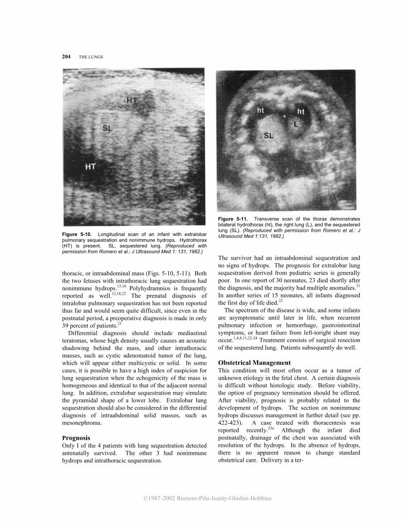
Figure 5-10. Longitudinal scan of an infant with extralobar pulmonary sequestration and nonimmune hydrops. Hydrothorax (HT) is present. SL, sequestered lung. (Reproduced with permission from Romero et al.: J Ultrasound Med 1: 131, 1982.) thoracic, or intraabdominal mass (Figs. 5-10, 5-11). Both the two fetuses with intrathoracic lung sequestration had nonimmune hydrops.13,18 Polyhydramnios is frequently reported as well.13,18,22 The prenatal diagnosis of intralobar pulmonary sequestration has not been reported thus far and would seem quite difficult, since even in the postnatal period, a preoperative diagnosis is made in only 39 percent of patients.21 Differential diagnosis should include mediastinal teratomas, whose high density usually causes an acoustic shadowing behind the mass, and other intrathoracic masses, such as cystic adenomatoid tumor of the lung, which will appear either multicystic or solid. In some cases, it is possible to have a high index of suspicion for lung sequestration when the echogenicity of the mass is homogeneous and identical to that of the adjacent normal lung. In addition, extralobar sequestration may simulate the pyramidal shape of a lower lobe. Extralobar lung sequestration should also be considered in the differential diagnosis of intraabdominal solid masses, such as mesonephroma. Prognosis Only I of the 4 patients with lung sequestration detected antenatally survived. The other 3 had nonimmune hydrops and intrathoracic sequestration.
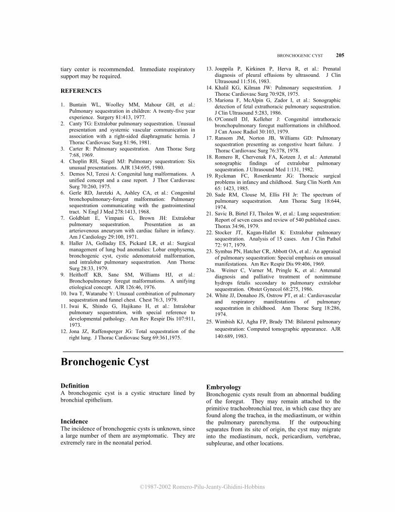
Figure 5-11. Transverse scan of the thorax demonstrates bilateral hydrothorax (ht), the right lung (L), and the sequestered lung (SL). (Reproduced with permission from Romero et al.: J Ultrasound Med 1:131, 1982.)
The survivor had an intraabdominal sequestration and no signs of hydrops. The prognosis for extralobar lung sequestration derived from pediatric series is generally poor. In one report of 30 neonates, 23 died shortly after the diagnosis, and the majority had multiple anomalies.21 In another series of 15 neonates, all infants diagnosed the first day of life died.22 The spectrum of the disease is wide, and some infants are asymptomatic until later in life, when recurrent pulmonary infection or hemorrhage, gastrointestinal symptoms, or heart failure from left-toright shunt may occur.1,4,8,11,22-24 Treatment consists of surgical resection of the sequestered lung. Patients subsequently do well. Obstetrical Management This condition will most often occur as a tumor of unknown etiology in the fetal chest. A certain diagnosis is difficult without histologic study. Before viability, the option of pregnancy termination should be offered. After viability, prognosis is probably related to the development of hydrops. The section on nonimmune hydrops discusses management in further detail (see pp. 422-423). A case treated with thoracentesis was reported recently.23a Although the infant died postnatally, drainage of the chest was associated with resolution of the hydrops. In the absence of hydrops, there is no apparent reason to change standard obstetrical care. Delivery in a ter-
©1987-2002 Romero-Pilu-Jeanty-Ghidini-Hobbins
BRONCHOGENIC CYST 205
tiary center is recommended. Immediate respiratory support may be required. REFERENCES 1. Buntain WL, Woolley MM, Mahour GH, et al.:
Pulmonary sequestration in children: A twenty-five year experience. Surgery 81:413, 1977.
2. Canty TG: Extralobar pulmonary sequestration. Unusual presentation and systemic vascular communication in association with a right-sided diaphragmatic hernia. J Thorac Cardiovasc Surg 81:96, 1981.
3. Carter R: Pulmonary sequestration. Ann Thorac Surg 7:68, 1969.
4. Choplin RH, Siegel MJ: Pulmonary sequestration: Six unusual presentations. AJR 134:695, 1980.
5. Demos NJ, Teresi A: Congenital lung malformations. A unified concept and a case report. J Thor Cardiovasc Surg 70:260, 1975.
6. Gerle RD, Jaretzki A, Ashley CA, et al.: Congenital bronchopulmonary-foregut malformation: Pulmonary sequestration communicating with the gastrointestinal tract. N Engl J Med 278:1413, 1968.
7. Goldblatt E, Vimpani G, Brown JH: Extralobar pulmonary sequestration. Presentation as an arteriovenous aneurysm with cardiac failure in infancy. Am J Cardiology 29:100, 1971.
8. Haller JA, Golladay ES, Pickard LR, et al.: Surgical management of lung bud anomalies: Lobar emphysema, bronchogenic cyst, cystic adenomatoid malformation, and intralobar pulmonary sequestration. Ann Thorac Surg 28:33, 1979.
9. Heithoff KB, Sane SM, Williams HJ, et al.: Bronchopulmonary foregut malformations. A unifying etiological concept. AJR 126:46, 1976.
10. Iwa T, Watanabe Y: Unusual combination of pulmonary sequestration and funnel chest. Chest 76:3, 1979.
11. Iwai K, Shindo G, Hajikano H, et al.: Intralobar pulmonary sequestration, with special reference to developmental pathology. Am Rev Respir Dis 107:911, 1973.
12. Jona JZ, Raffensperger JG: Total sequestration of the right lung. J Thorac Cardiovasc Surg 69:361,1975.
13. Jouppila P, Kirkinen P, Herva R, et al.: Prenatal diagnosis of pleural effusions by ultrasound. J Clin Ultrasound 11:516, 1983.
14. Khalil KG, Kilman JW: Pulmonary sequestration. J Thorac Cardiovasc Surg 70:928, 1975.
15. Mariona F, McAlpin G, Zador I, et al.: Sonographic detection of fetal extrathoracic pulmonary sequestration. J Clin Ultrasound 5:283, 1986.
16. O'Connell DJ, Kelleher J: Congenital intrathoracic bronchopulmonary foregut malformations in childhood. J Can Assoc Radiol 30:103, 1979.
17. Ransom JM, Norton JB, Williams GD: Pulmonary sequestration presenting as congestive heart failure. J Thorac Cardiovasc Surg 76:378, 1978.
18. Romero R, Chervenak FA, Kotzen J, et al.: Antenatal sonographic findings of extralobar pulmonary sequestration. J Ultrasound Med 1:131, 1982.
19. Ryckman FC, Rosenkrantz JG: Thoracic surgical problems in infancy and childhood. Surg Clin North Am 65: 1423, 1985.
20. Sade RM, Clouse M, Ellis FH Jr: The spectrum of pulmonary sequestration. Ann Thorac Surg 18:644, 1974.
21. Savic B, Birtel FJ, Tholen W, et al.: Lung sequestration: Report of seven cases and review of 540 published cases. Thorax 34:96, 1979.
22. Stocker JT, Kagan-Hallet K: Extralobar pulmonary sequestration. Analysis of 15 cases. Am J Clin Pathol 72: 917, 1979.
23. Symbas PN, Hatcher CR, Abbott OA, et al.: An appraisal of pulmonary sequestration: Special emphasis on unusual manifestations. Am Rev Respir Dis 99:406, 1969.
23a. Weiner C, Varner M, Pringle K, et al.: Antenatal diagnosis and palliative treatment of nonimmune hydrops fetalis secondary to pulmonary extralobar sequestration. Obstet Gynecol 68:275, 1986.
24. White JJ, Donahoo JS, Ostrow PT, et al.: Cardiovascular and respiratory manifestations of pulmonary sequestration in childhood. Ann Thorac Surg 18:286, 1974.
25. Wimbish KJ, Agha FP, Brady TM: Bilateral pulmonary sequestration: Computed tomographic appearance. AJR 140:689, 1983.
Bronchogenic Cyst Definition A bronchogenic cyst is a cystic structure lined by bronchial epithelium. Incidence The incidence of bronchogenic cysts is unknown, since a large number of them are asymptomatic. They are extremely rare in the neonatal period.
Embryology Bronchogenic cysts result from an abnormal budding of the foregut. They may remain attached to the primitive tracheobronchial tree, in which case they are found along the trachea, in the mediastinum, or within the pulmonary parenchyma. If the outpouching separates from its site of origin, the cyst may migrate into the mediastinum, neck, pericardium, vertebrae, subpleurae, and other locations.
©1987-2002 Romero-Pilu-Jeanty-Ghidini-Hobbins
206 THE LUNGS
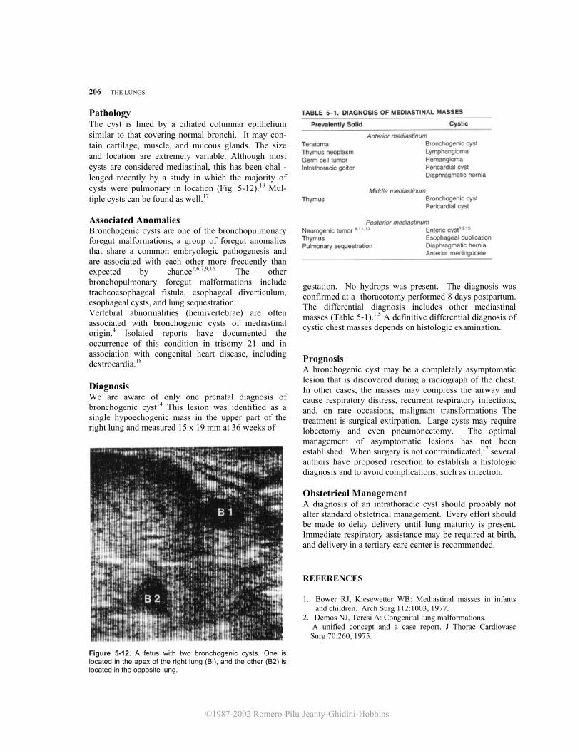
Pathology The cyst is lined by a ciliated columnar epithelium similar to that covering normal bronchi. It may con- tain cartilage, muscle, and mucous glands. The size and location are extremely variable. Although most cysts are considered mediastinal, this has been chal -lenged recently by a study in which the majority of cysts were pulmonary in location (Fig. 5-12).18 Mul- tiple cysts can be found as well.17
Associated Anomalies Bronchogenic cysts are one of the bronchopulmonary foregut malformations, a group of foregut anomalies that share a common embryologic pathogenesis and are associated with each other more frecuently than expected by chance2,6,7,9,16. The other bronchopulmonary foregut malformations include tracheoesophageal fistula, esophageal diverticulum, esophageal cysts, and lung sequestration. Vertebral abnormalities (hemivertebrae) are often associated with bronchogenic cysts of mediastinal origin.4 Isolated reports have documented the occurrence of this condition in trisomy 21 and in association with congenital heart disease, including dextrocardia.18 Diagnosis We are aware of only one prenatal diagnosis of bronchogenic cyst14 This lesion was identified as a single hypoechogenic mass in the upper part of the right lung and measured 15 x 19 mm at 36 weeks of
Figure 5-12. A fetus with two bronchogenic cysts. One is located in the apex of the right lung (Bl), and the other (B2) is located in the opposite lung.
gestation. No hydrops was present. The diagnosis was confirmed at a thoracotomy performed 8 days postpartum. The differential diagnosis includes other mediastinal masses (Table 5-1).1,5 A definitive differential diagnosis of cystic chest masses depends on histologic examination. Prognosis A bronchogenic cyst may be a completely asymptomatic lesion that is discovered during a radiograph of the chest. In other cases, the masses may compress the airway and cause respiratory distress, recurrent respiratory infections, and, on rare occasions, malignant transformations The treatment is surgical extirpation. Large cysts may require lobectomy and even pneumonectomy. The optimal management of asymptomatic lesions has not been established. When surgery is not contraindicated,17 several authors have proposed resection to establish a histologic diagnosis and to avoid complications, such as infection. Obstetrical Management A diagnosis of an intrathoracic cyst should probably not alter standard obstetrical management. Every effort should be made to delay delivery until lung maturity is present. Immediate respiratory assistance may be required at birth, and delivery in a tertiary care center is recommended.
REFERENCES
1. Bower RJ, Kiesewetter WB: Mediastinal masses in infants and children. Arch Surg 112:1003, 1977.
2. Demos NJ, Teresi A: Congenital lung malformations. A unified concept and a case report. J Thorac Cardiovasc
Surg 70:260, 1975.
©1987-2002 Romero-Pilu-Jeanty-Ghidini-Hobbins
BRONCHOGENIC CYST 207
3. Eraklis AJ, Griscom NT, McGovern JB: Bronchogenic
cysts of the mediastinum in infancy. N Engl J Med 281: 1150, 1969.
4. Fallon M, Gordon ARG, Lendrum AC: Mediastinal cysts of foregut origin associated with vertebral abnormalities. Br J Surg 41:520, 1954.
5. Felman AH: The Pediatric Chest. Radiological, Clinical and Pathological Observations. Springfield, IL, Chas. C Thomas, 1983, p 167.
6. Gerle RD, Jaretzki A, Ashley CA, et al.: Congential bronchopulmonary-foregut malformation: Pulmonary sequestration communicating with the gastrointestinal tract. N Engl J Med 278:1413, 1968.
7. Haller JA, Golladay ES, Pickard LR, et al.: Surgical management of lung bud anomalies: Lobar emphysema, bronchogenic cyst, cystic adenomatoid malformation, and intralobar pulmonary sequestration. Ann Thorac Surg 28:33, 1979.
8. Halperin DS, Oberhansli I, Siegrist CA, et al.: Intrathoracic neuroblastoma presenting with neonatal cardiorespiratory distress. Chest 85:822, 1984.
9. Heithoff KB, Sane SM, Williams HJ, et al.: Bronchopulmonary foregut malformations. A unifying etiological concept. AJR 126:46, 1976.
10. Hobbins JC, Grannum PAT, Berkowitz RL, et al.:
Ultrasound in the diagnosis of congenital anomalies. Am J Obstet Gynecol 134:331, 1979. 11. Illescas FF, Williams RL: Neonatal neuroblastoma pre
senting with respiratory distress. J Assoc Can Radiol 35:310, 1984.
12. Knochel JQ, Lee TG, Melendez MG, et al.: Fetal anomalies involving the thorax and abdomen. Radiol Clin North Am 20:297, 1982.
13. Massad M, Haddad F, Slim M, et al.: Spinal cord compression in neuroblastoma. Surg Neurol 23:567, 1985.
14. Mayden KL, Tortora M, Chervenak FA, et al.: The antenatal sonographic detection of lung masses. Am J Obstet Gynecol 148:349, 1984.
15. Newnham JP, Crues JV, Vinstein AL, et al.: Sonographic diagnosis of thoracic gastroenteric cyst in utero. Prenat Diagn 4:467, 1984.
16. O'Connell DJ, Kelleher J: Congenital intrathoracic bronchopulmonary foregut malformations in childhood. J Can Assoc Radiol 30:103, 1979.
17. Phelan PD, Landau LI, Olinsky A: Respiratory Illness in Children, 2d ed. Oxford, Blackwell Scientific, 1982, pp 397-416.
18. Ramenofsky ML, Leape LL, McCauley RGK: Bronchogenic cyst. J Pediatr Surg 14:219, 1979.