Embed Size (px)
Citation preview
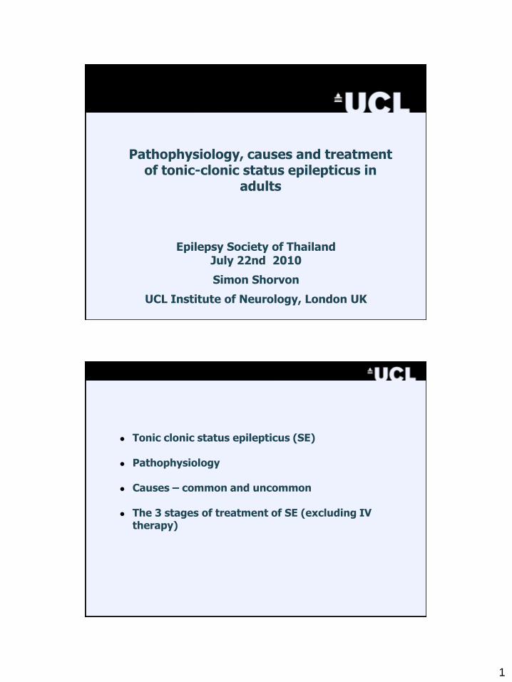
1
Epilepsy Society of Thailand July 22nd 2010
Simon Shorvon
UCL Institute of Neurology, London UK
Pathophysiology, causes and treatment of tonic-clonic status epilepticus in
adults
Tonic clonic status epilepticus (SE)
Pathophysiology
Causes – common and uncommon
The 3 stages of treatment of SE (excluding IV therapy)
2
Tonic-clonic status epilepticus
Incidence approximately 18-36 cases per 100,000 persons per year. 0.1% of all A&E visits.Rates higher in children, learning disability, structural cerebral pathology, frontal pathology
65% of cases occur de novo, without prior historyof epilepsy, due to acute cerebral event (vascular, trauma, infection) or acute metabolic/drug-inducedcause
In pre-existing epilepsy, TCSE is often precipitated by drug reduction/withdrawal, intercurrent illness, metabolic disturbance, progressive disease.
SE occurs in 5% of all adults and 10-25% of all children with epilepsy
Mortality rate – 10-20%
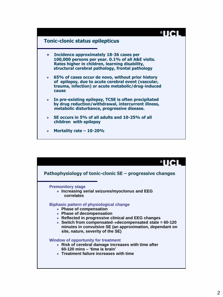
Pathophysiology of tonic-clonic SE – progressive changes
Premonitory stage Increasing serial seizures/myoclonus and EEG
correlates
Biphasic pattern of physiological change Phase of compensation Phase of decompensation Reflected in progressive clinical and EEG changes Switch from compensateddecompensated state = 60-120
minutes in convulsive SE (an approximation, dependant on site, nature, severity of the SE)
Window of opportunity for treatment Risk of cerebral damage increases with time after
60-120 mins – ‘time is brain’ Treatment failure increases with time
3
4
5
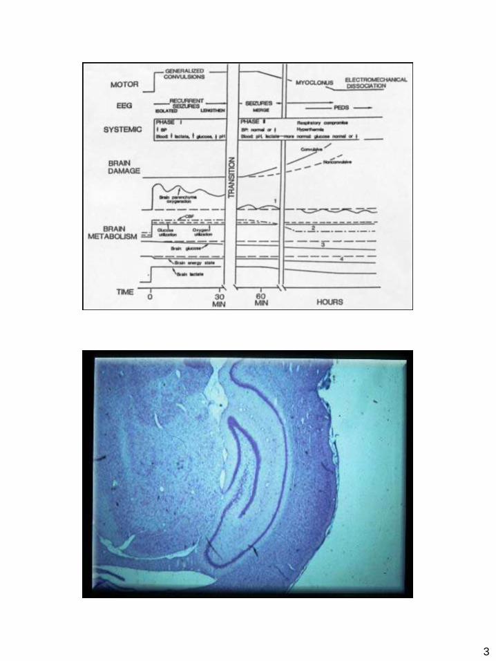
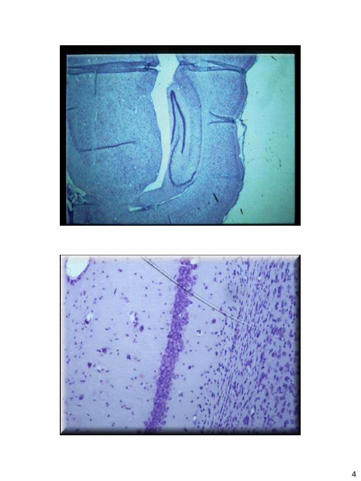
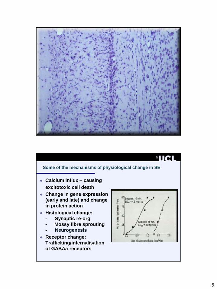
Some of the mechanisms of physiological change in SE
Calcium influx – causing
excitotoxic cell death
Change in gene expression
(early and late) and change
in protein action
Histological change:
- Synaptic re-org
- Mossy fibre sprouting
- Neurogenesis
Receptor change:
Trafficking/internalisation
of GABAa receptors
6
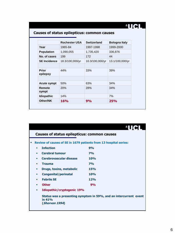
Causes of status epilepticus: common causes
Rochester USA Switzerland Bologna Italy
Year 1965-84 1997-1998 1999-2000
Population 1,090,055 1,735,420 336,876
No. of cases 199 172 44
SE Incidence 18.3/100,000/yr 10.3/100,000/yr 13.1/100,000/yr
Prior
epilepsy
44% 33% 39%
Acute sympt 50% 63% 34%
Remote
sympt
20% 28% 34%
Idiopathic 14% 7%
Other/NK 16% 9% 25%
Causes of status epilepticus: common causes
Review of causes of SE in 1679 patients from 13 hospital series:
Infection 9%
Cerebral tumour 7%
Cerebrovascular disease 10%
Trauma 7%
Drugs, toxins, metabolic 15%
Congenital/perinatal 10%
Febrile SE 12%
Other 9%
Idiopathic/cryptogenic 19%
Status was a presenting symptom in 59%, and an intercurrent event in 41%(Shorvon 1994)
7
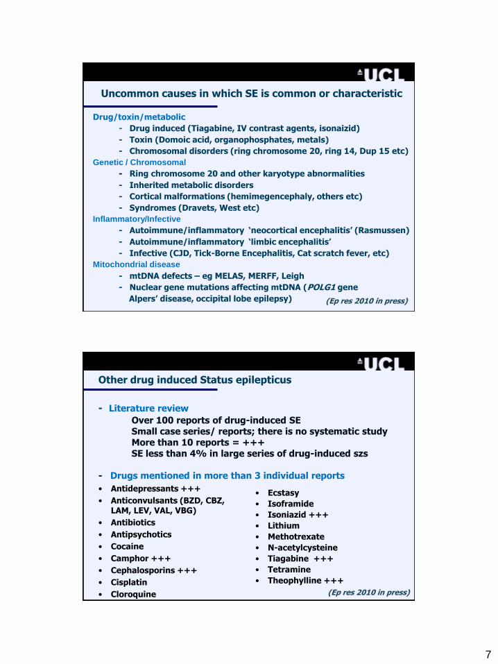
Uncommon causes in which SE is common or characteristic
Drug/toxin/metabolic
- Drug induced (Tiagabine, IV contrast agents, isonaizid)
- Toxin (Domoic acid, organophosphates, metals)
- Chromosomal disorders (ring chromosome 20, ring 14, Dup 15 etc)
Genetic / Chromosomal
- Ring chromosome 20 and other karyotype abnormalities
- Inherited metabolic disorders
- Cortical malformations (hemimegencephaly, others etc)
- Syndromes (Dravets, West etc)
Inflammatory/Infective
- Autoimmune/inflammatory „neocortical encephalitis‟ (Rasmussen)
- Autoimmune/inflammatory „limbic encephalitis‟
- Infective (CJD, Tick-Borne Encephalitis, Cat scratch fever, etc)
Mitochondrial disease
- mtDNA defects – eg MELAS, MERFF, Leigh
- Nuclear gene mutations affecting mtDNA (POLG1 gene
Alpers‟ disease, occipital lobe epilepsy) (Ep res 2010 in press)
Other drug induced Status epilepticus
- Literature review
Over 100 reports of drug-induced SESmall case series/ reports; there is no systematic studyMore than 10 reports = +++SE less than 4% in large series of drug-induced szs
- Drugs mentioned in more than 3 individual reports
• Antidepressants +++
• Anticonvulsants (BZD, CBZ, LAM, LEV, VAL, VBG)
• Antibiotics
• Antipsychotics
• Cocaine
• Camphor +++
• Cephalosporins +++
• Cisplatin
• Cloroquine
• Ecstasy
• Isoframide
• Isoniazid +++
• Lithium
• Methotrexate
• N-acetylcysteine
• Tiagabine +++
• Tetramine
• Theophylline +++
(Ep res 2010 in press)
8
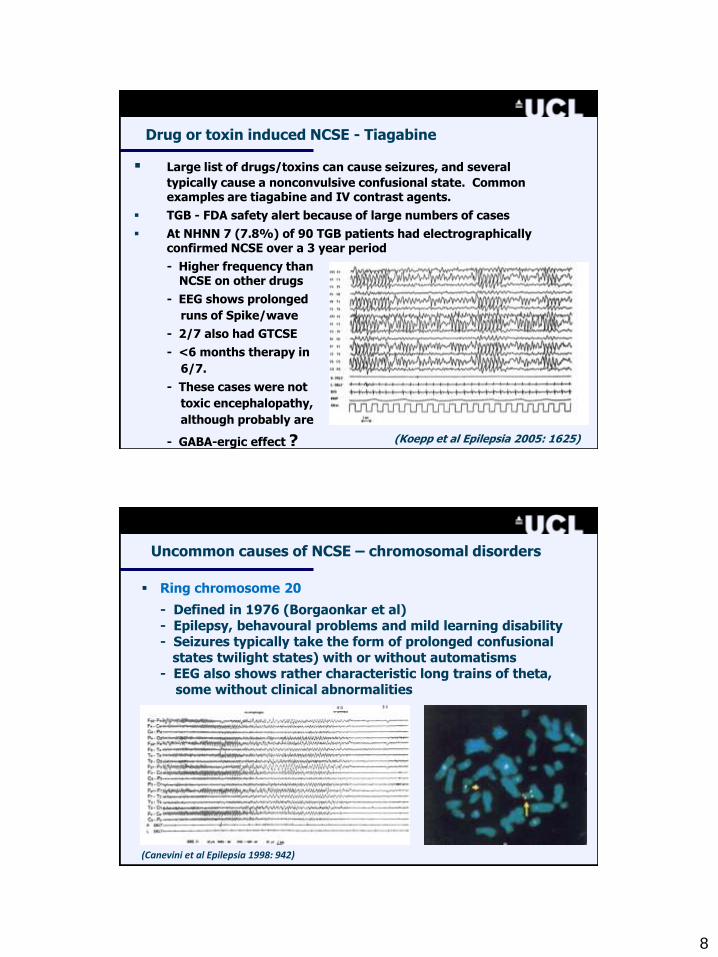
Drug or toxin induced NCSE - Tiagabine
Large list of drugs/toxins can cause seizures, and several
typically cause a nonconvulsive confusional state. Common examples are tiagabine and IV contrast agents.
TGB - FDA safety alert because of large numbers of cases
At NHNN 7 (7.8%) of 90 TGB patients had electrographically confirmed NCSE over a 3 year period
- Higher frequency thanNCSE on other drugs
- EEG shows prolonged
runs of Spike/wave
- 2/7 also had GTCSE
- <6 months therapy in
6/7.
- These cases were not
toxic encephalopathy,
although probably are
- GABA-ergic effect ? (Koepp et al Epilepsia 2005: 1625)
Uncommon causes of NCSE – chromosomal disorders
Ring chromosome 20
- Defined in 1976 (Borgaonkar et al)- Epilepsy, behavoural problems and mild learning disability- Seizures typically take the form of prolonged confusional
states twilight states) with or without automatisms- EEG also shows rather characteristic long trains of theta,
some without clinical abnormalities
(Canevini et al Epilepsia 1998: 942)
9
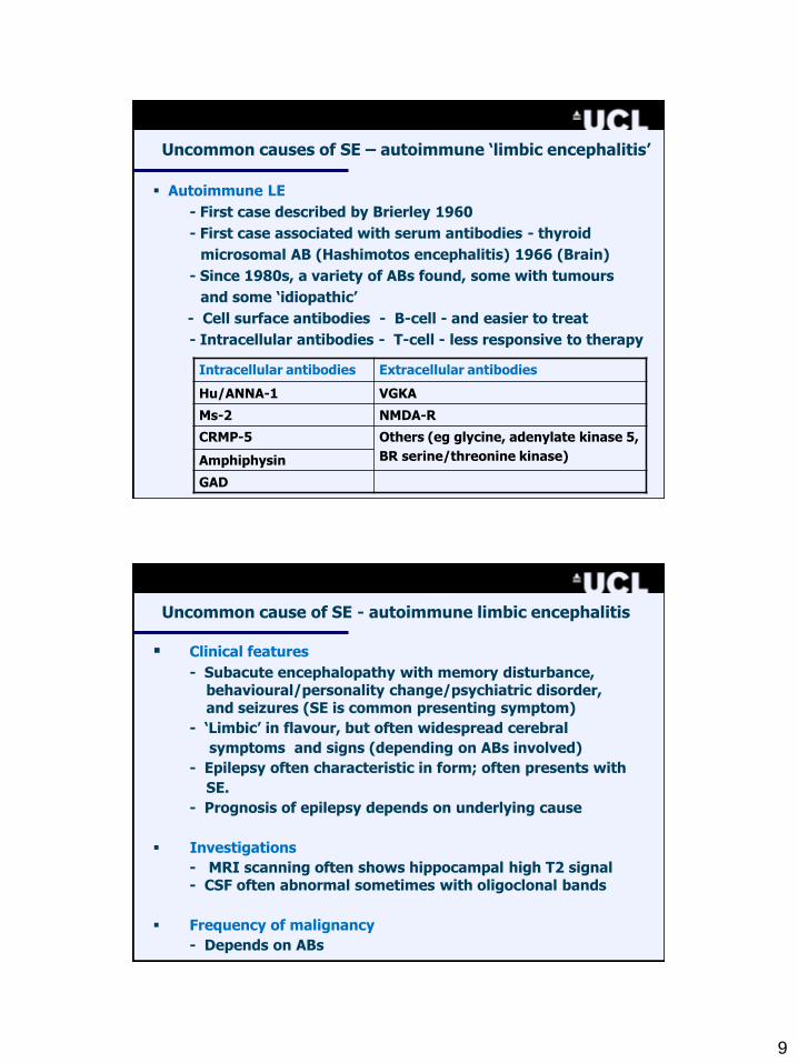
Uncommon causes of SE – autoimmune „limbic encephalitis‟
Autoimmune LE
- First case described by Brierley 1960
- First case associated with serum antibodies - thyroid
microsomal AB (Hashimotos encephalitis) 1966 (Brain)
- Since 1980s, a variety of ABs found, some with tumours
and some „idiopathic‟
- Cell surface antibodies - B-cell - and easier to treat
- Intracellular antibodies - T-cell - less responsive to therapy
Intracellular antibodies Extracellular antibodies
Hu/ANNA-1 VGKA
Ms-2 NMDA-R
CRMP-5 Others (eg glycine, adenylate kinase 5,
BR serine/threonine kinase)Amphiphysin
GAD
Uncommon cause of SE - autoimmune limbic encephalitis
Clinical features
- Subacute encephalopathy with memory disturbance, behavioural/personality change/psychiatric disorder, and seizures (SE is common presenting symptom)
- „Limbic‟ in flavour, but often widespread cerebral
symptoms and signs (depending on ABs involved)
- Epilepsy often characteristic in form; often presents with
SE.
- Prognosis of epilepsy depends on underlying cause
Investigations
- MRI scanning often shows hippocampal high T2 signal - CSF often abnormal sometimes with oligoclonal bands
Frequency of malignancy
- Depends on ABs
10
Uncommon causes of SE – autoimmune limbic encephalitis
Neoplastic autoimmune LE
- Neurological symptoms precede tumoural symptoms in
66%; LE associated with other signs
- Anti Hu: small cell lung cancer
10% have LE, others cerebellar, PN, autonomic
- Ma-2: intratubal germ cell tumours of testes
Other features include hypothalamic and brainstem signs
- Amphyphysin: LE and „stiff person syndrome
- GAD: LE with „stiff person syndrome‟
Non-neoplastic autoimmune LE
- Voltage-gated potassium channel antibodies
- NMDA-R antibodies First case described by Brierley 1960
- Hashimotos encephalitis (STREAT)
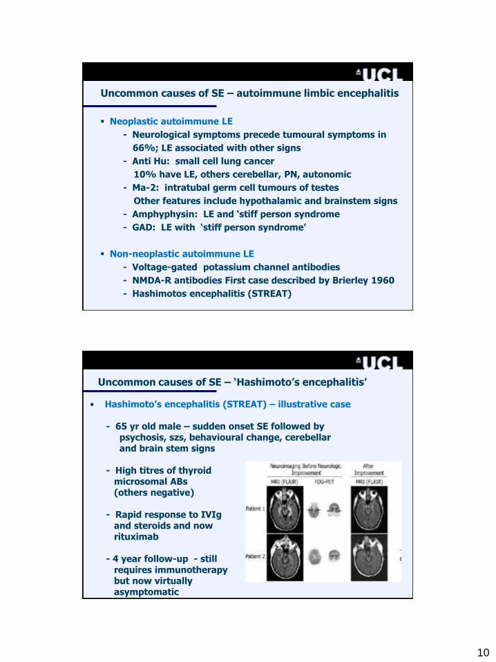
Uncommon causes of SE – „Hashimoto‟s encephalitis‟
• Hashimoto‟s encephalitis (STREAT) – illustrative case
- 65 yr old male – sudden onset SE followed bypsychosis, szs, behavioural change, cerebellar and brain stem signs
- High titres of thyroid microsomal ABs(others negative)
- Rapid response to IVIg and steroids and now rituximab
- 4 year follow-up - stillrequires immunotherapybut now virtually asymptomatic
11
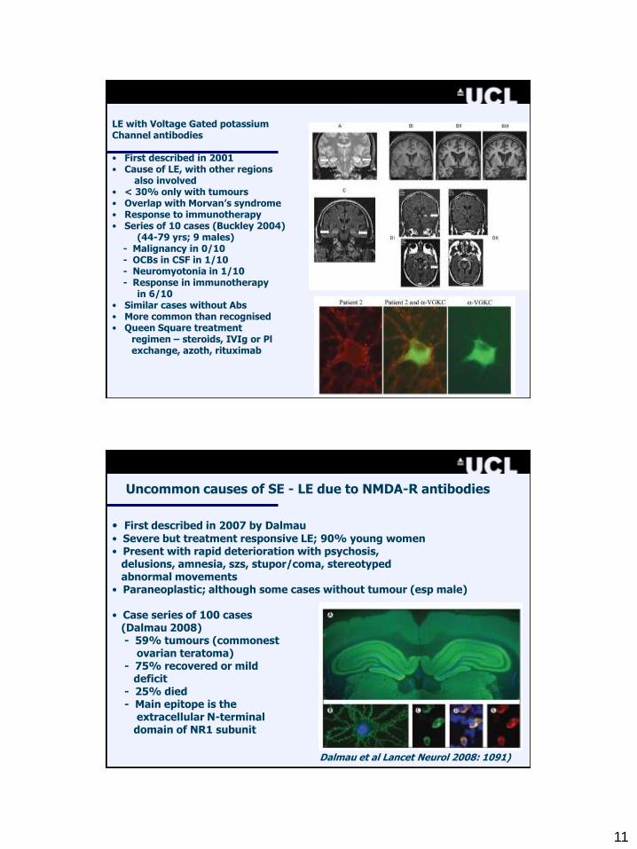
LE with Voltage Gated potassiumChannel antibodies
• First described in 2001• Cause of LE, with other regions
also involved • < 30% only with tumours• Overlap with Morvan‟s syndrome• Response to immunotherapy• Series of 10 cases (Buckley 2004)
(44-79 yrs; 9 males)- Malignancy in 0/10- OCBs in CSF in 1/10- Neuromyotonia in 1/10- Response in immunotherapy
in 6/10• Similar cases without Abs• More common than recognised• Queen Square treatment
regimen – steroids, IVIg or Plexchange, azoth, rituximab
Uncommon causes of SE - LE due to NMDA-R antibodies
• First described in 2007 by Dalmau
• Severe but treatment responsive LE; 90% young women• Present with rapid deterioration with psychosis,
delusions, amnesia, szs, stupor/coma, stereotypedabnormal movements
• Paraneoplastic; although some cases without tumour (esp male)
• Case series of 100 cases (Dalmau 2008)- 59% tumours (commonest
ovarian teratoma)- 75% recovered or mild
deficit- 25% died- Main epitope is the
extracellular N-terminaldomain of NR1 subunit
Dalmau et al Lancet Neurol 2008: 1091)
12
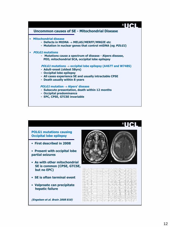
Uncommon causes of SE - Mitochondrial Disease
• Mitochondrial disease- Defects in MtDNA MELAS/MERFF/MNGIE etc- Mutation in nuclear genes that control mtDNA (eg POLG1)
• POLG1 mutations- Mutations cause a spectrum of disease - Alpers disease,
PEO, mitochondrial SCA, occipital lobe epilepsy
POLG1 mutations occipital lobe epilepsy (A467T and W748S)- Adult-onset (oldest 58yrs)- Occipital lobe epilepsy- All cases experience SE and usually intractable CPSE- Death usually within 8 years
POLG1 mutation Alpers‟ disease- Subacute presentation, death within 12 months- Occipital predominance - EPC, CPSE, GTCSE invariable
POLG1 mutations causing Occipital lobe epilepsy
• First described in 2008
• Present with occipital lobepartial seizures
• As with other mitochondrialSE is common (CPSE, GTCSE,but no EPC)
• SE is often terminal event
• Valproate can precipitate hepatic failure
(Engelsen et al. Brain 2008 818)
13
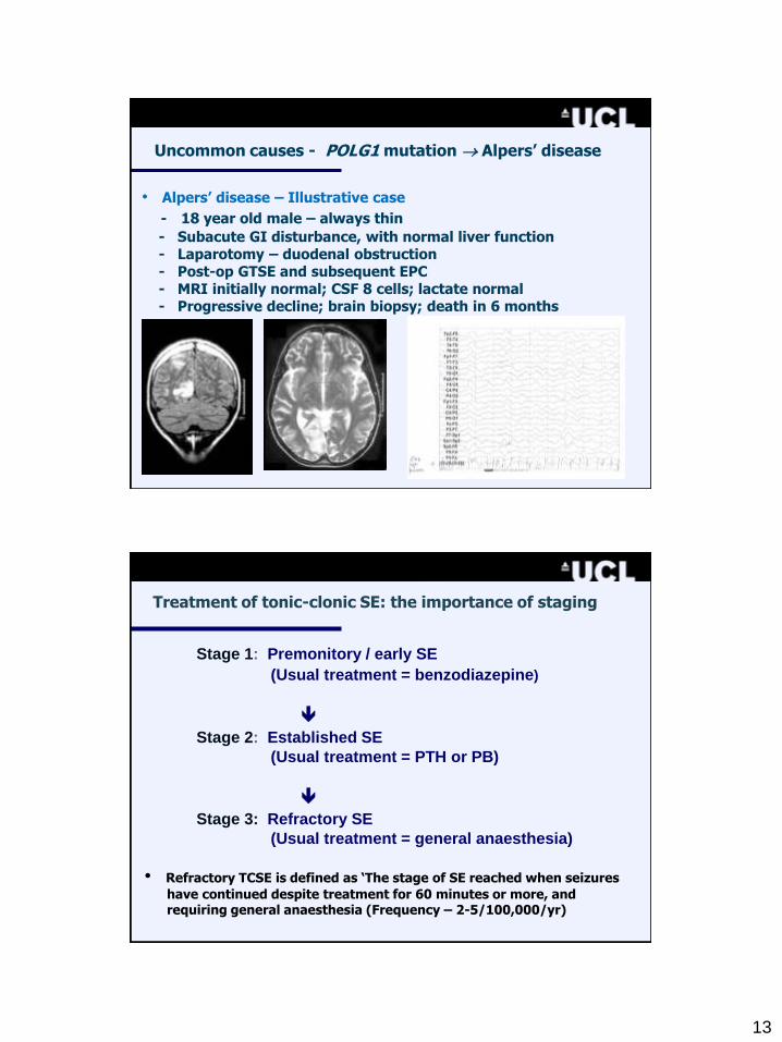
Uncommon causes - POLG1 mutation Alpers‟ disease
• Alpers‟ disease – Illustrative case
- 18 year old male – always thin
- Subacute GI disturbance, with normal liver function- Laparotomy – duodenal obstruction- Post-op GTSE and subsequent EPC- MRI initially normal; CSF 8 cells; lactate normal- Progressive decline; brain biopsy; death in 6 months
Treatment of tonic-clonic SE: the importance of staging
Stage 1: Premonitory / early SE
(Usual treatment = benzodiazepine)
Stage 2: Established SE
(Usual treatment = PTH or PB)
Stage 3: Refractory SE
(Usual treatment = general anaesthesia)
• Refractory TCSE is defined as „The stage of SE reached when seizures
have continued despite treatment for 60 minutes or more, andrequiring general anaesthesia (Frequency – 2-5/100,000/yr)
14
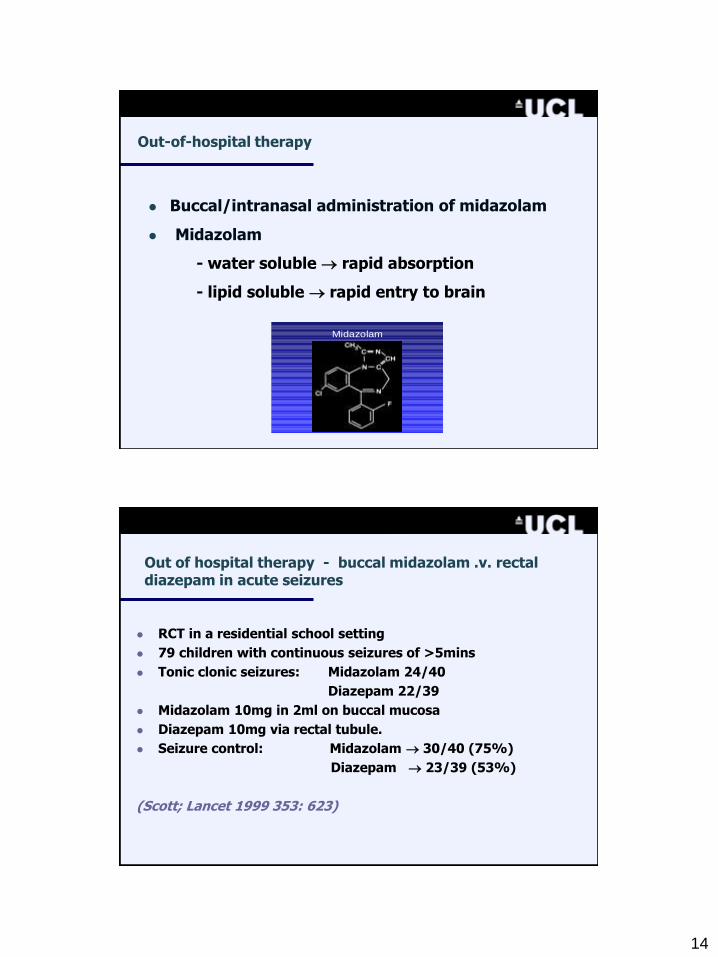
Out-of-hospital therapy
Buccal/intranasal administration of midazolam
Midazolam
- water soluble rapid absorption
- lipid soluble rapid entry to brain
Midazolam
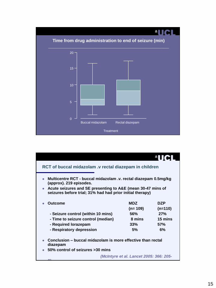
Out of hospital therapy - buccal midazolam .v. rectal diazepam in acute seizures
RCT in a residential school setting
79 children with continuous seizures of >5mins
Tonic clonic seizures: Midazolam 24/40
Diazepam 22/39
Midazolam 10mg in 2ml on buccal mucosa
Diazepam 10mg via rectal tubule.
Seizure control: Midazolam 30/40 (75%)
Diazepam 23/39 (53%)
(Scott; Lancet 1999 353: 623)
15
10
20
15
5
0Buccal midazolam Rectal diazepam
Treatment
Time from drug administration to end of seizure (min)
_____________________________________________
RCT of buccal midazolam .v rectal diazepam in children
Multicentre RCT - buccal midazolam .v. rectal diazepam 0.5mg/kg (approx). 219 episodes.
Acute seizures and SE presenting to A&E (mean 30-47 mins of seizures before trial; 31% had had prior initial therapy)
Outcome MDZ DZP
(n= 109) (n=110)
- Seizure control (within 10 mins) 56% 27%
- Time to seizure control (median) 8 mins 15 mins
- Required lorazepam 33% 57%
- Respiratory depression 5% 6%
Conclusion – buccal midazolam is more effective than rectal diazepam
50% control of seizures >30 mins
(McIntyre et al. Lancet 2005: 366: 205-9)
16
Stage 2 – established SE: post-BZD AED therapy
RCTs in established SE
- Diazepam/phenytoin vs phenobarbital - 2 RCTs (n=222)
- Phenytoin vs phenobarbital - 1 RCT (n= 186)
- Diazepam/phenytoin vs phenytoin - 1 RCT (n= 196)
Conclusions (4 RCTs):
1. No significant differences
2. trend to favour DZP/PHT over PB
3. trend to favour PB over PHT
Stage 2 – established SE
Licensed medications - phenytoin and phenobarbital - Both carry risk of cardiovascular and cerebral depression- Both cause hypotension - Both are EIAEDs (and PB also self induction) - Both interact with other drugs - PTH carries cardiac risk and risk of infusion site damage- Both have poor pharmacokinetics
Unlicensed alternatives: - Valproate at least 8 open case series (>200 pts) and
one RCT (VPA vs PTH) - Levetiracetam at least 20 open case series (>150 patients)- Control rates equivalent historically to PHT/PB- No cardiovascular toxicity- No hypotension - No cardiac risk- No risk to infusion site- LEV has no enzyme induction nor drug interactions
17
Treatment of refractory stage of SE (>90mins): conventional protocol
Thiopentone 100-240 mg IV bolus over 20 secs then
50 mg boluses every 2-3 mins then IV infusion 3-5 mg/kg/hr to obtain burst suppression.
or Propofol 2 mg/kg IV bolus, repeated, then IV infusion
of 5-10 mg/kg/hr to obtain burst suppression.or
Midazolam 0.1–0.3mg/kg IV bolus at a rate notexceeding 4mg/min, then IV infusion 0.05-0.4mg/kg/hr to obtain burst suppression.
Alternatives: Pentobarbitone, ketamine, etomidate, desflurane, isoflurane
08KP0090 / May 2008
Stage of refractory SE – which is the best general anaesthetic
Midazolam Barbiturate Propofol
Short term mortality (7 series)
17-69% 20-55% 26-88%
Accumulation +++ ++++ 0
Tolerance ++++ ++ 0
AED action ++++ ++++ 0
Elim half-life 6hr 24-36hr 1-2hr
Other problems „infusion syndrome‟
08KP0090 / May 2008
18
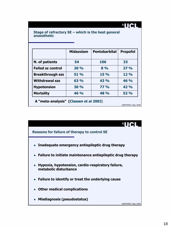
Stage of refractory SE – which is the best general anaesthetic
Midazolam Pentobarbital Propofol
N. of patients 54 106 33
Failed sz control 20 % 8 % 27 %
Breakthrough szs 51 % 15 % 12 %
Withdrawal szs 63 % 43 % 46 %
Hypotension 30 % 77 % 42 %
Mortality 46 % 48 % 52 %
A “meta-analysis” (Classen et al 2002)08KP0090 / May 2008
Reasons for failure of therapy to control SE
Inadequate emergency antiepileptic drug therapy
Failure to initiate maintenance antiepileptic drug therapy
Hypoxia, hypotension, cardio-respiratory failure, metabolic disturbance
Failure to identify or treat the underlying cause
Other medical complications
Misdiagnosis (pseudostatus)08KP0090 / May 2008

19
3rd London Innsbruck Colloquium on Acute Seizures and Status Epilepticus
Oxford UK : 7th - 9th April 2011
For further information:
www.statusepilepticus2011.eu