Embed Size (px)
Citation preview
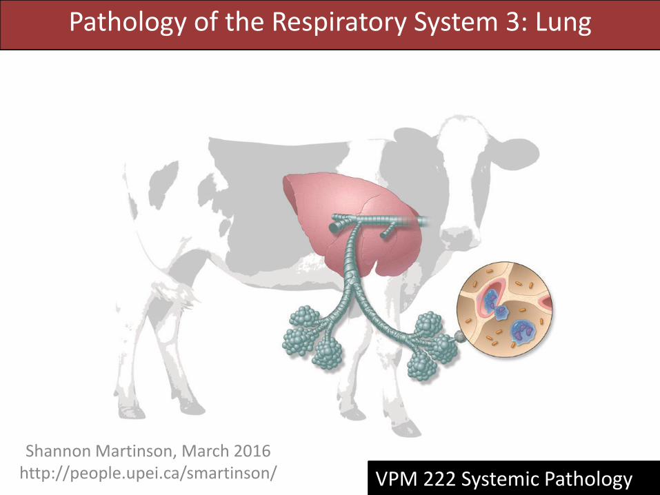
Pathology of the Respiratory System 3: Lung
Shannon Martinson, March 2016 http://people.upei.ca/smartinson/ VPM 222 Systemic Pathology
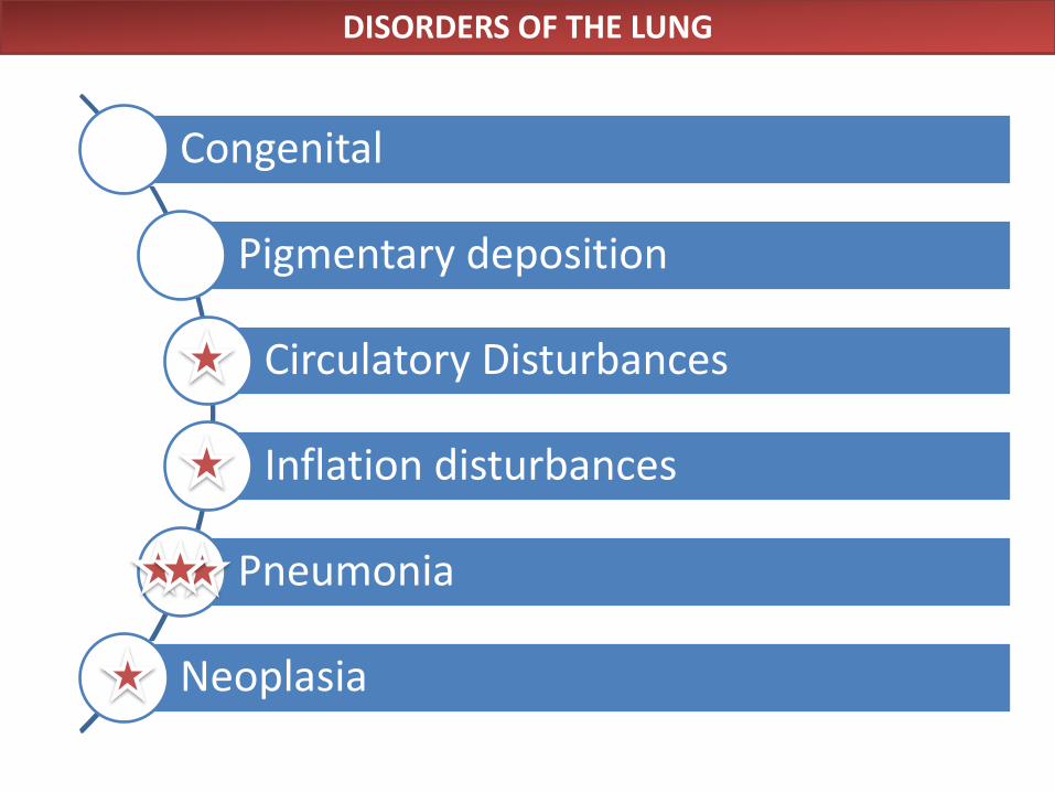
DISORDERS OF THE LUNG
Congenital
Pigmentary deposition
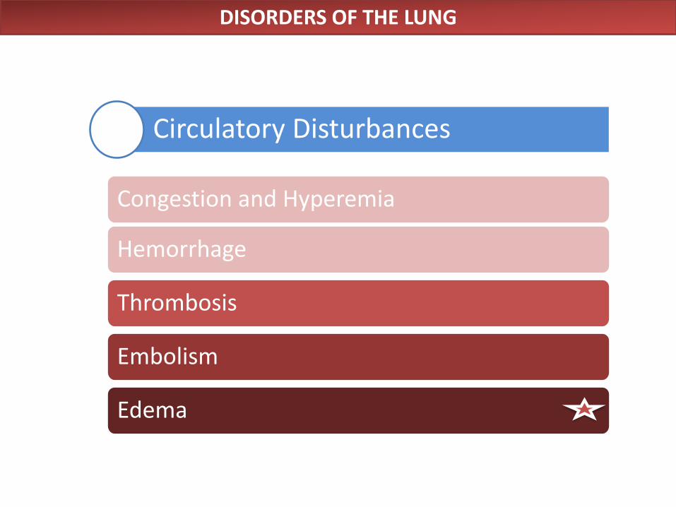
Circulatory Disturbances
Inflation disturbances
Pneumonia
Neoplasia
DISORDERS OF THE LUNG
Circulatory Disturbances
Congestion and Hyperemia
Hemorrhage
Thrombosis
Embolism
Edema
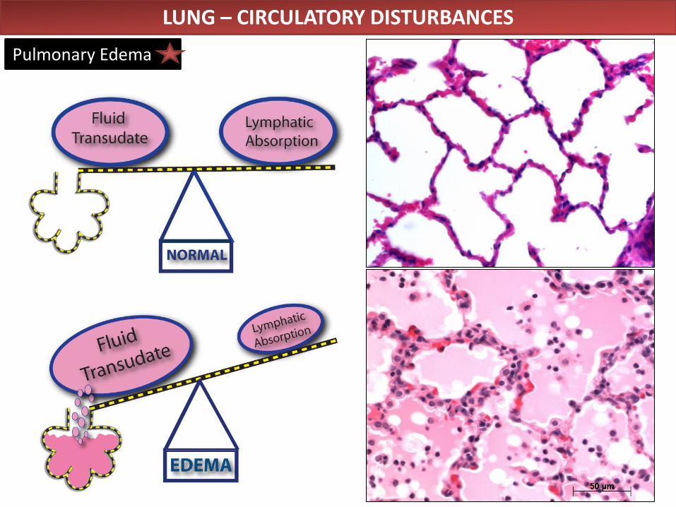
LUNG – CIRCULATORY DISTURBANCES
Pulmonary Edema
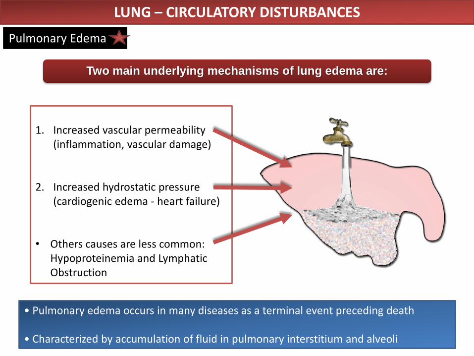
• Pulmonary edema occurs in many diseases as a terminal event preceding death • Characterized by accumulation of fluid in pulmonary interstitium and alveoli
1. Increased vascular permeability
(inflammation, vascular damage)
2. Increased hydrostatic pressure (cardiogenic edema - heart failure)
• Others causes are less common: Hypoproteinemia and Lymphatic Obstruction
Two main underlying mechanisms of lung edema are:
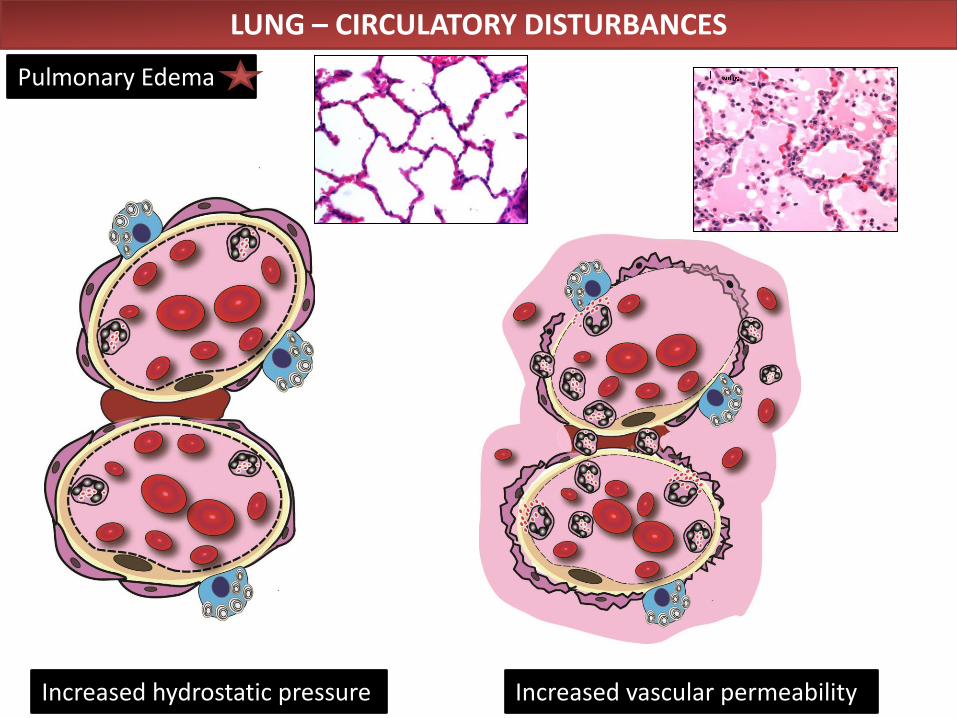
LUNG – CIRCULATORY DISTURBANCES
Pulmonary Edema
LUNG – CIRCULATORY DISTURBANCES
Pulmonary Edema
Increased vascular permeability Increased hydrostatic pressure
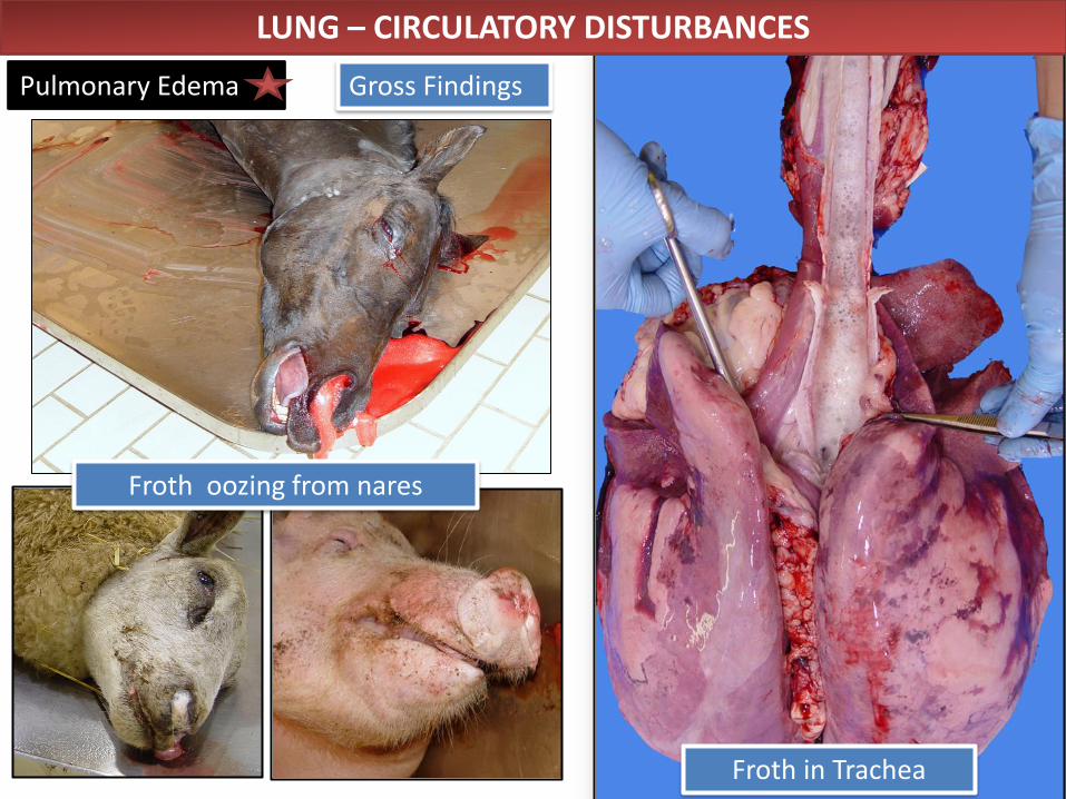
LUNG – CIRCULATORY DISTURBANCES
Pulmonary Edema Gross Findings
Froth oozing from nares
Froth in Trachea
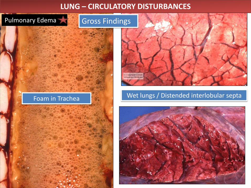
Foam in Trachea Wet lungs / Distended interlobular septa
LUNG – CIRCULATORY DISTURBANCES
Pulmonary Edema Gross Findings
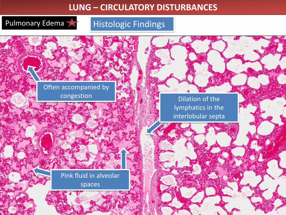
LUNG – CIRCULATORY DISTURBANCES
Pulmonary Edema Histologic Findings
Often accompanied by congestion
Pink fluid in alveolar spaces
Dilation of the lymphatics in the interlobular septa
Normal Fetal Lung
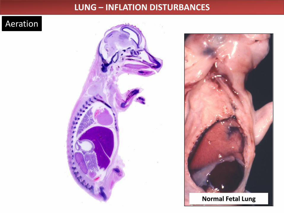
LUNG – INFLATION DISTURBANCES
Aeration
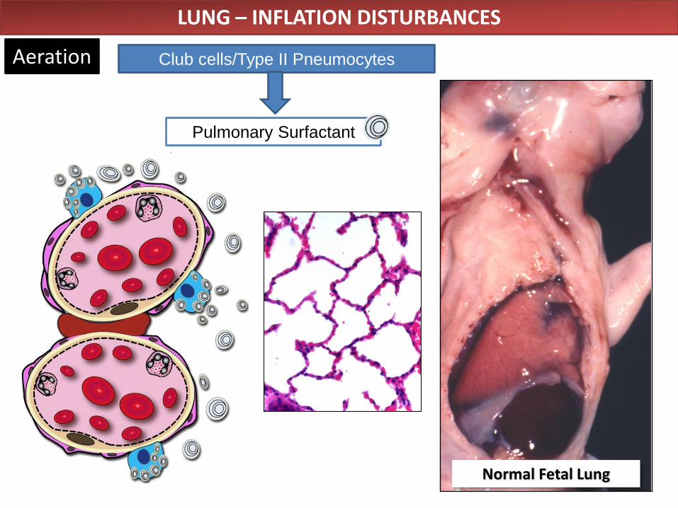
Club cells/Type II Pneumocytes Aeration
Normal Fetal Lung
LUNG – INFLATION DISTURBANCES
Pulmonary Surfactant
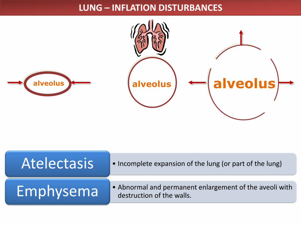
alveolus alveolus
LUNG – INFLATION DISTURBANCES
alveolus
• Incomplete expansion of the lung (or part of the lung) Atelectasis
• Abnormal and permanent enlargement of the aveoli with destruction of the walls. Emphysema
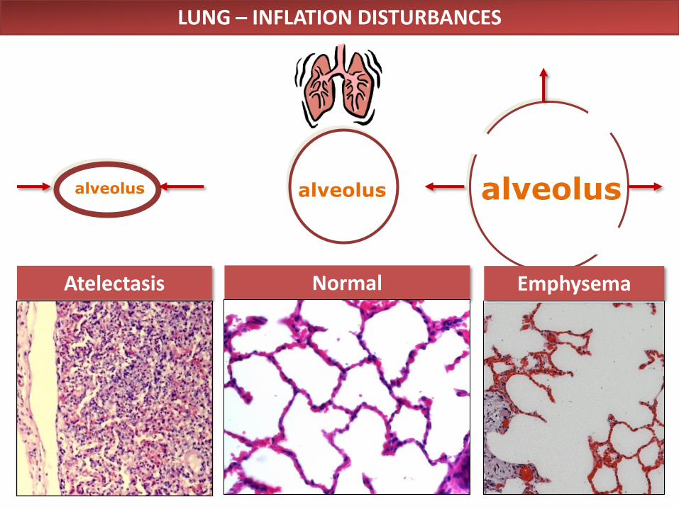
Normal
alveolus
Atelectasis
alveolus
LUNG – INFLATION DISTURBANCES
alveolus
Emphysema
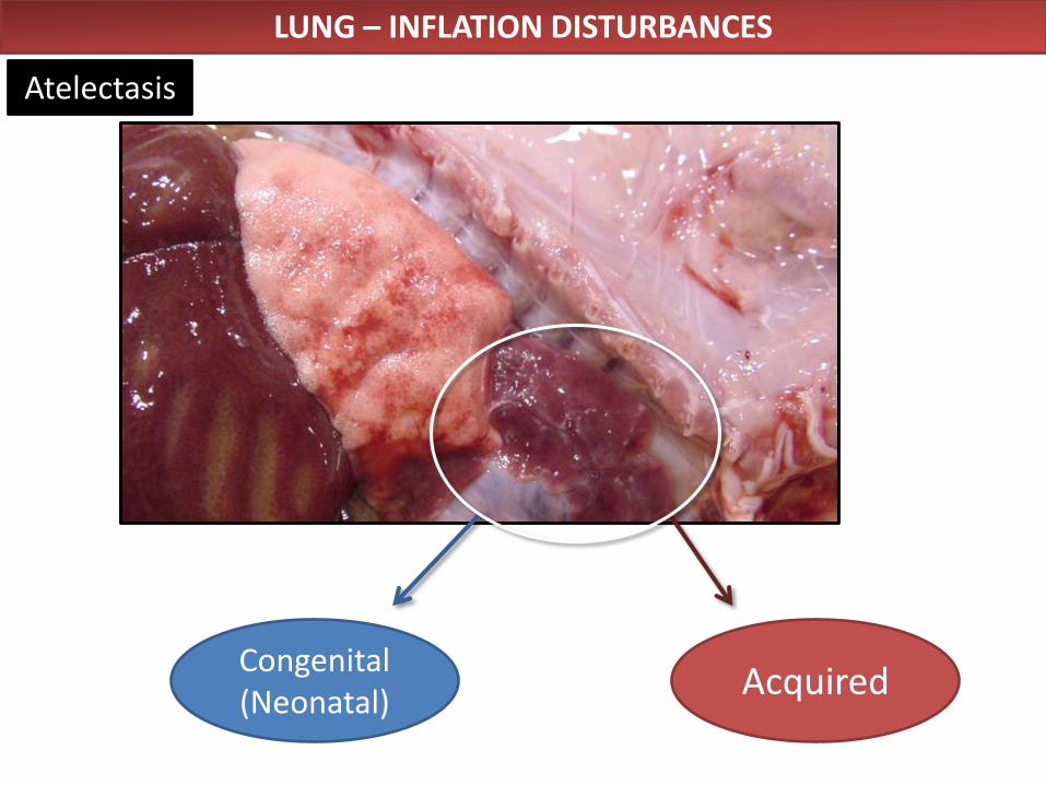
Congenital (Neonatal)
Acquired
LUNG – INFLATION DISTURBANCES
Atelectasis
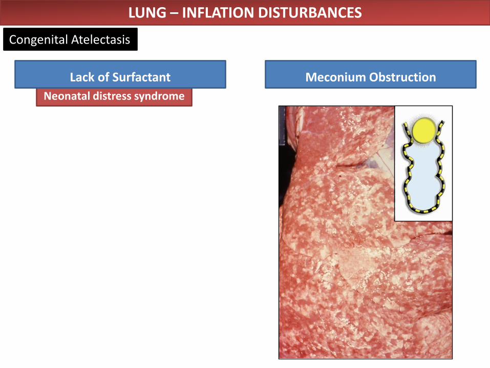
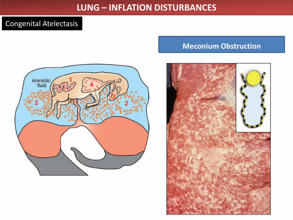
Meconium Obstruction
Neonatal distress syndrome
Lack of Surfactant
LUNG – INFLATION DISTURBANCES
Congenital Atelectasis
Meconium Obstruction
LUNG – INFLATION DISTURBANCES
Congenital Atelectasis
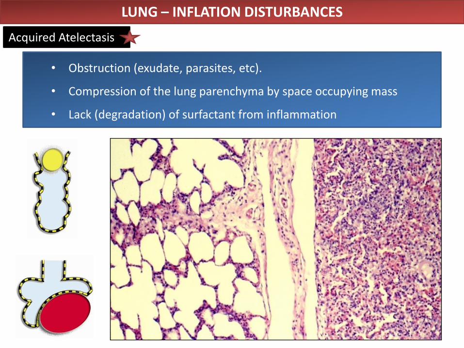
• Obstruction (exudate, parasites, etc).
• Compression of the lung parenchyma by space occupying mass
• Lack (degradation) of surfactant from inflammation
LUNG – INFLATION DISTURBANCES
Acquired Atelectasis
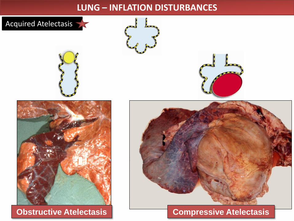
Compressive Atelectasis Obstructive Atelectasis
Acquired Atelectasis
LUNG – INFLATION DISTURBANCES
LUNG – INFLATION DISTURBANCES
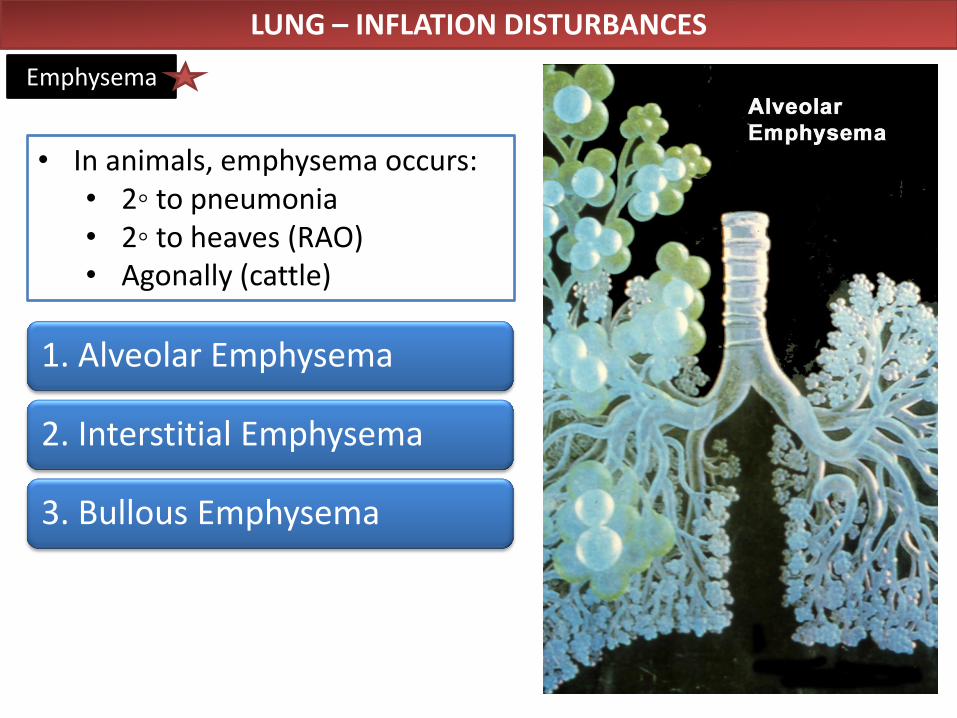
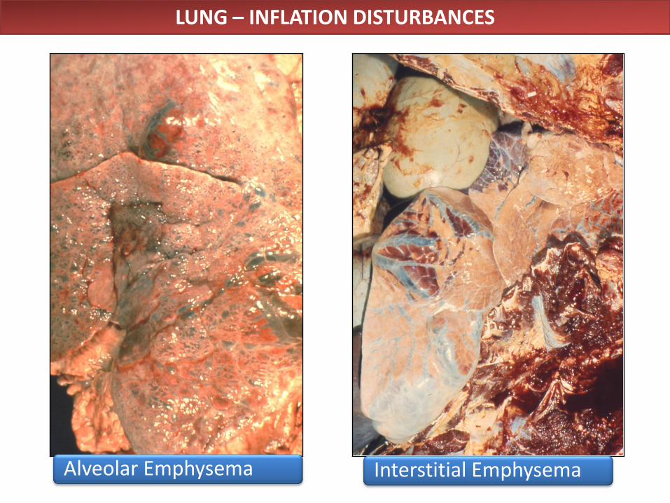
Emphysema
1. Alveolar Emphysema
2. Interstitial Emphysema
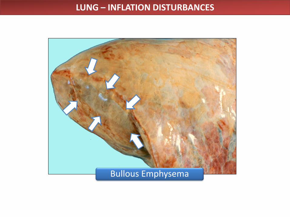
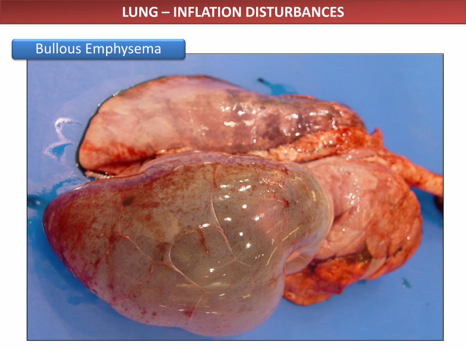
3. Bullous Emphysema
• In animals, emphysema occurs: • 2◦ to pneumonia • 2◦ to heaves (RAO) • Agonally (cattle)
Normal Emphysema
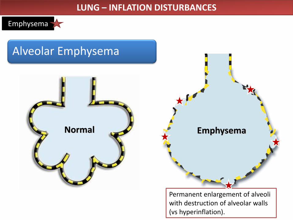
Permanent enlargement of alveoli with destruction of alveolar walls (vs hyperinflation).
LUNG – INFLATION DISTURBANCES
Emphysema
Alveolar Emphysema
LUNG – INFLATION DISTURBANCES
Alveolar Emphysema Interstitial Emphysema
LUNG – INFLATION DISTURBANCES
Bullous Emphysema
LUNG – INFLATION DISTURBANCES
Bullous Emphysema
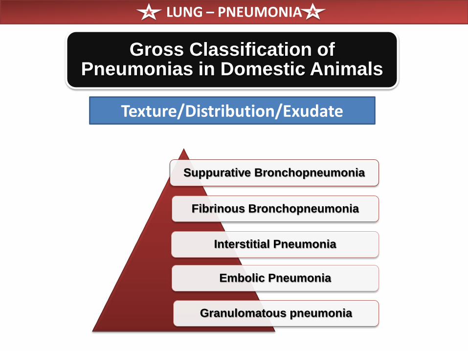
Gross Classification of Pneumonias in Domestic Animals
Texture/Distribution/Exudate
Suppurative Bronchopneumonia
Fibrinous Bronchopneumonia
Interstitial Pneumonia
Embolic Pneumonia
Granulomatous pneumonia
LUNG – PNEUMONIA
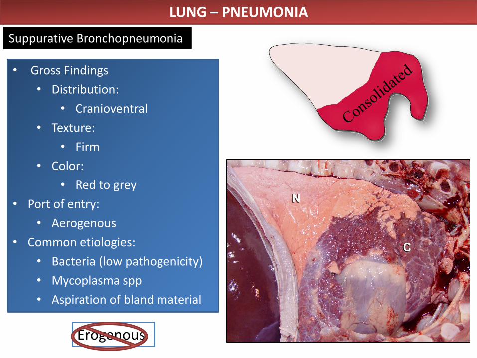
• Gross Findings
• Distribution:
• Cranioventral
• Texture:
• Firm
• Color:
• Red to grey
• Port of entry:
• Aerogenous
• Common etiologies:
• Bacteria (low pathogenicity)
• Mycoplasma spp
• Aspiration of bland material
Bovine lung
normal
LUNG – PNEUMONIA
Suppurative Bronchopneumonia
Erogenous
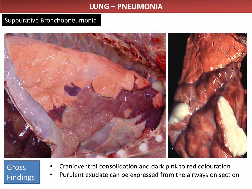
LUNG – PNEUMONIA
Suppurative Bronchopneumonia
• Cranioventral consolidation and dark pink to red colouration • Purulent exudate can be expressed from the airways on section
Gross Findings
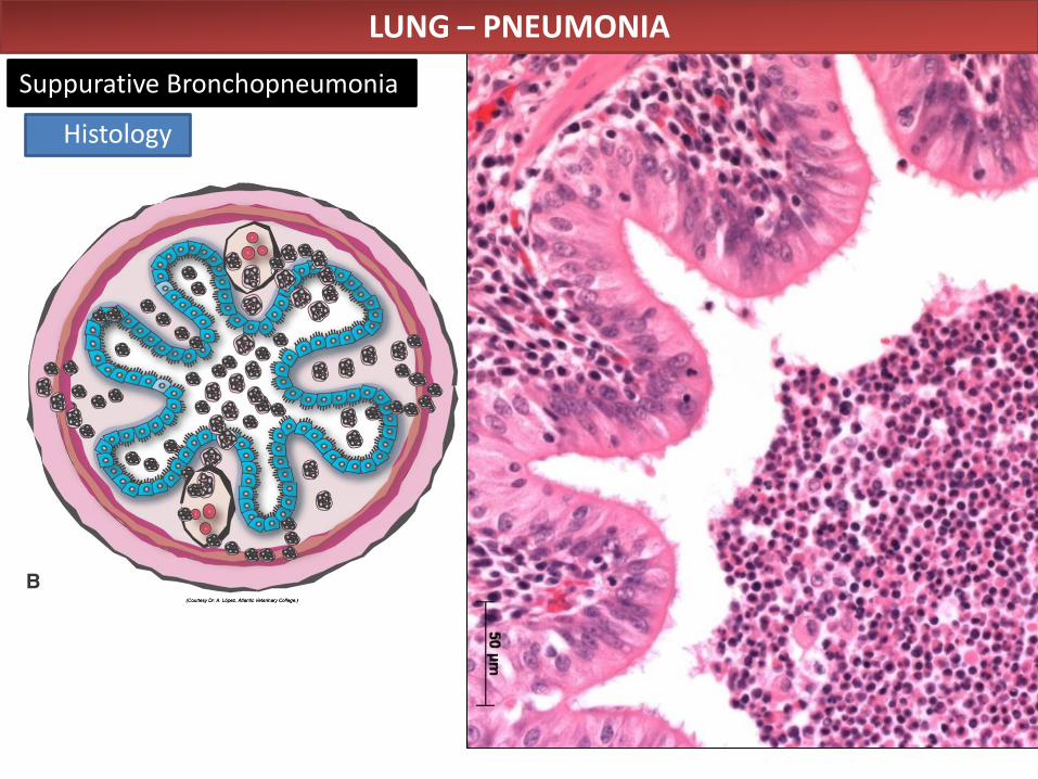
LUNG – PNEUMONIA
Suppurative Bronchopneumonia
Histology
• Abscesses
• Fibrosis, pleural adhesions
• Bronchiectasis
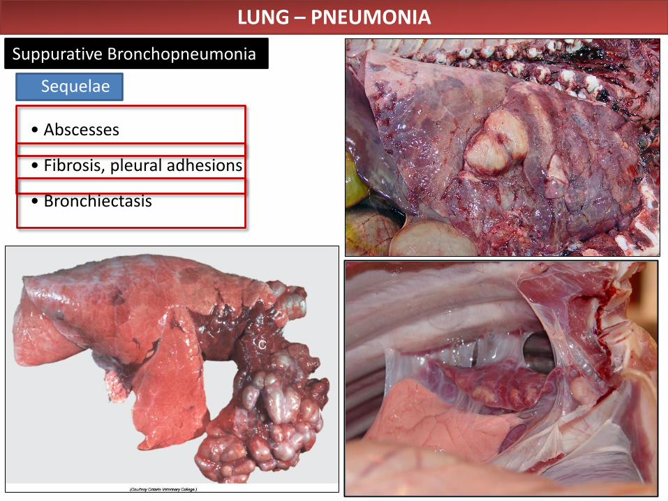
LUNG – PNEUMONIA
Suppurative Bronchopneumonia
Sequelae
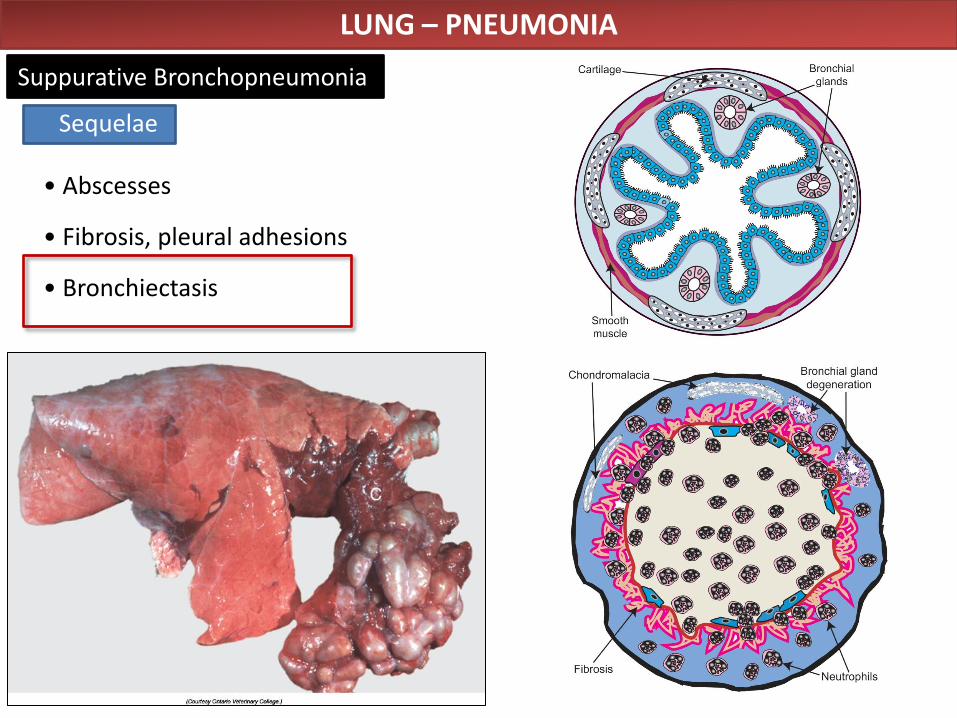
• Abscesses
• Fibrosis, pleural adhesions
• Bronchiectasis
LUNG – PNEUMONIA
Suppurative Bronchopneumonia
Sequelae
LUNG – PNEUMONIA
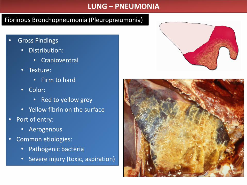
Fibrinous Bronchopneumonia (Pleuropneumonia)
• Gross Findings
• Distribution:
• Cranioventral
• Texture:
• Firm to hard
• Color:
• Red to yellow grey
• Yellow fibrin on the surface
• Port of entry:
• Aerogenous
• Common etiologies:
• Pathogenic bacteria
• Severe injury (toxic, aspiration)
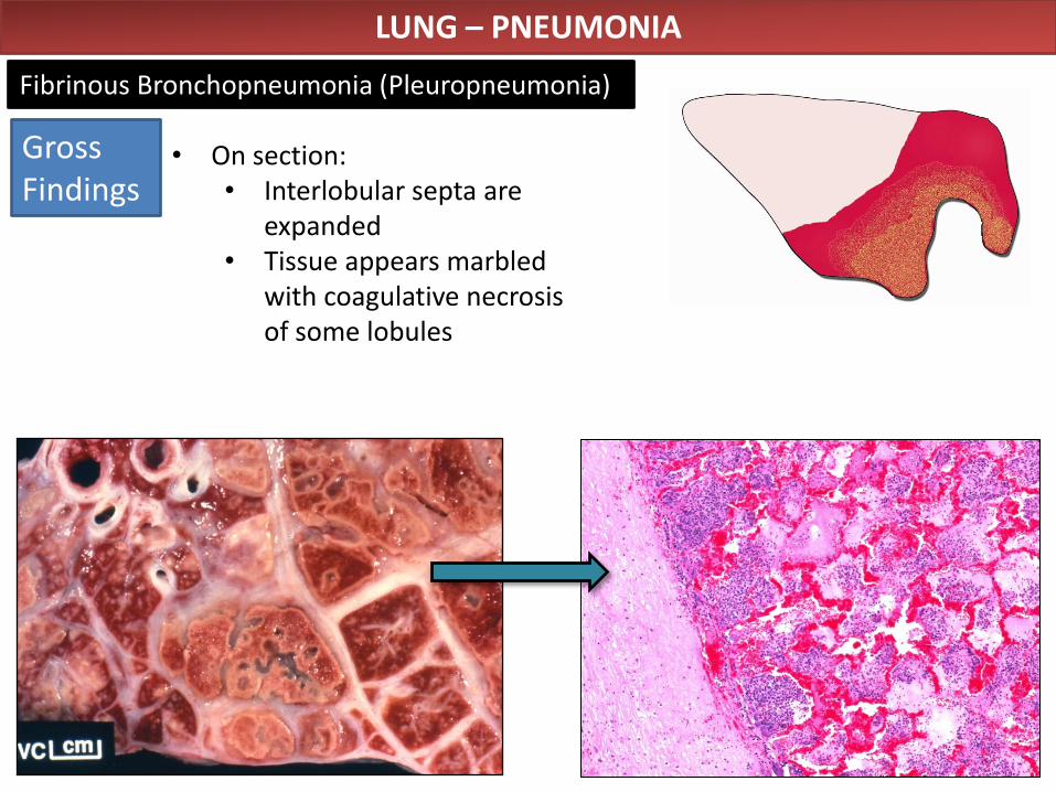
LUNG – PNEUMONIA
Fibrinous Bronchopneumonia (Pleuropneumonia)
• On section: • Interlobular septa are
expanded • Tissue appears marbled
with coagulative necrosis of some lobules
Gross Findings
LUNG – PNEUMONIA
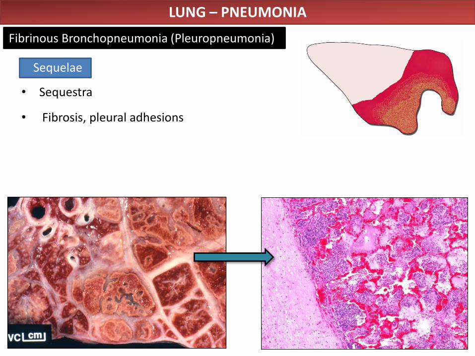
Fibrinous Bronchopneumonia (Pleuropneumonia)
• Sequestra
• Fibrosis, pleural adhesions
Sequelae
LUNG – PNEUMONIA
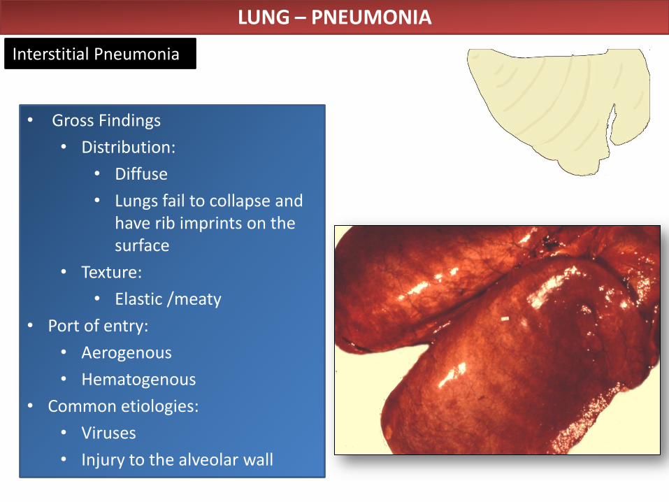
Interstitial Pneumonia
• Gross Findings
• Distribution:
• Diffuse
• Lungs fail to collapse and have rib imprints on the surface
• Texture:
• Elastic /meaty
• Port of entry:
• Aerogenous
• Hematogenous
• Common etiologies:
• Viruses
• Injury to the alveolar wall
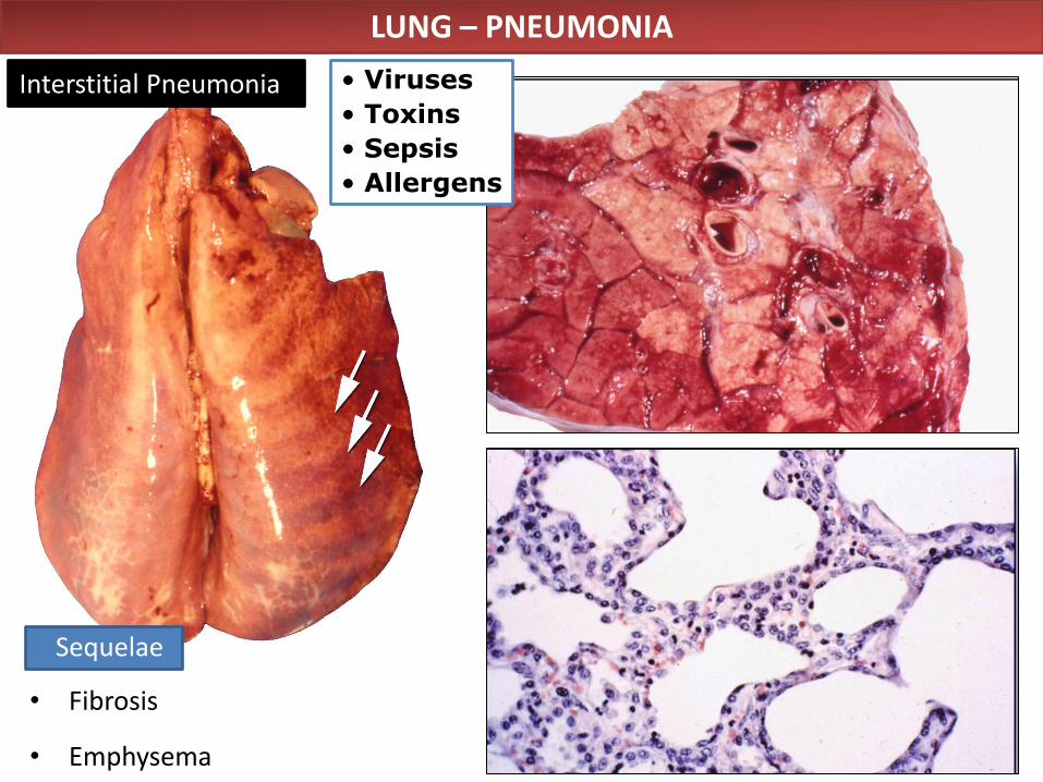
• Viruses
• Toxins
• Sepsis
• Allergens
LUNG – PNEUMONIA
Interstitial Pneumonia
• Fibrosis
• Emphysema
Sequelae
LUNG – PNEUMONIA
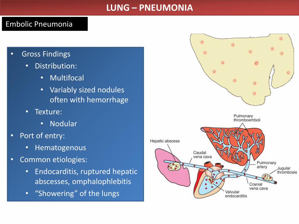
Embolic Pneumonia
• Gross Findings
• Distribution:
• Multifocal
• Variably sized nodules often with hemorrhage
• Texture:
• Nodular
• Port of entry:
• Hematogenous
• Common etiologies:
• Endocarditis, ruptured hepatic abscesses, omphalophlebitis
• “Showering” of the lungs
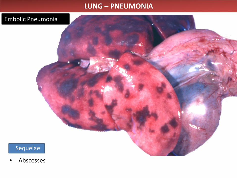
LUNG – PNEUMONIA
Embolic Pneumonia
• Abscesses
Sequelae
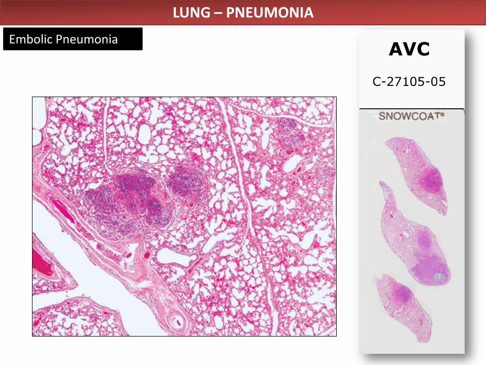
LUNG – PNEUMONIA
Embolic Pneumonia
LUNG – PNEUMONIA
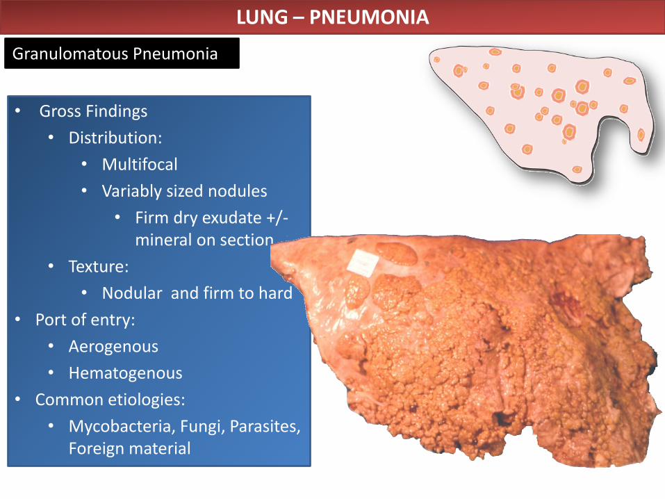
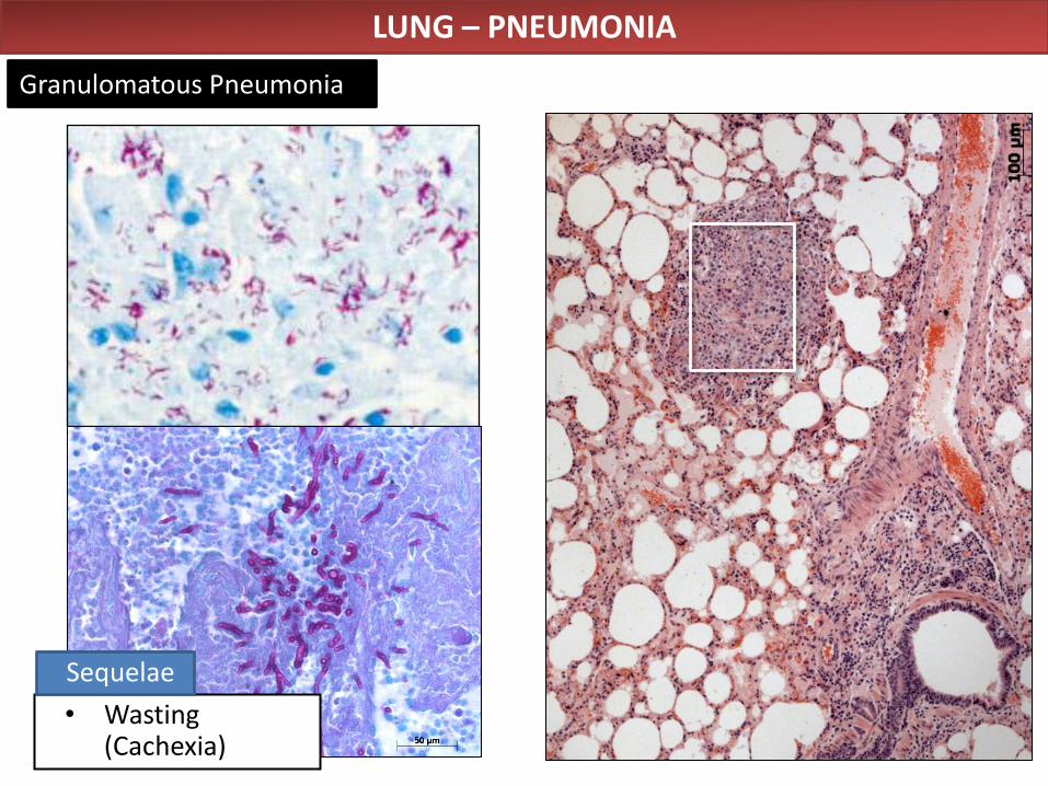
Granulomatous Pneumonia
• Gross Findings
• Distribution:
• Multifocal
• Variably sized nodules
• Firm dry exudate +/- mineral on section
• Texture:
• Nodular and firm to hard
• Port of entry:
• Aerogenous
• Hematogenous
• Common etiologies:
• Mycobacteria, Fungi, Parasites, Foreign material
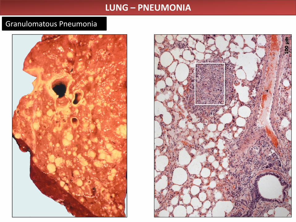
LUNG – PNEUMONIA
Granulomatous Pneumonia
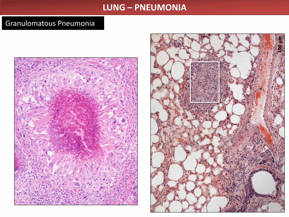
LUNG – PNEUMONIA
Granulomatous Pneumonia
LUNG – PNEUMONIA
Granulomatous Pneumonia
• Wasting (Cachexia)
Sequelae

A special thanks to Dr. Alfonso López for providing the
material for these lectures!


![Uveitic macular edema: a stepladder treatment paradigm€¦ · of macular edema [1,3–4], this review will focus on uveitic macular edema specifically. Uveitic macular edema Macular](https://img.pdfslide.us/doc/110x75/5ed770e44d676a3f4a7efe51/uveitic-macular-edema-a-stepladder-treatment-paradigm-of-macular-edema-13a4.jpg)