Embed Size (px)
Citation preview
Pathology of the Liver and Biliary Tract – 1 Normal Liver; Hepatic Injury, Response, and Failure
Shannon Martinson, August 2017 http://people.upei.ca/smartinson/
WELCOME!
• Dr Boute is the coordinator
• Course content on Moodle / pathologists websites
• Textbook for the course?
• Buy at bookstore or from your friends
• Available at the library!
• Download a chapter at a time for free
• Labs – enter through the PM lab
• Safety gear
• Wear boots
• Grey labcoats (available in pm)
• Bring gloves

OUTLINE
Normal anatomy & function
Hepatobiliary injury and responses
Manifestations of hepatic failure
Developmental anomalies and miscellaneous lesions
Circulatory disturbances
Metabolic & nutritional disturbances
Infectious diseases of the liver (hepatitis)
Toxin-induced liver diseases
Diseases of uncertain cause
Proliferative lesions of the liver
Diseases of the gallbladder and bile ducts
INTRODUCTION - GENERAL CONSIDERATIONS
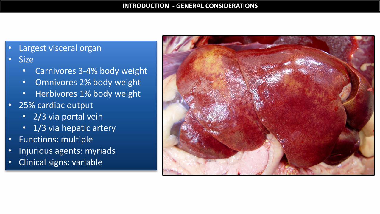
• Largest visceral organ • Size
• Carnivores 3-4% body weight • Omnivores 2% body weight • Herbivores 1% body weight
• 25% cardiac output • 2/3 via portal vein • 1/3 via hepatic artery
• Functions: multiple • Injurious agents: myriads • Clinical signs: variable
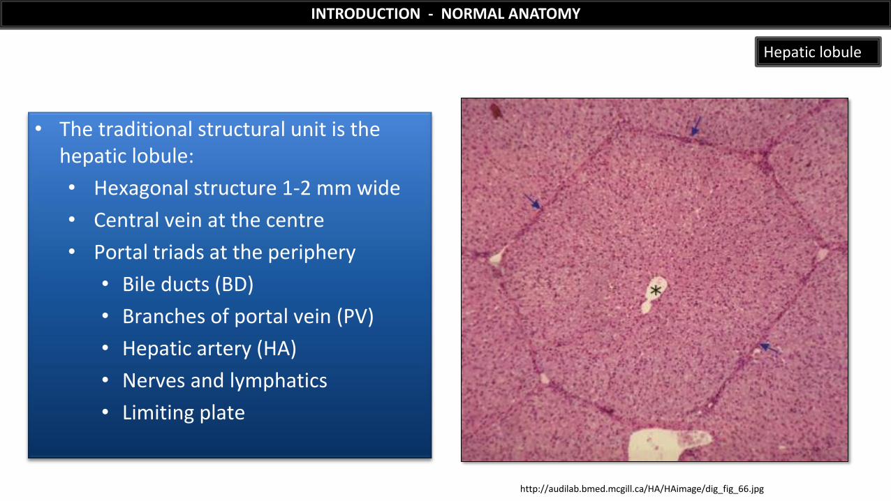
• The traditional structural unit is the hepatic lobule:
• Hexagonal structure 1-2 mm wide
• Central vein at the centre
• Portal triads at the periphery
• Bile ducts (BD)
• Branches of portal vein (PV)
• Hepatic artery (HA)
• Nerves and lymphatics
• Limiting plate
INTRODUCTION - NORMAL ANATOMY
Hepatic lobule
http://audilab.bmed.mcgill.ca/HA/HAimage/dig_fig_66.jpg
http://audilab.bmed.mcgill.ca/HA/HAimage/dig_fig_66.jpg
INTRODUCTION - NORMAL ANATOMY
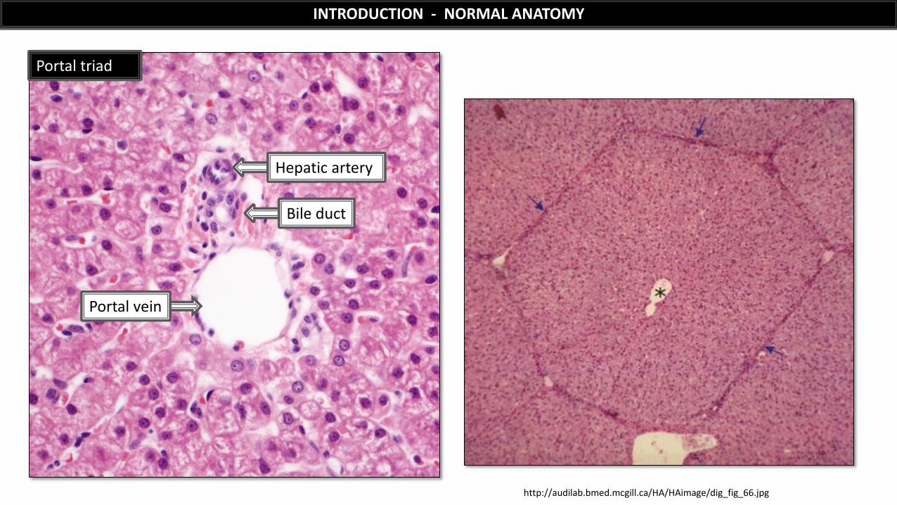
Hepatic artery
Portal vein
Portal triad
Bile duct
INTRODUCTION - NORMAL ANATOMY
Robbins and Cotran Pathologic Basis of Disease (2010), 8th ed., Elsevier
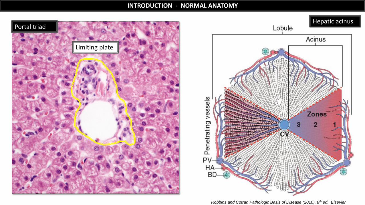
Hepatic acinus
Limiting plate
Portal triad
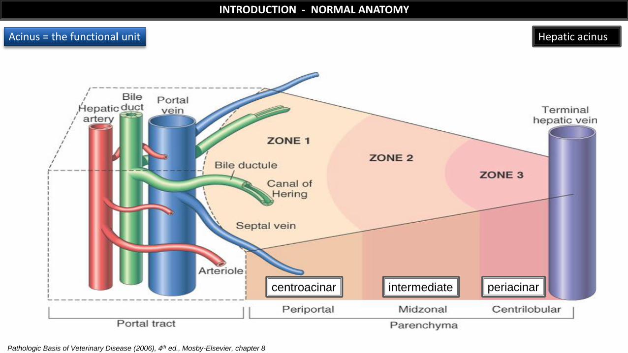
centroacinar
Pathologic Basis of Veterinary Disease (2006), 4th ed., Mosby-Elsevier, chapter 8
INTRODUCTION - NORMAL ANATOMY
Hepatic acinus
intermediate periacinar
Acinus = the functional unit
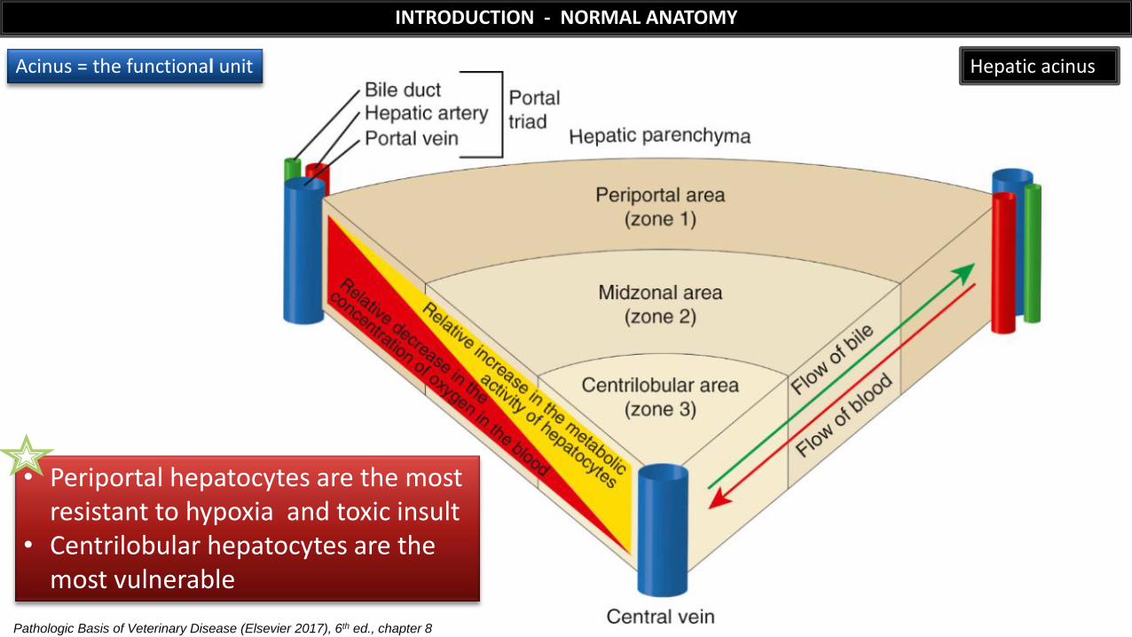
Hepatic acinus Acinus = the functional unit
INTRODUCTION - NORMAL ANATOMY
Pathologic Basis of Veterinary Disease (Elsevier 2017), 6th ed., chapter 8
• Periportal hepatocytes are the most resistant to hypoxia and toxic insult
• Centrilobular hepatocytes are the most vulnerable
Pathologic Basis of Veterinary Disease (2012), 5th ed., Mosby-Elsevier, chapter 8
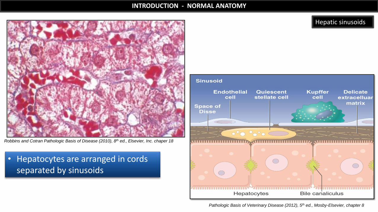
INTRODUCTION - NORMAL ANATOMY
Hepatic sinusoids
Robbins and Cotran Pathologic Basis of Disease (2010), 8th ed., Elsevier, Inc. chaper 18
• Hepatocytes are arranged in cords separated by sinusoids
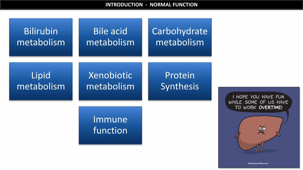
INTRODUCTION - NORMAL FUNCTION
Bilirubin metabolism
Bile acid metabolism
Carbohydrate metabolism
Lipid metabolism
Xenobiotic metabolism
Protein Synthesis
Immune function
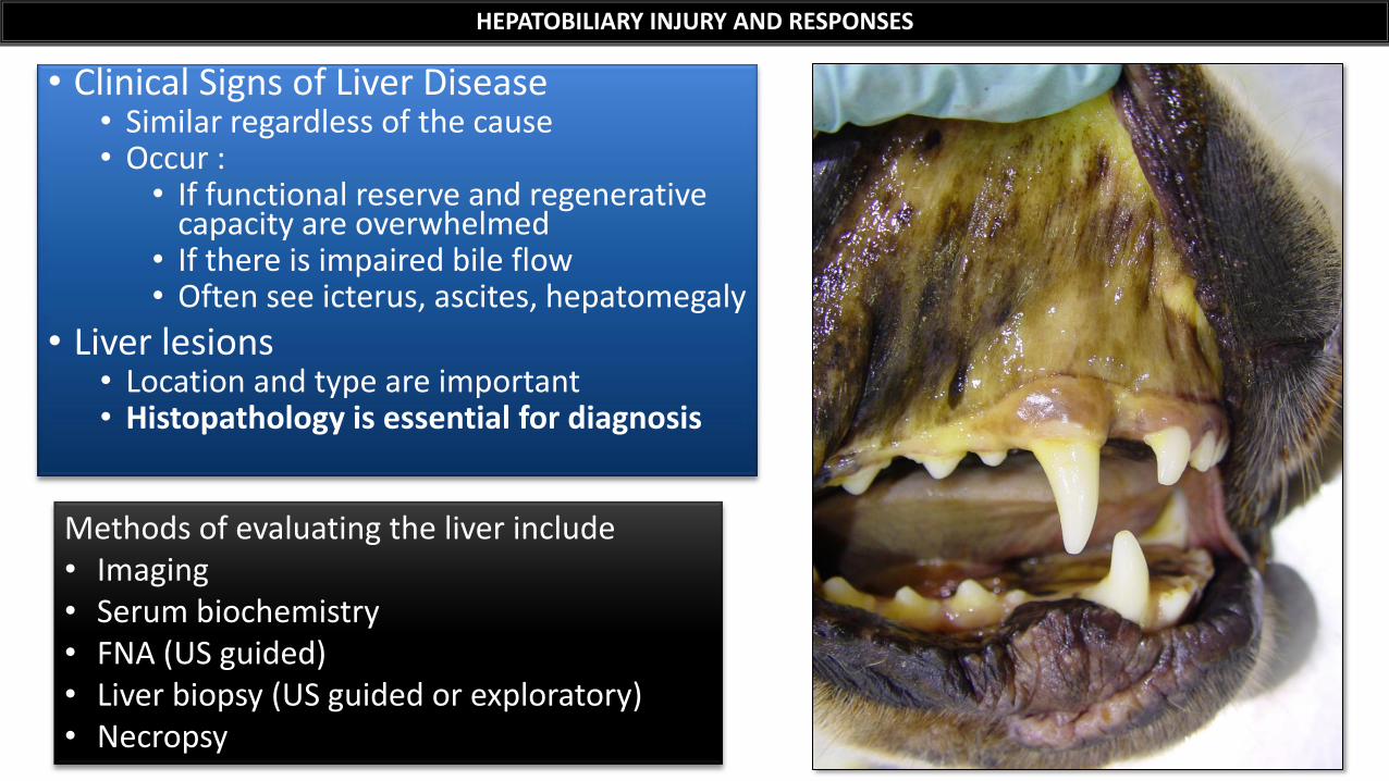
• Clinical Signs of Liver Disease • Similar regardless of the cause • Occur :
• If functional reserve and regenerative capacity are overwhelmed
• If there is impaired bile flow • Often see icterus, ascites, hepatomegaly
• Liver lesions • Location and type are important • Histopathology is essential for diagnosis
HEPATOBILIARY INJURY AND RESPONSES
Methods of evaluating the liver include • Imaging • Serum biochemistry • FNA (US guided) • Liver biopsy (US guided or exploratory) • Necropsy
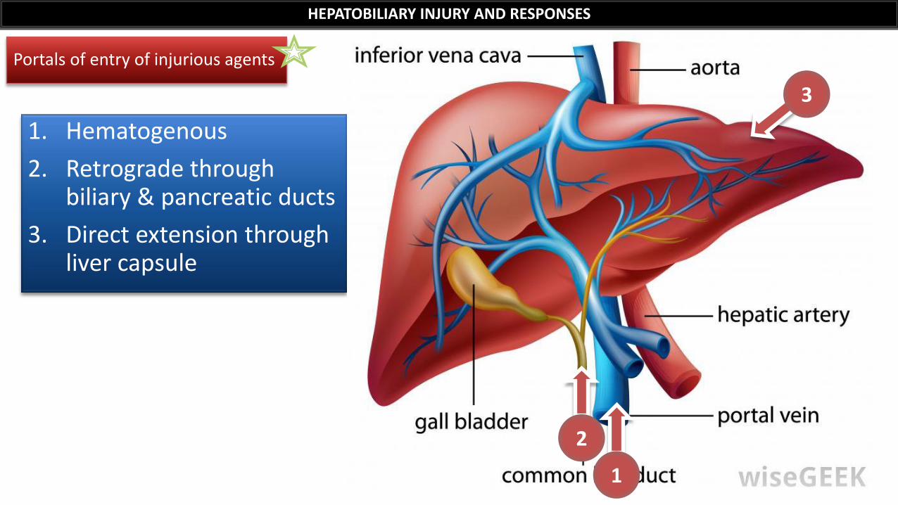
1. Hematogenous
2. Retrograde through biliary & pancreatic ducts
3. Direct extension through liver capsule
HEPATOBILIARY INJURY AND RESPONSES
Portals of entry of injurious agents
1
2
3
HEPATOBILIARY INJURY AND RESPONSES
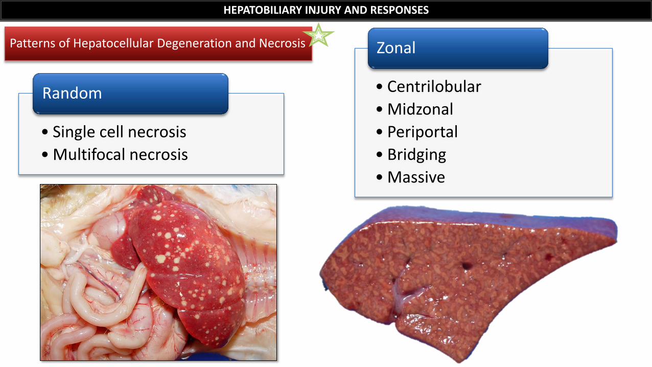
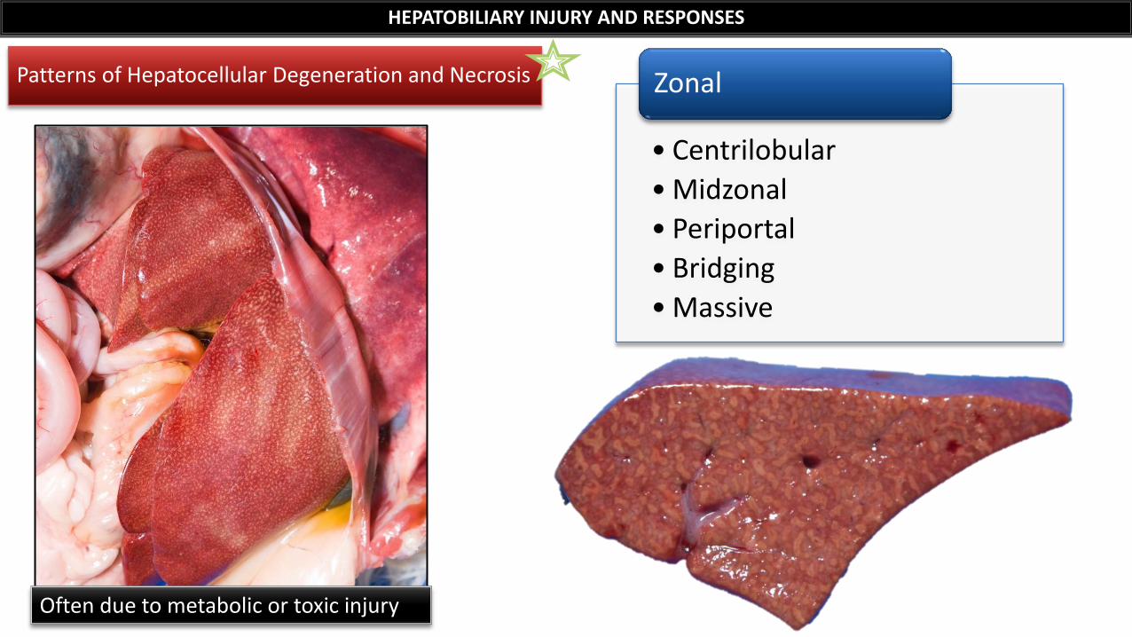
Patterns of Hepatocellular Degeneration and Necrosis
• Centrilobular
• Midzonal
• Periportal
• Bridging
• Massive
Zonal
• Single cell necrosis
• Multifocal necrosis
Random
HEPATOBILIARY INJURY AND RESPONSES
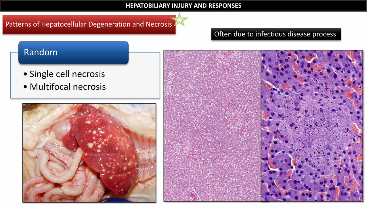
Patterns of Hepatocellular Degeneration and Necrosis
• Single cell necrosis
• Multifocal necrosis
Random
Often due to infectious disease process
HEPATOBILIARY INJURY AND RESPONSES
Patterns of Hepatocellular Degeneration and Necrosis
• Centrilobular
• Midzonal
• Periportal
• Bridging
• Massive
Zonal
Often due to metabolic or toxic injury
HEPATOBILIARY INJURY AND RESPONSES
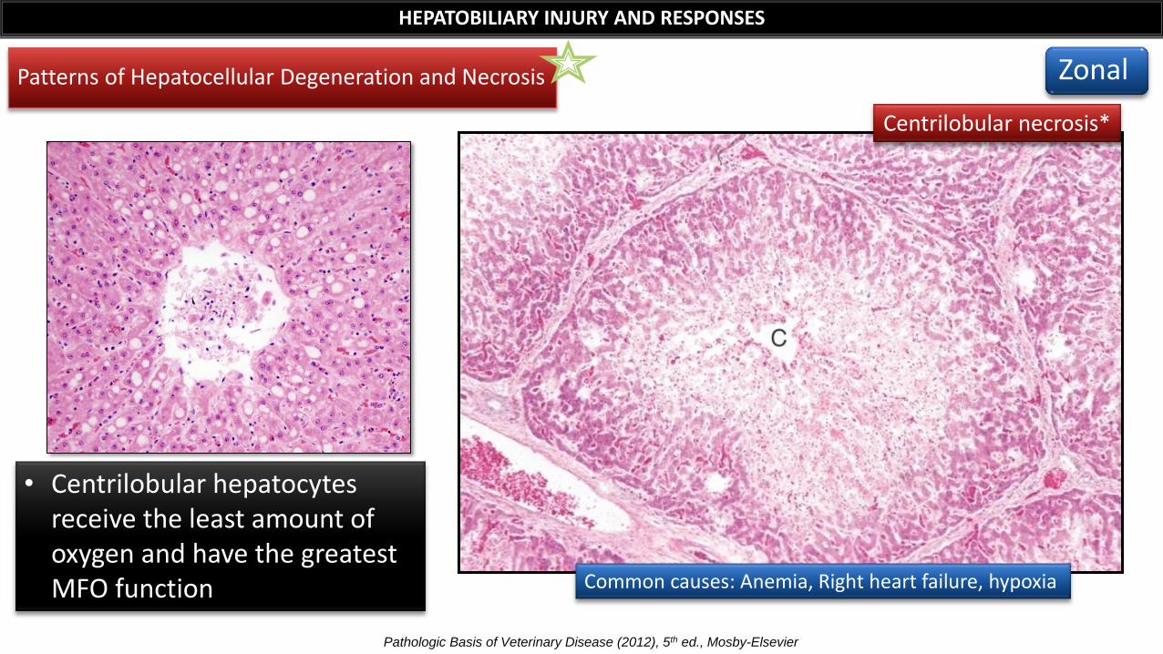
Centrilobular necrosis*
Pathologic Basis of Veterinary Disease (2012), 5th ed., Mosby-Elsevier
Patterns of Hepatocellular Degeneration and Necrosis
Common causes: Anemia, Right heart failure, hypoxia
• Centrilobular hepatocytes receive the least amount of oxygen and have the greatest MFO function
Zonal
HEPATOBILIARY INJURY AND RESPONSES
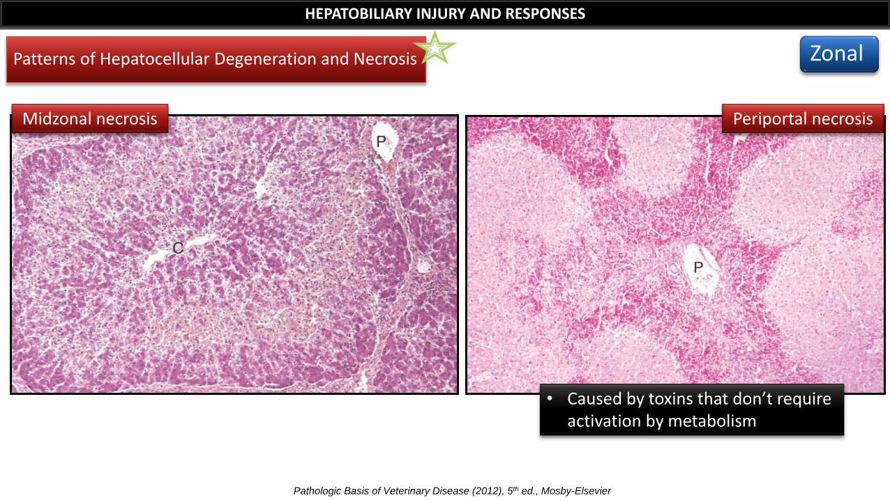
Midzonal necrosis Periportal necrosis
Patterns of Hepatocellular Degeneration and Necrosis
Pathologic Basis of Veterinary Disease (2012), 5th ed., Mosby-Elsevier
• Caused by toxins that don’t require activation by metabolism
Zonal
Patterns of Hepatocellular Degeneration and Necrosis
HEPATOBILIARY INJURY AND RESPONSES
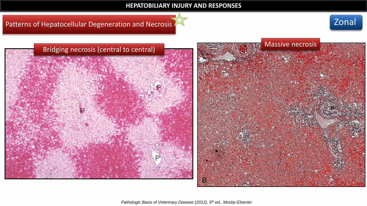
Bridging necrosis (central to central) Massive necrosis
Pathologic Basis of Veterinary Disease (2012), 5th ed., Mosby-Elsevier
Zonal
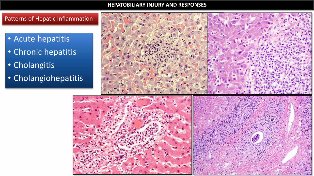
• Acute hepatitis
• Chronic hepatitis
• Cholangitis
• Cholangiohepatitis
HEPATOBILIARY INJURY AND RESPONSES
Patterns of Hepatic Inflammation
HEPATOBILIARY INJURY AND RESPONSES
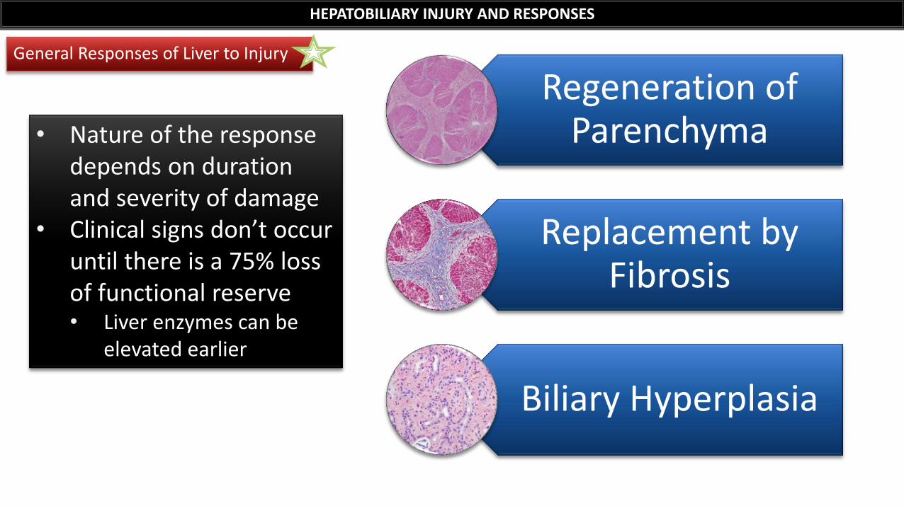
General Responses of Liver to Injury
• Nature of the response depends on duration and severity of damage
• Clinical signs don’t occur until there is a 75% loss of functional reserve • Liver enzymes can be
elevated earlier
Regeneration of Parenchyma
Replacement by Fibrosis
Biliary Hyperplasia
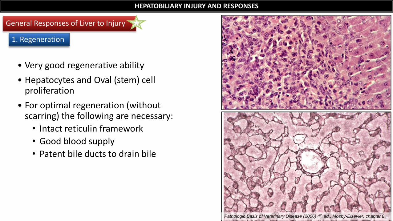
• Very good regenerative ability
• Hepatocytes and Oval (stem) cell proliferation
• For optimal regeneration (without scarring) the following are necessary:
• Intact reticulin framework
• Good blood supply
• Patent bile ducts to drain bile
HEPATOBILIARY INJURY AND RESPONSES
General Responses of Liver to Injury
1. Regeneration
Pathologic Basis of Veterinary Disease (2006) 4th ed., Mosby-Elsevier, chapter 8
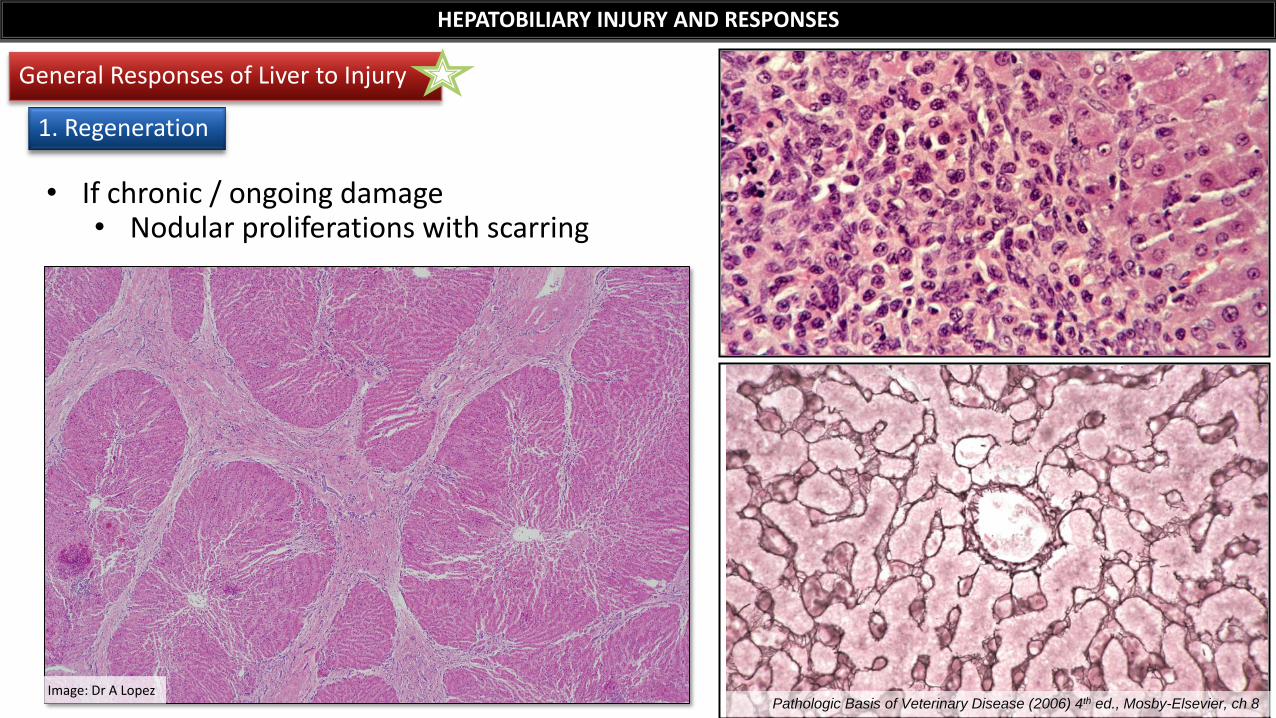
HEPATOBILIARY INJURY AND RESPONSES
General Responses of Liver to Injury
1. Regeneration
• If chronic / ongoing damage • Nodular proliferations with scarring
Pathologic Basis of Veterinary Disease (2006) 4th ed., Mosby-Elsevier, ch 8 Image: Dr A Lopez
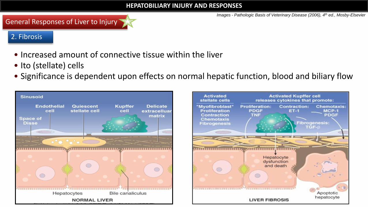
• Increased amount of connective tissue within the liver • Ito (stellate) cells • Significance is dependent upon effects on normal hepatic function, blood and biliary flow
Images - Pathologic Basis of Veterinary Disease (2006), 4th ed., Mosby-Elsevier
HEPATOBILIARY INJURY AND RESPONSES
General Responses of Liver to Injury
2. Fibrosis
HEPATOBILIARY INJURY AND RESPONSES
General Responses of Liver to Injury
2. Fibrosis
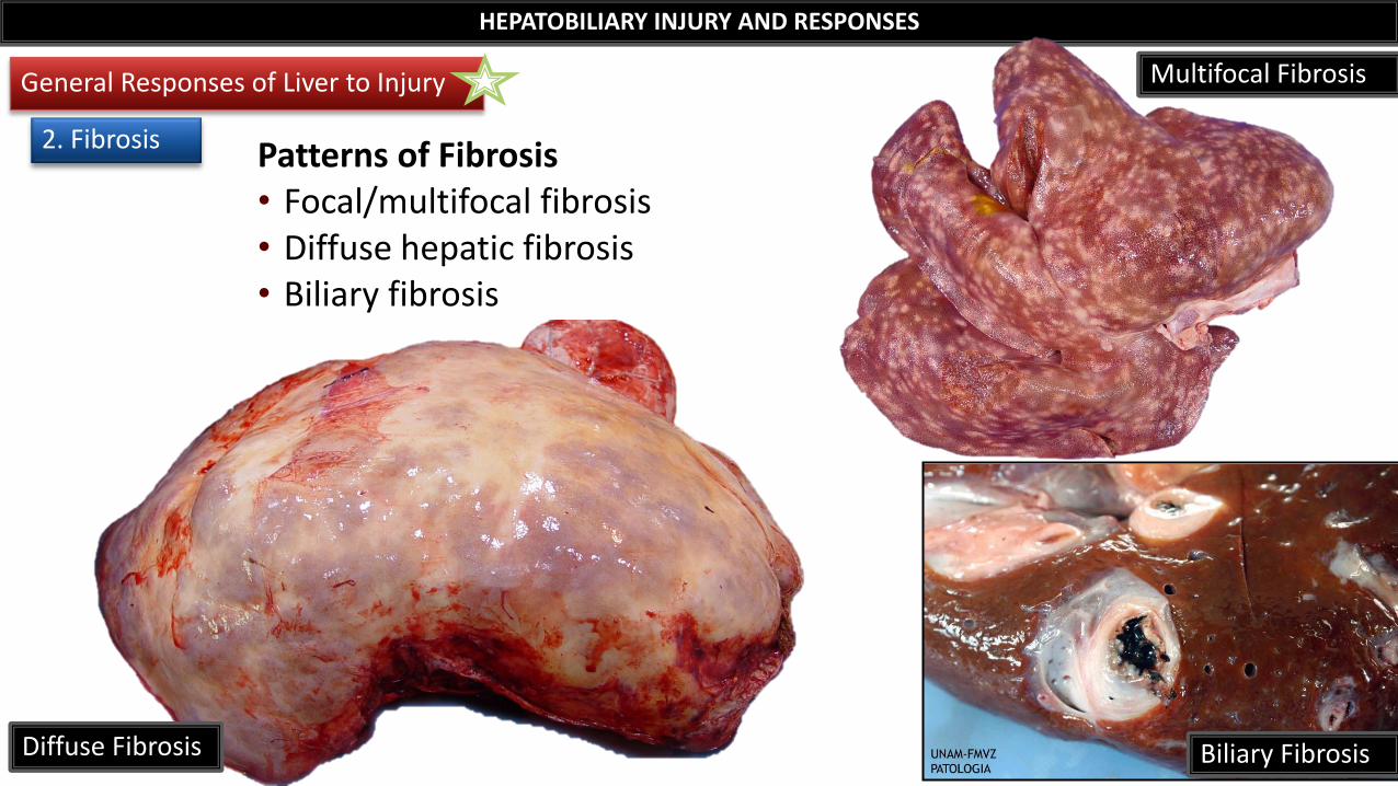
Biliary Fibrosis
Multifocal Fibrosis
Patterns of Fibrosis • Focal/multifocal fibrosis • Diffuse hepatic fibrosis • Biliary fibrosis
Diffuse Fibrosis
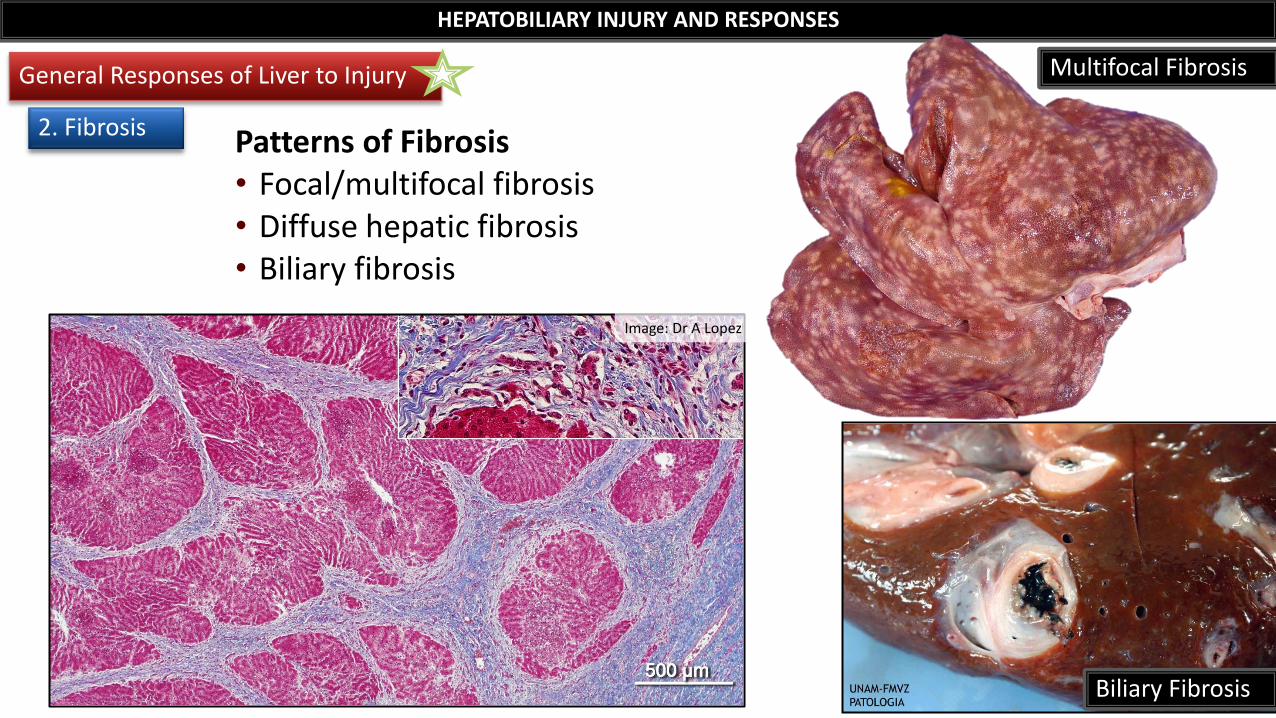
Patterns of Fibrosis • Focal/multifocal fibrosis • Diffuse hepatic fibrosis • Biliary fibrosis
HEPATOBILIARY INJURY AND RESPONSES
General Responses of Liver to Injury
2. Fibrosis
Multifocal Fibrosis
Biliary Fibrosis
Image: Dr A Lopez
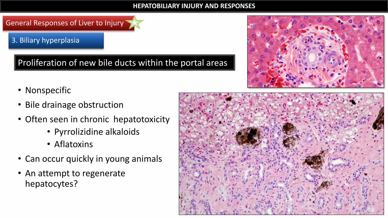
• Nonspecific
• Bile drainage obstruction
• Often seen in chronic hepatotoxicity
• Pyrrolizidine alkaloids
• Aflatoxins
• Can occur quickly in young animals
• An attempt to regenerate hepatocytes?
Proliferation of new bile ducts within the portal areas
HEPATOBILIARY INJURY AND RESPONSES
General Responses of Liver to Injury
3. Biliary hyperplasia
• Final irreversible result of hepatic disease • Distortion of the architecture
• Cause cannot be determined
• Characterized by 3 components*:
1. Nodular regeneration
2. Fibrosis
3. Bile duct hyperplasia
HEPATOBILIARY INJURY AND RESPONSES
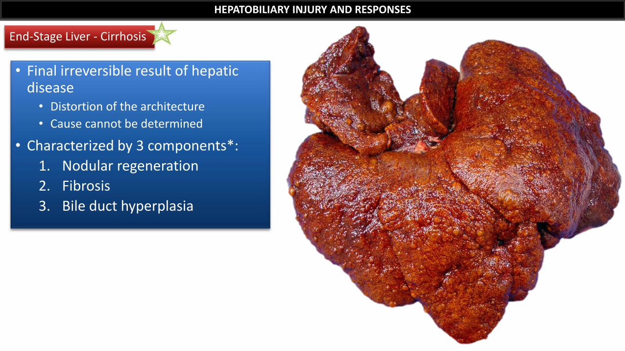
End-Stage Liver - Cirrhosis
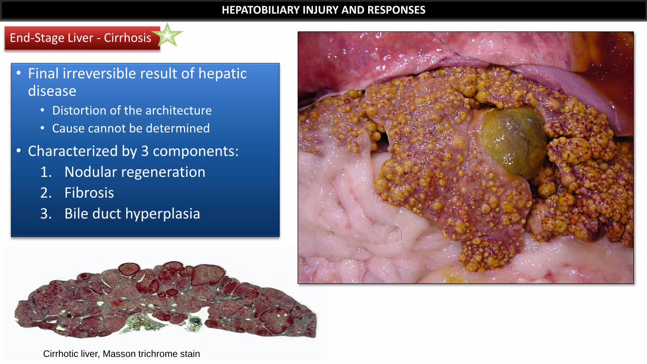
• Final irreversible result of hepatic disease • Distortion of the architecture
• Cause cannot be determined
• Characterized by 3 components:
1. Nodular regeneration
2. Fibrosis
3. Bile duct hyperplasia
Cirrhotic liver, Masson trichrome stain
HEPATOBILIARY INJURY AND RESPONSES
End-Stage Liver - Cirrhosis
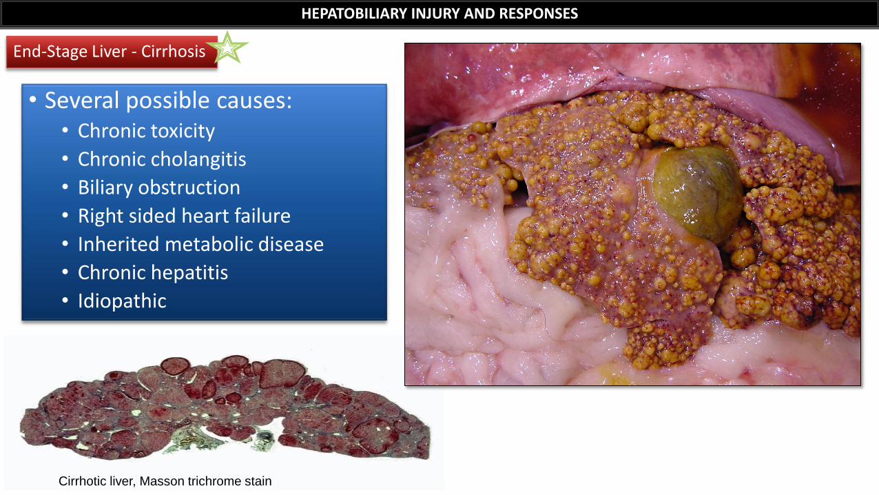
• Several possible causes: • Chronic toxicity
• Chronic cholangitis
• Biliary obstruction
• Right sided heart failure
• Inherited metabolic disease
• Chronic hepatitis
• Idiopathic
Cirrhotic liver, Masson trichrome stain
HEPATOBILIARY INJURY AND RESPONSES
End-Stage Liver - Cirrhosis
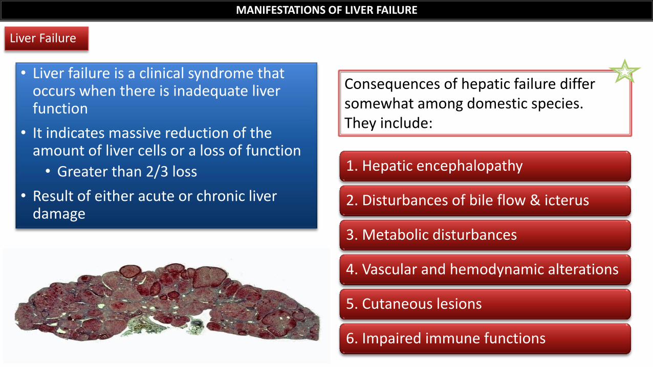
• Liver failure is a clinical syndrome that occurs when there is inadequate liver function
• It indicates massive reduction of the amount of liver cells or a loss of function
• Greater than 2/3 loss
• Result of either acute or chronic liver damage
MANIFESTATIONS OF LIVER FAILURE
1. Hepatic encephalopathy
2. Disturbances of bile flow & icterus
3. Metabolic disturbances
4. Vascular and hemodynamic alterations
5. Cutaneous lesions
6. Impaired immune functions
Consequences of hepatic failure differ somewhat among domestic species. They include:
Liver Failure
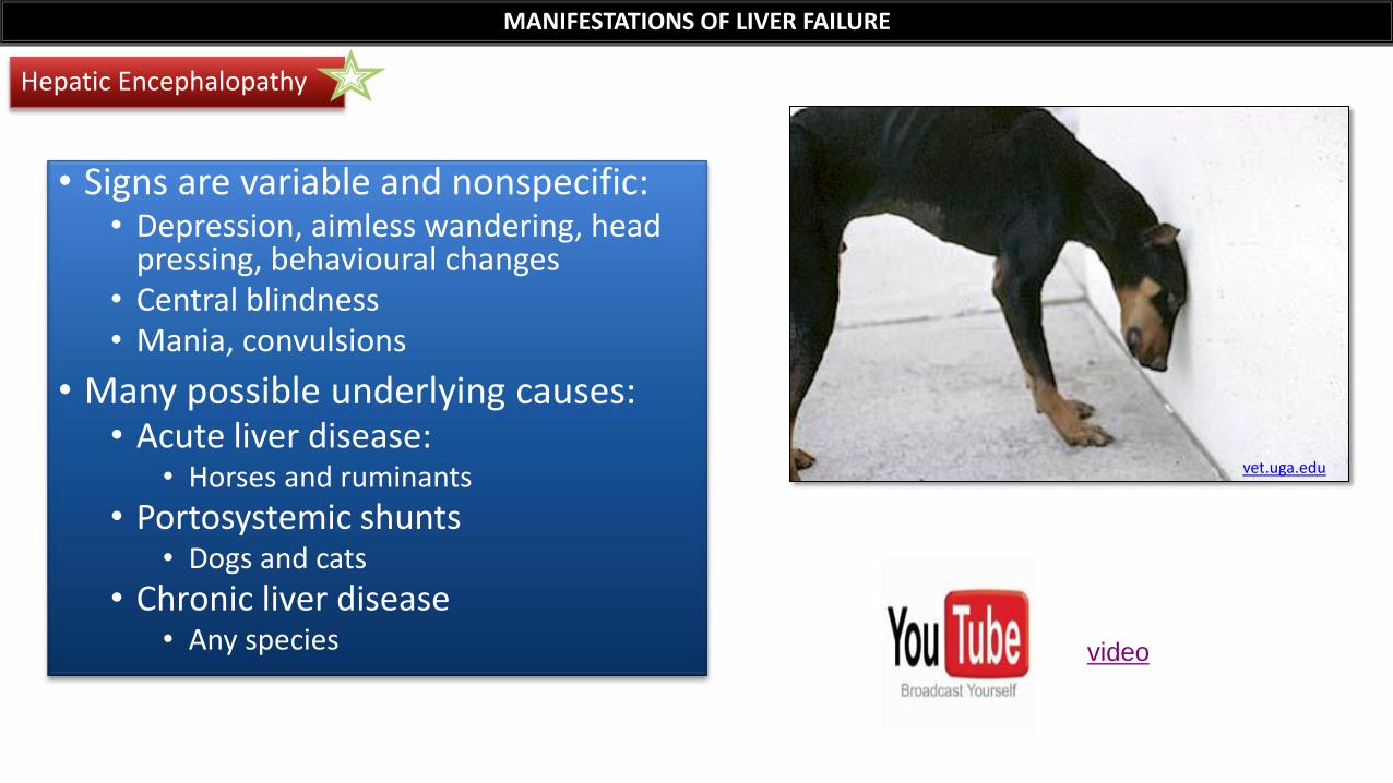
• Signs are variable and nonspecific: • Depression, aimless wandering, head
pressing, behavioural changes • Central blindness • Mania, convulsions
• Many possible underlying causes: • Acute liver disease:
• Horses and ruminants
• Portosystemic shunts • Dogs and cats
• Chronic liver disease • Any species
Hepatic Encephalopathy
MANIFESTATIONS OF LIVER FAILURE
vet.uga.edu
video

Hepatic Encephalopathy
MANIFESTATIONS OF LIVER FAILURE
• Blood accumulation of neurotoxic substances bypassing the liver and reaching the brain
• Clinical signs are more severe after feeding
Pathogenesis
• Main substance is ammonia
• Other factors:
• +/- Imbalance of inhibitory & excitatory amino acid neurotransmitters
• +/- Increased brain concentration of benzodiazepines
Neurotoxic substances
www.nadis.org.uk/bulletins/nervous-diseases-in-cattle.aspx
Disturbances of Bile Flow
MANIFESTATIONS OF LIVER FAILURE
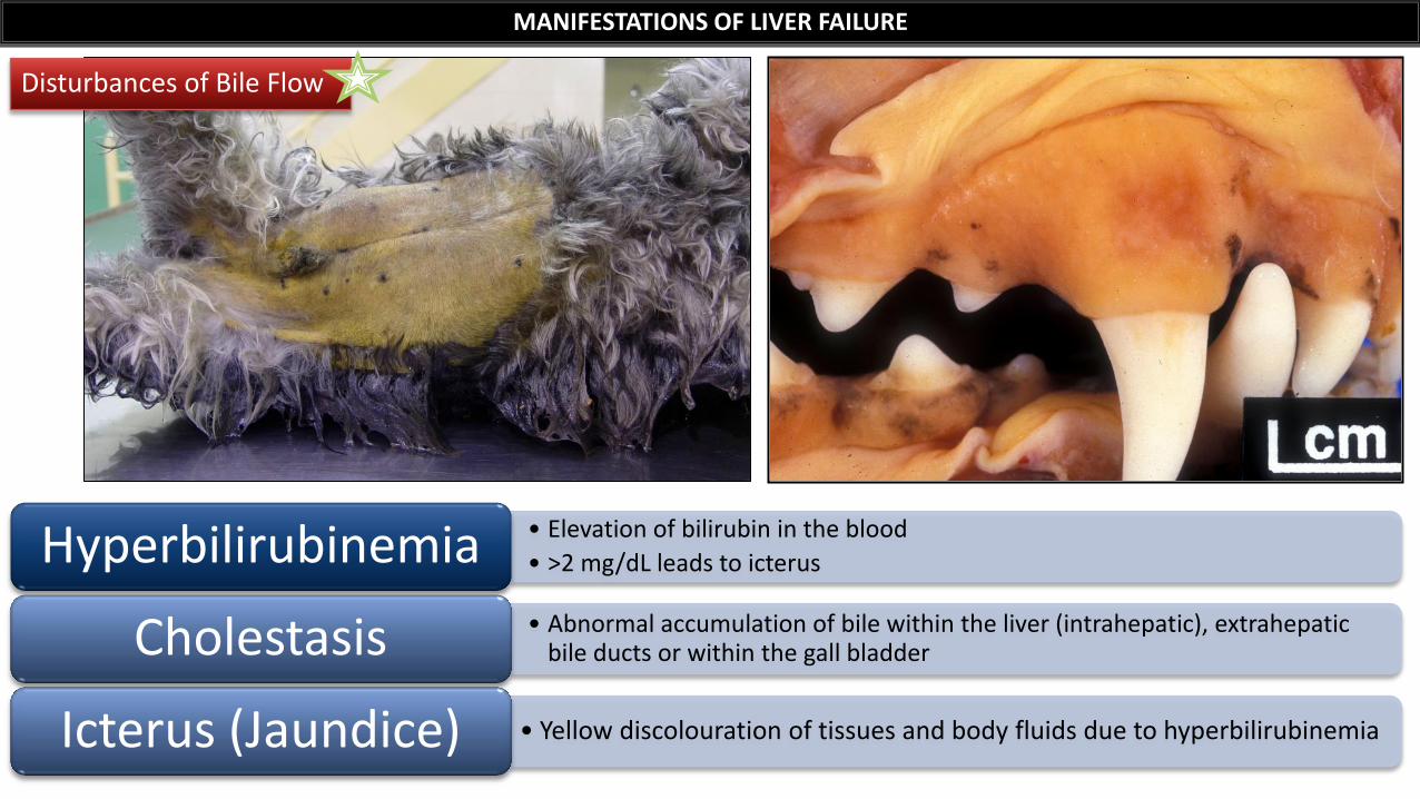
• Elevation of bilirubin in the blood
• >2 mg/dL leads to icterus Hyperbilirubinemia • Abnormal accumulation of bile within the liver (intrahepatic), extrahepatic
bile ducts or within the gall bladder Cholestasis
• Yellow discolouration of tissues and body fluids due to hyperbilirubinemia Icterus (Jaundice)
Disturbances of Bile Flow
MANIFESTATIONS OF LIVER FAILURE
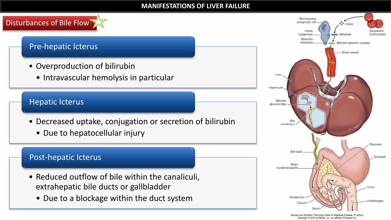
• Overproduction of bilirubin
• Intravascular hemolysis in particular
Pre-hepatic Icterus
• Decreased uptake, conjugation or secretion of bilirubin
• Due to hepatocellular injury
Hepatic Icterus
• Reduced outflow of bile within the canaliculi, extrahepatic bile ducts or gallbladder
• Due to a blockage within the duct system
Post-hepatic Icterus
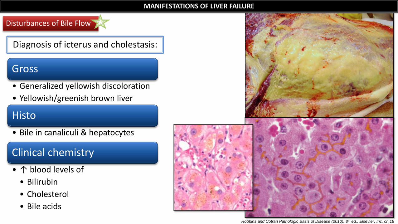
Diagnosis of icterus and cholestasis:
Gross
• Generalized yellowish discoloration
• Yellowish/greenish brown liver
Histo
• Bile in canaliculi & hepatocytes
Clinical chemistry
• ↑ blood levels of
• Bilirubin
• Cholesterol
• Bile acids
Disturbances of Bile Flow
MANIFESTATIONS OF LIVER FAILURE
Robbins and Cotran Pathologic Basis of Disease (2010), 8th ed., Elsevier, Inc. ch 18
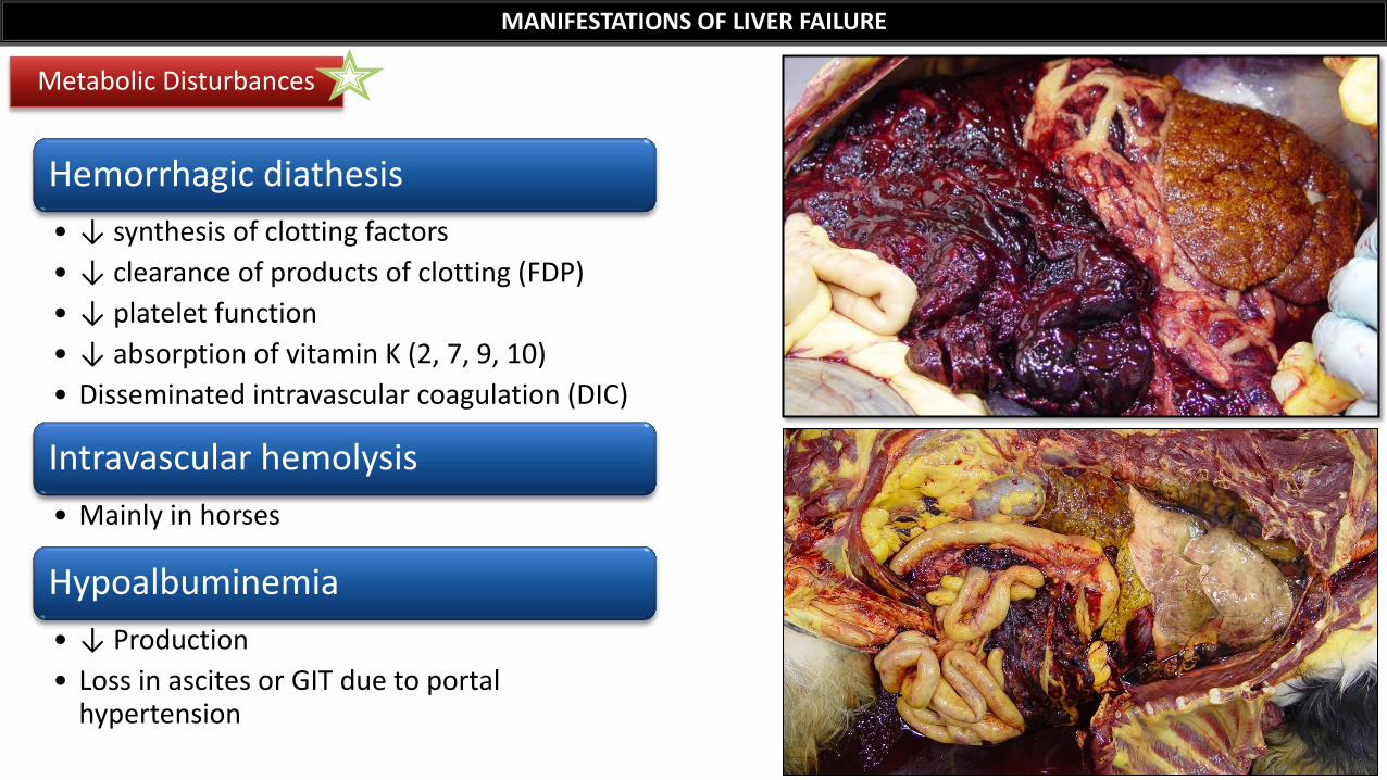
Hemorrhagic diathesis
• ↓ synthesis of clotting factors
• ↓ clearance of products of clotting (FDP)
• ↓ platelet function
• ↓ absorption of vitamin K (2, 7, 9, 10)
• Disseminated intravascular coagulation (DIC)
Intravascular hemolysis
• Mainly in horses
Hypoalbuminemia
• ↓ Production
• Loss in ascites or GIT due to portal hypertension
Metabolic Disturbances
MANIFESTATIONS OF LIVER FAILURE
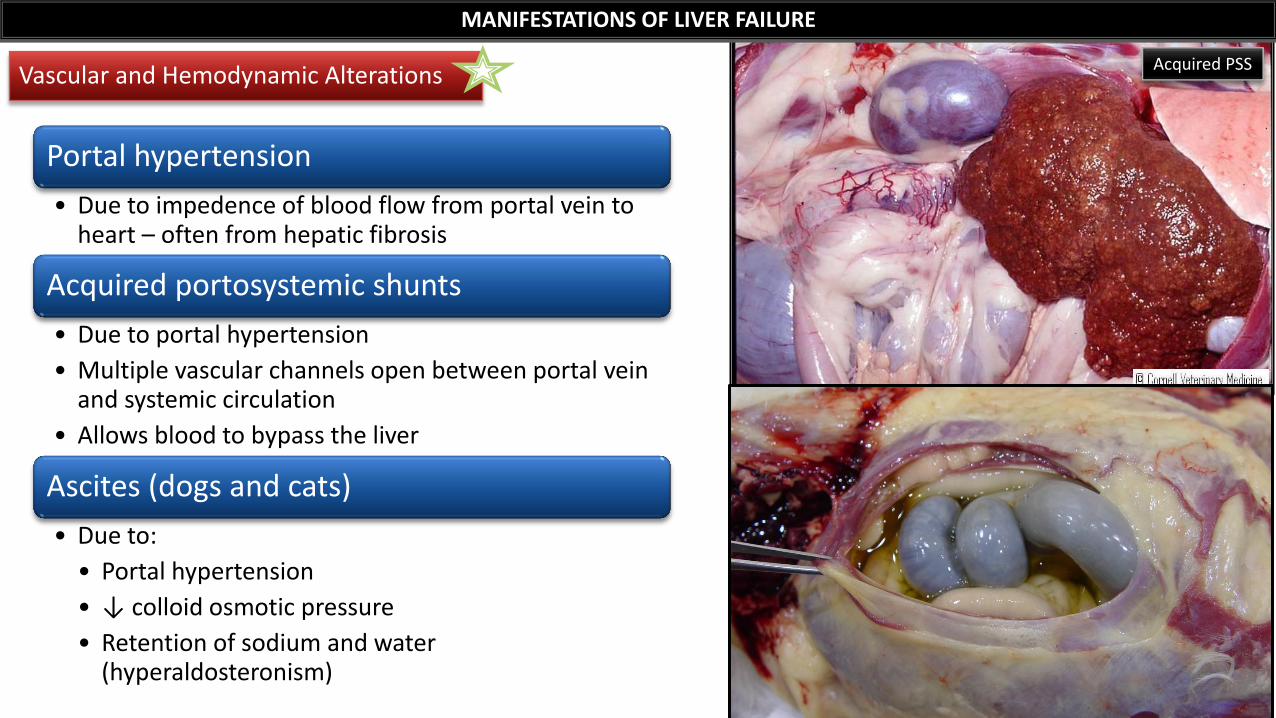
Portal hypertension
• Due to impedence of blood flow from portal vein to heart – often from hepatic fibrosis
Acquired portosystemic shunts
• Due to portal hypertension
• Multiple vascular channels open between portal vein and systemic circulation
• Allows blood to bypass the liver
Ascites (dogs and cats)
• Due to:
• Portal hypertension
• ↓ colloid osmotic pressure
• Retention of sodium and water (hyperaldosteronism)
Vascular and Hemodynamic Alterations
MANIFESTATIONS OF LIVER FAILURE
Acquired PSS
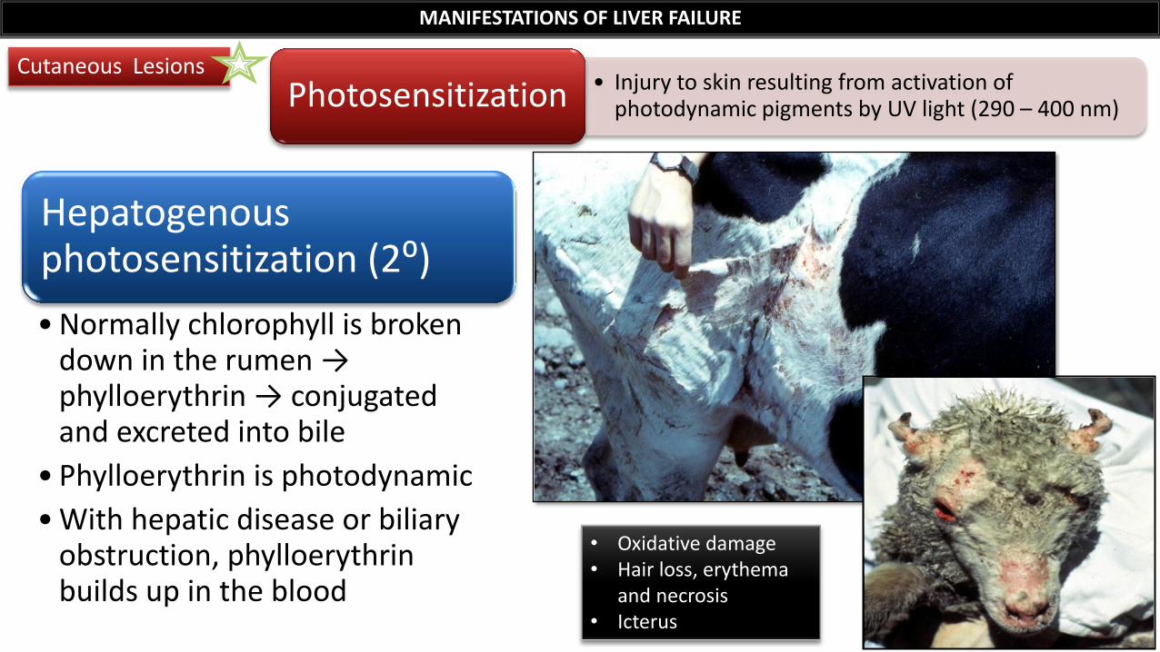
Hepatogenous photosensitization (2⁰)
• Normally chlorophyll is broken down in the rumen → phylloerythrin → conjugated and excreted into bile
• Phylloerythrin is photodynamic
• With hepatic disease or biliary obstruction, phylloerythrin builds up in the blood
Cutaneous Lesions
MANIFESTATIONS OF LIVER FAILURE
• Injury to skin resulting from activation of photodynamic pigments by UV light (290 – 400 nm) Photosensitization
• Oxidative damage • Hair loss, erythema
and necrosis • Icterus
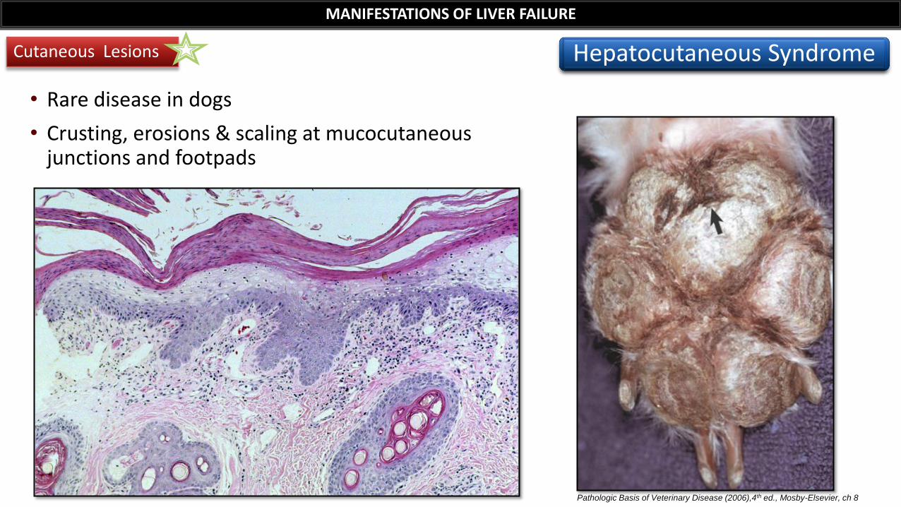
• Rare disease in dogs
• Crusting, erosions & scaling at mucocutaneous junctions and footpads
Pathologic Basis of Veterinary Disease (2006),4th ed., Mosby-Elsevier, ch 8
Cutaneous Lesions
MANIFESTATIONS OF LIVER FAILURE
Hepatocutaneous Syndrome
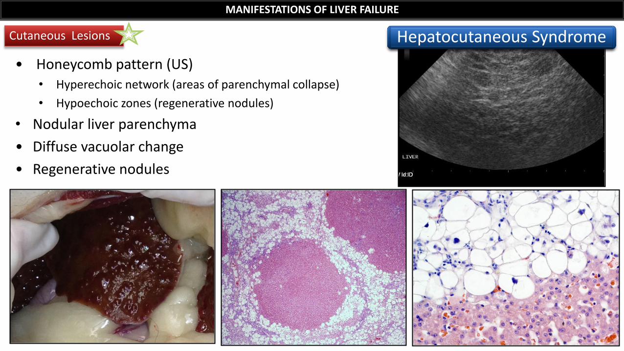
• Honeycomb pattern (US) • Hyperechoic network (areas of parenchymal collapse)
• Hypoechoic zones (regenerative nodules)
• Nodular liver parenchyma
• Diffuse vacuolar change
• Regenerative nodules
Cutaneous Lesions
MANIFESTATIONS OF LIVER FAILURE
Hepatocutaneous Syndrome