Embed Size (px)
Citation preview
CLINIC FOR OPHTHALMOLOGY
UNIVERSITY OF LÜBECK
DIRECTOR: PROF. DR. SALVATORE GRISANTI
PATHOGENESIS OF AGE RELATED MACULAR DEGENERATION (AMD)
Immunohistochemical evaluation of the macrophage
activation pattern in human choroidal neovascular membranes
Thesis
for
the acquisition of doctorate
at University of Lübeck
- Medical Faculty -
Presented by
Khaled Nassar
Born in ―El Menoufia‖
Egypt
Lübeck 2010

1. Referee: Prof. Dr. med. Salvatore Grisanti
2. Referee: Prof.Dr.med.Hartmut Merz
Day of oral examination: 13.04.2011
Approved for printing. Lübeck, in 13.04.2011
I
TABLE OF CONTENTS
TABLE OF CONTENTS
1 INTRODUCTION…………………………………………………………….. 1
1.1 INTRODUCTION…………………………………………............... 1
1.2 SCIENTIFIC BACKGROUND……………………………………... 2
1.2.1 Clinical definitions…………………………………………... 2
1.2.2 Epidemiology ……………………………..………………... 3
1.2.3 Diagnosis and ancillary testing …………………...………. 4
1.2.4 Natural history ……………………………..……………..... 4
1.2.5 Treatment……………………………………………………. 5
1.2.6 The pathogenesis of CNV…………………..…………….. 8
1.2.7 Aetiology ……………………………………….………….... 11
1.2.8 Macrophages and immune competent cells in AMD……. 14
2 THESIS AIMS………………………………………………………………. 23
3 MATERIALS AND METHODS…………………………………………….. 24
3.1 CNV SPECIMENS………………………………………………….. 24
3.2 HISTOPATHOLOGICAL METHODS…………………………….. 24
3.3 PHOTOGRAPHY…………………………………………………… 26
3.4 ANALYSIS…………………………………………………….......... 27
3.5 STATISTICS………………………..………………………………. 27
4 RESULTS…………………………………………………………………….. 28
4.1 CLINICAL CHARACTERIZATION…….………………………….. 28
4.2 IMMUNOHISTOPATHOLOGIC FINDING……………………….. 29
4.3 MACROPHAGE ACTIVATION PATTERN………………………. 33
5 DISCUSSION………………………………………………………………… 40
6 SUMMARY………………………………………………………………….... 53
7 BIBLIOGRAPHY…………………………………………………………….. 54
II
TABLE OF CONTENTS
8 ATTACHMENTS…………………………………………………………….. 81
8.1 PATIENT CHARACTERISTICS…………………………………... 81
8.2 MATERIALS PREPARATION ……………..…………….............. 83
8.3 STAINING PROTOCOL…………………………………………… 84
8.4 ABBREVIATIONS………………………………….………….…… 89
9 ACKNOWLEDGMENTS……………………………………………………. 91
10 CURRICULUM VITAE………………………………………………………. 92
11 PUBLICATIONS LIST……………………………………………………… 95
1
1 INTRODUCTION
1 INTRODUCTION
1.1 Introduction
The retina is the light‐sensitive part of the eye. It is a thin film of tissue
covering most of the inner wall of the eye. The macula is the central part of the
retina which has the highest concentration of cone photoreceptors required to
resolve fine detail. Age related macular degeneration (AMD) is a progressive late
onset disease affecting the central macula, which is responsible for fine central
vision needed for driving, reading, and recognizing people’s faces. It is the leading
cause of irreversible blindness in western countries. It is estimated that the
number of people with AMD will double by the year 2020. Current treatment
options for AMD are limited, mostly to the late neovascular stage of the disease,
with no treatment currently available for the late atrophic form of disease.
Growing evidence suggests that inflammatory and immunologic events
might play a role in AMD. Several human histological studies have suggested the
participation of macrophages in choroidal neovascular membrane (CNV)
formation. The macrophage is a highly adaptive and responsive cell which is able
to recognize alterations in its micro-environment and maintain tissue homeostasis.
Macrophages play a central role in a remarkable range of biological and
pathological processes. The precise mechanisms of macrophage involvement in
AMD are still not fully understood. Macrophage heterogeneity raises an important
question regarding the beneficial and harmful roles of macrophages in AMD
pathogenesis.
2
2 INTRODUCTION
1.2 Scientific Background
1.2.1 Clinical definitions
1.2.1.1 Age-related macular degeneration (AMD)
AMD refers to spectrum of diseases associated with visual loss, retinal
pigment epithelium (RPE) changes, drusen, geographical atrophy (GA), and CNV
which usually occur in patients over the age of 50. A unified classification of AMD
is not yet present; however, for simplicity it can be clinically and histologically
classified into two major subtypes: dry and wet AMD. Dry AMD progresses more
slowly and manifests with drusen, GA of RPE, and photoreceptor dysfunction and
degeneration. Wet AMD on the other hand has a key feature of CNV, the growth of
new blood vessels from the choroid into the region underlying the RPE or
extending past the RPE into the subretinal space and the retina. This CNV can
lead to leakage of blood into the subretinal space, which, along with RPE atrophy
and photoreceptor degeneration, leads to vision loss (Bird et al., 1995; Age
Related Eye Disease Study Research Group (AREDS) report Nr. 6, 2001).
1.2.1.2 Drusen
Cells of the RPE continuously ingest photoreceptor outer segments that are
shed throughout life. The residue of intracellular digestion may eventually fill the
cell. Drusen are extracellular deposits that lie between the basement membrane of
the RPE and the inner collagenous zone of Bruch's membrane (Burns and
Feeney-Burns, 1980). Drusen are either small hard drusen which are not
associated with an increased risk for the development of CNV (Bressler et al.,
1990) and are not age-related (Klein et al., 1992) or large soft drusen which their
presence increases the risk for development of RPE abnormalities, GA, and CNV
formation (Bressler et al., 1990; Klein et al., 1997).
1.2.1.3 Atrophic macular degeneration
The atrophic form of macular degeneration has been called GA because
the areas of RPE atrophy tend to form well-demarcated borders that do not relate
to specific anatomic structures (Blair, 1975). Atrophic macular degeneration leads
to significant visual loss in almost all cases; patients describe a gradual and subtle
blurring of vision that relates to the degree of foveal involvement. A minimum of
175 μm of the retina should be involved in order to classify a patient as having GA
(Bird et al., 1995).
3
3 INTRODUCTION
1.2.1.4 Choroidal neovascularization (CNV)
CNV refers to the growth of abnormal new vessels of choroidal origin
beneath the sensory retina or RPE (Campochiaro and Glaser, 1986). CNV is an
abnormality found in many diseases in which the integrity of the RPE, Bruch's
membrane, and choriocapillaris has been compromised. The effect on vision from
CNV derives from its tendency to leak fluid beneath and into the sensory retina, to
bleed, and to create a fibrovascular disciform scar in the macular region (Schatz et
al., 1979).
1.2.2 Epidemiology
AMD can be classified broadly into two categories: non-neovascular (dry)
and neovascular (wet). Although non-neovascular AMD accounts for
approximately 80% of all diagnosed cases, neovascular AMD is responsible for
nearly 80% of significant visual disability associated with this disease. GA, the
most severe non-neovascular manifestation of AMD, causes approximately 21% of
the cases of legal blindness in North America (Leibowitz et al., 1980). The average
age at onset of visual loss is about 75 years. After the age of 50, the incidence
steadily increases, with more than one third of people in the ninth decade of life
affected. The visual impact is significant; the Salisbury Eye Evaluation Study
reported the prevalence of blindness, defined as visual acuity (VA) 20/200 or
worse, associated with AMD as 0.38% in individuals aged 70–79 years and 1.15%
in individuals aged 80–84 years (Muñoz et al., 2000).
In Germany forecasts show that the number of patients with advanced AMD
will increase from 710,000 at present to over a million by 2020. During the same
period, the number of patients with neovascular AMD will increase from 485,000 to
700,000. There are currently 50,000 new cases of neovascular AMD every year in
Germany, and it has only just become possible to treat these with the new anti-
vascular endothelial growth factor (VEGF) drugs. The new treatment options will
cause additional annual costs of 1.1-2.9 billion euro for Germany alone (Knauer
and Pfeiffer, 2006). No significant gender predilection has been identified for AMD.
The Framingham Eye Study showed a slightly higher incidence of AMD in
Caucasian women compared to men (Leibowitz et al., 1980). The Health and
Nutrition Examination Survey (HANES) found no difference (Klein and Klein,
1982).
4
4 INTRODUCTION
1.2.3 Diagnosis and ancillary testing
Clinical examination is usually sufficient to establish a diagnosis of AMD.
Subtle macular abnormalities, especially subretinal fluid, are best detected by
stereoscopic slit-lamp biomicroscopic examination using a contact lens.
Fluorescein angiography is useful in any patient in whom CNV is suspected to
determine the characteristics of the lesion and the patient's potential qualification
for available therapeutic modalities. Determination of the presence of CNV and
evaluation of the extent, location and composition of its components are critical in
deciding whether treatment is indicated and if so, which therapeutic modality is
appropriate. If a lesion is well demarcated, its location may be determined by the
closest point to the centre of the foveal avascular zone (FAZ). Lesion location is
classified angiographically as follows:
1. Extrafoveal (=200 µm and < 2500 µm from the centre of the FAZ).
2. Juxtafoveal (1–199 µm from the centre of the FAZ).
3. Subfoveal (under the centre of the FAZ).
Based on angiographic patterns of fluorescence, components of CNV
lesions may be categorized as either classic or occult. Classic CNV is
characterized by bright uniform early hyperfluorescence and exhibiting leakage in
the late phase which obscures the boundaries. Occult CNV is recognized
angiographically by one of two patterns: fibrovascular Pigment epithelium
detachment (PED), or late leakage from an undetermined source (Chamberlin et
al., 1989; 1Macular Photocoagulation Study, 1991; 2Macular Photocoagulation
Study, 1991).
1.2.4 Natural history
The risk of visual loss in eyes that initially manifest drusen or RPE
abnormalities varies, depending on the characteristics of the macula and the
status of the fellow eye. In eyes of patients older than 65 years that have bilateral
drusen but no significant visual loss initially, the risk of a new atrophic lesion or
neovascular lesion that results in visual loss has been reported as 9% at 1 year,
16% at 2 years, and 24% at 3 years (Holz et al., 1994). Confluent drusen, focal
hyperpigmentation of the RPE, and extrafoveal areas of chorioretinal atrophy are
three clinical findings that increase the risk for the subsequent development of
visual loss. In individuals who already have neovascular AMD in one eye, the risk
of developing CNV in the fellow eye is estimated at about 7–10% per year. If the
5
5 INTRODUCTION
fellow eye has no large drusen or focal RPE hyperpigmentation, the 5-year risk of
developing CNV is only 10%. When both large drusen and RPE
hyperpigmentation are present, however, the 5-year risk increases to
approximately 60% (Bressler et al., 1990).
1.2.4.1 Occult Choroidal Neovascularization
Natural history studies of occult CNV demonstrate a poor visual outcome
associated with these lesions. One retrospective study reviewed 84 eyes with
occult CNV and showed a 63% rate of moderate visual loss over an average of 28
months; average visual acuity declined from 20/80 to 20/250 over this interval
(Bressler et al., 1988). The verteporfin in photodynamic therapy (VIP) study
followed 93 eyes with purely occult subfoveal CNV in a placebo control arm. At 12
months, 73% of these eyes experienced visual loss from baseline, with 32%
manifesting severe visual loss. At 24 months, 79% experienced visual loss from
baseline, with a 43% rate of severe visual loss (Verteporfin in Photodynamic
Therapy report 2, 2001; Bressler, 2002).
1.2.4.2 Classic Choroidal Neovascularization
The natural history of subfoveal classic CNV can be discerned by
evaluating the control arms of the Macular Photocoagulation Study (MPS) and the
Treatment of Age-Related Macular Degeneration with Photodynamic Therapy
(TAP) Study. In the MPS, visual acuity in untreated eyes harbouring classic CNV
decreased an average of 1.9 lines at 3 months and 4.4 lines at 24 months. Severe
visual loss was noted in 11% of eyes at 3 months and 37% at 24 months. The TAP
Study showed a similar trend (Photodynamic Therapy Study, 1999; Bressler,
2001).
1.2.5 Treatment
The management of either type of AMD continues to be a challenge for
patients, ophthalmologists, and the healthcare system. Recently, the progress
made in the comprehension of the basic pathological mechanisms in both types of
AMD has led to novel developments in therapeutic strategies resulting in a
widening of the available treatment options and improved prognostic perspectives
(Augustin et al., 2009). Treatment options span a broad range of therapeutic
approaches, including thermal laser photocoagulation (1Macular Photocoagulation
Study, 1991), surgical approaches as excision, displacement, or transplantation
6
6 INTRODUCTION
(De Juan and Machemer, 1988; Stone and Sternberg, 2002; Lüke et al., 2009),
radiation therapy (Bellmann et al., 2003), transpupillary thermotherapy
(Subramanian and Reichel, 2003), feeder-vessel laser (Shiraga et al., 1998), and
new treatments targeting the CNV component and its pathogenic cascade, such
as photodynamic therapy (PDT) with verteporfin (Harding, 2001), anti-
inflammatory drugs: triamcinolone acetonide (Gopal and Sharma, 2007),
anecortave acetate (D’Amico et al., 2003), squalamine (Higgins et al., 2000), and
more recently anti-VEGF therapies: ranibizumab (Ferrara et al., 2006), macugen
(Drolet et al., 2000), and bevacizumab (Rosenfeld et al., 2005). Other treatment
modalities include gene therapy (Gehlbach et al., 2003). The next discussion will
summarise the different treatments which were received by the recruited patients.
1.2.5.1 Photodynamic Therapy (PDT)
PDT using verteporfin entails a two-part process with the photosensitizer
(verteporfin) injected intravenously first. This is followed by controlled exposure to
blue laser light, at 689 nM, 600 mW/cm2, 50 J/cm2, for 83 seconds.
Neovascularization is eradicated when the verteporfin, which accumulates in the
choroidal vessels, is activated by the laser light and generates reactive oxygen
species. These attach to localized endothelial cells, causing platelet binding and
aggregation. This blocks further blood flow through the vessels, and in fairly quick
order, atrophy of the neovascularization follows. Blood vessels that have been
eradicated in this way do not grow back, although other vessels will still be formed
within the subretinal space due to continued expression of VEGF (Augustin et al.,
2009). The TAP study and VIP study showed that CNV patients, particularly those
with a predominantly classic component, have a reduced risk of moderate visual
loss at 12 and 24 months (Lim, 2002). More recently the VIP study has
demonstrated some benefit for patients with pure occult CNV after 2 years of
treatment with PDT (Verteporfin in Photodynamic Therapy Study report 2, 2001).
In PDT, microvascular injury induces inflammation, hypoxia, and the expression of
angiogenic and survival molecules including VEGF-A, which could lead to CNV
persistence and recurrence. For that reason, it is likely that multiple rounds of AMD
retreatment are necessary (Spaide et al., 2003). Immunohistological examination
of surgically extracted membranes suggests that revascularization after PDT is
caused by angiogenesis rather than recanalization (2Grisanti et al., 2004;
Petermeier et al., 2006). Matrix metalloproteinases-9 (MMP-9) plays a role in
7
7 INTRODUCTION
recurrence after PDT. PDT induced an early, temporary decrease in MMP-9 and
endostatin expression. At longer intervals, MMP-9 increase is possibly associated
with the angiogenic process responsible for recurrence after PDT (Tatar et al.,
2006; 1Tatar et al., 2007).
1.2.5.2 Anti-VEGF Therapy
A different approach to the treatment of ocular neovascularization is
antiangiogenic therapy with targeted molecular therapy. One of the potential
targets for antiangiogenic therapy is VEGF. VEGF not only makes a fundamental
contribution to the neovascular processes but it also participates in the related
physiological pathways (Grisanti and Tatar, 2008).
1.2.5.2.1 Ranibizumab (rhuFab) (Lucentis)
Ranibizumab (Lucentis, Genentech/Novartis, Inc, South San Francisco,
California) is a humanized, recombinant monoclonal antibody fragment designed
to recognise all five human isoforms of VEGF. In animal studies, it has been
shown to penetrate through all retinal layers and inhibit VEGF-A, thereby
decreasing vascular permeability and blocking angiogenesis (Krzystolik et al.,
2002). In a 2-year study, ranibizumab provided greater clinical benefit than
verteporfin PDT in patients with age-related macular degeneration with new-onset,
predominantly classic CNV with lower rates of serious adverse reactions and
complications (Brown et al., 2009).
1.2.5.2.2 Bevacizumab (Avastin®)
Bevacizumab (avastin®; Genentech, Inc, South San Francisco, California)
is a full length humanized murine monoclonal antibody directed against human
VEGF-A and thus a closely-related drug to ranibizumab. It was approved by the
Food and Drug Administration (FDA) in 2004 for the intravenous treatment of
metastatic colorectal cancer. Its potential for the treatment of CNV was first tested
by Michels and his colleagues via intravenous infusion in a 12-week, open-label,
uncontrolled study, (Michels et al., 2005). Striking effects were observed on both
visual acuity, the optical coherence tomography (OCT) and the angiographic
characteristics of the neovascular lesions. However, patients experienced a mean
increase of 12 mmHg in systolic blood pressure. This systemic side effect,
combined with the promising visual and anatomic results from the intravenous
infusion of bevacizumab, led investigators to consider the intravitreal injection of
8
8 INTRODUCTION
bevacizumab (Rosenfeld et al., 2005).
Bevacizumab significantly reduces VEGF-induced permeability and
proliferation of cultured endothelial cells (Peters et al., 2007). Published case
series of bevacizumab treatment for different neovascular ocular pathologies
indicated positive anatomical and functional effects. A potential side-effect is the
apparently increased incidence of RPE tears (5 to 10%), after large and
hemorrhagic pigment epithelium detachment. It has been postulated that the fast
resolution of fluid and/or contraction of the fibrous tissue may cause the rip
(Grisanti and Ziemssen, 2007). Examination of surgically extracted human CNV
that had prior treatment with bevacizumab showed a shift within the angiogenic
balance in terms of decreased VEGF predominance over endostatin (2Tatar et al.,
2009).
1.2.5.3 Anti-inflammatory Therapy
CNV usually contain histopathologic evidence of inflammation, especially
macrophages. These cells secrete numerous proangiogenic factors, including
VEGF, prostaglandins, MMP, and others (Grossniklaus et al., 2002). Intraocular
injection of triamcinolone acetonide (TA) was effective in reducing the leakage
from CNV (Rechtman et al., 2003). Combined treatment with intravitreal TA and
PDT has shown improvement in visual acuity and less-frequent requirement for re-
treatments in short-term follow-up (Spaide et al., 2003). In addition, it seems to
exert angiogenesis inhibitory effects on CNV, which is achieved by enhancing
endostatin expression rather than by suppressing VEGF expression (1Tatar et al.,
2008).
1.2.6 The Pathogenesis of Choroidal Neovascularization:
CNV is the process leading to ―wet‖ AMD. It is the result of abnormal
choroidal vessels growing through Bruch’s membrane and proliferating beneath
either the RPE or the neurosensory retina. These abnormal vessels then either
leak and/or bleed. The result is a rapid impairment of retinal function resulting in
visual distortion and loss of vision. Histologically, CNV starts as an ingrowth of
capillaries through Bruch`s membrane and is typically associated with basal
laminar or linear deposits (Green and Key, 1977). With time, these capillaries
mature into arteries and veins, followed by fibrosis (Green and Enger, 1993). Gass
initially subdivided CNV into two groups, based on where these vessels were
9
9 INTRODUCTION
found within the retina. CNV type I referred to the vessels located in the sub-RPE
space (between the RPE and Bruch`s membrane), whilst in CNV type II the
vessels were located in the subsensory retinal space (between the neural retina
and the underlying RPE). CNV type I was thought to be due to diseased RPE,
where its attachment to the underlying Bruch`s membrane is loosened, thereby
allowing vessels to infiltrate the sub-RPE space. This type of CNV was typically
thought to occur in diseases such as AMD. CNV type II was attributed to
conditions causing a breach in the RPE that allowed the ingrowth of vessels from
the choroid into the sub-retinal space (Gass, 1994).
Clinically, the CNV is often seen as a grey-green membrane beneath the
sensory retina. Serous detachment of the retina or subretinal hemorrhage is often
associated, due to fluid leakage or hemorrhage from the CNV itself. In case of
severe bleeding, large areas of subretinal hemorrhage may occur with associated
sudden and catastrophic central visual loss. The blood may also extend into the
vitreal cavity, resulting in a vitreal hemorrhage (Green and Enger, 1993).
Based on the pattern of leakage from the abnormal vascular complex as
seen on fundus fluorescein angiography (FFA), CNV is classified into ―classic‖ and
―occult‖ varieties. In both types of CNV, visual loss results from leakage from the
newly formed blood vessels or hemorrhage or both. With the advent of submacular
surgery, when CNV is surgically excised from beneath the retina, correlation
between the clinical and histological appearance of CNV in AMD has become
possible. Several studies have now confirmed that well defined ―classic‖ CNV on
FFA correlates best with CNV type II, with the fibrovascular complex situated in
the subretinal space; whilst ill-defined ―occult‖ CNV on FFA corresponded best
with CNV type I, where most fibrovascular tissue was located in the sub-RPE
space (Grossniklaus and Gass, 1998; Lafaut et al., 2000). These studies also
found that conditions resulting in a breach in the RPE were almost always
associated with classic CNV type II, whilst CNV due to AMD could either be
classic type II, occult type I, or a combination of both (Bressler et al., 1987;
Grossniklaus et al., 1994; Grossniklaus and Gass, 1998; Grossniklaus and Green,
1998).
Histological analyses of CNV with cell markers have disclosed that the
cellular components of these membranes include RPE, inflammatory cells,
vascular endothelium, glial cells, myofibroblasts, and fibrocytes (Lopez et al.,
10
10 INTRODUCTION
1991; Grossniklaus et al., 1992). Extracellular components have included several
types of collagen (collagen types I, III, IV, and V), fibronectin, laminin,
glycosaminoglycans (GAGs), and lipid (Grossniklaus et al., 1992). The formation
of a disciform scar is always associated with poor central vision and is considered
to be the end stage of CNV. Histologically, disciform scars predominantly develop
from CNV, where it is thought that leakage and hemorrhage from the abnormal
vascular complex lead to fibrous tissue proliferation and a fibrovascular scar with
endothelium lined vascular channels (Lopez et al., 1991). The scar itself often has
two components, one within layers of Bruch`s membrane and the other between
the neural retina and RPE (Sarks, 1976; Green and Enger, 1993). Cells invading
Bruch`s membrane may also alter it and release angiogenic factors. Macrophages
increase in number (Killingsworth et al., 1990) and are thought by some to be the
factor common to all diseases with neovascularization (BenEzra, 1978).
Macrophages promote the growth of endothelial cells, pericytes, and
fibroblasts. Furthermore, macrophage derived prostaglandins, especially
prostaglandin E, may be a strong stimulus to neovascularization (Polverini et al.,
1977). Activated macrophages produce enzymes such as collagenases and
elastases, and may erode Bruch`s membrane by a combination of mechanical
disruption, phagocytosis, and extracellular release of enzymes. With increasing
age, this cellular response is not seen until Bruch`s membrane has membranous
debris present beneath the RPE basement membrane and seems to occur
preferentially beneath hard drusen (Killingsworth et al., 1990). Other cells may
also play a role in Bruch`s membrane damage and the promotion of new vessels
to invade (Heriot et al., 1984; Penfold et al., 1984). Endothelial cell processes
have also been found to invade Bruch`s membrane normally (Guymer et al.,
2004). The mechanism which initiates and modulates the normal rate of basement
membrane dissolution and endothelial cell protrusion and the conversion of this
phenomenon to neovascularization is unknown. Grisanti and colleagues studied
the endoglin expression in surgically excised CNV. Endoglin expression was
elevated in vascular endothelial cells contained within CNV. They suggested a
persisting post-mitotic activation in an advanced stage of this neovascular tissue
(1Grisanti et al., 2004). The angiogenesis process is likely to be determined by the
relative concentrations of various growth factors and the nature of the collagen
and inter-fiber matrix of Bruch`s membrane. Some have suggested that the
11
11 INTRODUCTION
basement membrane may bind these factors, thereby modulating their immediate
effect (Glaser, 1988). Ample evidence suggests the existence of many factors in
Bruch`s membrane with the potential to modify cell behaviour (Sarks, 1976;
Feeney-Burns and Ellersieck, 1985; Loffler and Lee, 1986; Van der Schaft et al.,
1991). Angiogenic growth factors that may have a role in the formation of CNV
secondary to AMD thus far include VEGF, basic fibroblast growth factor (bFGF),
nitric oxide, and angiopoietins. Of these, the role of VEGF appears to be the most
prominent. VEGF is a pro-angiogenic growth factor that is essential for normal
embryonic tissue growth and is expressed by RPE cells in a paracrine fashion in
the normal maintenance of the underlying choriocapillaris. Elevated intra-ocular
levels of VEGF have also been implicated in the development of CNV secondary
to AMD (Lopez et al., 1996; Kliffen et al., 1997). VEGF expression is increased by
tissue hypoxia and oxidative stress and these factors have also been implicated in
the pathogenesis of AMD. Indeed VEGF is a chemoattractant for vascular
endothelial cells precursors, inducing their mobilization and promoting their
differentiation (Asahara et al., 1999); VEGF induces CNV enlargement also by
stimulating endothelial cells expression of MMPs, which degrade the extracellular
matrix and facilitate neovascular tissue invasion (Lamoreaux et al., 1998). Lastly,
VEGF represents a potent chemotactic signal for macrophages (Clauss et al.,
1990; Barleon et al., 1996). However, the exact mechanism of VEGF over-
expression in AMD remains to be fully elucidated. Nevertheless, treatments aimed
at reducing intra-ocular levels of VEGF using various anti-VEGF monoclonal
antibodies have been tested in multi-centre, controlled clinical trials with efficacy in
the treatment of CNV in AMD (Bressler, 2009).
1.2.7 Aetiology
The cause and pathogenesis of AMD is still unknown. Several theories
have been proposed and the most widely considered are discussed below. The
general consensus is that AMD has a multi-factorial causation, where both genetic
and environmental factors play a part.
1.2.7.1 Genetics
AMD can be considered as a genetically complex disorder of the
photoreceptor- RPE - Bruch’s membrane - choriocapillaris complex (1Ambati et al.,
2003). A genetic influence on AMD pathology is well known from family and twin
studies (De Jong et al., 1997; Gorin et al., 1999). First-degree relatives of patients
12
12 INTRODUCTION
with AMD, compared with first-degree relatives in families without the disorder, are
at increased risk of the condition (Seddon et al., 1997; Seddon et al., 2005), are
affected at a younger age, and have an increased lifetime risk of late AMD (Klaver
et al., 1998; Assink et al., 2005).
Variants in the complement factor H (CFH) gene at 1q31 have been shown
by several independent studies to be associated with a significantly increased risk
of AMD in the Caucasian population. These findings imply that the innate immune
system might be implicated in AMD pathogenesis (Edwards et al., 2005). Variants
within CFH and LOC387715 may contribute to the increased risk of advanced
AMD largely or entirely through their impact on precursors, such as drusen and/or
other RPE/Bruch’s membrane changes (Schmitz-Valckenberg et al., 2006).
1.2.7.2 Environmental Risk Factors
The greatest risks for developing AMD are increasing age and a family
history of AMD (Smith et al., 1996). The most consistently found modifiable risk
factor, however, is smoking, which increases the incidence of AMD two to five
times, or progression from early AMD to CNV in current smokers (Smith et al.,
1996; Smith et al., 2001). Other studies have implicated hypertension, carotid-
cardiovascular disease, and high cholesterol levels (Macular Photocoagulation
Study, 1994; Vingerling et al., 1995; AREDS report Nr. 8, 2001). A positive
relationship was found between early AMD and a high body mass index (BMI)
(Klein et al., 2001). A number of studies have also linked increased cholesterol or
total dietary fat intake to both early and late AMD when analyzed as the risk
associated with the highest versus lowest quartiles of intake (Mares-Perlman et
al., 1995; Hyman et al., 2000). Conversely, increased intake of long chain omega-
3 polyunsaturated fatty acids and fish has been associated with reduced risk of
early AMD (Seddon et al., 2001). Although AMD shares risk factors with
atherosclerosis, the actual association between AMD and atherosclerosis is
inconsistent. Some studies have demonstrated positive links between AMD and
atherosclerosis (Vingerling et al., 1995), cardiovascular disease (CVD), and
cerebrovascular disease (Vidaurri et al., 1984).
1.2.7.3 Theories of AMD pathogenesis
1.2.7.3.1 Vascular Theory
The observation that AMD and atherosclerosis share risk factors and
13
13 INTRODUCTION
pathogenetic mechanisms, has led to the development of a hypothesis that is
identified as a hemodynamic or vascular model of the pathogenesis of AMD. It
holds that AMD is a vascular disorder characterised by impairment of choroidal
perfusion of the RPE. This model, evolved over four decades, has now been
updated to incorporate recently reported evidence that the changes affecting
Bruch’s membrane in old age and AMD involve lipoproteins processed by the
RPE. The model proposes that these lipoproteins accumulate in drusen and in
Bruch’s membrane because the choriocapillaris do not clear them (Friedman,
2004).
The strength of this model is that it neatly explains the association between
CVD and AMD, as both have been found to share similar risk factors
(hypertension, smoking, obesity, and high dietary fat intake), with some studies
showing an increased prevalence of AMD in those with CVD (Vingerling et al.,
1995; AREDS report Nr. 8, 2001). Decreased choroidal blood flow and increased
lipid content of Bruch`s membrane with increasing age have also been well
documented (Friedman et al., 1995).
1.2.7.3.2 Oxidation Theory
Reactive oxygen species cause oxidative damage to cytoplasmic and
nuclear elements of cells and cause changes to the extracellular matrix. The
degree of oxidative damage is restricted by a range of potent antioxidants and the
repair of damaged elements. However, some oxidative damage will occur and the
accumulation of this damage throughout life is believed to be a major contributory
factor in tissue aging. The retina is a typical example in which oxidative damage
manifests in what we term ―retinal aging‖ and includes loss of retinal cells,
accumulation of lipofuscin within the RPE, drusen formation, and accumulation of
degradative products in Bruch’s membrane and changes in choroidal capillaries.
Once these changes become excessive they are believed to contribute to the
onset of AMD (Winkler et al., 1999).
1.2.7.3.3 Inflammation
Inflammation has been recently implicated in a number of degenerative
diseases associated with aging, including atherosclerosis and Alzheimer disease
(Parihar and Hemnani, 2004). The role of inflammation in AMD pathogenesis is a
rapidly evolving area of research, particularly with discovery of CFH gene in 2005,
14
14 INTRODUCTION
and has been reviewed extensively (Maller et al., 2006). A number of recent
epidemiological studies have found an association between AMD and increased
levels of blood inflammatory markers. For example, AMD has been linked to high
leukocyte count (Klein et al., 1993), high plasma fibrinogen level (Smith et al.,
1998), oxidized low density lipoprotein (LDL), and elevated C-reactive protein
(CRP) (Seddon et al., 2004). With regard to CRP, elevated levels of this
inflammatory marker have been found to be an independent risk factor for the
development of CVD (Li and Chen, 2003) and recently it has been associated with
increased risk of advanced AMD (Seddon et al., 2004). Inflammatory cells may be
involved in the breakdown of Bruch’s membrane, RPE atrophy, and CNV.
Accumulation of inflammatory cells has also been demonstrated in choroidal
vessels associated with drusen and disciform scars (Penfold et al., 2001). The
recent finding implicating the CFH gene in AMD (Edwards et al., 2005) greatly
strengthens the inflammatory theory of AMD, as single-nucleotide polymorphism
(SNP) within this gene could result in a decreased regulation of the complement
cascade and an uncontrolled inflammation. Another supporting genetic finding in
favour of this inflammatory theory is the recent link between variants encoding
Toll-like receptor 4 (TLR4) and an increased susceptibility to developing AMD
(Zareparsi et al., 2005). As smoking is known to activate the complement pathway,
the progression of AMD in those with impaired CFH may therefore be accelerated.
However, what triggers the inflammatory pathway is still not known. The fact that
the CFH gene is involved in the alternative complement pathway has led some to
speculate that some microorganisms may act as a trigger and indeed some work
has implicated Chlamydia in AMD. It is possible that a number of organisms could
be the trigger that activates the alternate complement pathway which is then
unable to terminate effectively in those with defective CFH, leading to chronic
inflammation and disease (Kalayoglu et al., 2003).
1.2.8 Macrophages and immune competent cells in AMD
1.2.8.1 Morphological evidence
1.2.8.1.1 Macrophages, Bruch’s membrane breaks and early AMD
Sarks noted that in eyes with early AMD, macrophages were found
adjacent to breaks in Bruch’s membrane and in association with subclinical CNV,
which considered as the earliest evidence of neovascularization (Sarke, 1976).
These observations were reinforced by similar follow‐up findings (Van der Schaft
15
15 INTRODUCTION
et al., 1993). Stromal and choroidal leukocytes, including macrophages, were
found in increased numbers by Penfold and his colleagues once eyes have
developed a continuous layer of basal laminar deposit (BLD). Mean macrophage
counts were significantly higher in eyes with continuous BLD compared to normal
aged eyes (Penfold et al., 1984; Penfold et al., 1985). Later reports found that
macrophages appeared to be attracted to membranous debris deposition within
Bruch’s membrane in early AMD (Killingsworth et al., 1990). Macrophages were
also found accompanying both ―active‖ and ―inactive‖ subclinical CNV (Sarks et al.,
1997). These morphological observations suggest a link between macrophage
infiltration and the earliest stages of the neovascular process, possibly by causing
breaks in Bruch’s membrane which allow the in‐growth of newly formed vessels.
1.2.8.1.2 Macrophages in advanced AMD lesions
An ultrastructural study of membranes from eyes with neovascular AMD
found leukocytes and macrophages within neovascular structures, at breaks in
Bruch’s membrane, and in contact with activated pericytes further reinforcing the
view that macrophages induce new vessel growth. Macrophage association with
the neovascular process appeared to persist in burnt‐out disciform lesions
(Penfold et al., 1987). A large histopathological survey of surgically excised
subfoveal CNV lesions confirmed that macrophages were the most frequently
found cell type after RPE in both neovascular membranes and disciform scars
(Grossniklaus et al., 2005). Interestingly, giant cells appear to be the most
common type of leukocyte found in eyes with GA (Penfold et al., 1985; Penfold et
al., 1986), whereas the breaks in Bruch’s membrane commonly found in eyes with
disciform scars are absent. This observation further emphasises the association of
Bruch’s membrane breaks with the in‐growth of neovascular tissue. Sarks
proposed that macrophages and giant cells are attracted to Bruch’s membrane
when the phagocytic capacity of RPE is exceeded (Sarks et al., 1997).
1.2.8.2 Macrophages and choroidal neovascularisation
With the compelling morphological data that macrophages are intimately
involved with AMD lesions, and particularly the suggestion that they may facilitate
neovascularisation, investigators sought evidence of macrophage expression of
angiogenic mediators. Oh and his colleagues found that macrophages in surgically
excised neovascular membranes, identified by cluster of differentiation 68 (CD68)
16
16 INTRODUCTION
labelling, expressed the proinflammatory cytokines interleukin‐1β (IL‐1β) and
tumour necrosis factor alpha (TNFα), while RPE cells admixed in the same lesion
expressed VEGF. They suggested that the presence of activated,
pro‐inflammatory macrophages may induce VEGF production by RPE, promoting
CNV development (Oh et al., 1999). Grossniklaus and his colleagues also
examined surgically excised CNV membranes and they observed that
CD68‐positive macrophages expressed VEGF and tissue factor, while RPE cells
expressed monocyte chemoattractant protein-1 (MCP-1) (Grossniklaus et al.,
2002). These authors concluded that RPE might be important for macrophage
recruitment and that recruited macrophages expressed two growth factors
essential for neovascularisation.
Further evidence of the ability of macrophages to induce neovascularisation
was provided by animal model studies. The laser photocoagulation animal model,
most commonly used to approximate the CNV found in AMD, causes breaks in
Bruch’s membrane. Neovascular lesions typically form within one week of
photocoagulation. Choroidal vascular endothelial cells migrated into the subretinal
space via the laser‐induced defects in Bruch’s membrane in monkey eyes three
days after photocoagulation and macrophage infiltration was observed three to
seven days after photocoagulation (Ishibashi et al., 1997). In the same study,
VEGF expression was first found in macrophages and later in RPE and Muller
cells. Tsutsumi and colleagues explored this relationship further when they
compared macrophage infiltration and extent of CNV after laser photocoagulation
in a chemokine receptor 2 (CCR2) knockout mouse model (Tsutsumi et al., 2003).
CCR2, the receptor for MCP-1, is normally expressed by macrophages and is
essential for macrophage trafficking. Both the number of infiltrating macrophages
and the extent of CNV were significantly less in the knockout mice compared to
wild type mice. These observations suggested that the neovascularisation process
depends much more on recruited and not resident macrophages. Further
strengthening this observation, Sakurai and his colleagues found that depletion of
circulating monocytes using intravenous clodronate reduced both CNV lesion
volume and leakage (as measured by fluorescein angiography) (Sakurai et al.,
2003). To demonstrate that the recruited macrophages were derived from
circulating monocytes, Caicedo and his colleagues used fluorescently‐labelled
monocytes transplanted into the bone marrow of irradiated mice (Caicedo et al.,
17
17 INTRODUCTION
2005). Care was taken to reduce the laser intensity and duration to limit its effect
on Bruch’s membrane and minimise retinal injury. Blood‐derived monocytes in this
study infiltrated the retina overlying CNV three days after laser photocoagulation.
Activation of the overlying Muller cells occurred secondary to macrophage
infiltration. Muller cell activation was abolished after depletion of circulating
monocytes using clodronate.
In summary, the laser‐induced model of CNV has produced a number of
important observations. Firstly, it is likely that circulating monocytes, and not
resident immune cells, are responsible for the inflammatory damage immediately
after Bruch’s membrane disruption. Indeed, infiltrating macrophages may activate
local cells (Caicedo et al., 2005). Secondly, recruited macrophages express VEGF
and the extent of neovascularisation and leakage depends on the extent of
macrophage trafficking and infiltration. However, laser photocoagulation tends to
destroy the outer retina, along with Bruch’s membrane and choroid. So, laser
induced model of CNV is an acute traumatic model which is very unlike the CNV
caused by AMD. Not surprisingly, the first cells to infiltrate laser‐induced CNV
lesions are neutrophils, which are conspicuously absent in AMD lesions.
1.2.8.3 Choroidal macrophage recruitment and turnover and AMD
1.2.8.3.1 CCL2, CCR2 and Cx3CR1 Knockout mice and AMD like lesions
In 2003, Ambati and colleagues reported that knockout mice deficient in
either MCP‐1, also known as chemokine ligand 2 (CCL2) or its receptor CCR2
developed AMD‐like lesions when left to age beyond 9 months. These lesions
included RPE lipofuscin accumulation, RPE degeneration, photoreceptor fall out,
and the development of drusen and CNV; features not seen in wild type mice even
beyond 24 months of age (2Ambati et al., 2003).
Evidence of immunoglobulin G (IgG) and complement 3c (C3c) in the
choroidal vessel walls of the knockout mice suggested an immune complex
deposition. Complement 5 (C5), serum amyloid P protein, and advanced glycation
end products were also immunolocalised on RPE or the choroids of knockout
mice. In wild‐type mice, an age‐dependent increase in CCL2 expression by RPE
cells, together with an age‐dependent increase in choroidal macrophages,
suggested that CCL2–CCR2 interaction was critical to normal choroidal
macrophage recruitment. They also found that choroidal macrophages from wild
18
18 INTRODUCTION
type mice were able to degrade C5 and IgG deposited on the choroids or RPE of
knockout mice. Together, data from this report suggested that resident choroidal
macrophages play a critical role in the elimination of opsonised debris in Bruch’s
membrane (2Ambati et al., 2003).
Macrophages expressing scavenger receptors for oxidised lipoproteins are
found in human AMD eyes. Disruption of the normal recruitment or turnover of
choroidal macrophages may lead to persistence of both the debris and the
opsonising molecules C5 and IgG, resulting in lesions similar to those found in
AMD. AMD‐like lesions were also found in mice deficient in CCL‐2 and Chemokine
(C-X3-C motif) receptor 1 (CXC3R1), fractalkine, a chemokine receptor involved in
leukocyte trafficking (Tuo et al., 2004; Tuo et al., 2007), further reinforcing this
view. These reports highlight the differences between age‐related pathological
changes and those that result from laser disruption of Bruch’s membrane in mouse
models. While CNV is reduced in CCL‐2 (MCP‐1) knockout mice after laser
photocoagulation, it develops spontaneously when the same mice are left to age
beyond 9 months.
1.2.8.3.2 IL-10 knockout mice and laser-induced CNV
An interesting but counterintuitive finding in interleukin‐10 (IL‐10) knockout
mice was recently reported by Apte and colleagues. IL‐10 is a major
immunosuppressive cytokine that programs macrophages along the
non‐inflammatory alternatively activated macrophages (M2) pathway. These
authors reported that the knockout mice developed less extensive laser‐induced
CNV compared to wild type mice. Lack of IL‐10 would be expected to permit
macrophage programming along the pro‐inflammatory pathway, resulting in more
CNV. The authors explained their unexpected observations by proposing that it is
the inhibition of normal macrophage recruitment (e.g. by local IL‐10) that promotes
CNV (Apte et al., 2006).
1.2.8.3.3 Polymorphisms in leukocyte recruitment and turnover genes
A number of genetic studies have supported the observation that normal
macrophage recruitment and function are important in the maintenance of a
healthy and debris‐free Bruch’s membrane. Goverdhan and colleagues examined
polymorphisms in the human leukocyte antigen (HLA) gene and AMD risk. They
found one at‐risk allele and two protective alleles, as well as differential expression
19
19 INTRODUCTION
of HLA system antigens in the choroid (Goverdhan et al., 2005). HLAs are
important molecules for macrophage recognition of antigens. The HLA genes are
the most polymorphic in the human genome, and the mechanism by which they
confer susceptibility to AMD is not yet clear. These findings provide further
evidence for the involvement of immune or inflammatory processes in AMD
pathogenesis. A single gene polymorphism in the CX3CR1 gene was also found to
be associated with AMD (Tuo et al., 2004).
1.2.8.3.4 Serum myeloid cells and AMD
There is some epidemiological evidence implicating circulating myeloid cells
in AMD, suggesting a higher systemic inflammatory ―set point‖. A higher white cell
count was found in patients with neovascular AMD and disciform scars compared
to controls in a small case‐control study in 1986 (Blumenkranz et al., 1986). High
white cell count was also associated with neovascular AMD (Klein et al., 1993)
and with the 10‐year incidence of large (>125μm) drusen in the Beaver Dam Study
(Klein et al., 2003). Finally, monocytes isolated from patients with neovascular
AMD expressed more TNFα when stimulated with RPE blebs in vitro compared to
controls (Cousins et al., 2004).
1.2.8.3.5 Choroidal dendritic cells and drusen biogenesis
Although dendritic cell networks are likely to be present in the normal
human choroid, the functional role of choroidal dendritic cells is at present poorly
characterised. Hageman and associates have proposed that sub‐RPE debris can
attract the processes of cells on the choroid side of Bruch’s membrane. Based on
that, they proposed that these cells share immunophenotypic features with
dendritic cells. These authors propose that dendritic cell processes are attracted to
injured RPE or diffuse sub‐RPE debris, becoming a focus for deposition of
inflammatory proteins which eventually results in the formation of drusen.
Unfortunately they reported CD68 and HLA‐DR immuno-reactivity in
drusen, an immunophenotype more typical of macrophages. While it is an
interesting pathogenic mechanism, there has so far been insufficient evidence to
support it (Hageman et al., 2001).
1.2.8.4 Heterogeneity of macrophages
Macrophages are cells derived from bone marrow-derived monocytes that
20
20 INTRODUCTION
have homed into tissues. Blood monocytes, which can engage in pinocytic and
phagocytic activities, migrate in response to various chemotactic factors and
express fragment crystallisable-gamma (Fc-ɣ) receptors and iC3b complement.
These have been considered to be immature forms of tissue-resident
macrophages but can be seen also as a population of circulating macrophages.
Further differentiation takes place in tissues and the resulting macrophage
populations are being referred to as resident macrophages (Ma et al., 2003).
Leenen and his colleagues have reviewed a variety of immunological markers
recognized by a selected panel of monoclonal antibodies that are characteristic of
macrophage precursors and immature macrophages, mature macrophages,
macrophage subsets, and interferon gamma (IF-γ)-stimulated macrophages
(Leenen et al., 1994). Normal macrophages (resident macrophages) can be found
in many tissues. Local populations of macrophages are maintained by proliferation
of resident progenitor cells and influx of monocytes from blood. Inflammatory
macrophages that are derived exclusively from monocytes have similar properties.
Some types of macrophages identified, either by expression of distinct surface
markers or by discrete secretory products, may be associated with distinct disease
states. Tissue macrophages stand guard against foreign invaders and are able to
instantly defend as well as sending signals for recruitment and presenting antigen
to other immunological cells. Critical reason macrophages are so effective as a
first line of defence is that they are distribute throughout the body in various
organs, tissues, and fluids (Rutherford et al., 1993).
Macrophages, through their secretory products, control inflammation,
changes in the composition of the extracellular matrix, tissue reorganization,
angiogenesis, and other processes. Macrophages constitute an important link
between the innate and adaptive immune systems through the presentation of
antigens to T-cells and production of cytokines and chemokines. Tissue
macrophages play a critical role in wound healing by producing chemoattractants
which recruit and activate additional macrophages, growth factors which promote
cellular proliferation, proteases, and extracellular matrix molecules, and factors
restraining tissue growth once repair is completed. Macrophages have the
capacity to secrete a plethora of cytokines, growth factors, and other mediators
which can affect many other cell types (Douglas and Hassan, 1990).
Macrophages display great diversity of phenotype and function resulting
21
21 INTRODUCTION
from their ability to adapt to the local environment (1Gordon et al., 1992). It is the
exposure to particular tissues, cell types, and physiological states that leads tissue
macrophages to vary maturationally, functionally, and metabolically as evidenced
by their differential response to stimulation and their range of distinguishing
markers (Gordon et al., 1986). For example, although phagocytosis is a hallmark
of macrophage activity, skin-associated macrophages, Langerhans cells, are
poorly phagocytic (Gordon, 1995). In addition, cytokine production, receptor
expression, and perioxidatic activity are highly variable between macrophage
subtypes. The diversity in macrophage phenotype and function has compounded
the difficulty in the interpretation of the ever expanding volume of data concerning
these cells. Since spatially distinct macrophages do not respond uniformly,
assumptions about one population based on the evidence of another can be
misleading. In truth, even macrophages within a single tissue do not behave
similarly (Daems et al., 1976; 2Gordon et al., 1992). Pathological conditions and
inflammatory events influence macrophage response and activation state. For
example, a Gram-negative bacterial infection may lead to recruitment of fully
mature, fully activated cells (2Gordon et al., 1992; Kinnaert et al., 1996). On the
other hand, it has been reported that tumour-associated macrophages are more
immature and in some cases unresponsive (Dinapoli et al., 1996).
In broad terms, macrophages can be activated along a ―classical‖ or
―alternate‖ pathway. The term classically activated macrophages (sometimes
abbreviated as caMPhi; also called M1 macrophages) refers to macrophages that
have undergone cellular activation in response to lipopolysaccharides (LPS) or
IFN-γ and more generally by Type 1 helper T cells (Th1) cytokines which includes
IFN-γ and TNF-α (Gordon, 2003). These macrophages express the enzyme
inducible nitric oxide synthase (iNOS), which allows them to convert arginine to
nitric oxide, producing nitrite, peroxynitrites, and superoxides. These powerful
oxidants cause lipid peroxidation of affected cell membranes, leading to cell death.
The term alternatively activated macrophages (sometimes abbreviated as
aaMPhi; called also M2 macrophages) refers to macrophages that have
undergone cellular activation in response to IL4 and IL13 or glucocorticoids (Loke
et al., 2002). Alternate activation of macrophages switches on a different metabolic
program, which is transforming growth factor ß-1 (TGF-β1) dependent.
Macrophages thus programmed to express the enzyme arginase, which converts

22
22 INTRODUCTION
arginine to ornithine, promoting cell division and repair (Mills et al., 2000). M2
generated in vivo have a gene expression profile distinct from other macrophage
populations. Both types of activated cells can co-exist and show differences in
their capacity to secrete mediators and to express receptors (Mantovani et al.,
2004).
Thus, these cells participate to different extents in pro-inflammatory and
anti-inflammatory immune reactions. Particle-mediated cell activation of
macrophages also differs from type 1 or type 2 activated cells in gene expression
patterns (Gordon, 2003, Martinez et al., 2008). The term macrophage deactivation
refers to a reversal of the activated state. This process requires the presence of a
variety of mediators. The different types of macrophages constitute a
heterogeneous population of cells that differ in their origin and differentiation stage
and may have been subject to different tissue-specific micro-environmental
influences. Frequently, any distinctions made to describe degrees of cell activation
are more or less arbitrary. Depending in large part on the nature of the stimuli,
macrophages may be primed only for selected functions and may not attain the full
spectrum of functional capacities. Distinct subpopulations of macrophages or
differently activated macrophages display various functions in immune reactions
and this may be reflected by differences in the factors they produce, and the
mediators they respond to, under different circumstances. Numerous studies have
been carried out to identify genes expressed or repressed specifically in
macrophages in response to a variety of stimuli (Mantovani et al., 2004).
23
23 THESIS AIMS
2 THESIS AIMS
Over the past two decades, there has been significant progress in the
pathophysiology and treatment of AMD. Recently, it has become clear that many
chronic degenerative diseases associated with aging demonstrate important
immune and inflammatory components. Although several human histological
studies have suggested the participation of macrophages in CNV formation, the
precise mechanisms are still not fully understood. It is still not possible to clearly
demonstrate whether macrophages accumulate near CNV because they play a
causative role in CNV or because they serve as an adaptive response against
CNV-associated pathology.
Macrophages heterogeneity raises an important question regarding the role
of macrophage in AMD, especially in light of conflicting theories regarding their
beneficial and harmful roles in AMD pathogenesis. Further studies in the M1 and
M2 subsets of macrophages as well as the differential role of these macrophages
in AMD are necessary to parse out the various functions of macrophages in
preventing and/or inducing AMD pathology.
This study aims at understanding the effect of the CNV related variables,
patient variables and the treatment effect on the macrophage activation pattern
through evaluation of M1 and M2 patterns of activation in surgically excised
choroidal neovascular membrane specimens.
24
24 MATERIALS AND METHODS
3 MATERIALS AND METHODS:
3.1 CNV specimens
This study is a retrospective review of interventional case series of 58
surgically excised CNVs derived from 58 eyes of 58 AMD patients. Before surgery,
therapy options were discussed with patients. Each patient gave written informed
consent after the nature of the treatment procedure and the risks and benefits of
all treatment alternatives were discussed in details. The study followed the
guidelines of the Declaration of Helsinki as revised in Tokyo and Venice. The 58
patients were divided into 9 subgroups, table 1. The groups represent different
variables as regards to the angiographic classification (G1-3) and the different
applied medical treatment modalities (G4-G8). CNV specimens of the first three
groups with no prior treatment were used as the control group (G9) of CNV with
prior treatment (G4-G8). A detailed description of the patient’s characteristics is
given in the attachment number 8.1.
Table 1. Study groups
Group Description
Group 1 Patients received no treatment, classic CNV, (n=5).
Group 2 Patients received no treatment, occult CNV, (n=10).
Group 3 Patients received no treatment, hemorrhagic CNV, (n=10).
Group 4 Patients treated with verteporfin photodynamic therapy (PDT), (n=9).
Group 5 Patients treated with bevacizumab (Avastin®), (n=10).
Group 6 Patients treated with ranibizumab (Lucentis®), (n=4).
Group 7 Patients treated with triamcinolone acetonide (TA), (n=5).
Group 8 Patients treated with bevacizumab and PDT combination, (n=5).
Group 9 None treated CNV (G1-3, n=25) used as control group for CNV with
prior treatment.
3.2 Histopathological Methods
Within minutes after surgery, excised CNVs were fixed in formalin 3.7% for
24-36 hours and embedded in paraffin. Serial sections of the embedded tissue
specimens were then cut at 4 µm intervals. CNV were examined for the pattern of
macrophage activation using three specific antibodies for macrophages (namely
CD68, CCR7, and CD163 antibodies) staining for pan M, M1, M2 macrophages,
respectively, table 2.
25
25 MATERIALS AND METHODS
Tabel 2. Antibodies used to characterise macrophages and its phenotypes
Antibody Macrophage
Name *Abb. Name Abb.
Cluster of differentiation 68 CD68 Pan macrophages. M
Chemokine receptor 7 CCR7 Classically activated macrophages. M1
Cluster of differentiation 163 CD163 Alternative activated macrophages M2
*Abb. : Abbreviation
The tissue sections were prepared for a two-steps indirect
immunohistochemical staining by deparaffinization with xylene and rehydration
through a graded ethanol series. Endogenous peroxidises were blocked through
incubation in 0.3% H2O2 in 0.1% sodium azide for 10 minutes. A heat-mediated
antigen retrieval technique that included a 30-min boil in 0.01 Molar citrate buffer,
pH 6.0, was used. To prevent non-specific antibody binding, the slides were
incubated for one hour with a blocking buffer of 10% normal goat serum (ab7481,
Abcam plc, Cambridge, UK) at room temperature. The primary antibodies used
were CD68 without dilution (mAb Mouse, clone PG-M1, IS613, Dako GmbH,
Hamburg, Germany), CCR7 (mAb Rabbit, N-term, ab32527, Abcam plc,
Cambridge, UK) antibody at 1:250 dilutions, diluted in blocking buffer and CD163
(mAb Mouse, RM3/1, ab17051, Abcam plc, Cambridge, UK) at 1:100 dilutions,
diluted in phosphate buffered saline (PBS). Anti-rabbit and mouse horseradish
peroxidase (HRP) polymer secondary antibody was used as the secondary
antibody (Goat, ab2891, Abcam plc, Cambridge, UK). Staining was concluded with
3-amino-9-ethylcarbazole (AEC) staining kit (AEC101, Sigma-Aldrich Gmbh,
Munich, Germany) and Mayer's hematoxylin (CARL ROTH GmbH + Co. KG,
Karlsruhe, Germany) followed by application of Fluoromount™ aqueous based
mounting medium (Sigma-Aldrich Gmbh, Munich, Germany) then the slides were
covered with cover slip. Cells with red-rose insoluble precipitates in the form of
bands or dots were considered positive cells. Human tonsil was used as positive
control. The blocking serum substituted the primary antibody for negative control.
For every membrane two sections were stained for each antibody and one section
was used as negative control. A detailed discussion of the used protocol is given
in the attachment number 8.2 and 8.3.
26
26 MATERIALS AND METHODS
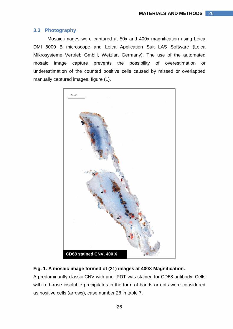
3.3 Photography
Mosaic images were captured at 50x and 400x magnification using Leica
DMI 6000 B microscope and Leica Application Suit LAS Software (Leica
Mikrosysteme Vertrieb GmbH, Wetzlar, Germany). The use of the automated
mosaic image capture prevents the possibility of overestimation or
underestimation of the counted positive cells caused by missed or overlapped
manually captured images, figure (1).
Fig. 1. A mosaic image formed of (21) images at 400X Magnification.
A predominantly classic CNV with prior PDT was stained for CD68 antibody. Cells
with red–rose insoluble precipitates in the form of bands or dots were considered
as positive cells (arrows), case number 28 in table 7.
CD68 stained CNV, 400 X
20 µm
27
27 MATERIALS AND METHODS
3.4 Analysis
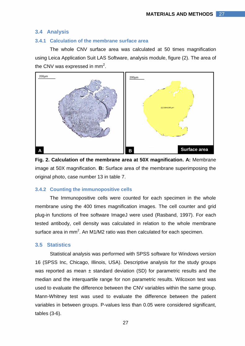
3.4.1 Calculation of the membrane surface area
The whole CNV surface area was calculated at 50 times magnification
using Leica Application Suit LAS Software, analysis module, figure (2). The area of
the CNV was expressed in mm2.
Fig. 2. Calculation of the membrane area at 50X magnification. A: Membrane
image at 50X magnification. B: Surface area of the membrane superimposing the
original photo, case number 13 in table 7.
3.4.2 Counting the immunopositive cells
The Immunopositive cells were counted for each specimen in the whole
membrane using the 400 times magnification images. The cell counter and grid
plug-in functions of free software ImageJ were used (Rasband, 1997). For each
tested antibody, cell density was calculated in relation to the whole membrane
surface area in mm2. An M1/M2 ratio was then calculated for each specimen.
3.5 Statistics
Statistical analysis was performed with SPSS software for Windows version
16 (SPSS Inc, Chicago, Illinois, USA). Descriptive analysis for the study groups
was reported as mean ± standard deviation (SD) for parametric results and the
median and the interquartile range for non parametric results. Wilcoxon test was
used to evaluate the difference between the CNV variables within the same group.
Mann-Whitney test was used to evaluate the difference between the patient
variables in between groups. P-values less than 0.05 were considered significant,
tables (3-6).
A B Surface area
200µm 200µm
28
28 RESULTS
4 RESULTS
4.1 Clinical characterization
This study retrospectively reviewed 58 eyes of 58 consecutive patients who
had surgical extraction of CNV between 2004 and 2007. In all patients the cause
of CNV development was AMD. The patient characteristics are described in
attachment 8.1. The CNVs of the 58 patients, 32 females and 26 males, were
divided into 9 groups as mentioned in the methodology, table 1. The mean age of
the patients was 74.94 ± SD 7.16 years. In 34 patients the right eyes were
operated on and in 24 patients the left eyes were operated on. The mean value of
preoperative VA was 0.74 ± SD 0.82 LogMar notation. The angiographic diagnosis
of the membranes was classic CNV in 15 membranes, occult CNV in 25
membranes and hemorrhagic CNV in 18 membranes. In 55 patients the
membrane was located subfoveal, in 1 patient the membrane was located
subfoveal and juxtafoveal, in 1 patient the membrane was located juxtafoveal and
peripapillary and in 1 patient the membrane was located juxtafoveal and
subfoveal. The membrane size was less than 2 optic disc diameter (DD) in 20.7%
of the membranes, less than 3 DD in 48.3% and more than 3 DD in 31%. At the
time of surgery, 43% patients had received no prior treatment. 15.5 % of patients
had received PDT monotherapy, 17.2 % of patients had received intravitreal
bevacizumab monotherapy. 6.9 % of patients had received intravitreal
ranibizumab monotherapy once. 8.6 % of patients had received intravitreal TA
monotherapy. 8.6% of patients had received combined bevacizumab and PDT
treatment. The mean period between the medical treatment and the surgical
extraction of the membrane in each group is shown in table (3).
Table 3. The mean value of the duration (in days) between the date of the last
given treatment and the surgical intervention
N Mean Std. Deviation
Group 4 (G4) 9 93 51
Group 5 (G5) 10 49 64
Group 6 (G6) 4 18 17
Group 7 (G7) 5 15 22
Group 8 (PDT) (G8) 5 189 118
Group 8 (Avastin) (G8) 5 111 123
G4: Patients treated with PDT. G5: Patients treated with bevacizumab (Avastin®). G6: Patients treated with Ranibizumab (Lucentis®). G7: Patients treated with triamcinolone. G8: Patient treated with combined PDT and bevacizumab.
29
29 RESULTS
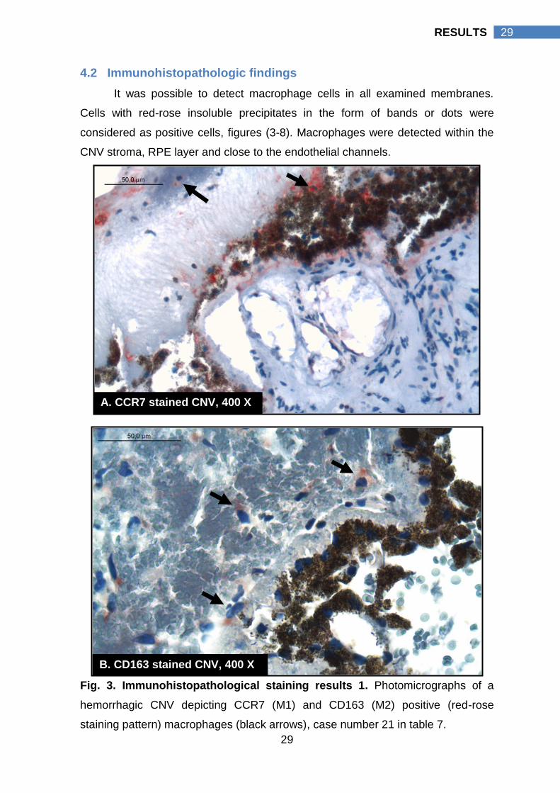
4.2 Immunohistopathologic findings
It was possible to detect macrophage cells in all examined membranes.
Cells with red-rose insoluble precipitates in the form of bands or dots were
considered as positive cells, figures (3-8). Macrophages were detected within the
CNV stroma, RPE layer and close to the endothelial channels.
Fig. 3. Immunohistopathological staining results 1. Photomicrographs of a
hemorrhagic CNV depicting CCR7 (M1) and CD163 (M2) positive (red-rose
staining pattern) macrophages (black arrows), case number 21 in table 7.
A. CCR7 stained CNV, 400 X
B. CD163 stained CNV, 400 X
30
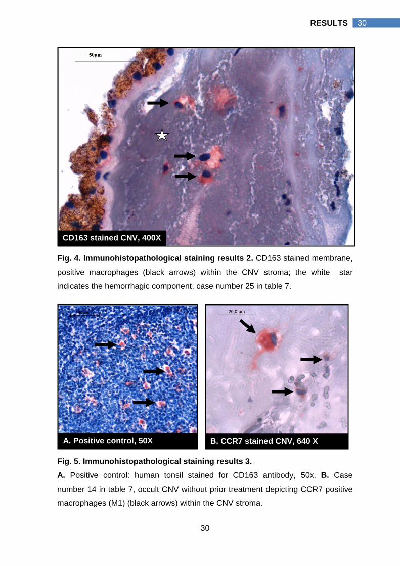
30 RESULTS
Fig. 4. Immunohistopathological staining results 2. CD163 stained membrane,
positive macrophages (black arrows) within the CNV stroma; the white star
indicates the hemorrhagic component, case number 25 in table 7.
Fig. 5. Immunohistopathological staining results 3.
A. Positive control: human tonsil stained for CD163 antibody, 50x. B. Case
number 14 in table 7, occult CNV without prior treatment depicting CCR7 positive
macrophages (M1) (black arrows) within the CNV stroma.
B. CCR7 stained CNV, 640 X A. Positive control, 50X
CD163 stained CNV, 400X
31
31 RESULTS
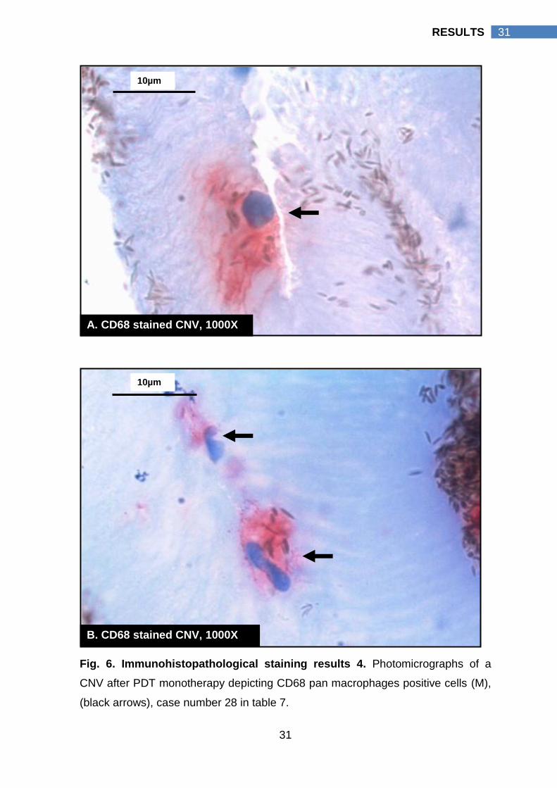
Fig. 6. Immunohistopathological staining results 4. Photomicrographs of a
CNV after PDT monotherapy depicting CD68 pan macrophages positive cells (M),
(black arrows), case number 28 in table 7.
A. CD68 stained CNV, 1000X
B. CD68 stained CNV, 1000X
10µm
10µm
32
32 RESULTS
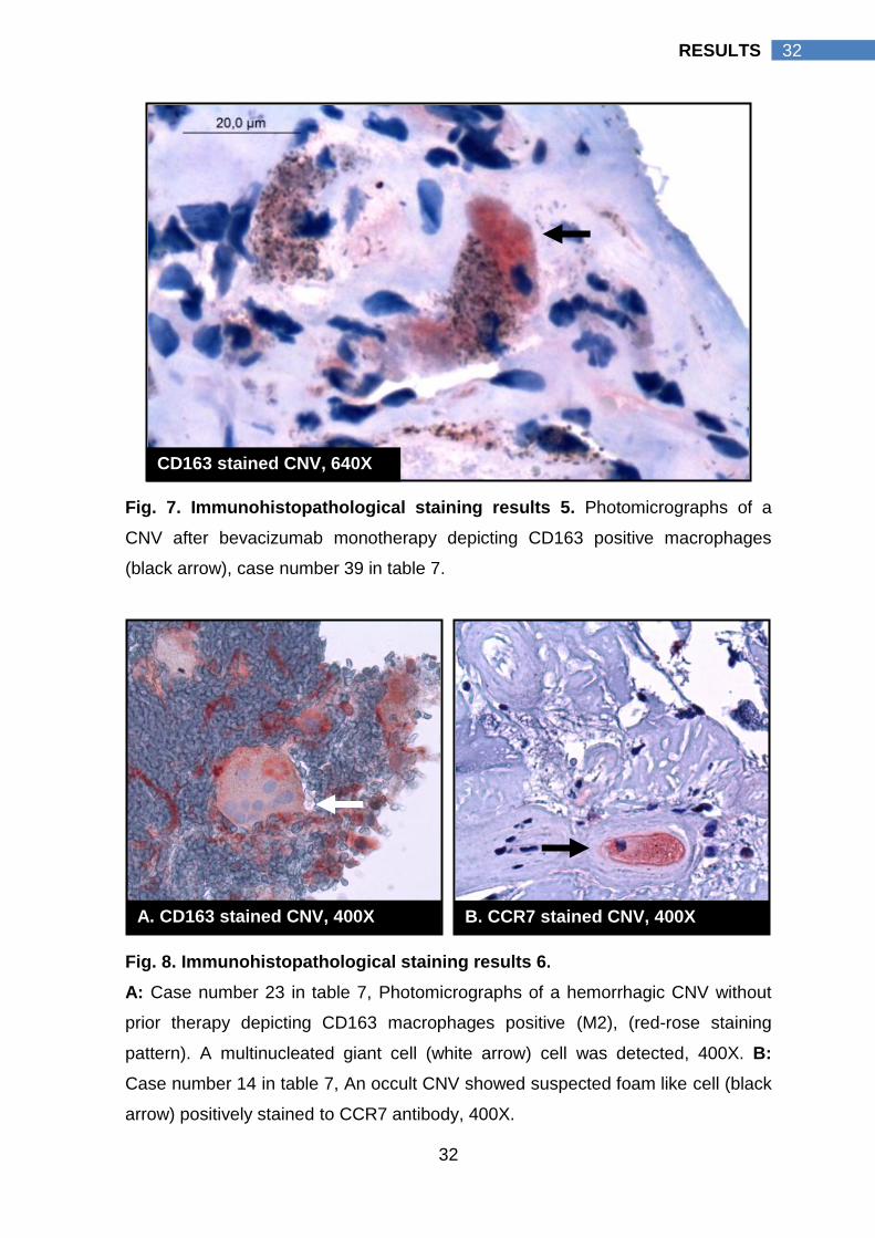
Fig. 7. Immunohistopathological staining results 5. Photomicrographs of a
CNV after bevacizumab monotherapy depicting CD163 positive macrophages
(black arrow), case number 39 in table 7.
Fig. 8. Immunohistopathological staining results 6.
A: Case number 23 in table 7, Photomicrographs of a hemorrhagic CNV without
prior therapy depicting CD163 macrophages positive (M2), (red-rose staining
pattern). A multinucleated giant cell (white arrow) cell was detected, 400X. B:
Case number 14 in table 7, An occult CNV showed suspected foam like cell (black
arrow) positively stained to CCR7 antibody, 400X.
CD163 stained CNV, 640X
A. CD163 stained CNV, 400X B. CCR7 stained CNV, 400X
33
33 RESULTS
4.3 Macrophage activation pattern
Macrophage cell density was represented as the number of macrophage
cells in relation to the surface area of the membrane in mm2. The median value of
cell density (cell/mm2) for each macrophage phenotype and the M1/M2 cell density
ratio in each group were calculated, tables (4-6) & figures (9-12).
In G1 “Classic CNV, n = 5”, the median value of the M1 cell density (125
cell/mm2 interquartile range 64 cell/mm2 - 225 cell/mm2 ) was slightly higher than
the median value of its M2 cell density (112 cell/mm2 interquartile range 62
cell/mm2 - 210 cell/mm2, p value 0.893). In G2 “Occult CNV, n= 10”, the median
value of the M1 cell density (136 cell/mm2 interquartile range 112 cell/mm2 - 197
cell/mm2) was slightly lower than the median value of its M2 cell density (156
cell/mm2 interquartile range 98 cell/mm2 - 224 cell/mm2, p value 0.959). In G3
“Hemorrahgic CNV, n= 10”, the median value of the M2 cell density (928
cell/mm2 interquartile range 400 cell/mm2 - 1263 cell/mm2) was significantly higher
than the median value of its M1 cell density (211 cell/mm2 interquartile range 109
cell/mm2 - 417 cell/mm2, p value 0.005). As well, M2 cell density of hemorrhagic
CNV (G3) (928 cell/mm2 interquartile range 400 cell/mm2 - 1263 cell/mm2) was
significantly higher than M2 cell density of classic CNV (112 cell/mm2 interquartile
range 62 cell/mm2 - 210 cell/mm2, p value) and occult CNV (156 cell/mm2
interquartile range 98 cell/mm2 - 224 cell/mm2), p values, 0.014 and 0.003
respectively. A more global view for the pattern of macrophage activation was
presented by G9, which represent all non treated CNV specimens (n=25). In G9 a
statistically significant higher M2 cell density (204 cell/mm2 interquartile range 107
cell/mm2 - 807 cell/mm2) than its M1 cell density (159 cell/mm2 interquartile range
105 cell/mm2 - 264 cell/mm2) was detected, p value 0.012.
To emphasize, in CNV with no prior treatment (G9), M2 cells were the
prevalent macrophage cell type that was more pronounced in hemorrhagic CNV
(G3). Hemorrhagic CNV had a statistically significant higher M2 cell density
compared to its M1 cell density; classic CNV M2 cell density (G1) and occult CNV
M2 cell density (G2).
CNV treated with PTD (G4, n= 9) was extracted at a relatively longer
interval following treatment (Mean 93 days ± SD 51). Following PDT, the median
value of M1 cell density was higher than its M2 cell density (143 cell/mm2
34
34 RESULTS
interquartile range 115 cell/mm2 - 236 cell/mm2 versus 131 cell/mm2 interquartile
range 50 cell/mm2 - 200 cell/mm2, p value 0.26).
However the median value of M1 cell density was lower compared to
control group (G9) (143 cell/mm2 interquartile range 115 cell/mm2 - 236 cell/mm2
versus 159 cell/mm2 interquartile range 105 cell/mm2 - 204 cell/mm2, p value
0.984). The median value of M2 cell density was significantly lower than that of the
control group (131 cell/mm2 interquartile range 50 cell/mm2 - 200 cell/mm2 versus
204 cell/mm2 interquartile range 107 cell/mm2 - 807 cell/mm, p value 0.044).
M1/M2 ratio of PDT treated CNV (1.75 interquartile range 1.03 – 3.37) was
significantly higher than the control group (0.58 interquartile range 0.36 – 1.24), p
value 0.01.
To emphasize, in PDT treated CNV (G4), activation of M2 cells were
significantly suppressed compared to the control. A significantly high M1/M2 ratio
was present compared to the control group which was caused by the prevalence
of M1 cells.
Following treatment with bevacizumab (Avastin®) “G5, n= 10”, CNVs
were extracted after therapy with mean value of duration of 49 days ± SD 64. The
median value of the M2 cell density was lower than the median value of its M1 cell
density (37 cell/mm2 interquartile range 17 cell/mm2 - 167 cell/mm2 versus 81
cell/mm2 interquartile range 51 cell/mm2 - 94 cell/mm2, p value 0.799).
However the median value of M, M1 and M2 cell densities was significantly
lower compared to control group (G9). M cell density was significantly lower in
bevacizumab treated CNV (G5) compared to control (G9) (182 cell/mm2
interquartile range 66 cell/mm2 - 318 cell/mm2 versus 311 cell/mm2 interquartile
range 215 cell/mm2 - 517 cell/mm2, p value 0.026). The median value of M1 cell
density was significantly lower than that of the control group (81 cell/mm2
interquartile range 51 cell/mm2 - 94 cell/mm2 versus 159 cell/mm2 interquartile
range 105 cell/mm2 - 264 cell/mm, p value 0.003). The median value of M2 cell
density was significantly lower than that of the control group (37 cell/mm2
interquartile range 18 cell/mm2 - 167 cell/mm2 versus 204 cell/mm2 interquartile
range 107 cell/mm2 - 807 cell/mm, p value 0.003).
To emphasize, bevacizumab therapy (G5) suppresses macrophages
infiltration to the CNV. Bevacizumab caused a statistically significant reduction of
35
35 RESULTS
M, M1, and M2 macrophages (p value 0.026, 0.003, 0.003 respectively, Mann
Whitney test) with more reduction of angiogenic M2 cells compared to the control
group.
Following treatment with ranibizumab (Lucentis®) “G6, n=4”, CNVs
were extracted after therapy with mean value of duration of 18 days ± SD 17. The
median value of the M1 cell density was higher than the median value of its M2
cell density (125 cell/mm2 interquartile range 85 cell/mm2 - 149 cell/mm2 versus 29
cell/mm2 interquartile range 19 cell/mm2 - 51 cell/mm2, p value 0.068).
M cell density was significantly lower in ranibizumab treated CNV (G6)
compared to control G (9) (178 cell/mm2 interquartile range 138 cell/mm2 - 189
cell/mm2 versus 311 cell/mm2 interquartile range 215 cell/mm2 - 517 cell/mm2, p
value 0.027). The median value of M1 cell density was lower than that of the
control group (125 cell/mm2 interquartile range 85 cell/mm2 - 149 cell/mm2 versus
159 cell/mm2 interquartile range 105 cell/mm2 - 264 cell/mm, p value 0.229). The
median value of M2 cell density was significantly lower than that of the control
group (29 cell/mm2 interquartile range 19 cell/mm2 - 51 cell/mm2 versus 204
cell/mm2 interquartile range 107 cell/mm2 - 807 cell/mm, p value 0.003). There
were no statistically significant differences in ranibizumab M, M1, M2 cell densities
and M1/M2 ratios compared to bevacizumab treated group (G5). The median M1
/M2 ratio was significantly increased (3.87 interquartile range 1.97-8.67) compared
to the control group (0.58 interquartile range 0.36-1.24), p value 0.007.
To emphasize, ranibizumab therapy (G6) caused a statistically significant
reduction of the median M cell density and the median M2 density compared to the
control group. The median M1 /M2 ratio was significantly increased compared to
the control group. The M1 cells remain relatively unaffected compared to the
control.
Following treatment with TA “G7, n=5”, CNVs were extracted after
therapy with mean value of duration of 15 days ± SD 22. The median value of the
M1 cell density was significantly lower than the median value of its M2 cell density
(33 cell/mm2 interquartile range 21 cell/mm2 - 52 cell/mm2 versus 54 cell/mm2
interquartile range 19 cell/mm2 - 92 cell/mm2, p value 0.043).
M cell density was significantly lower in TA treated CNV (G7) compared to
control Group (G9) (163 cell/mm2 interquartile range 97 cell/mm2 - 187 cell/mm2
36
36 RESULTS
versus 311 cell/mm2 interquartile range 215 cell/mm2 - 517 cell/mm2, p value
0.013). The median value of M1 cell density was significantly lower than that of the
control group (33 cell/mm2 interquartile range 22 cell/mm2 - 52 cell/mm2 versus
159 cell/mm2 interquartile range 105 cell/mm2 - 264 cell/mm, p value 0.003). The
median value of M2 cell density was significantly lower than that of the control
group (54 cell/mm2 interquartile range 19 cell/mm2 - 54 cell/mm2 versus 204
cell/mm2 interquartile range 107 cell/mm2 - 807 cell/mm, p value 0.006).
To emphasize, TA (G7) caused a significant reduction of pan M, M1, and
M2 macrophages cell densities compared to the control group. The M2 cell density
was significantly higher compared to M1 following triamcinolone treatment.
Following treatment with combined PDT and bevacizumab “G8, n=5”,
CNVs were extracted after therapy with mean value of duration of 189 days ± SD
118 for PDT and 111 days ± SD 123 for bevacizumab. The median value of the
M1 cell density was lower than the median value of its M2 cell density (89 cell/mm2
interquartile range 44 cell/mm2 - 172 cell/mm2 versus 148 cell/mm2 interquartile
range 106 cell/mm2 285 cell/mm2, p value 0.345).
M cell density was higher in combined PDT and bevacizumab treated CNV
(G8) compared to control (G9) (424 cell/mm2 interquartile range 384 cell/mm2 -
615 cell/mm2 versus 311 cell/mm2 interquartile range 215 cell/mm2 - 517 cell/mm2,
p value 0.140). The median value of M1 cell density was lower than that of the
control group (89 cell/mm2 interquartile range 44 cell/mm2 - 172 cell/mm2 versus
159 cell/mm2 interquartile range 105 cell/mm2 - 264 cell/mm, p value 0.080). The
median value of M2 cell density was lower than that of the control group (148
cell/mm2 interquartile range 106 cell/mm2 - 285 cell/mm2 versus 204 cell/mm2
interquartile range 107 cell/mm2 - 807 cell/mm, p value 0.42).
To emphasize, combined anti-VEGF and PDT (G8) showed no significant
change in the M1/M2 ratio, pan M, M1 or M2 macrophages cell densities when
compared to the control group.
37
37 RESULTS
Table 4. The pattern of macrophage activation represented as cell density (cell/mm2)
Pan macrophage(M)
Classically activated macrophages (M1)
Alternatively activated macrophages (M2) M1/M2 ratio
Percentiles Percentiles Percentiles Percentiles
Groups n M* 25 50 75 M 25 50 75 M 25 50 75 M 25 50 75
G1 5 266 197 266 422 125 64 125 225 112 62 112 210 0.69 0.48 0.69 3.40
G2 10 290 161 290 394 136 112 136 197 156 98 156 224 1.12 0.56 1.12 1.80
G3 10 517 234 517 946 211 109 211 417 928 400 928 1263 0.32 0.22 0.32 0.50
G4 9 366 87 366 500 143 115 143 236 131 50 131 200 1.75 1.03 1.75 3.37
G5 10 182 66 182 318 81 51 81 94 37 18 37 167 2.28 0.50 2.28 4.01
G6 4 178 138 178 189 125 85 125 149 29 19 29 51 3.87 1.97 3.87 8.67
G7 5 163 97 163 187 33 21 33 52 54 19 54 92 0.74 0.48 0.74 1.80
G8 5 424 384 424 615 89 44 89 172 148 106 148 285 0.44 0.21 0.44 2.10
G9 25 311 215 311 517 159 105 159 264 204 107 204 807 0.58 0.36 0.58 1.24
n: Number of CNV membranes. G1: No treatment, classic AMD CNV. G2: No treatment, occult CNV. G3: No treatment, hemorrhagic
CNV. G4: Patients treated with PDT. G5: Patients treated with bevacizumab (Avastin®). G6: Patients treated with ranibizumab
(Lucentis®). G7: Patients treated with triamcinolone. G8: Patient treated with combined PDT and bevacizumab. G9: No treatment
control group. M*: Median value of the macrophage cell density.
38
38 RESULTS
Table 6. The significance of the difference in the macrophage activation
between the study groups using the Mann-Whitney test, p values (Asymp. Sig)
less than 0.05 were regarded as significant differences.
M M1 M2 Ratio
G1&G2 0.903 0.624 0.540 0.800
G1&G3 0.221 0.270 0.014* 0.027*
G2&G3 0.112 0.290 0.003* 0.002*
G4&G9 0.424 0.984 0.044* 0.010*
G5&G9 0.026* 0.003* 0.003* 0.077
G5&G6 0.690 0.095 0.917 0.074
G6&G9 0.027* 0.229 0.003* 0.007*
G7&G9 0.013* 0.003* 0.006* 0.359
G8&G9 0.140 0.080 0.420 0.359
Significant p value
G1: No treatment, classic AMD CNV. G2: No treatment, occult CNV. G3: No
treatment, hemorrhagic CNV. G4: Patients treated with PDT. G5: Patients treated
with bevacizumab (Avastin®). G6: Patients treated with Ranibizumab (Lucentis®).
G7: Patients treated with triamcinolone. G8: Patient treated with combined PDT and
bevacizumab. G9: No treatment control group.
Table 5. The significance of the difference between M1 and M2 cell density
within the same group using the Wilcoxon test.
Group Asymptomatic Significant (2-tailed)
G1 0.893
G2 0.959
G3 0.005*
G4 0.260
G5 0.799
G6 0.068
G7 0.043*
G8 0.345
G9 0.012*
*Significant p value
G1: No treatment, classic AMD CNV. G2: No treatment, occult CNV. G3: No
treatment, hemorrhagic CNV. G4: Patients treated with PDT. G5: Patients treated
with bevacizumab (Avastin®). G6: Patients treated with Ranibizumab (Lucentis®).
G7: Patients treated with triamcinolone. G8: Patient treated with combined PDT and
bevacizumab. G9: No treatment control group.
39
39 RESULTS
Fig. 9. The median values of the pan macrophage (M) cell density
Fig. 10. The median values of the cell density of the classically activated
macrophage (M1)
40
40 RESULTS
Fig. 11. The median values of the cell density of the alternatively activated
macrophage (M2)
Fig. 12. The median values of the M1/M2 ratio, red line indicate equal cell
density of both phenotypes (M1/M2 ratio).
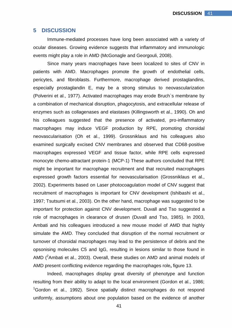
41
41 DISCUSSION
5 DISCUSSION
Immune-mediated processes have long been associated with a variety of
ocular diseases. Growing evidence suggests that inflammatory and immunologic
events might play a role in AMD (McGonagle and Georgouli, 2008).
Since many years macrophages have been localized to sites of CNV in
patients with AMD. Macrophages promote the growth of endothelial cells,
pericytes, and fibroblasts. Furthermore, macrophage derived prostaglandins,
especially prostaglandin E, may be a strong stimulus to neovascularization
(Polverini et al., 1977). Activated macrophages may erode Bruch`s membrane by
a combination of mechanical disruption, phagocytosis, and extracellular release of
enzymes such as collagenases and elastases (Killingsworth et al., 1990). Oh and
his colleagues suggested that the presence of activated, pro‐inflammatory
macrophages may induce VEGF production by RPE, promoting choroidal
neovascularisation (Oh et al., 1999). Grossniklaus and his colleagues also
examined surgically excised CNV membranes and observed that CD68‐positive
macrophages expressed VEGF and tissue factor, while RPE cells expressed
monocyte chemo-attractant protein-1 (MCP-1) These authors concluded that RPE
might be important for macrophage recruitment and that recruited macrophages
expressed growth factors essential for neovascularisation (Grossniklaus et al.,
2002). Experiments based on Laser photocoagulation model of CNV suggest that
recruitment of macrophages is important for CNV development (Ishibashi et al.,
1997; Tsutsumi et al., 2003). On the other hand, macrophage was suggested to be
important for protection against CNV development. Duvall and Tso suggested a
role of macrophages in clearance of drusen (Duvall and Tso, 1985). In 2003,
Ambati and his colleagues introduced a new mouse model of AMD that highly
simulate the AMD. They concluded that disruption of the normal recruitment or
turnover of choroidal macrophages may lead to the persistence of debris and the
opsonising molecules C5 and IgG, resulting in lesions similar to those found in
AMD (2Ambati et al., 2003). Overall, these studies on AMD and animal models of
AMD present conflicting evidence regarding the macrophages role, figure 13.
Indeed, macrophages display great diversity of phenotype and function
resulting from their ability to adapt to the local environment (Gordon et al., 1986;
1Gordon et al., 1992). Since spatially distinct macrophages do not respond
uniformly, assumptions about one population based on the evidence of another
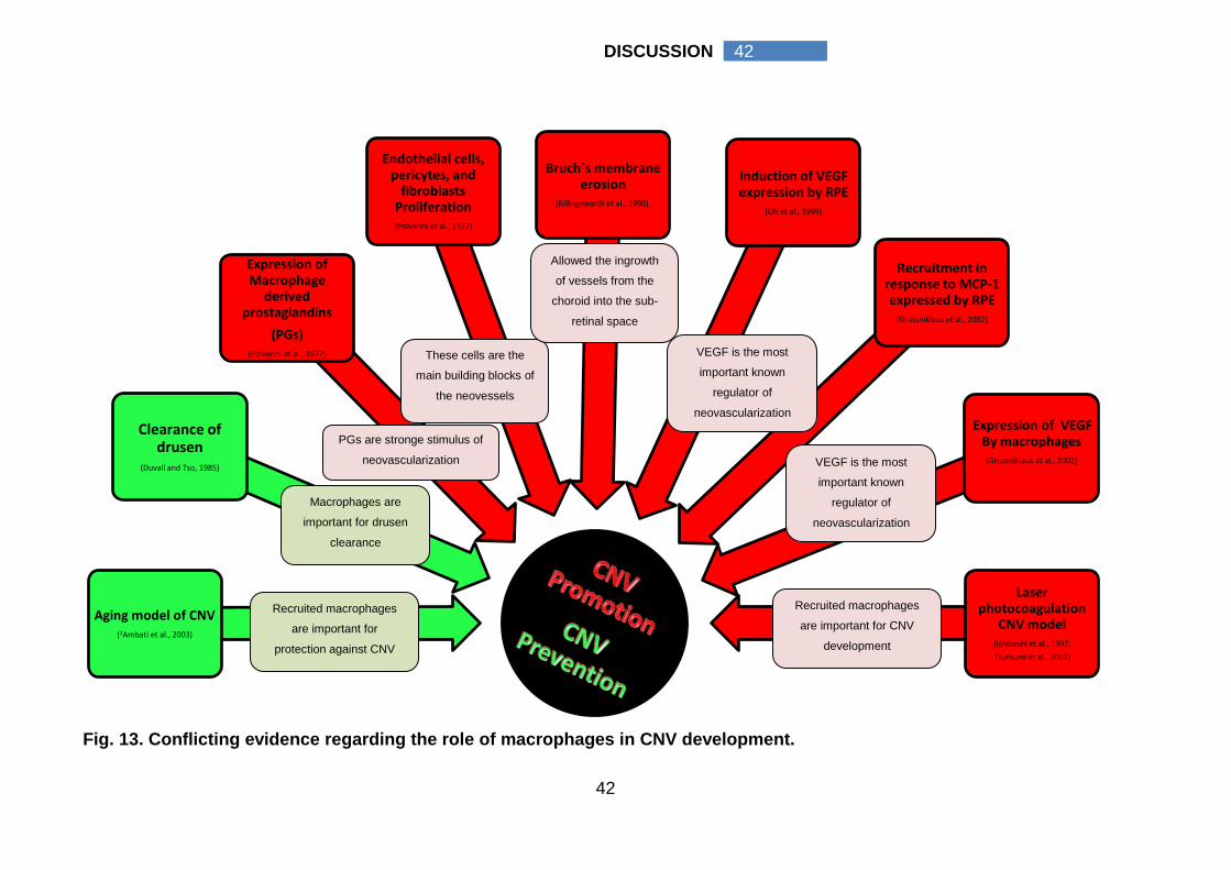
42
42 DISCUSSION
Fig. 13. Conflicting evidence regarding the role of macrophages in CNV development.
Clearance of drusen
(Duvall and Tso, 1985)
Aging model of CNV(2Ambati et al., 2003)
Expression of Macrophage
derived prostaglandins
(PGs)(Polverini et al., 1977)
Recruitment in response to MCP-1 expressed by RPE
(Grossniklaus et al., 2002)
Endothelial cells, pericytes, and
fibroblasts Proliferation(Polverini et al., 1977)
Bruch`s membrane erosion
(Killingsworth et al., 1990).
Induction of VEGF expression by RPE
(Oh et al., 1999)
Laser photocoagulation
CNV model(Ishibashi et al., 1997;
Tsutsumi et al., 2003)
Expression of VEGF By macrophages
(Grossniklaus et al., 2002)
PGs are stronge stimulus of
neovascularization
These cells are the
main building blocks of
the neovessels
Allowed the ingrowth
of vessels from the
choroid into the sub-
retinal space
VEGF is the most
important known
regulator of
neovascularization
Recruited macrophages
are important for CNV
development
Recruited macrophages
are important for
protection against CNV
VEGF is the most
important known
regulator of
neovascularization
Macrophages are
important for drusen
clearance
43
43 DISCUSSION
can be misleading (Daems et al., 1976; 2Gordon et al., 1992). In order to
understand their role in human CNV development it is necessary to identify and
distinguish the phenotype of the involved cells.
Macrophages can be distinguished as classically activated (M1) or
alternatively activated (M2) cells. These different types express specialized and
polarized functional properties in response to cytokines and microbial products
(Ma et al., 2003). M1 macrophages are involved in type I inflammation, tissue
destruction, killing intracellular parasites and tumour resistance. They can produce
powerful oxidants cause lipid peroxidation of affected cell membranes, leading to
cell death (Gordon, 2003). M2 macrophages are programmed to express the
enzyme arginase, which converts arginine to ornithine, promoting cell division,
repair (Mills et al., 2000) and angiogenesis (Kodelja et al., 1997). Thus,
macrophages participate to different extents in pro-inflammatory and anti-
inflammatory immune reactions. However, both types of activated cells can co-
exist and show differences in their capacity to secrete mediators and to express
receptors (Mantovani et al., 2004), figure (14).
Fig. 14. Macrophage polarized activation.
A simplified view of immune and tissue-derived signals inducing classical (M1) and
alternative (M2) macrophage polarized activation. The main functional effects on
macrophage functions, molecular markers and effector molecules are schematically
represented in both cases. ―Abbreviations: CXCL10: Chemokine (C-X-C motif) ligand 10,
TNF-α: Tumour necrosis factor alpha, CCL22: Chemokine ligand 22, ROI: Reactive
oxygen intermediate, RNI: Reactive nitrogen intermediate.‖
44
44 DISCUSSION
The question addressed by the present study was to describe the pattern of
macrophage activation in relation to the CNV variables. This present study
demonstrates that the macrophages were activated within the CNV either as M1 or
M2 subtype with a variable degree of activation of each type. The sum of the
activated macrophages (M1+M2) was not always equal to the pan macrophage
cell count. This in equality may be due to the presence of some macrophages in
the deactivated form that stain for CD68 (Gordon, 2003).
The types of the CNV characteristics as well as the prior treatments have
influence on the subtype of the macrophage activation. Although macrophage
detection and/or its density in CNV were previously described (Lopez et al., 1991;
Dastgheib and Green, 1994; Penfold et al., 2001; Schnurrbusch et al., 2001;
Grossniklaus et al., 2002; Tsutsumi et al., 2003; 2Ambati et al., 2003; Csaky et al.,
2004; Grossniklaus et al., 2005; Suzuki et al., 2007; 2Tatar et al., 2007; 1,2Tatar et
al., 2009), the author is unaware of previous reports that describe the pattern of
macrophage activation in human CNV.
In this study we showed that M2 macrophages were significantly higher
compared to M1 macrophages in non treated CNV (G9, n= 25), p value 0.012. The
type of the CNV had an influence on the macrophage activation. Hemorrhagic
CNV (G3, n=10) had a statistically significant higher M2 cell density compared to
its M1 cell density; classic CNV M2 cell density, and occult CNV M2 cell density, p
value = 0.005, 0.013, and 0.003 respectively. This high M2 density can have more
than one explanation.
First explanation, it could be a cause of the hemorrhage within the CNV. It
was reported that macrophages produce various angiogenic cytokines, including
TNF-α, IL-1, bFGF, VEGF, and TGF-ß and thus play a key role in angiogenesis
(Oh et al., 1999). Of the two major macrophages subtypes, M2 macrophages are
well equipped to play an important role in protracted macrophages associated
angiogenic processes (Kodelja et al., 1997). In addition, a high M2 cell density was
present in other diseases which share with CNV some of its angiogenic
hemorrhagic complications such as endometriosis (Bacci et al., 2009),
atherosclerosis (Bouhlel et al., 2007, Slevin et al., 2009), and also growing and
metastatic tumors (Lamagna et al., 2006; Lin et al., 2006). The increased
susceptibility for hemorrhage can be related to the fact that the developed
neovessels are immature and fragile which could contribute to the development of
45
45 DISCUSSION
an unstable haemorrhagic rupture-prone environment (Moulton, 2001; Slevin et
al., 2009).
Second explanation is that the high density of M2 macrophages can be
formed in response to hemorrhage. This can be supported by the fact that
endocytosis of foreign and necrotic debris as well as scavenging components of
damaged cells are well known functions of macrophages (Lamagna et al., 2006).
CD163 is a scavenger receptor cysteine-rich (SRCR), a hemoglobin scavenger
receptor exclusively expressed in the monocyte-macrophage system with a
particularly high expression in M2 macrophages (Law et al., 1993; Kristiansen et
al., 2001, Gordon, 2003). CD163 plays a major role in dampening the
inflammatory response and in scavenging components of damaged cells
(Moestrup and Moller, 2004; Borda et al., 2008). CD163-mediated endocytosis of
haptoglobin-hemoglobin complexes formed upon red blood cell haemolysis leads
to lysosomal degradation of the ligand protein and metabolism of heme by
cytosolic heme oxygenase. CD163 regulates inflammation by at least two ways.
Firstly, it is reported to directly induce intracellular signaling leading to secretion of
anti-inflammatory cytokines. Secondly, and perhaps even more importantly, the
CD163-mediated delivery of hemoglobin to the macrophage may fuel an anti-
inflammatory response because heme metabolites have potent anti-inflammatory
effects (Borde et al., 2008; Pilling et al., 2009).
A Third explanation can be a combination of the previously mentioned
explanations. In this explanation the author postulates that local M2 macrophages
can proliferate and accelerate the angiogenesis of such hemorrhage prone
neovessels then once hemorrhage occurs; it starts to carry out the scavenger
function. The presence of the M2 cells in non hemorrhagic CNV as presented in
our study can support this explanation.
The above results are important for several reasons. First, they suggest that
the alternative activation of macrophages could be a determinant factor for the
susceptibility to hemorrhagic complications for CNV patients which are a major
cause of poor visual prognosis in these patients (Hochman et al., 1997). Second,
they suggest that it may be possible to avoid hemorrhagic complications of human
CNV though local inhibition of alternatively activated macrophage. Third, since
there is a continuous shedding of the extracellular domain of CD163 which leads
to substantial amounts of soluble receptor in plasma (Borde et al., 2008; Pilling et
46
46 DISCUSSION
al., 2009), the author suggest that the M2 macrophages activity can be evaluated
through an estimation of the plasma level of soluble CD163 receptor and in turn
the susceptibility to haemorrhagic complications within CNV. Fourth, it brings
attention to a possibility of involvement of peroxisome proliferator-activated
receptor-gamma (PPAR-ɣ) in hemorrhagic CNV. There is growing evidence of the
involvement of PPAR receptors in AMD pathogenesis (Herzlich et al., 2008).
Bouhlel and associates reported that PPAR-γ activation primes primary human
monocytes for M2 differentiation and M2 marker expression was positively
correlated with that of PPAR-γ in human atherosclerotic lesions (Bouhlel et al.,
2007). In addition, the similarity between the atherosclerosis and the development
of AMD is well established (Friedman, 2004; Kamei et al., 2007).
Examination of membranes treated with photodynamic therapy (G4, n=9)
revealed a statistically significant reduction of M2 cells compared to control, p
value, 0.04 with a statistically significant high M1/M2 ratio. An increased M1/M2
ratio indicates prevalence of M1 in PDT treated membranes. PDT causes up-
regulation of TNF-α (Schmidt-Erfurth and Hasan, 2000), and IFN-ɣ (Gollnick et al.,
2006). Both cytokines are major stimuli for macrophage to be activated through
the classic pathway (M1) (Mosser, 2003). Post PDT prevalent M1 macrophages
could be due the effect of TNF-α and IFN-ɣ on local macrophages.
She and his colleagues suggested that PDT-induced inflammation in the
CNV lesion might activate macrophages to produce iNOS, which results in
excessive nitric oxide release and subsequently in photoreceptor apoptosis (She
et al., 2007). The iNOS is one of the M1 macrophages products that causes to cell
death (Gordon, 2003). Detection of M1 macrophage phenotype after long interval
following PDT treatment (93 days ± SD 51), may indicate a need for long acting
treatment strategies that target M1 macrophages following PDT.
M1 cells specifically express CCR7 (Martinez et al., 2006). That CCR7 has
been identified as a key regulator of homeostatic T-cell trafficking to secondary
lymphoid organs. However, recent studies suggest that this receptor also can be
involved in the abnormal recruitment of T-cells from the circulation to sites of
inflammation (Forster et al., 1999; Sallusto et al., 2000). Macrophages expressing
CCR7 have been implicated in inflammatory skin disorders, (Ohl et al., 2004),
were found within the inflamed atherosclerotic vessel wall (Waldo et al., 2008) and
they are known to mediate inflammation-associated tumour progression (Mburu et
47
47 DISCUSSION
al., 2006). CCR7 expressing macrophages are exposed to increased levels of
Chemokine ligand 19 (CCL19) and Chemokine ligand 21 (CCL21), potentially
promoting inflammation, MMP activation, and thrombus formation (Serra et al.,
2004; Burman et al., 2005). M1 cells could have a potential role in angiogenesis
through MMP activation (Song et al., 2000; Ito et al., 2009).
Reduced M2 cells density following PDT could be explained in two ways.
First, as we mention above, PDT causes up-regulation of TNF-α (Schmidt-Erfurth
and Hasan, 2000) and IFN-ɣ (Gollnick et al., 2006), so macrophages are activated
through classic pathway and no longer activated through the alternative pathway.
Second, Although PDT cause up regulation of IL10 (a major stimulus of
macrophage activation via alternative pathway) (Pazos and Nader, 2007), this
stimulation is of a limited duration (72 and 120 h) post-PDT (Simkin et al., 2000).
In our study the mean value of the duration between treatment and surgery was
(93 days ± SD 51).
There are several activators of angiogenesis including VEGF, bFGF, TGF-
ß, angiopoietin-1, and angiopoetin-2. However, it appears that VEGF signalling
represents a critical rate-limiting step (Kaiser, 2006). The VEGF family currently
has seven members: VEGF-A to VEGF-F and placental growth factor (Waisbourd
et al., 2007). VEGF-A is the most important known regulator of angiogenesis
(Kaiser, 2006). Different isoforms can be produced by alternative exon splicing of
the VEGF-A, of these isoforms VEGF-165 is probably the major pathogenic
species (Robinson and Stringer, 2001). Three VEGF receptors have been
identified so far: VEGFR-1, VEGFR-2, and VEGFR-3 (Waisbourd et al., 2007).
Two anti-VEGF therapies are currently approved for intraocular use in
patients with AMD and CNV: pegaptanib sodium (Macugen) and ranibizumab
(Lucentis). Pegaptanib is a Ribonucleic acid (RNA) aptamer which specifically
binds to and blocks the VEGF-165 isoform (Waisbourd et al., 2007). Ranibizumab
is a recombinant humanized antibody fragment which inhibits all the biological
active forms of VEGF (Raftery et al., 2007). Bevacizumab (Avastin®) is another
anti-VGEF therapy which is a full length pan anti-VEGF humanized monoclonal
antibody. It was approved for use in colon cancer but not for AMD (Steinbrook,
2006). Despite the lack of regulatory approval, it has been widely used off label for
CNV and induces significant improvements in both visual acuity and retinal
morphology (Spaide et al., 2006; Emerson et al., 2007).
48
48 DISCUSSION
This study found that bevacizumab (Avastin®) caused a statistically
significant reduction of pan macrophages, p value 0.026; M1 macrophages, p
value 0.003, and M2 macrophages, p value 0.003 with more reduction of M2 cells.
The significant reduction of both types of macrophages can be a direct result of
the reduction of the pan macrophage.
VEGF is a major chemo-attractant for inflammatory cells, including
macrophages, neutrophils, dendritic cells, myeloid-derived suppressor cells, and
T-cells (Ancelin et al., 2004; Huang et al., 2007; Dineen et al., 2008; Ko et al.,
2009; Ozao-Choy et al., 2009; Shin et al., 2009). Hence a low macrophage
infiltration following bevacizumab was previously reported. In tumour xenograft
models, anti-VEGF therapy leads to a reduction in macrophage infiltration
(Salnikov et al., 2006; Whitehurst et al., 2007; Dineen et al., 2008; Roland et al.,
2009). The study carried out by Ronald and his colleagues showed that anti-VEGF
therapy with bevacizumab inhibited the growth of an established breast tumour
with reduced tumour microvessels density and limitation of the infiltration of
tumour-associated macrophages (Roland et al., 2009). Salnikov and his
colleagues also found that at 5 mg/kg dose, bevacizumab reduced the density of
macrophages in xenograft carcinoma (Salnikov et al., 2006). Tog and his
colleagues stated a significant correlation between tumour associated
macrophages (TAM) and angiogenesis in renal cell carcinoma and they expected
a reduction of TAM following anti-VEGF therapy (Toge et al., 2009). Tsutsui and
his colleagues concluded that there is a close association between TAM
infiltrations and both the VEGF expression and microvascular density. A
prognostic implication of TAM infiltration is attributed to their involvement in tumour
angiogenesis (Tsutsui et al., 2005).
Kiss and colleagues evaluated the effect of intravitreal bevacizumab on
anterior chamber inflammatory activity. They found that no inflammatory response
was detected clinically and by the laser flare meter after intravitreal bevacizumab
administration in addition a slight reduction of the anterior chamber flare was
present (Kiss et al., 2006).
Tatar and colleagues studied the effect of bevacizumab on the macrophage
cell density in human CNV. They found a significantly higher macrophage cell
density in bevacizumab treated CNV compared to control groups. The density of
macrophages in the bevacizumab treated group ranged between (interquartile
49
49 DISCUSSION
range 321- 61821 cell/mm2, median = 4661 cells/mm2) (2Tatar et al., 2008). This
cell density is considerably high compared to our findings of pan macrophage cell
density, (interquartile range 66-318 cell/mm2, median = 182 cell/mm2). The current
study tried to improve the reliability of the cell counting through the use of mosaic
software from Leica System which allowed the imaging of the whole membrane at
high magnification levels without the overlapping of images which is likely to occur
in membranes with large sizes. The captured images were then enrolled as a
single stack to be manually counted with ImagJ software (Rasband, 1997).
Through the use of this software we avoid the biases caused by the overlap or
missing parts of the membrane that would occur with the use of manual
photography. A simple example of a mosaic image (formed of 21 images) is given
in figure (7).
This current study also evaluated the effect of combined photodynamic
therapy and bevacizumab treatment on the macrophage activation. Results
showed no significant change in the M1/M2 ratio, pan macrophages, M1
macrophages or M2 macrophages when compared to the control group. This can
be explained by the anti-inflammatory effect of the bevacizumab could reduce the
inflammation caused by the photodynamic therapy. These results are consistent
with finding reported by Maier et al. They reported that PDT combined with
injection of intravitreal bevacizumab tends to be more effective compared to PDT
monotherapy by reducing the post-PDT increase of vascular growth and
inflammatory factors (Maier et al., 2008).
Ranibizumab is a recombinant humanized antibody fragment which inhibits
all the biological active forms of VEGF (Raftery et al., 2007). There was no
significant difference in the macrophage activation pattern between ranibizumab
and bevacizumab. We found that ranibizumab caused a statistically significant
reduction of the median M cell density, p value 0.027 and the median M2 density,
p value=0.003 compared to the control group. The median M1 /M2 ratio was
significantly increased compared to the control group, p value=0.009. The M1 cells
(pro-inflammatory macrophage) were not suppressed. Ranibizumab causes
suppression of macrophage recruitment through its anti-VEGF action (Salnikov et
al., 2006; Whitehurst et al., 2007; Dineen et al., 2008; Roland et al., 2009). Further
reduction of macrophage activation can occur through inability of ranibizumab to
50
50 DISCUSSION
activate complement system, as it cannot bind complement because it lacks the
Fc region (Gaudreault et al., 2005; Ferrara et al., 2006). Complement activation is
essential for the activation of the macrophages via humeral pathway (Gordon
2003).
Although a lower incidence of inflammation was expected in patients
treated with ranibizumab (Gaudreault et al., 2005; Ferrara et al., 2006), evidence
of intraocular inflammation was still detectable (Brown et al., 2006; Rosenfeld et
al., 2006; Kourlas and Abrams, 2007; Brown et al., 2009). Rosenfeld et al.
reported an incidence of iridocyclitis (83%) and injection-site reactions (72%)
following intravitreal injection of ranibizumab which did not increase with repeated
injections, despite increasing the ranibizumab doses (Rosenfeld et al., 2006).
We suggest that this degree of inflammation can be explained by the
presence of M1 (pro inflammatory macrophage). This suggestion is supported by
the detection of IL-6, IL-1ß, and TNF-α cytokines following ranibizumab injection.
Campochiaro et al found that IL-6, IL-1ß, and TNF-α were present in detectable
amounts one month following ranibizumab injection in patients with vein
occlusions and that their aqueous level was unaffected by the resolution of the
macular oedema caused by the ranibizumab (Campochiaro et al., 2009). IL-6, IL-
1ß, and TNF-α are the main proinflammatory cytokines produced by M1 cells
(Gordon 2003).
Triamcinolone acetonide (TA) is a synthetic steroid of the glucocorticoid
family with the hydrogen atom in the ninth position replaced with fluorine; its
glucocorticoid potency is five times hydrocortisone (Gopal and Sharma, 2007). In
this study we examined five occult CNV with TA prior to treatment. The CNV were
extracted 15 ± 22 days after treatment. We found that there was a significant
reduction of pan macrophages M, M1, and M2 macrophages cell density
compared to control, p value 0.013, 0.003, and 0.006 respectively.
A low macrophage infiltration following TA therapy is well documented in
previous studies. Singhal and colleagues found that TA attenuated
macrophage/microglia accumulation associated retinal ganglion cell death and
facilitates survival of Müller stem cell grafts (Singhal et al., 2010). Jahangier and
his colleagues found that intra-articular treatment with glucocorticoids was
associated with a considerable reduction of macrophage infiltration in the
synovium which resulted in decreased arthritis activity at 6 months follow up
51
51 DISCUSSION
(Jahangier et al., 2006). Young and his colleagues found that intra-articular
glucocorticoids may reduce CD68 positive macrophage infiltration into the synovial
lining layer, 1 month following treatment (Young et al., 2001). In vitro, TA has been
shown to downregulate the expression of TNF-α, VEGF, IL1, and MMP and
subsequently influences neovascularization (Kim et al., 2007). Down regulation of
TNF-α can be the cause of low M1 cell density because it is one of the major
signals required for classic activation of macrophages (M1) (Gordon, 2003). The
reduction of the M1 and M2 types can be secondary to the overall reduction of
macrophages caused by TA. In our study we found that M2 cell density was
significantly higher compared to M1 following TA treatment, p value 0.043. This is
explained by the role of glucocorticoids in alternative activation of macrophages.
Monocytes can be a source of M2 alternatively activated macrophages in
response to IL-4, IL-10, IL-13, TGF-β1, or glucocorticoids (Gordon, 2003).
Tatar et al. evaluated the early effects of TA (5 ± SD 2.34 days) on
macrophage infiltration to CNV. They found that TA treated CNV had a
macrophage density similar to the control CNV (1Tatar et al., 2009). In our study
we evaluated the delayed effects of TA (15 ± SD 22 days) on macrophage
infiltration. We found that TA caused a reduction of macrophages infiltration to the
CNV with a selective shift towards activation via an alternative pathway (M2). This
discrepancy can be due to the difference in the point of time in which the CNV
were examined. The mean value of the duration between the treatment and the
extraction was 5± SD 2.34 days in Tatar’s study, and 15 ± SD 22 days in this
study. We argue that the extra time between treatment and extraction in this study
has allowed the therapeutic action of TA to become evident, hence the significant
reduction of macrophage cell density (Jonas et al., 2004; Jonas et al., 2005).
In addition to the description of the macrophage activation profile in AMD
related CNV; we were able to detect foam-like cells and giant cells within the
membranes. Foam cells were more prevalent in M1 macrophages. Macrophage
foam cell formation in the arterial intima is the hallmark of early atherosclerosis
(Ross, 1999). Ocular foam cells can be formed as macrophages take up the
oxidized phospholipids caused by oxidative stress (Suzuki et al., 2007) or due to
the activation of TLR secondary to pathogen exposure (Cao et al., 2007). This
finding can support the vascular theory of CNV development.
There were some limitations in the present study. Firstly, the determination
52
52 DISCUSSION
of M1 versus M2 phenotype was based upon a limited number of cell surface
markers. Secondly, it is unknown whether macrophages that were not committed
to the M1 or M2 phenotype were recruited to the site of CNV and then stimulated
to differentiate locally or whether phenotype-committed macrophages were
selectively recruited to sites of neovascularization depending upon the antigens
that were present. However, this study was able to introduce an evidence of the
presence of macrophage heterogenicity in human CNV and highlight the effect of
the CNV type and the prior treatment on the activation pattern of macrophage.
In conclusion, Macrophages play major and different roles in CNV
development. The identification and understanding of different activation patterns
may be important for distinct therapeutic strategies.
53
53 SUMMARY
6 SUMMARY
Inflammatory processes play an integral and causative role in the
pathogenesis of AMD. Evidence suggests that macrophages are involved in CNV
pathogenesis; however, conflicting functions were present. Macrophages express
specialized activation patterns which are referred as classically activated
macrophages (M1) and alternatively activated macrophages (M2). In this study
we retrospectively examined 58 surgically excised CNV for the pattern of
macrophage activation using immunohistochemical technique. CD68, CCR7, and
CD163 antibodies were used to stain for M, M1 and M2 macrophages,
respectively. Macrophage activation pattern was examined in CNV specimens
belonging to patients with and without prior treatment
In CNV with no prior treatment, M2 cells were the prevalent macrophage
cell type. Hemorrhagic CNV had a higher M2 cell density compared to its M1;
classic CNV M2 and occult CNV M2 cell densities. High M2 cells density can be
responsible for CNV growth via secretion of angiogenesis cytokines. The
increased M2 cells density of hemorrhagic CNV can be an indirect cause of
hemorrhage, a response to hemorrhage or both.
Photodynamic therapy suppressed M2 cells activation with a significant
high M1/M2 ratio caused by the prevalence of M1 cells compared to the control. It
resulted in a state of inflammation caused by the relatively high M1 cells (pro-
inflammatory) and low M2 cells (anti-inflammatory, angiogenic).
Bevacizumab anti-VEGF therapy suppressed infiltration of all macrophages
subtypes probably by blocking the chemo-attractant effect of VEGF. It promotes
an anti-inflammatory and anti-angiogenic effects. Combined Bevacizumab anti-
VEGF and PDT attenuate the inflammatory state caused by PDT. Ranibizumab
anti-VEGF therapy suppressed the overall macrophage infiltration, However M1
cells were more prevalent.This suppression can be explained by its ability to block
VEGF and inability to cause activation of monocytes into macrophages via
humoral pathway as it cannot activate the complement system. Triamcinolone
acetate therapy caused a reduction of macrophages’ infiltration with selective
shift towards the M2 activation.
54
54 BIBLIOGRAPHY
7 Bibliography
AREDS report Nr. 6: Age‐Related Eye Disease Study Research Group report
number 6: The age‐related eye disease study system for classifying age‐related
macular degeneration from stereoscopic colour fundus photographs: The
age‐related eye disease study report number 6. Am J Ophthalmol 132, 668‐681
(2001)
AREDS report Nr. 8: Age-Related Eye Disease Study Research Group report
number 8: A randomized, placebo-controlled, clinical trial of high dose
supplementation with vitamins C and E, beta carotene, and zinc for age-related
macular degeneration and vision loss: The age‐related eye disease study report
number 8. Arch Ophthalmol 119, 1417–1436 (2001)
1Ambati J, Ambati BK, Yoo SH, Ianchulev S, Adamis AP: Age-related macular
degeneration: etiology, pathogenesis, and therapeutic strategies. Surv Ophthalmol
48, 257–293 (2003)
2Ambati J, Anand A, Fernandez S, Sakurai E, Lynn BC, Kuziel WA, Rollins BJ,
Ambati BK: An animal model of age-related macular degeneration in senescent
Ccl2- or Ccr2-deficient mice. Nat Med 9, 1390-1397 (2003)
Ancelin M, Chollet-Martin S, Hervé MA, Legrand C, El Benna J, Perrot-Applanat
M: Vascular endothelial growth factor VEGF189 induces human neutrophil
chemotaxis in extravascular tissue via an autocrine amplification mechanism. Lab
Invest 84, 502–512 (2004)
Apte RS, Richter J, Herndon J, Ferguson TA: Macrophages inhibit
neovascularization in a murine model of age-related macular degeneration. PLoS
Med 3, e310 (2006)
Asahara T, Takahashi T, Masuda H, Kalka C, Chen D, Iwaguro H, Inai Y, Silver
M, Isner JM: VEGF contributes to postnatal neovascularization by mobilizing bone
marrow-derived endothelial progenitor cells. EMBO J 15, 3964-3972 (1999)
55
55 BIBLIOGRAPHY
Assink JJ, Klaver CC, Houwing-Duistermaat JJ, Wolfs RC, van Duijn CM, Hofman
A, de Jong PT: Heterogeneity of the genetic risk in age-related macular disease: a
population-based familial risk study. Ophthalmology 112, 482–487 (2005)
Augustin AJ, Scholl S, Kirchhof J: Treatment of neovascular age-related macular
degeneration: Current therapies. Clin Ophthalmol 3, 175-182 (2009)
Bacci M, Capobianco A, Monno A, Cottone L, Di Puppo F, Camisa B, Mariani M,
Brignole C, Ponzoni M, Ferrari S, Panina-Bordignon P, Manfredi AA, Rovere-
Querini P: Macrophages are alternatively activated in patients with endometriosis
and required for growth and vascularization of lesions in a mouse model of
disease. Am J Pathol 175, 547-556 (2009)
Barleon B, Sozzani S, Zhou D, Weich HA, Mantovani A, Marmé D: Migration of
human monocytes in response to vascular endothelial growth factor (VEGF) is
mediated via the VEGF receptor flt-1. Blood 15, 3336-3343 (1996)
Bellmann C, Unnebrink K, Rubin GS, Miller D, Holz FG: Visual acuity and contrast
sensitivity in patients with neovascular age-related macular degeneration. Results
from the Radiation Therapy for Age- Related Macular Degeneration (RAD-) Study.
Graefes Arch Clin Exp Ophthalmol 241, 968–974 (2003)
BenEzra D: Neovasculogenic ability of prostaglandins, growth factors, and
synthetic chemoattractants. Am J Ophthalmol 86, 455–461 (1978)
Bird AC, Bressler NM, Bressler SB, Chisholm IH, Coscas G, Davis MD, de Jong
PT, Klaver CC, Klein BE, Klein R: An international classification and grading
system for age-related maculopathy and age-related macular degeneration. The
International ARM Epidemiological Study Group. Surv Ophthalmol 39, 367–374
(1995)
Blair CJ: Geographic atrophy of the retinal pigment epithelium: A manifestation of
senile macular degeneration. Arch Ophthalmol 9, 19–25 (1975)
56
56 BIBLIOGRAPHY
Blumenkranz MS, Russell SR, Robey MG, Kott-Blumenkranz R, Penneys N: Risk
factors in age-related maculopathy complicated by choroidal neovascularization.
Ophthalmology 93, 552-558 (1986)
Borda JT, Alvarez X, Mohan M, Hasegawa A, Bernardino A, Jean S, Aye P,
Lackner AA: CD163, a marker of perivascular macrophages, is up-regulated by
microglia in simian immunodeficiency virus encephalitis after haptoglobin-
hemoglobin complex stimulation and is suggestive of breakdown of the blood-brain
barrier. Am J Pathol 172, 725-737 (2008)
Bouhlel MA, Derudas B, Rigamonti E, Dièvart R, Brozek J, Haulon S, Zawadzki
C, Jude B, Torpier G, Marx N, Staels B, Chinetti-Gbaguidi G: PPAR-gamma
activation primes human monocytes into alternative M2 macrophages with anti-
inflammatory properties. Cell Metab 6, 137–143 (2007)
Bressler NM, Bressler SB, Gragoudas ES: Clinical characteristics of choroidal
neovascular membranes. Arch Ophthalmol 105, 209–213 (1987)
Bressler NM, Frost LA, Bressler SB, Murphy RP, Fine SL: Natural course of
poorly defined choroidal neovascularization associated with macular degeneration.
Arch Ophthalmol 106, 1537–1542 (1988)
Bressler SB, Maguire MG, Bressler NM, Fine SL: The Macular Photocoagulation
Study Group: Relationship of drusen and abnormalities of the retinal pigment
epithelium to the prognosis of neovascular macular degeneration. Arch
Ophthalmol 108, 1442–1447 (1990)
Bressler NM: Photodynamic therapy of subfoveal choroidal neovascularization in
age-related macular degeneration with Verteporfin: two-year results of 2
randomized clinical trials—TAP report 2. Arch Ophthalmol 119, 198–207 (2001)
Bressler NM: Verteporfin therapy of subfoveal choroidal neovascularization in
age-related macular degeneration: two-year results of a randomized clinical trial
including lesions with occult with no classic choroidal neovascularization.
57
57 BIBLIOGRAPHY
Verteporfin in Photodynamic Therapy report 2. Am J Ophthalmol 133, 168–169
(2002)
Bressler NM: Antiangiogenic approaches to age-related macular degeneration
today. Ophthalmology 116, S15-23 (2009)
Brown DM, Kaiser PK, Michels M, Soubrane G, Heier JS, Kim RY, Sy JP,
Schneider S; ANCHOR Study Group: Ranibizumab versus verteporfin for
neovascular age-related macular degeneration. N Engl J Med 355, 1432-1444
(2006)
Brown DM, Michels M, Kaiser PK, Heier JS, Sy JP, Ianchulev T: ANCHOR Study
Group. Ranibizumab versus verteporfin photodynamic therapy for neovascular
age-related macular degeneration: Two-year results of the ANCHOR study.
Ophthalmology 116, 57-65 (2009)
Burman A, Haworth O, Hardie DL, Amft EN, Siewert S, Jackson DG, Salmon M,
Buckley CD: A chemokine-dependent stromal induction mechanism for aberrant
lymphocyte accumulation and compromised lymphatic return in rheumatoid
arthritis. J Immunol 174, 1693–1700 (2005)
Burns RP, Feeney-Burns L: Clinico-morphologic correlations of drusen of Bruch's
membrane. Trans Am Ophthalmol Soc 78, 206–225 (1980)
Caicedo A, Espinosa-Heidmann DG, Piña Y, Hernandez EP, Cousins SW: Blood-
derived macrophages infiltrate the retina and activate Muller glial cells under
experimental choroidal neovascularization. Exp Eye Res 81, 38-47 (2005)
Cao F, Castrillo A, Tontonoz P, Re F, Byrne GI: Chlamydia pneumoniae-induced
macrophage foam cell formation is mediated by Toll-like receptor 2. Infect Immun
75, 753-759 (2007)
Campochiaro PA, Glaser BM: A retina-derived stimulator(s) of retinal pigment
epithelial cell and astrocyte proliferation. Exp Eye Res 43, 449–457 (1986)
58
58 BIBLIOGRAPHY
Campochiaro PA, Choy DF, Do DV, Hafiz G, Shah SM, Nguyen QD, Rubio R,
Arron JR: Monitoring ocular drug therapy by analysis of aqueous samples.
Ophthalmology 116, 2158-2164 (2009)
Chamberlin JA, Bressler NM, Bressler SB, Elman MJ, Murphy RP, Flood TP,
Hawkins BS, Maguire MG, Fine SL: The use of fundus photographs and
fluorescein angiograms in the identification and treatment of choroidal
neovascularization in the Macular Photocoagulation Study. The Macular
Photocoagulation Study Group. Ophthalmology 96, 1526–1534 (1989)
Clauss M, Gerlach M, Gerlach H, Brett J, Wang F, Familletti PC, Pan YC, Olander
JV, Connolly DT, Stern D. Vascular permeability factor: a tumor-derived
polypeptide that induces endothelial cell and monocyte procoagulant activity, and
promotes monocyte migration. J Exp Med 1, 1535-1545 (1990)
Cousins SW, Espinosa-Heidmann DG, Csaky KG: Monocyte activation in patients
with age-related macular degeneration: a biomarker of risk for choroidal
neovascularization? Arch Ophthalmol 122, 1013-1018 (2004)
Csaky K, Baffi J, Chan CC, Byrnes GA: Clinicopathologic correlation of
progressive fibrovascular proliferation associated with occult choroidal
neovascularization in age-related macular degeneration. Arch Ophthalmol 122,
650–652 (2004)
Daems WT, Koerten HK, Soranzo MR: Differences between monocyte-derived
and tissue macrophages. Adv Exp Med Biol 73, 27-40 (1976)
D’Amico DJ, Goldberg MF, Hudson H, Jerdan JA, Krueger S, Luna S, Robertson
SM, Russell S, Singerman L, Slakter JS, Sullivan EK, Yannuzzi L, Zilliox P:
Anecortave acetate as monotherapy for the treatment of subfoveal lesions in
patients with exudative age-related macular degeneration (AMD): interim (month
6) analysis of clinical safety and efficacy. Retina 23, 14–23 (2003)
Dastgheib K, Green WR: Granulomatous reaction to Bruch's membrane in age-
59
59 BIBLIOGRAPHY
related macular degeneration. Arch Ophthalmol 112, 813–818 (1994)
De Jong PT, Klaver CC, Wolfs RC, Assink JJ, Hofman A: Familial aggregation of
age-related maculopathy. Am J Ophthalmol 124, 862–863 (1997)
De Juan E Jr, Machemer R: Vitreous surgery for hemorrhagic and fibrous
complications of age-related macular degeneration. Am J Ophthalmol 15, 25-29
(1988)
Dinapoli MR, Calderon CL, Lopez DM: The altered tumoricidal capacity of
macrophages isolated from tumor-bearing mice is related to reduce expression of
the inducible nitric oxide synthase gene. J Exp Med 183, 1323-1329 (1996)
Dineen SP, Lynn KD, Holloway SE, Miller AF, Sullivan JP, Shames DS, Beck AW,
Barnett CC, Fleming JB, Brekken RA: Vascular endothelial growth factor receptor
2 mediates macrophage infiltration into orthotopic pancreatic tumors in mice.
Cancer Res 68, 4340–4346 (2008)
Douglas SD, Hassan NF: Morphology of monocytes and macrophages. In:
Williams WJ, Beutler E, Erslev AJ, Lichtman MA: Haematology. 4. edition, 858–
868, McGraw-Hill, New York 1990
Drolet DW, Nelson J, Tucker CE, Zack PM, Nixon K, Bolin R, Judkins MB, Farmer
JA,Wolf JL, Gill SC, Bendele RA: Pharmacokinetics and safety of an anti-vascular
endothelial growth factor aptamer (NX1838) following injection into the vitreous
humor of rhesus monkeys. Pharm Res 17, 1503–1510 (2000)
Duvall J, Tso MO: Cellular mechanisms of resolution of drusen after laser
coagulation. An experimental study. Arch Ophthalmol 103, 694-703 (1985)
Edwards AO, Ritter R III, Abel KJ, Manning A, Panhuysen C, Farrer LA:
Complement factor H polymorphism and age-related macular degeneration.
Science 308, 421–424 (2005)
60
60 BIBLIOGRAPHY
Emerson MV, Lauer AK, Flaxel CJ, Wilson DJ, Francis PJ, Stout JT, Emerson
GG, Schlesinger TK, Nolte SK & Klein ML: Intravitreal bevacizumab (Avastin)
treatment of neovascular agerelated macular degeneration. Retina 27, 439–444
(2007)
Feeney-Burns L, Ellersieck MR: Age-related changes in the ultrastructure of
Bruch’s membrane. Am J Ophthalmol 100, 686–697 (1985)
Ferrara N, Damico L, Shams N, Lowman H, Kim R: Development of ranibizumab,
an anti-vascular endothelial growth factor antigen binding fragment, as therapy for
neovascular age-related macular degeneration. Retina 26, 859–870 (2006)
Forster R, Schubel A, Breitfeld D, Kremmer E, Renner-Muller I, Wolf E, Lipp M:
CCR7 coordinates the primary immune response by establishing functional
microenvironments in secondary lymphoid organs. Cell 99, 23–33 (1999)
Friedman E, Krupsky S, Lane AM, Oak SS, Friedman ES, Egan K, Gragoudas
ES: Ocular blood flow velocity in age-related macular degeneration.
Ophthalmology 102, 640–646 (1995)
Friedman E: Update of the vascular model of AMD. Br J Ophthalmol 88, 161–163
(2004)
Gaudreault J, Fei D, Rusit J, Suboc P, Shiu V: Preclinical pharmacokinetics of
Ranibizumab (rhuFabV2) after a single intravitreal administration. Invest
Ophthalmol Vis Sic 46, 726–733 (2005)
Gass JD: Biomicroscopic and histopathologic considerations regarding the
feasibility of surgical excision of subfoveal neovascular membranes Trans Am
Ophthalmol Soc 92, 91-111 (1994)
Gehlbach P, Demetriades AM, Yamamoto S, Deering T, Duh EJ, Yang HS,
Cingolani C, Lai H, Wei L, Campochiaro PA: Periocular injection of an adenoviral
vector encoding pigment epithelium-derived factor inhibits choroidal
61
61 BIBLIOGRAPHY
neovascularization. Gene Ther 10, 637–646 (2003)
Glaser BM: Extracellular modulating factors and the control of intraocular
neovascularization: an overview. Arch Ophthalmol 106, 603–607 (1988)
Gollnick SO, Owczarczak B, Maier P: Photodynamic therapy and anti-tumor
immunity. Lasers Surg Med 38, 509-515 (2006)
Gopal L, Sharma T: Use of intravitreal injection of triamcinolone acetonide in the
treatment of age-related macular degeneration. Indian J Ophthalmol 55, 431-435
(2007)
Gordon S, Crocker PR, Morris L, Lee SH, Perry VH, Hume DA: Localization and
function of tissue macrophages. Ciba Found Symp 118, 54-67 (1986)
1Gordon S, Lawson L, Rabinowitz S, Crocker PR, Morris L, Perry VH: Antigen
markers of macrophage differentiation in murine tissues. Curr Top Microbiol
Immunol 181, 1-37 (1992)
2Gordon S, Fraser I, Nath D, Hughes D, Clarke S: Macrophages in tissues and in
vitro. Curr Opin Immunol 4, 25-32 (1992)
Gordon S: The macrophage. Bioessays 17, 977-986 (1995)
Gordon S: Alternative activation of macrophages. Nat Rev Immunol 3, 23-35
(2003)
Gorin MB, Breitner JC, De Jong PT, Hageman GS, Klaver CC, Kuehn MH,
Seddon JM: The genetics of age-related macular degeneration. Mol Vis 3, 5-29
(1999)
Goverdhan SV, Howell MW, Mullins RF, Osmond C, Hodgkins PR, Self J, Avery
K, Lotery AJ: Association of HLA class I and class II polymorphisms with age-
related macular degeneration. Invest Ophthalmol Vis Sic 46, 1726-1734 (2005)
62
62 BIBLIOGRAPHY
Green WR, Key SN III: Senile macular degeneration: a histopathologic study.
Trans Am Ophthalmol Soc 75, 180–254 (1977)
Green WR, Enger C: Age-related macular degeneration histopathologic studies:
the 1992 Lorenz E. Zimmerman Lecture. Ophthalmology 100, 1519–1535 (1993)
1Grisanti S, Canbek S, Kaiserling E, Adam A, Lafaut B, Gelisken F, Szurman P,
Henke-Fahle S, Oficjalska-Mlynczak J, Bartz-Schmidt, KU: Expression of Endoglin
in Choroidal Neovascularization and Human Eyes. Exp Eye Res 78, 207-213
(2004)
2Grisanti S, Tatar O, Canbek S, Lafaut B, Gelisken F, Inhoffen W, Szurman P,
Aisenbrey S, Oficjalska-Mlynczak J, Bartz-Schmidt, KU: Immunohistopathologic
Evaluation Of Choroidal Neovascular Membranes Following Verteporfin-
Photodynamic Therapy. Am J Ophthalmol 137, 914-923 (2004)
Grisanti S, Ziemssen F: Bevacizumab: off-label use in ophthalmology. Indian J
Ophthalmol 55, 417-420 (2007)
Grisanti S, Tatar O: The role of vascular endothelial growth factor and other
endogenous interplayers in age-related macular degeneration. Prog Retin Eye
Res 27, 372-390 (2008).
Grossniklaus HE, Martinez JA, Brown VB, Lambert HM, Sternberg P Jr, Capone
A Jr, Aaberg TM, Lopez PF: Immunohistochemical and histochemical properties of
surgically excised subretinal neovascular membranes in age-related macular
degeneration. Am J Ophthalmol 114, 464–472 (1992)
Grossniklaus HE, Hutchinson AK, Capone A Jr, Woolfson J, Lambert HM:
Clinicopathologic features of surgically excised choroidal neovascular membranes.
Ophthalmology 101, 1099–1111 (1994)
Grossniklaus HE, Gass JD: Clinicopathologic correlations of surgically excised
type 1 and type 2 submacular choroidal neovascular membranes. Am J
63
63 BIBLIOGRAPHY
Ophthalmol 126, 59–69 (1998)
Grossniklaus HE, Green WR: Histopathologic and ultrastructural findings of
surgically excised choroidal neovascularization. Submacular Surgery Trials
Research Group. Arch Ophthalmol 116, 745–749 (1998)
Grossniklaus HE, Ling JX, Wallace TM, Dithmar S, Lawson DH, Cohen C, Elner
VM, Elner SG, Sternberg P Jr: Macrophage and retinal pigment epithelium
expression of angiogenic cytokines in choroidal neovascularization. Mol Vis 8,
119–126 (2002)
Grossniklaus HE, Miskala PH, Green WR, Bressler SB, Hawkins BS, Toth C,
Wilson DJ, Bressler NM: Histopathologic and ultrastructural features of surgically
excised subfoveal choroidal neovascular lesions: submacular surgery trials report
no. 7. Arch Ophthalmol 123, 914-921 (2005)
Guymer RH, Bird AC, Hageman GS: Cytoarchitecture of choroidal capillary
endothelial cells. Invest Ophthalmol Vis Sic 45, 1660–1666 (2004)
Hageman GS, Luthert PJ, Victor Chong NH, Johnson LV, Anderson DH, Mullins
RF: An integrated hypothesis that considers drusen as biomarkers of immune-
mediated processes at the RPE Bruch’s membrane interface in aging and age-
related macular degeneration. Prog Retin Eye Res 20, 705–732 (2001)
Harding S: Photodynamic therapy in the treatment of subfoveal choroidal
neovascularisation. Eye 15, 407–412 (2001)
Heriot WJ, Henkind P, Bellhorn RW, Burns MS: Choroidal neovascularization can
digest Bruch’s membrane: a prior break is not essential. Ophthalmology 91, 1603–
1608 (1984)
Herzlich AA, Tuo J, Chan CC: Peroxisome proliferator-activated receptor and
age-related macular degeneration. PPAR Res 2008, 389507(2008)
64
64 BIBLIOGRAPHY
Higgins RD, Sanders RJ, Yan Y, Zasloff M, Williams JI: Squalamine improves
retinal neovascularization. Invest Ophthalmol Vis Sic 41, 1507–1512 (2000)
Hochman MA, Seery CM, Zarbin MA: Pathophysiology and management of
subretinal hemorrhage. Surv Ophthalmol 42, 195-213 (1997)
Holz FG, Wolfensberger TJ, Piguet B, Gross-Jendroska M, Wells JA, Minassian
DC, Chisholm IH, Bird AC: Bilateral macular drusen in age-related macular
degeneration. Prognosis and risk factors. Ophthalmology 101, 1522–1528 (1994)
Huang Y, Chen X, Dikov MM, Novitskiy SV, Mosse CA, Yang L, Carbone DP:
Distinct roles of VEGFR-1 and VEGFR-2 in the aberrant hematopoiesis associated
with elevated levels of VEGF. Blood 110, 624–631 (2007)
Hyman L, Schachat AP, He Q, Leske MC: Hypertension, cardiovascular disease,
and age-related macular degeneration. Age-Related Macular Degeneration Risk
Factors Study Group. Arch Ophthalmol 118, 351–358 (2000)
Ishibashi T, Hata Y, Yoshikawa H, Nakagawa K, Sueishi K, Inomata H:
Expression of vascular endothelial growth factor in experimental choroidal
neovascularization. Graefes Arch Clin Exp Ophthalmol 235, 159-167 (1997)
Ito TK, Ishii G, Saito S, Yano K, Hoshino A, Suzuki T, Ochiai A: Degradation of
soluble VEGF receptor-1 by MMP-7 allows VEGF access to endothelial cells.
Blood 113, 2363-2369 (2009)
Jahangier ZN, Jacobs JW, Kraan MC, Wenting MJ, Smeets TJ, Bijlsma JW,
Lafeber FP, Tak PP: Pretreatment macrophage infiltration of the synovium predicts
the clinical effect of both radiation synovectomy and intra-articular glucocorticoids.
Ann Rheum Dis 65, 1286-1292 (2006)
Jonas JB, Akkoyun I, Budde WM, Kreissig I, Degenring RF: Intravitreal reinjection
of triamcinolone for exudative age-related macular degeneration. Arch Ophthalmol
122, 218-222 (2004)
65
65 BIBLIOGRAPHY
Jonas JB, Degenring RF, Kreissig I, Friedemann T, Akkoyun I: Exudative age-
related macular degeneration treated by intravitreal triamcinolone acetonide. A
prospective comparative nonrandomized study. Eye 19, 163-170 (2005)
Kaiser PK: Antivascular endothelial growth factor agents and their development:
therapeutic implications in ocular diseases. Am J Ophthalmol 142, 660–668 (2006)
Kalayoglu MV, Galvan C, Mahdi OS, Byrne GI, Mansour S: Serological
association between Chlamydia pneumoniae infection and age-related macular
degeneration. Arch Ophthalmol 121, 478–482 (2003)
Kamei M, Yoneda K, Kume N, Suzuki M, Itabe H, Matsuda K, Shimaoka T,
Minami M, Yonehara S, Kita T, Kinoshita S: Scavenger receptors for oxidized
lipoprotein in age-related macular degeneration. Invest Ophthalmol Vis Sic 48,
1801-1807 (2007)
Killingsworth MC, Sarks JP, Sarks SH: Macrophages related to Bruch’s
membrane in age-related macular degeneration. Eye 4, 613–621 (1990)
Kim YH, Choi MY, Kim YS, Park CH, Lee JH, Chung IY, Yoo JM, Choi WS, Cho
GJ, Kang SS: Triamcinolone acetonide protects the rat retina from STZ-induced
acute inflammation and early vascular leakage. Life Sic 81, 1167–1173 (2007)
Kinnaert P, De Wilde JP, Bournonville B, Husson C, Salmon I: Direct activation of
human peritoneal mesothelial cells by heat-killed microorganisms. Ann Surg 224,
749-754 (1996)
Kiss C, Michels S, Prager F, Weigert G, Geitzenauer W, Schmidt-Erfurth U:
Evaluation of anterior chamber inflammatory activity in eyes treated with
intravitreal bevacizumab. Retina 26, 877-881 (2006)
Klaver CC, Wolfs RC, Assink JJ, van Duijn CM, Hofman A, de Jong PT: Genetic
risk of age-related maculopathy. Population-based familial aggregation study. Arch
Ophthalmol 116, 1646–1651 (1998)
66
66 BIBLIOGRAPHY
Klein BE, Klein R: Cataracts and macular degeneration in older Americans: Arch
Ophthalmol 100, 571–573 (1982)
Klein BE, Klein R, Lee KE, Jensen SC: Measures of obesity and age-related eye
diseases. Ophthalmic Epidemiol 8, 251–262 (2001)
Klein R, Klein BE, Linton KP: Prevalence of age-related maculopathy. The Beaver
Dam Eye Study. Ophthalmology 99, 933–343 (1992)
Klein R, Klein BE, Franke T: The relationship of cardiovascular disease and its
risk factors to age-related maculopathy: The Beaver Dam Eye Study.
Ophthalmology 100, 406–414 (1993)
Klein R, Klein BE, Jensen SC, Meuer SM: The 5-year incidence and progression
of age-related maculopathy: The Beaver Dam Eye Study. Ophthalmology 104, 7–
21 (1997)
Klein R, Klein BE, Tomany SC, Cruickshanks KJ: Association of emphysema,
gout, and inflammatory markers with long-term incidence of age-related
maculopathy. Arch Ophthalmol 121, 674-678 (2003)
Kliffen M, Sharma HS, Mooy CM, Kerkvliet S, de Jong PT: Increased expression
of angiogenic growth factors in age related maculopathy. Br J Ophthalmol 81,
154–62 (1997)
Knauer C, Pfeiffer N: Blindness in Germany--today and in 2030. Ophthalmologe
103, 735-741 (2006)
Ko JS, Zea AH, Rini BI, Ireland JL, Elson P, Cohen P, Golshayan A, Rayman PA,
Wood L, Garcia J, Dreicer R, Bukowski R, Finke JH: Sunitinib mediates reversal of
myeloid-derived suppressor cell accumulation in renal cell carcinoma patients. Clin
Cancer Res 15, 2148–2157 (2009)
Kodelja V, Müller C, Tenorio S, Schebesch C, Orfanos CE, Goerdt S: Differences
67
67 BIBLIOGRAPHY
in angiogenic potential of classically vs alternatively activated macrophages.
Immunobiology 197, 478-493 (1997)
Kourlas H, Abrams P: Ranibizumab for the treatment of neovascular age-related
macular degeneration: a review. Clin Ther 29, 1850-1861 (2007)
Kristiansen M, Graversen JH, Jacobsen C, Sonne O, Hoffman HJ, Law SK,
Moestrup SK: Identification of the haemoglobin scavenger receptor. Nature 409,
198-201 (2001)
Krzystolik MG, Afshari MA, Adamis AP, Gaudreault J, Gragoudas ES, Michaud
NA, Li W, Connolly E, O'Neill CA, Miller JW: Prevention of experimental choroidal
neovascularization with intravitreal anti-vascular endothelial growth factor antibody
fragment. Arch Ophthalmol 120, 338–346 (2002)
Lafaut BA, Bartz-Schmidt KU, Vanden Broecke C, Aisenbrey S, De Laey JJ,
Heimann K: Clinicopathological correlation in exudative age related macular
degeneration: histological differentiation between classic and occult choroidal
neovascularisation. Br J Ophthalmol 84, 239–243 (2000)
Lamagna C, Aurrand-Lions M, Imhof BA: Dual role of macrophages in tumor
growth and angiogenesis. J Leukoc Biol 80, 705-713 (2006)
Lamoreaux WJ, Fitzgerald ME, Reiner A, Hasty KA, Charles ST: Vascular
endothelial growth factor increases release of gelatinase A and decreases release
of tissue inhibitor of metalloproteinases by microvascular endothelial cells in vitro.
Microvasc Res 55, 29-42 (1998)
Law SK, Micklem KJ, Shaw JM: A new macrophage differentiation antigen which
is a member of the scavenger receptor superfamily. Eur J Immunol 23, 2320-2325
(1993)
Leenen PJ, de Bruijn MF, Voerman JS, Campbell PA, van Ewijk W: Markers of
mouse macrophage development detected by monoclonal antibodies. J Immunol
68
68 BIBLIOGRAPHY
Methods 174, 5-19 (1994)
Leibowitz HM, Krueger DE, Maunder LR, Milton RC, Kini MM, Kahn HA,
Nickerson RJ, Pool J, Colton TL, Ganley JP, Loewenstein JI, Dawber TR: The
Framingham Eye Study monograph: an ophthalmological and epidemiological
study of cataract, glaucoma, diabetic retinopathy, macular degeneration, and
visual acuity in a general population of 2631 adults, 1973–1975. Surv Ophthalmol
24, 335–610 (1980)
Li JJ, Chen XJ: Simvastatin inhibits interleukin-6 release in human monocytes
stimulated by C-reactive protein and lipopolysaccharide. Coron Artery Dis 14,
329–334 (2003)
Lim JI: Photodynamic therapy for choroidal neovascular disease: photosensitizers
and clinical trials. Ophthalmol Clin North Am 15, 473–478 (2002)
Lin EY, Li JF, Gnatovskiy L, Deng Y, Zhu L, Grzesik DA, Qian H, Xue XN, Pollard
JW: Macrophages regulate the angiogenic switch in a mouse model of breast
cancer. Cancer Res 66, 11238-11246 (2006)
Loffler KU, Lee WR: Basal linear deposit in the human macula. Graefes Arch Clin
Exp Ophthalmol 224, 493–501 (1986)
Loke P, Nair MG, Parkinson J, Guiliano D, Blaxter M, Allen JE: IL-4 dependent
alternatively-activated macrophages have a distinctive in vivo gene expression
phenotype. BMC Immunol, 3-7 (2002)
Lopez PF, Grossniklaus HE, Lambert HM, Aaberg TM, Capone A Jr, Sternberg P
Jr, L'Hernault N: Pathologic features of surgically excised subretinal neovascular
membranes in age-related macular degeneration. Am J Ophthalmol 112, 647–656
(1991)
Lopez PF, Sippy BD, Lambert HM, Thach AB, Hinton DR: Transdifferentiated
retinal pigment epithelial cells are immunoreactive for vascular endothelial growth
69
69 BIBLIOGRAPHY
factor in surgically excised age-related macular degeneration-related choroidal
neovascular membranes. Invest Ophthalmol Vis Sic 37, 855–868 (1996)
Lüke M, Ziemssen F, Völker M, Altpeter E, Beutel J, Besch D, Bartz-Schmidt KU,
Gelisken F: Full macular translocation (FMT) versus photodynamic therapy (PDT)
with verteporfin in the treatment of neovascular age-related macular degeneration:
2-year results of a prospective, controlled, randomised pilot trial (FMT-PDT).
Graefes Arch Clin Exp Ophthalmol 247, 745-754 (2009)
Ma J, Chen T, Mandelin J, Ceponis A, Miller NE, Hukkanen M, Ma GF, Konttinen
YT: Regulation of macrophage activation. Cell Mol Life Sci 60, 2334-2346 (2003)
1Macular Photocoagulation Study (MPS): Laser photocoagulation of subfoveal
neovascular lesions in age-related macular degeneration. Results of a randomized
clinical trial. Macular Photocoagulation Study Group. Arch Ophthalmol 109, 1220–
1231 (1991)
2Macular Photocoagulation Study (MPS): Laser photocoagulation of subfoveal
recurrent neovascular lesions in age-related macular degeneration. Results of a
randomized clinical trial. Macular Photocoagulation Study Group. Arch Ophthalmol
109, 1232–1241(1991)
Macular Photocoagulation Study (MPS): Laser photocoagulation for juxtafoveal
choroidal neovascularization. Five-year results from randomized clinical trials.
Macular Photocoagulation Study Group. Arch Ophthalmol 112, 500–509 (1994)
Maier M, Haas K, Feucht N, Fiore B, Winkler von Mohrenfels C, Clos A, Lohmann
C: Photodynamic therapy with verteporfin combined with intravitreal injection of
bevacizumab for occult and classic CNV in AMD. Klin Monbl Augenheilkd 225,
653-659 (2008)
Maller J, George S, Purcell S, Fagerness J, Altshuler D, Daly MJ, Seddon JM:
Common variation in three genes, including a non coding variant in CFH, strongly
influences risk of age-related macular degeneration. Nat Genet 38,1055–1059
70
70 BIBLIOGRAPHY
(2006)
Mares-Perlman JA, Brady WE, Klein R, Vanden Langenberg GM, Klein BE, Palta
M: Dietary fat and age-related maculopathy. Arch Ophthalmol 113, 743–748
(1995)
Mantovani A, Sica A, Sozzani S, Allavena P, Vecchi A, Locati M: The chemokine
system in diverse forms of macrophage activation and polarization. Trends
Immunol 25, 677–686 (2004)
Martinez FO, Gordon S, Locati M, Mantovani A: Transcriptional profiling of the
human monocyte-to-macrophage differentiation and polarization: new molecules
and patterns of gene expression. J Immunol 177, 7303-7311 (2006)
Martinez FO, Sica A, Mantovani A, Locati M: Macrophage activation and
polarization. Front Biosci 13, 453–461 (2008)
Mburu YK, Wang J, Wood MA, Walker WH, Ferris RL: CCR7 mediates
inflammation-associated tumor progression. Immunol Res 36, 61-72 (2006)
McGonagle D, Georgouli T: The importance of 'Mechnikov's thorn' for an
improved understanding of 21st century medicine and immunology: a view from
the eye. Scand J Immunol 68,129-139 (2008)
Michels S, Rosenfeld JR, Puliafito CA, Marcus EN, Venkatraman AS: Systemic
bevacizumab (Avastin) therapy for neovascular age-related macular degeneration:
twelve-week results of an uncontrolled open-label clinical study. Ophthalmology
112, 1035–1047 (2005)
Mills CD, Kincaid K, Alt JM, Heilman MJ, Hill AM: M-1/M-2 macrophages and the
Th1/Th2 paradigm. J Immunol 164, 6166-6173 (2000)
Moestrup SK, Møller HJ: CD163: a regulated hemoglobin scavenger receptor with
a role in the anti-inflammatory response. Ann Med 36, 347-354 (2004)
71
71 BIBLIOGRAPHY
Mosser DM: The many faces of macrophage activation. J Leukoc Biol 73, 209-212
(2003)
Mosser DM, Edwards JP: Exploring the full spectrum of macrophage activation.
Nat Rev Immunol 8, 958–969 (2008)
Moulton KS: Plaque angiogenesis and atherosclerosis. Curr Atheroscler Rep 3,
225-233 (2001)
Muñoz B, West SK, Rubin GS, Schein OD, Quigley HA, Bressler SB, Bandeen-
Roche K: Causes of blindness and visual impairment in a population of older
Americans: The Salisbury Eye Evaluation Study. Arch Ophthalmol 118, 819–825
(2000)
Oh H, Takagi H, Takagi C, Suzuma K, Otani A, Ishida K, Matsumura M, Ogura Y,
Honda Y: The potential angiogenic role of macrophages in the formation of
choroidal neovascular membranes. Invest Ophthalmol Vis Sic 40, 1891-1898
(1999)
Ohl L, Mohaupt M, Czeloth N, Hintzen G, Kiafard Z, Zwirner J, Blankenstein T,
Henning G, Förster R: CCR7 governs skin dendritic cell migration under
inflammatory and steady-state conditions. Immunity 21, 279-288 (2004)
Ozao-Choy J, Ma G, Kao J, Wang GX, Meseck M, Sung M, Schwartz M, Divino
CM, Pan PY, Chen SH: The novel role of tyrosine kinase inhibitor in the reversal of
immune suppression and modulation of tumor microenvironment for immune-
based cancer therapies. Cancer Res 69, 2514–2522 (2009)
Parihar MS, Hemnani T: Alzheimer's disease pathogenesis and therapeutic
interventions. J Clin Neurosci 11, 456-467 (2004)
Pazos MC, Nader HB: Effect of photodynamic therapy on the extracellular matrix
and associated components. Braz J Med Biol Res 40, 1025-1035 (2007)
72
72 BIBLIOGRAPHY
Penfold P, Killingsworth M, Sarks S: An ultrastructural study of the role of
leucocytes and fibroblasts in the breakdown of Bruch’s membrane. Aust J
Ophthalmol 12, 23–31 (1984)
Penfold PL, Killingsworth MC, Sarks SH: Senile macular degeneration: the
involvement of immunocompetent cells. Graefes Arch Clin Exp Ophthalmol 223,
69-76 (1985)
Penfold PL, Killingsworth MC, Sarks SH: Senile macular degeneration: the
involvement of giant cells in atrophy of the retinal pigment epithelium. Invest
Ophthalmol Vis Sic 27, 364–371 (1986)
Penfold PL, Provis JM, Billson FA: Age-related macular degeneration:
ultrastructural studies of the relationship of leucocytes to angiogenesis. Graefes
Arch Clin Exp Ophthalmol 225, 70-76 (1987)
Penfold PL, Madigan MC, Gillies MC, Provis JM: Immunological and aetiological
aspects of macular degeneration. Prog Retin Eye Res 20, 385–414 (2001)
Petermeier K, Tatar O, Inhoffen W, Völker M, Lafaut BA, Henke-Fahle S, Gelisken
F, Bopp S, Bartz-Schmidt KU, Grisanti S: Verteporfin photodynamic therapy
induced apoptosis in choroidal neovascular membranes. Br J Ophthalmol 90,
1034-1039 (2006)
Peters S, Julien S, Heiduschka P, Grisanti S, Ziemssen F, Adler M, Schraermeyer
U, Bartz-Schmidt KU: Antipermeability and antiproliferative effects of standard and
frozen bevacizumab on choroidal endothelial cells. Br J Ophthalmol 91, 827-831
(2007)
Photodynamic Therapy (TAP) Study: Photodynamic therapy of subfoveal
choroidal neovascularization in age-related macular degeneration with Verteporfin:
one-year results of 2 randomized clinical trials—TAP report. Treatment of Age-
Related Macular Degeneration with Photodynamic Therapy (TAP) Study Group.
Arch Ophthalmol 117, 1329–1345 (1999)
73
73 BIBLIOGRAPHY
Pilling D, Fan T, Huang D, Kaul B, Gomer RH: Identification of markers that
distinguish monocyte-derived fibrocytes from monocytes, macrophages, and
fibroblasts. PLoS One 4, e7475 (2009)
Polverini PJ, Cotran PS, Gimbrone MA Jr, Unanue ER: Activated macrophages
induce vascular proliferation. Nature 269, 804–806 (1977)
Raftery J, Clegg A, Jones J, Tan SC & Lotery A: Ranibizumab (Lucentis) versus
bevacizumab (Avastin): modelling cost effectiveness. Br J Ophthalmol 91, 1244–
1246 (2007)
Rasband S: ImageJ, U. S. National Institutes of Health, Bethesda, Maryland,
USA, http://rsb.info.nih.gov/ij/ (1997-2009).
Rechtman E, Allen VD, Danis RP, Pratt LM, Harris A, Speicher MA: Intravitreal
triamcinolone for choroidal neovascularization in ocular histoplasmosis syndrome.
Am J Ophthalmol 136, 739–741 (2003)
Robinson CJ, Stringer SE: The splice variants of vascular endothelial growth
factor (VEGF) and their receptors. J Cell Sci 114, 853–865 (2001)
Roland CL, Dineen SP, Lynn KD, Sullivan LA, Dellinger MT, Sadegh L, Sullivan
JP, Shames DS, Brekken RA: Inhibition of vascular endothelial growth factor
reduces angiogenesis and modulates immune cell infiltration of orthotopic breast
cancer xenografts. Mol Cancer Ther 8, 1761-1771 (2009)
Rosenfeld PJ, Moshfeghi AA, Puliafito CA: Optical coherence tomography
findings after an intravitreal injection of bevacizumab (Avastin) for neovascular
age-related macular degeneration. Ophthalmic Surg Lasers Imaging 36, 331–335
(2005)
Rosenfeld PJ, Heier JS, Hantsbarger G, Shams N: Tolerability and efficacy of
multiple escalating doses of ranibizumab (Lucentis) for neovascular age-related
macular degeneration. Ophthalmology 113, 623.e1 (2006)
74
74 BIBLIOGRAPHY
Ross R: Atherosclerosis is an inflammatory disease. Am Heart J 138, 419–420
(1999)
Rutherford MS, Witsell A, Schook LB: Mechanisms generating functionally
heterogeneous macrophages: chaos revisited. J Leukoc Biol 53, 602-618 (1993)
Sakurai E, Anand A, Ambati BK, van Rooijen N, Ambati J: Macrophage depletion
inhibits experimental choroidal neovascularization. Invest Ophthalmol Vis Sic 44,
3578-3585 (2003)
Sallusto F, Mackay CR, Lanzavecchia A: The role of chemokine receptors in
primary, effector, and memory immune responses. Annu Rev Immunol 18, 593–
620 (2000)
Salnikov AV, Heldin NE, Stuhr LB, Wiig H, Gerber H, Reed RK, Rubin K:
Inhibition of carcinoma cell-derived VEGF reduces inflammatory characteristics in
xenograft carcinoma. Int J Cancer 119, 2795-2802 (2006)
Sarks JP, Sarks SH, Killingsworth MC: Morphology of early choroidal
neovascularisation in age-related macular degeneration: correlation with activity.
Eye 11, 515-522 (1997)
Sarks SH: Aging and degeneration in the macular region: A clinico-pathological
study. Br J Ophthalmol 60, 324–341 (1976)
Schatz H, Yannuzzi LA, Gitter KA: Subretinal neovascularization. In Yannuzzi LA,
Gitter KA, Schatz H: The Macula: A Comprehensive Text and Atlas. 1. edition,
180–201, Williams & Wilkins, Baltimore 1979
Schmitz-Valckenberg S, Bindewald-Wittich A, Dolar-Szczasny J, Dreyhaupt J,
Wolf S, Scholl HP, Holz FG: Correlation between the area of increased
autofluorescence surrounding geographic atrophy and disease progression in
patients with AMD. Invest Ophthalmol Vis Sic 47, 2648–2654 (2006)
75
75 BIBLIOGRAPHY
Schmidt-Erfurth U, Hasan T: Mechanisms of action of photodynamic therapy with
verteporfin for the treatment of age-related macular degeneration. Surv
Ophthalmol 45, 195-214 (2000)
Schnurrbusch UE, Welt K, Horn LC, Wiedemann P, Wolf S: Histological findings
of surgically excised choroidal neovascular membranes after photodynamic
therapy. Br J Ophthalmol 85, 1086-1091 (2001)
Seddon JM, Ajani UA, Mitchell BD: Familial aggregation of age-related
maculopathy. Am J Ophthalmol 123,199–206 (1997)
Seddon JM, Rosner B, Sperduto RD, Yannuzzi L, Haller JA, Blair NP, Willett W:
Dietary fat and risk for advanced age-related macular degeneration. Arch
Ophthalmol 119, 1191–1199 (2001)
Seddon JM, Gensler G, Milton RC, Klein ML, Rifai N: Association between C-
reactive protein and age-related macular degeneration. J Am Med Assoc 291,
704–710 (2004)
Seddon JM, Cote J, Page WF, Aggen SH, Neale MC: The US twin study of age-
related macular degeneration: relative roles of genetic and environmental
influences. Arch Ophthalmol 123, 321–327 (2005)
Serra HM, Baena-Cagnani CE, Eberhard Y: Is secondary lymphoid-organ
chemokine (SLC/CCL21) much more than a constitutive chemokine? Allergy 59,
1219–1223 (2004)
She H, Nakazawa T, Matsubara A, Hisatomi T, Young TA, Michaud N, Connolly E,
Hafezi-Moghadam A, Gragoudas ES, Miller JW: Reduced photoreceptor damage
after photodynamic therapy through blockade of nitric oxide synthase in a model of
choroidal neovascularization. Invest Ophthalmol Vis Sic 48, 2268-2877 (2007)
Shiraga F, Ojima Y, Matsuo T, Takasu I, Matsuo N: Feeder vessel
photocoagulation of subfoveal choroidal neovascularization secondary to age-
76
76 BIBLIOGRAPHY
related macular degeneration. Ophthalmology 105, 662–669 (1998)
Shin JY, Yoon IH, Kim JS, Kim B, Park CG: Vascular endothelial growth factor-
induced chemotaxis and IL-10 from T cells. Cell Immunol 256, 72–78 (2009)
Singhal S, Lawrence JM, Salt TE, Khaw PT, Limb GA: Triamcinolone attenuates
macrophage/microglia accumulation associated with NMDA-induced RGC death
and facilitates survival of Müller stem cell grafts. Exp Eye Res 90, 308-315 (2010)
Simkin GO, Tao JS, Levy JG, Hunt DW: IL-10 contributes to the inhibition of
contact hypersensitivity in mice treated with photodynamic therapy. J Immunol 1,
2457-2462 (2000)
Slevin M, Krupinski J, Badimon L: Controlling the angiogenic switch in developing
atherosclerotic plaques: Possible targets for therapeutic intervention. J
Angiogenes Res 21, 1-4 (2009)
Smith W, Mitchell P, Leeder SR: Smoking and age-related maculopathy. The Blue
Mountains Eye Study. Arch Ophthalmol 114, 1518–1523 (1996)
Smith W, Mitchell P, Leeder SR, Wang JJ: Plasma fibrinogen levels, other
cardiovascular risk factors, and age-related maculopathy: The Blue Mountains Eye
Study. Arch Ophthalmol 116, 583–587 (1998)
Smith W, Assink J, Klein R, Mitchell P, Klaver CC, Klein BE, Hofman A, Jensen S,
Wang JJ, de Jong PT: Risk factors for age-related macular degeneration: pooled
findings from three continents. Ophthalmology 108, 697–704 (2001)
Song E, Ouyang N, Hörbelt M, Antus B, Wang M, Exton MS: Influence of
alternatively and classically activated macrophages on fibrogenic activities of
human fibroblasts. Cell Immunol 204, 19-28 (2000)
Spaide RF, Sorenson J, Maranan L: Combined photodynamic therapy with
verteporfin and intravitreal triamcinolone acetonide for choroidal
77
77 BIBLIOGRAPHY
neovascularization. Ophthalmology 110, 1517–1525 (2003)
Spaide RF, Laud K, Fine HF, Klancnik JM Jr, Meyerle CB, Yannuzzi LA, Sorenson
J, Slakter J, Fisher YL & Cooney MJ: Intravitreal bevacizumab treatment of
choroidal neovascularization secondary to age-related macular degeneration.
Retina 26, 383–390 (2006)
Steinbrook R: The price of sight–ranibizumab, bevacizumab, and the treatment of
macular degeneration. N Engl J Med 355, 1409–1412 (2006)
Stone TW, Sternberg P Jr: Submacular surgery trials update. Ophthalmol Clin
North Am 15, 479–488 (2002)
Subramanian ML, Reichel E: Current indications of transpupillary thermotherapy
for the treatment of posterior segment diseases. Curr Opin Ophthalmol 14, 155–
158 (2003)
Suzuki M, Kamei M, Itabe H, Yoneda K, Bando H, Kume N, Tano Y: Oxidized
phospholipids in the macula increase with age and in eyes with age-related
macular degeneration. Mol Vis 13, 772–778 (2007)
Tatar O, Adam A, Shinoda K, Stalmans P, Eckardt C, Lüke M, Bartz-Schmidt KU,
Grisanti S: Expression of VEGF and PEDF in choroidal neovascular membranes
following verteporfin photodynamic therapy. Am J Ophthalmol 142, 95-104 (2006)
1Tatar O, Adam A, Shinoda K, Yoeruek E, Szurman P, Bopp S, Eckardt C, Bartz-
Schmidt KU, Grisanti S: Matrix metalloproteinases in human choroidal neovascular
membranes excised following verteporfin photodynamic therapy. Br J Ophthalmol
91, 1183-1189 (2007)
2Tatar O, Adam A, Shinoda K, Yoeruek E, Szurman P, Bopp S, Eckardt C, Bartz-
Schmidt KU, Grisanti S: Influence of verteporfin photodynamic therapy on
inflammation in human choroidal neovascular membranes secondary to age-
related macular degeneration. Retina 27, 713-723 (2007)
78
78 BIBLIOGRAPHY
1Tatar O, Shinoda K, Kaiserling E, Pertile G, Eckardt C, Mohr A, Yoeruek E,
Szurman P, Bartz-Schmidt KU, Grisanti S: Early effects of triamcinolone on
vascular endothelial growth factor and endostatin in human choroidal
neovascularization. Arch Ophthalmol 126,193-199 (2008).
2Tatar O, Yoeruek E, Szurman P, Bartz-Schmidt KU; Tübingen Bevacizumab
Study Group, Adam A, Shinoda K, Eckardt C, Boeyden V, Claes C, Pertile G,
Scharioth GB, Grisanti S: Effect of bevacizumab on inflammation and proliferation
in human choroidal neovascularization. Arch Ophthalmol 126, 782-790 (2008)
1Tatar O, Adam A, Shinoda K, Kaiserling E, Boeyden V, Claes C, Eckardt C,
Eckert T, Pertile G, Scharioth GB, Yoeruek E, Szurman P, Bartz-Schmidt KU,
Grisanti S: Early effects of intravitreal triamcinolone acetonide on inflammation and
proliferation in human choroidal neovascularization. Arch Ophthalmol 127, 275-
281 (2009)
2Tatar O, Shinoda K, Kaiserling E, Claes C, Eckardt C, Eckert T, Pertile G,
Boeyden V, Scharioth GB, Yoeruek E, Szurman P, Bartz-Schmidt KU, Tuebingen
Bevacizumab Study Group, Grisanti S: Implications of bevacizumab on vascular
endothelial growth factor and endostatin in human choroidal neovascularisation. Br
J Ophthalmol 93, 159-165 (2009)
Toge H, Inagaki T, Kojimoto Y, Shinka T, Hara I: Angiogenesis in renal cell
carcinoma: the role of tumor-associated macrophages. Int J Urol 16, 801-807
(2009)
Tsutsui S, Yasuda K, Suzuki K, Tahara K, Higashi H, Era S: Macrophage
infiltration and its prognostic implications in breast cancer: the relationship with
VEGF expression and microvessel density. Oncol Rep 14, 425-431 (2005)
Tsutsumi C, Sonoda KH, Egashira K, Qiao H, Hisatomi T, Nakao S, Ishibashi M,
Charo IF, Sakamoto T, Murata T, Ishibashi T: The critical role of ocular-infiltrating
macrophages in the development of choroidal neovascularization. J Leukoc Biol
74, 25-32 (2003)
79
79 BIBLIOGRAPHY
Tuo J, Smith BC, Bojanowski CM, Meleth AD, Gery I, Csaky KG, Chew EY, Chan
CC: The involvement of sequence variation and expression of CX3CR1 in the
pathogenesis of age-related macular degeneration. FASEB J 18, 1297-1299
(2004)
Tuo J, Bojanowski CM, Zhou M, Shen D, Ross RJ, Rosenberg KI, Cameron DJ,
Yin C, Kowalak JA, Zhuang Z, Zhang K, Chan CC: Murine ccl2/cx3cr1 deficiency
results in retinal lesions mimicking human age-related macular degeneration.
Invest Ophthalmol Vis Sic 48, 3827-3836 (2007)
Van der Schaft TL, de Bruijn WC, Mooy CM, Ketelaars DA, de Jong PT: Is basal
laminar deposit unique for age-related macular degeneration? Arch Ophthalmol
109, 420–425 (1991)
Van der Schaft TL, Mooy CM, de Bruijn WC, de Jong PT: Early stages of age-
related macular degeneration: an immunofluorescence and electron microscopy
study. Br J Ophthalmol 77, 657-661 (1993)
Verteporfin in Photodynamic Therapy report 2: Verteporfin therapy of subfoveal
choroidal neovascularization in age-related macular degeneration: two-year results
of a randomized clinical trial including lesions with occult with no classic choroidal
neovascularization. Am J Ophthalmol 131, 541–560 (2001)
Vidaurri JS, Pe'er J, Halfon ST, Halperin G, Zauberman: Association between
drusen and some of the risk factors for coronary artery disease. Ophthalmologica
188, 243–247 (1984)
Vingerling JR, Dielemans I, Bots ML, Hofman A, Grobbee DE, de Jong PT: Age-
related macular degeneration is associated with atherosclerosis: The Rotterdam
Study. Am J Epidemiol 142, 404–409 (1995)
Waisbourd M, Loewenstein A, Goldstein M & Leibovitch I: Targeting vascular
endothelial growth factor: a promising strategy for treating age-related macular
degeneration. Drugs Aging 24, 643–662(2007)
80
80 BIBLIOGRAPHY
Waldo SW, Li Y, Buono C, Zhao B, Billings EM, Chang J, Kruth HS: Heterogeneity
of human macrophages in culture and in atherosclerotic plaques. Am J Pathol 172,
1112-1126 (2008)
Whitehurst B, Flister MJ, Bagaitkar J, Volk L, Bivens CM, Pickett B, Castro-Rivera
E, Brekken RA, Gerard RD, Ran S: Anti-VEGF-A therapy reduces lymphatic
vessel density and expression of VEGFR-3 in an orthotopic breast tumor model.
Int J Cancer 121, 2181–2191 (2007)
Winkler BS, Boulton ME, Gottsch JD, Sternberg P: Oxidative damage and age-
related macular degeneration. Molecular Vision 5, 32 (1999)
Young L, Katrib A, Cuello C, Vollmer-Conna U, Bertouch JV, Roberts-Thomson
PJ, Ahern MJ, Smith MD, Youssef PP: Inhibition of vascular endothelial growth
factor reduces angiogenesis and modulates immune cell infiltration of orthotopic
breast cancer xenografts. Arthritis Rheum 44, 343-350 (2001)
Zareparsi S, Buraczynska M, Branham KE, Shah S, Eng D, Li M, Pawar H,
Yashar BM, Moroi SE, Lichter PR, Petty HR, Richards JE, Abecasis GR, Elner
VM, Swaroop A: Toll-like receptor 4 variant D299G is associated with susceptibility
to age-related macular degeneration. Hum Mol Genet 14, 1449–1455 (2005)
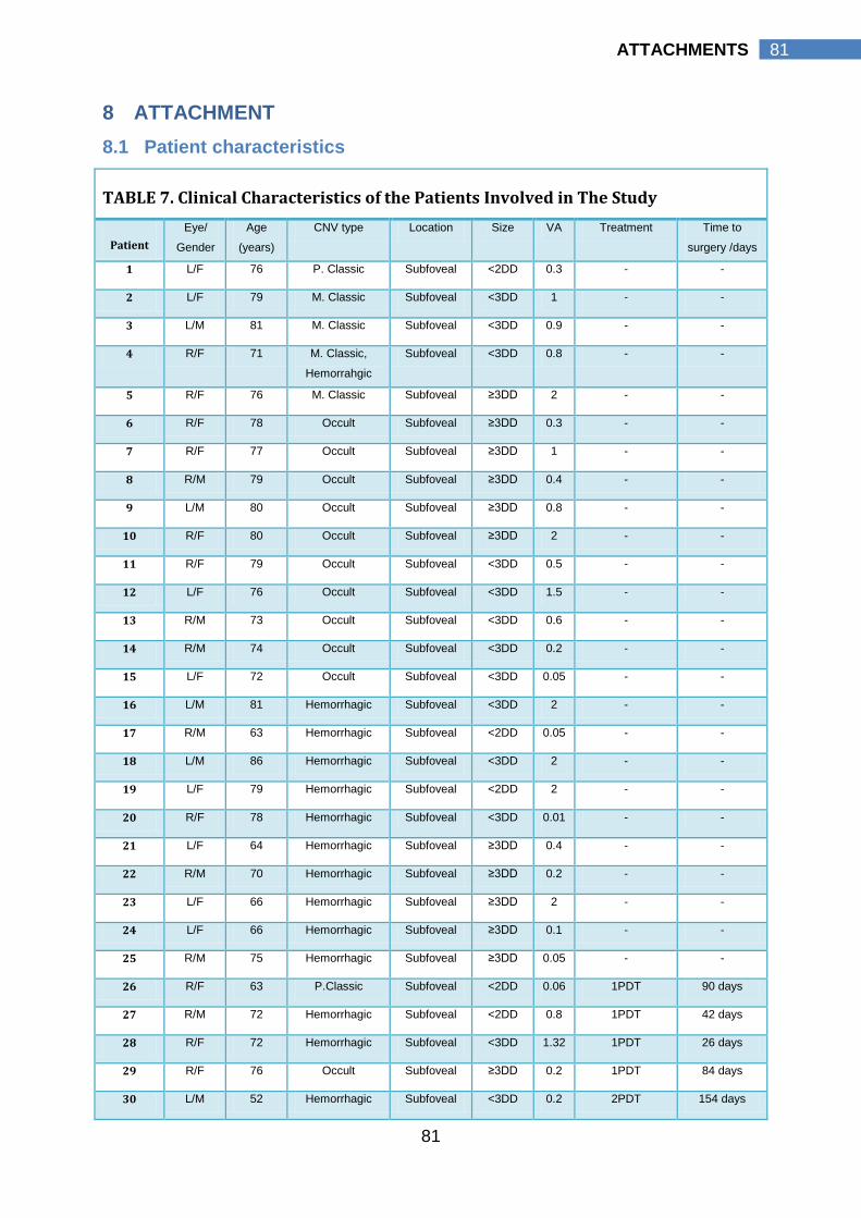
81
81 ATTACHMENTS
8 ATTACHMENT
8.1 Patient characteristics
TABLE 7. Clinical Characteristics of the Patients Involved in The Study
Patient
Eye/
Gender
Age
(years)
CNV type Location Size VA Treatment Time to
surgery /days
1 L/F 76 P. Classic Subfoveal <2DD 0.3 - -
2 L/F 79 M. Classic Subfoveal <3DD 1 - -
3 L/M 81 M. Classic Subfoveal <3DD 0.9 - -
4 R/F 71 M. Classic,
Hemorrahgic
Subfoveal <3DD 0.8 - -
5 R/F 76 M. Classic Subfoveal ≥3DD 2 - -
6 R/F 78 Occult Subfoveal ≥3DD 0.3 - -
7 R/F 77 Occult Subfoveal ≥3DD 1 - -
8 R/M 79 Occult Subfoveal ≥3DD 0.4 - -
9 L/M 80 Occult Subfoveal ≥3DD 0.8 - -
10 R/F 80 Occult Subfoveal ≥3DD 2 - -
11 R/F 79 Occult Subfoveal <3DD 0.5 - -
12 L/F 76 Occult Subfoveal <3DD 1.5 - -
13 R/M 73 Occult Subfoveal <3DD 0.6 - -
14 R/M 74 Occult Subfoveal <3DD 0.2 - -
15 L/F 72 Occult Subfoveal <3DD 0.05 - -
16 L/M 81 Hemorrhagic Subfoveal <3DD 2 - -
17 R/M 63 Hemorrhagic Subfoveal <2DD 0.05 - -
18 L/M 86 Hemorrhagic Subfoveal <3DD 2 - -
19 L/F 79 Hemorrhagic Subfoveal <2DD 2 - -
20 R/F 78 Hemorrhagic Subfoveal <3DD 0.01 - -
21 L/F 64 Hemorrhagic Subfoveal ≥3DD 0.4 - -
22 R/M 70 Hemorrhagic Subfoveal ≥3DD 0.2 - -
23 L/F 66 Hemorrhagic Subfoveal ≥3DD 2 - -
24 L/F 66 Hemorrhagic Subfoveal ≥3DD 0.1 - -
25 R/M 75 Hemorrhagic Subfoveal ≥3DD 0.05 - -
26 R/F 63 P.Classic Subfoveal <2DD 0.06 1PDT 90 days
27 R/M 72 Hemorrhagic Subfoveal <2DD 0.8 1PDT 42 days
28 R/F 72 Hemorrhagic Subfoveal <3DD 1.32 1PDT 26 days
29 R/F 76 Occult Subfoveal ≥3DD 0.2 1PDT 84 days
30 L/M 52 Hemorrhagic Subfoveal <3DD 0.2 2PDT 154 days
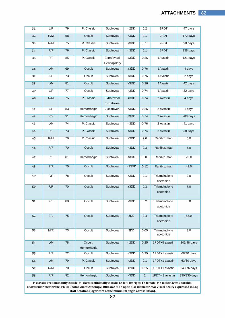
82
82 ATTACHMENTS
31 L/F 79 P. Classic Subfoveal <2DD 0.2 2PDT 47 days
32 R/M 58 Occult Subfoveal <3DD 0.1 2PDT 172 days
33 R/M 75 M. Classic Subfoveal <3DD 0.1 2PDT 90 days
34 R/F 76 P. Classic Subfoveal <3DD 0.1 2PDT 135 days
35 R/F 85 P. Classic Extrafoveal,
Peripapillary
≥3DD 0.26 1Avastin 121 days
36 L/M 69 Occult Subfoveal ≥3DD 0.76 1Avastin 4 days
37 L/F 73 Occult Subfoveal <3DD 0.76 1Avastin 2 days
38 L/M 81 Occult Subfoveal ≥3DD 0.26 1Avastin 42 days
39 L/F 77 Occult Subfoveal <3DD 0.74 1Avastin 32 days
40 R/M 75 P. Classic Extrafoveal,
Juxtafoveal
<3DD 0.74 2 Avastin 4 days
41 L/F 83 Hemorrhagic Juxtafoveal <3DD 0.26 2 Avastin 1 days
42 R/F 91 Hemorrhagic Subfoveal ≥3DD 0.74 2 Avastin 200 days
43 L/M 74 P. Classic Subfoveal <3DD 0.76 2 Avastin 41 days
44 R/F 73 P. Classic Subfoveal <3DD 0.74 2 Avastin 38 days
45 R/M 79 P. Classic Subfoveal <3DD 2.0 Ranibizumab 5.0
46 R/F 70 Occult Subfoveal <3DD 0.3 Ranibizumab 7.0
47 R/F 81 Hemorrhagic Subfoveal ≥3DD 3.0 Ranibizumab 20.0
48 R/F 70 Occult Subfoveal <33DD 0.12 Ranibizumab 42.0
49 F/R 78 Occult Subfoveal <2DD 0.1 Triamcinolone
acetonide
3.0
50 F/R 70 Occult Subfoveal ≥3DD 0.3 Triamcinolone
acetonide
7.0
51 F/L 80 Occult Subfoveal <3DD 0.2 Triamcinolone
acetonide
8.0
52 F/L 75 Occult Subfoveal 3DD 0.4 Triamcinolone
acetonide
55.0
53 M/R 73 Occult Subfoveal 3DD 0.05 Triamcinolone
acetonide
3.0
54 L/M 78 Occult,
Hemorrhagic
Subfoveal <2DD 0.25 1PDT+5 avastin 245/48 days
55 R/F 72 Occult Subfoveal <3DD 0.25 1PDT+1 avastin 68/40 days
56 L/M 79 P. Classic Subfoveal <2DD 0.1 1PDT+1 avastin 63/60 days
57 R/M 70 Occult Subfoveal <2DD 0.25 1PDT+1 avastin 240/76 days
58 R/F 92 Hemorrhagic Subfoveal ≥3DD 2 1PDT+ 2 avastin 330/330 days
P. classic: Predominantly classic; M. classic: Minimally classic; L= left; R= right; F= female; M= male; CNV= Choroidal
neovascular membrane; PDT= Photodynamic therapy; DD= size of an optic disc diameter. VA: Visual acuity expressed in Log
MAR notation (logarithm of the minimum angle of resolution).
83
83 ATTACHMENTS
8.2 Materials and stock solutions preparation protocol
8.2.1 Positive control:
Human tonsils were used as positive control for the three antibodies.
8.2.2 Washing buffer:
Tris Buffered Saline (TBS) washing buffer: 1XTBS/0.1% Tween-20 was the
washing buffer throughout the experiments.
8.2.2.1 To prepare stock solution of 10X TBS:
Add 24.2 g Trizma base and 80 g sodium chloride to 1L of
dH2O. Adjust pH to 7.6.
8.2.2.2 Working solution:
1XTBST/0.1% Tween-20: add 100ml 10XTBS to 900 ml
dH2O. Add 1 ml Tween-20 and mix well.
8.2.3 Antigen Retrieval Solution: 0.1M Sodium Citrate Buffer, pH 6.0
8.2.3.1 To prepare stock solutions:
Solution A. 0.1 M citric acid solutions: dissolve 21.0 g of
citric acid, monohydrate (C6H8O7.H2O) in 100 ml of dH2O.
Solution B. 0.1 M sodium citrate solutions: dissolve 29.4 g
trisodium citrate dihydrate (C6H5Na3O7.2H2O) in 100 ml of
dH2O.
8.2.3.2 Working solution:
Add 9 ml of Stock solution A and 41 ml of stock solution B
to 450 ml of dH2O. Adjust pH to 6.0.
8.2.4 Blocking buffer:
PBS (Dulbecco’s Phosphate Buffered Salts, 1X, catalogue #21-031-CV from
Mediatech, Inc.) + 10% goat serum
8.2.5 Primary antibody:
8.2.5.1 CD 163, Abcam, CD163 antibody [RM3/1] (ab17051)
Original concentration 0.1 mg/ml.
The primary antibody was diluted with PBS.
Prepare dilutes of 1:50, 1:100, and 1:200.
Test the prepared dilution for optimal dilution that produces the
best staining.
84
84 ATTACHMENTS
Best staining results obtained with dilution of 1:100.
8.2.5.2 CD68, Dako, FLEX Monoclonal Mouse Anti-Human CD68 Clone PG-M1
Ready-to-use monoclonal mouse antibody provided in liquid form
in a buffer containing stabilizing protein and 0.015 mol/L sodium
azide.
8.2.5.3 CCR7, (N-term) rabbit monoclonal [Y59] antibody
Recommended dilution 1:250, 1:100
The antibody was diluted with blocking buffer.
Best staining results obtained with dilution of 1:250.
8.2.5.4 Secondary antibody: Abcam, anti rabbit and mouse HRP polymer secondary
antibody (ab2891).
The secondary antibody used is ready to use prediluted, so no
need for further dilution.
8.2.6 AEC chromagen:
AEC chromagen was prepared as instructed in the
manufacture manual.
8.2.7 Phosphate Buffered Saline – PBS
1. 8 g NaCl
2. 0.2 g KCl
3. 1.44 g Na2HPO4
4. 0.24 g KH2PO4
5. 800 ml dH2O
6. pH - 7.4
7. dH2O up to 1 litter.
8.3 Staining Protocol
8.3.1 Specimen Cutting
8.3.1.1 Materials required
1. CNV specimens embedded in paraffin blocks.
2. 9 microscope glass slides.
3. Water bath.
4. Human tonsil was used as positive control.

85
85 ATTACHMENTS
8.3.1.2 Procedure
1. Serial cutting of the embedded tissues at 0.4µm thickness.
2. The cuts are transferred carefully to water bath where they are
brought to the glass slides.
3. Then the slides are left to dry at 58°C over night or at 65°C for 1
hour.
8.3.2 Deparafinization
8.3.2.1 Materials required
1. Xylene.
2. Ethanol (100%, 95%, 80%, 70%).
8.3.2.2 Procedure
1. Put the slides into a rack for IHC.
2. Dip the rack into 4 consecutive stain jars containing xylene to
remove paraffin (10 minutes).
3. Dip the rack into ethanol to remove xylene :
a. 100% ethanol 5 min.
b. 95% ethanol 5 min.
c. 80% ethanol 5 min.
d. 70% ethanol 5 min.
4. Prepare a container which is large enough to afford 2 racks. Put
the rack at one side of the container and, then, make water flow
from the other side. Don’t make the tissue side face the water
flow directly. Rinse the rack with tap water to remove ethanol for 5
minutes.
8.3.3 Washing solution
8.3.3.1 Materials required
1. TBST washing buffer: 1XTBS/0.1% Tween-20
8.3.3.2 Procedure
1. Slides are immersed for washing for 5 min.
8.3.4 Blocking of endogenous peroxidise
86
86 ATTACHMENTS
8.3.4.1 Materials required
1. 0.3%H2O2 in 0.1% sodium azide.
8.3.4.2 Procedure
1. Dip the rack in 0.3%H2O2 in 0.1% sodium azide for 10 minutes.
2. Rinse the rack with tap water for 15 minutes.
8.3.5 Antigen retrieval
8.3.5.1 Materials required
1. Sodium Citrate Buffer, pH 6.0.
2. dH2O.
8.3.5.2 Procedure
1. Place covered staining dish into the rice cooker. Add 120 mL
dH2O and press ―cook‖.
2. Place a staining dish containing Antigen Retrieval Solution
(Sodium Citrate Buffer, pH 6.0) in the rice cocker and turn it on
for 30 minutes.
3. Immerse slides into staining dish containing Antigen Retrieval
Solution (Sodium Citrate Buffer, pH 6.0).
4. When ―cook‖ is turned to ―warm‖ (about 20–30 min), unplug the
cooker and remove the staining dish to the bench top.
5. Allow to cool down, without cover, for 20 min.
8.3.6 Serum blocking
8.3.6.1 Materials required
1. Blocking solution: Goat Serum.
8.3.6.2 Procedure
1. Wash slides with TBST for 5 min on a shaker.
2. Block slides with the blocking solution for 1 hour, (don’t wash).
8.3.7 Primary antibody
8.3.7.1 Materials required
1. Chamber with a lid.
2. Marking pen.
87
87 ATTACHMENTS
3. Tissue for moistening.
4. Primary antibody.
8.3.7.2 Procedure
1. Dilute primary antibody if indicated (see under 8.1.61, 8.1.6.2
and 8.1.6.3).
2. Apply primary antibody to each section and incubate overnight
in the humidified chamber (4 ºC). Insure complete cover of the
tissue (a volume between 30µl:50µl is sufficient).
3. Wash slides three times with TBST (3 min each on a shaker).
8.3.8 Secondary antibody
8.3.8.1 Materials required
1. Secondary antibody.
8.3.8.2 Procedure
1. Apply to each section secondary HRP-conjugated
secondary anti-rabbit antibody diluted in the blocking
solution per manufacturer’s recommendation; incubate for 1
hour at room temperature.
2. Wash slides three times with TBST (3 min each on a shaker).
8.3.9 Chromagen
8.3.9.1 Materials required
1. AEC Chromagen.
2. Demineralised H2O.
8.3.9.2 Procedure
1. Add freshly prepared AEC substrate to the sections.
2. Incubate tissue sections with the substrate at room
temperature until suitable staining develops (generally 2–5
min).
3. Rinse sections with demineralised water (2min X 3).
88
88 ATTACHMENTS
8.3.10 Counter Staining
8.3.10.1 Materials required
1. Mayer’s Haematoxylin.
8.3.10.2 Procedure
1. Add haematoxylin to the sections.
2. Incubate tissue sections for 3 min.
3. Rinse sections with water for 3 min.
4. No further dehydration step is required.
8.3.11 Mounting
8.3.11.1 Materials required
1. Fluoromount™ aqueous based mounting medium.
2. Cover slips.
8.3.11.2 Procedure
1. Let the slid to adequately dry off.
2. Mount cover slips on slides using aqueous based mounting
medium.
89
89 ATTACHMENTS
8.4 Abbreviations
aaMphi Alternatively activated macrophages.
AEC 3-Amino-9-Ethyl Carbazole.
AMD Age related macular degeneration.
AREDS Age-related eye disease study research group.
bFGF Basic fibroblast growth factor, also known, FGF2 or FGF-β.
BLD Basal laminar deposit.
BMI Body mass index.
caMphi Classically activated macrophages.
C3c Complement 3c.
C5 Complement 5.
CCL2 Chemokine ligand 2.
CCL19 Chemokine ligand 19.
CCL21 Chemokine ligand 21.
CCR2 Chemokine receptor 2.
CCR7 Chemokine receptor 7.
CD68 Cluster of differentiation 68.
CD163 Cluster of differentiation 163.
CFH Complement factor H.
CNV Choroidal neovascularization, choroidal neovascular membrane.
CRP C-reactive protein.
CVD Cardiovascular diseases.
CX3CR1 Chemokine (C-X3-C motif) receptor 1.
DD Optic disc diameter.
FAZ Foveal avascular zone.
Fc Fragment crystallisable region.
FFA Fundus fluorescein angiography.
GA Geographic atrophy.
GAGs Glycosaminoglycans.
HANES The health and nutrition examination survey.
HLA Human leukocyte antigen system.
HLA‐DR Major histocompatibility complex, MHC class II.
HRP Horseradish peroxidase
iC3b iC3b is the proteolyticly inactive product of the complement cleavage
fragment C3b
IFN-ɣ Interferon gamma.
IgG Immunoglobulin G.
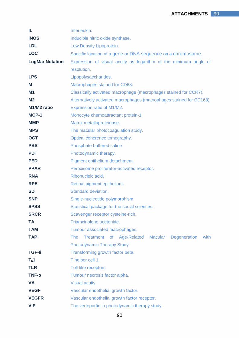
90
90 ATTACHMENTS
IL Interleukin.
iNOS Inducible nitric oxide synthase.
LDL Low Density Lipoprotein.
LOC Specific location of a gene or DNA sequence on a chromosome.
LogMar Notation Expression of visual acuity as logarithm of the minimum angle of
resolution.
LPS Lipopolysaccharides.
M Macrophages stained for CD68.
M1 Classically activated macrophage (macrophages stained for CCR7).
M2 Alternatively activated macrophages (macrophages stained for CD163).
M1/M2 ratio Expression ratio of M1/M2.
MCP-1 Monocyte chemoattractant protein-1.
MMP Matrix metalloproteinase.
MPS The macular photocoagulation study.
OCT Optical coherence tomography.
PBS Phosphate buffered saline
PDT Photodynamic therapy.
PED Pigment epithelium detachment.
PPAR Peroxisome proliferator-activated receptor.
RNA Ribonucleic acid.
RPE Retinal pigment epithelium.
SD Standard deviation.
SNP Single-nucleotide polymorphism.
SPSS Statistical package for the social sciences.
SRCR Scavenger receptor cysteine-rich.
TA Triamcinolone acetonide.
TAM Tumour associated macrophages.
TAP The Treatment of Age-Related Macular Degeneration with
Photodynamic Therapy Study.
TGF-ß Transforming growth factor beta.
Th1 T helper cell 1.
TLR Toll-like receptors.
TNF-α Tumour necrosis factor alpha.
VA Visual acuity.
VEGF Vascular endothelial growth factor.
VEGFR Vascular endothelial growth factor receptor.
VIP The verteporfin in photodynamic therapy study.
91
91 ACKNOWLEDGMENTS
9 ACKNOWLEDGMENTS
I am so grateful to Prof. Salvatore Grisanti for allowing me the opportunity to
conduct my research further at the University Eye Hospital, Lübeck. I would like to
express my deepest gratitude for his constructive and valuable suggestions. I also
appreciate his continuous motivation and friendly support throughout my work.
I would like to thank Prof. Dr. med. Hartmut Merz, Institute of Pathology, for
his beneficial help in the evaluation of the antibody concentration and positive control
choice. I would like to thank Mrs. Birgit Hüsing, Dr. Ralph Pries and Dr. Philipp
Steven, for their esteemed advice in immunohistochemical and immunofluorescent
staining technique. I also express my thanks to Dr. Martin Rudolf for his appreciated
support in imaging software and to Mrs. Christine Örün for her technical support.
I am also grateful to my colleagues at the laboratory, Dr. Matthias Lüke; Dr.
Julia Lüke; Dr. Aysegul Tura and Dr. Aizhan Tapenbayeva for always creating a
pleasant work atmosphere, for their sincere friendship and their gracious
encouragement. I also express my thanks to Mrs. Petra Hammermeister for her
immeasurable support during difficult times.
In particular I would like to express gratitude to Prof. M. Soliman, Prof. M.M.
Soliman and Prof. M. Kamal for their continuous encouragement.
I am also grateful to Dr. Ahmed El far and Mrs. Kristin Abdel Aal for their help
in reviewing of the English grammar and its usage in the thesis.
I would like thank my wife for her endless patience, unwavering support and
for her priceless advices. Finally, I would like to thank all the members of my family
for their endless support throughout this long journey.
92
92 CURRICULUM VITAE
10 CURRICULUM VITAE
10.1 PERSONAL DATA
Name Khaled Nassar
Birth date 16.10.1975
Nationality Egyptian
Marital status Married with one son.
Work address: Schleswig-Holstein University
Campus Lübeck, Clinic Eye Hospital
Department of ophthalmology
Ratzeburger Allee 160
Telephone:0049 (0)451-5004010
Fax: 0049 (0)451-5004952
Private address:
Alfstrasse18
23552 Lübeck
Deutschland
Telephone: 0049(0)4518831116
Cell phone: 0049(0)1744030067
e-mail: [email protected]
10.2 EDUCATION Date
Primary school, Monufia, Egypt 1981 1986
Prep school, Monufia, Egypt 1986 1989
High school, Monufia, Egypt 1989 1993
10.3 CERTIFICATIONS
1. Bachelor of Medicine, Bachelor of
Surgery (M.B., B.Ch.)
Cairo University, Egypt.
1.
Oct
1993
1.
Oct
1999
2. Master degree of Ophthalmology
(MSc)
Cairo University, Egypt.
1.
Mar
2000
1.
Nov
2003
3. Doctorate exam of Ophthalmology
(MD)
Cairo University, Egypt.
1.
May
2007
93
93 CURRICULUM VITAE
10.4 POST GRADUATE TRAINING AND CONFERENCES
The Wuerttemberg Ophthalmological
Association Meeting
University Hospital Tübingen
Centre for Ophthalmology, Tübingen, Germany
22.
Feb
2008
23.
Feb
2008
Pars-plana-Vitrectomy and Cataract-Surgery
Wet-Lab:
Hamburg University, Hamburg, Germany
19.
July
2008
Borsteler Autumn seminar for Pathology und
Biomedicine
Research Center Borstel, Borstel, Germany
12.
Sep
2008
13.
Sep
2008
Lübeck Ophthalmology meeting
Lübeck University, Lübeck, Germany
1.
Nov
2008
Meeting of the Vasculitis Interdiscipline
Centres
Lübeck University, Lübeck, Germany
16.
Jan
2009
12th VItreoretinal Symposium, Frankfurt-
Marburg 2009,
Johann Wolfgang Goethe University
Frankfurt/Main, Germany
27.
Aug
2009
29.
Aug
2009
Lübeck Ophthalmology meeting
Lübeck University, Lübeck, Germany
28.
Nov
2009
28.
Nov
2009
8th International Symposium on Ocular
Pharmacology and Therapeutics.
Rom, Italy.
3.
Dec
2009
6.
Dec
2009
ARVO 2010 Annual meeting: The future of
Eye and vision research.
2.
May
6.
May
94
94 CURRICULUM VITAE
Fort Lauderdale, Florida, USA 2010 2010
World Ophthalmology Congress 2010
(WOC® 2010)
Berlin, Germany
5.
June
2010
9.
June
2010
23rd International Congress of German
Ophthalmic Surgeons
Hamburg, Germany
21
Oct
2010
24
Oct
2010
10.5 POSTGRADUATE WORK
1. Research Fellow
Department of Ophthalmology
University of Lübeck, Germany
1.
May
2008
18.
Aug
2011
2. Research Fellow
Tübingen University
Department of Ophthalmology,
Vitreoretinal surgery Laboratory research
Department.
1.
Dec
2007
30.
April
2008
4. Assistant Lecturer of Ophthalmology,
Department of Ophthalmology
Fayoum University, Egypt
10.
Jan
2005
30.
Nov
2007
5. Ophthalmology Resident
Cairo University, Egypt
17.
May
2001
16.
May
2004
6. Transitional year
Cairo University, Egypt
1.
Mar
2000
28.
Feb
2001
95
95 PUBLICATION LIST
11 PUBLICATIONS LIST
11.1 Original work
1. Lüke J, Nassar K, Lüke M, Tura A, Merz H, Giannis A, Grisanti S. The Effect
of Adjuvant Dimethylenastron, a Mitotic Kinesin Eg5 Inhibitor, in Experimental
Glaucoma Filtration Surgery. Curr Eye Res. 2010 [Epub ahead of print]. IF
1.519.
2. Nassar K, El-Far E, Tatar O, Lüke J, Lüke M, Tura A, Grisanti S. Macrophage
Activation Pattern in Human Choroidal Neovascular Membranes. ARVO 2010
Annual Meeting, USA, Florida, 2010, Presentation abstract, 401/D1061.
3. Lüke M, Lüke J, Tura A, Nassar K, Grisanti S. The effects of pegaptanib
sodium on retinal function in isolated perfused vertebrate retina. Curr Eye
Res. 2010; 35:248-254. IF 1.519.
4. Beutel J, Wegner J, Wegner R, Ziemssen F, Nassar K, Rohrbach M, Hilgers,
Lüke M, Grisanti S. Possible implications of MCAM in metastasis of primary
uveal melanoma patients. Curr Eye Res, Curr Eye Res. 2009; 34:1004-1009.
IF 1.519.
5. Nassar K, Lüke J, Lüke M, Kamal M, Abd El-Nabi E, Soliman M, Rohrbach M,
Grisanti S. The novel use of decorin in prevention of proliferative
vitreoretinopathy. 8th ISOPT, Rome, 2009, Abstract: page 139.
6. Lüke J, Nassar K, Lüke M, Tura A, Giannis A, Grisanti S. The effect of
adjuvant Dimethylenastron, a mitotic kinesin Eg5 inhibitor, in experimental
glaucoma filtrating surgery. 8th ISOPT, Rome, 2009, Abstract: page 120.
11.2 Books contribution
1. Nassar K: Illustrator of Laser in Ophthalmology ―Principles Of Ophthalmic
surgery‖. In: Taha A (Editor in Chief), 1. edition, EL BLAGH, Cairo (2007)