Embed Size (px)
Citation preview

Chapter 23 Digestive System 1
Anatomy & Physiology II Dr. L. Bacha Chapter Outline (Marieb & Hoehn 2013)
Part I OVERVIEW OF THE DIGESTIVE SYSTEM
two groups of organs form the digestive system (see Fig. 23.1):
1. alimentary canal
what is it also called?
it is a continuous muscular tube that winds through the body
list the organs that make up alimentary canal:
why is food material in this tube technically outside the body?
2. accessory digestive organs list the accessory digestive organs:
Digestive Processes (see Figs. 23.2 and 23.3)
• list (and read about!) the six numbered digestive functions:
1. 2.
peristalsis is the major means of propulsion and involves what?
- what is its main effect?
3.
what does mechanical breakdown increase and what does it prepare food for?
Chapter 23 Digestive System 2
what do mechanical processes include?
- describe segmentation:
- what is the function of segmentation?
4.
involves enzymes that breakdown complex food molecules to what?
5.
the passage of substances from the lumen of the GI tract into what?
6.
Basic Functional Concepts - read and enjoy this section on basic functional concepts
Digestive System Organs: Relationships
Relationship of the Digestive Organs to the Peritoneum
most digestive organs reside in what cavity?
the abdominopelvic cavity contains the peritoneal cavity, which is lined by a serous membrane called the peritoneum:
what does the visceral peritoneum cover?
what does the parietal peritoneum line?
name the slitlike potential space between the parietal and visceral peritoneum:
Chapter 23 Digestive System 3
the serous membranes lining the peritoneal cavity continuously produce a serous fluid called peritoneal fluid
- what is the function of the peritoneal fluid in the peritoneal cavity?
what is a mesentery?
mesenteries suspend organs of the abdominopelvic cavity and attach some organs to others
what are the functions of mesenteries?
Blood Supply: The Splanchnic Circulation
what does the splanchnic circulation include?
- what does the hepatic portal circulation collect?

Chapter 23 Digestive System 4
Histology of the Alimentary Canal (see Fig. 23.6)
the walls of organs of the GI tract from the esophagus to the anal canal have the same 4 basic layers:
(1) The Mucosa ∙ it is a mucous membrane that lines the lumen of the GI tract ∙ the mucosa’s major functions are to do what?
∙ there are 3 layers within the mucosa
a. epithelium - lines the lumens of the organs; the type varies with the organ (stratified squamous or simple columnar)
b. lamina propria - areolar CT
c. muscularis mucosae – thin layer of smooth muscle
(2) The Submucosa ∙ areolar CT (3) The Muscularis Externa
∙ what is this layer responsible for?
∙ formed by either skeletal muscle (e.g. mouth, pharynx, part of the esophagus) OR smooth muscle (e.g.,
part of the esophagus, stomach, intestines)
∙ the muscularis externa is arranged in 2 layers (sometimes 3):
1) inner circular layer
2) outer longitudinal layer

Chapter 23 Digestive System 5
(4) The Serosa
serosa (= visceral peritoneum) ∙ it is the outermost layer; what is it formed of?
adventitia ∙ in some cases, the outermost layer is an adventitia rather than a serosa ∙ an adventitia is formed by CT only; not covered by a mesothelium; the CT blends with that of
surrounding structures ∙ e.g., binds the esophagus to surrounding structures in the thoracic cavity
Enteric Nervous System of the Alimentary Canal
name the neurons of the alimentary canal’s own in-house nerve supply:
- they communicate widely with one another to regulate what?
name the two major intrinsic nerve plexuses (ganglia interconnected by unmyelinated fibers) found in the walls of the alimentary canal:
what does the submucosal nerve plexus occupy?
what does the myenteric nerve plexus lie between?
enteric neuron of these plexuses provide the major nerve supply to the GI tract wall and control
what? is the enteric nervous system linked to the central nervous system?
- generally speaking, what is the effect of parasympathetic inputs?
- what is the effect of the sympathetic nervous system on digestive activities?

Chapter 23 Digestive System 6
Part II FUNCTIONAL ANATOMY OF THE DIGESTIVE SYSTEM
The Mouth and Associated Structures
The Mouth
what is the mouth also called?
what forms the superior boundary (the roof) of the mouth?
posteriorly it is continuous with what?
The Tongue
• what does the tongue occupy?
• the tongue is composed of interlacing bundles of what?
• describe the functions of the tongue:
• intrinsic muscles of the tongue
- intrinsic muscles are skeletal muscles within the tongue
- allow the tongue to do what?
• extrinsic muscles of the tongue - are skeletal muscles that originate on bones of the skull or the soft palate and insert onto the tongue - they alter the position of the tongue (protrude it, retract it, move it side to side)
• define lingual frenulum and indicate its functions:
• the upper surface and sides of the tongue are covered with a variety of projections called lingual papillae (see Fig. 23.8), some of which contain microscopic taste buds

Chapter 23 Digestive System 7
The Salivary Glands
• the salivary glands secrete components of saliva • list the functions of saliva:
• there are 3 pairs of extrinsic (major) salivary glands that secrete saliva into ducts that empty into the mouth (see Fig. 23.9):
1. parotid glands - describe the location of the parotid gland:
- each parotid duct opens into the oral cavity next to what tooth?
2. submandibular glands - the submandibular gland lies along what?
- its ducts opens into the floor of the oral cavity at the base of what?
3. sublingual glands
- the sublingual gland lies under what?
- it has numerous ducts that open into what?
Composition of Saliva
mostly water
numerous electrolytes
name the two enzymes in saliva:
also contains mucin, lysozymes, IgA antibodies, and metabolic wastes
Control of Salivation
the parasympathetic nervous system stimulates the secretion of saliva the sympathetic ns inhibits the secretion of saliva
Remember “flight-
or fight”…does your
mouth become more
moist or dry when
you are scared?

Chapter 23 Digestive System 8
The Teeth
• are located in sockets (alveoli) of what two bones?
• what is another word for chew?
Dentition and Dental Formula
2 sets of teeth in humans (see Fig. 23.10):
primary dentition - consists of deciduous teeth
permanent dentition - consists of permanent teeth describe the features and functions of the four different types of teeth:
∙ incisors -
∙ canines (cuspids) -
∙ premolars (bicuspids) and molars -
the dental formula is a shorthand way of indicating what?
- write the dental formula for the primary and permanent dentition:
Tooth Structure
locate the following parts on Fig. 23.11:
∙ crown - define the crown of a tooth:
∙ gingiva - gum
Chapter 23 Digestive System 9
∙ enamel - covers the crown of a tooth
- what makes enamel the hardest substance in the body?
∙ root
- define the root of a tooth:
∙ neck
- define the neck of a tooth:
∙ cementum - a calcified CT that covers the dentin of the root ∙ periodontal ligament
- fibrous connective tissue attached to the cementum and anchors the tooth in the alveolus ∙ name the fibrous joint that it forms:
- also acts as a shock absorber
∙ dentin - describe dentin:
∙ pulp cavity - a central cavity, surrounded by dentin, that contains pulp (soft tissue with CT, blood vessels and nerves)
∙ root canal - the extension of the pulp cavity into the root of the tooth
∙ apical foramen - an opening at the proximal end of each root - what does the apical foramen allow?
The Pharynx
the oropharynx and laryngopharynx are both common passageways for what?
it is lined by mucous membrane; has skeletal muscles that are involved in swallowing
Chapter 23 Digestive System 10
The Esophagus
describe the esophagus:
name the opening in the diaphragm through which the esophagus pierces to enter the abdomen:
some features of interest:
∙ the mucosa of the esophagus contains what type of epithelium?
∙ the superior 1/3 of the muscularis externa of the esophagus is formed by skeletal muscle; the mid portion is mixed, and the inferior 1/3 is formed by smooth muscle
∙ the circular muscle layers of smooth muscle in the muscularis externa in the superior and inferior parts of the esophagus are normally thickened and contracted, functioning as sphincters:
- the upper esophageal sphincter is between the pharynx and esophagus
gastroesophageal sphincter (lower esophageal sphincter or cardiac sphincter) between the esophagus and the stomach
Digestive Processes: Mouth to Esophagus
• list the four functions of the mouth:
• in contrast to the multifunctional mouth, the pharynx and esophagus merely serve as what?
Mastication (Chewing) ∙ mechanical breakdown of food begins with chewing in the mouth
Chapter 23 Digestive System 11
Deglutition (Swallowing)
∙ deglutition is a complicated process that involves the coordinated activity of what?
∙ list the two major phases of deglutition:
1. BUCCAL PHASE:
the buccal phase occurs where?
is this stage voluntary or involuntary?
what does the tongue do (as indicated in blue print in step 1 of Fig. 23.13)?
2. PHARYNGEAL-ESOPHAGEAL PHASE:
a. Pharyngeal Phase:
begins when the bolus passes into oropharynx
what does the soft palate do?
what does the larynx do?
what does the upper esophageal sphincter do?
- this allows food to enter the esophagus as muscles of the pharynx contract
b. Esophageal Phase:
the bolus is moved along the esophagus toward the stomach by wavelike
peristaltic contractions (see Fig. 23.13 and 23.3b)
what sphincter relaxes to allow the bolus to enter the stomach?
Chapter 23 Digestive System 12
The Stomach
the chemical breakdown of what begins in the stomach?
in the stomach, food is converted to a creamy paste called what?
the stomach lies in what quadrant of the abdominopelvic cavity?
Gross Anatomy
when empty, the mucosa and submucosa of the stomach has large, longitudinal folds called what?
list the four main regions of the stomach and note their location in Fig. 23.14:
the pyloric sphincter (pyloric valve) is between the pyloric region of the stomach and the duodenum - it controls what?
Microscopic Anatomy (see Fig. 23.15)
features of the mucosa of the stomach:
∙ the stomach is lined by simple columnar epithelial cells that produce mucus
∙ is the mucus alkaline or acidic?
∙ gastric pits
- inward extensions of the surface epithelium
cells that line the gastric pits undergo cell division to continuously replenish the epithelium of the stomach
∙ gastric glands
- gastric glands are continuations of the gastric pits lined by epithelial cells that secrete gastric juice
Types of Gland Cells
parietal cells and chief cells form the gastric glands in the fundic and body regions:
parietal cells
- what two substances do they secrete?
I know this is silly, but I remember that PARIETAL cells produce HCl because parietal ends in an “L”!
Chapter 23 Digestive System 13
- indicate the various functions of the extremely acidic contents of the stomach:
- intrinsic factor is required for what?
chief cells - what substance do chief cells produce?
- chief cells also secrete what?
gastric glands in the cardiac and pyloric regions
are mainly formed by cells that produce mucus
enteroendocrine cells are also present in gastric glands
- these are cells that produce hormones, such as histamine and gastrin
The Mucosal Barrier
What are the three factors that create this barrier?

Chapter 23 Digestive System 14
First, my summary of stimulation of HCL secretion by parietal cells in the stomach:
1. Parasympathetic nerve impulses stimulate the release of acetylcholine (ACh), which:
1. increases secretion of pepsinogen by chief cells 2. increases secretion of HCl by parietal cells 3. stimulates release of gastrin by enteroendocrine cells (G cells)
2. Gastrin produced by G cells increases gastric secretion
3. Histamine produced by mast cells stimulates the parietal cells to secrete HCl
(Note: the histamine receptors on the parietal cells are called H2 receptors, and are different from the H1 receptors of allergic reactions.)
Turn to Table 23.3 on page 885 and list the three major functions of the stomach:
Digestive Processes Occurring in the Stomach
chemical digestion in the stomach
proteins begin to be broken down by the enzyme pepsin (formed from pepsinogen, which is inactive)
- remember what cells of the gastric glands produce pepsinogen?
gastric lipases secreted by chief cells of the stomach that have a minor role in digesting lipids
what is the stomach’s only function that is essential to life?
what is intrinsic factor required for?
what is vitamin B12 needed for?
absorption in the stomach very limited absorption takes place in the stomach - e.g., some water, ions, alcohol, and aspirin
Regulation of Gastric Secretion
The production of acid and enzymes by the gastric mucosa can be controlled the nervous system and hormones:

Chapter 23 Digestive System 15
THERE ARE THREE OVERLAPPING PHASES THAT OCCUR DURING DIGESTION: Phase 1: Cephalic Phase
when does this phase occur?
what is it triggered by?
during this phase, parasympathetic nerves stimulate the salivary glands to secrete saliva and gastric
glands (via the vagus nerve) to secrete gastric juice Phase 2: Gastric Phase
begins once food reaches the stomach
neural and hormonal mechanisms regulate this phase to promote gastric secretion and motility:
Neural Regulation:
stretch receptors are stimulated by distension of the stomach by food and chemoreceptors detect an increase in pH (due to proteins entering the stomach, which buffer the acid in the stomach)
these receptors send impulses to the submucosal plexus of the stomach, where they activate the parasympathetic nervous system and enteric neurons
which send impulses to the muscularis externa and gastric glands, stimulating peristalsis and production of gastric juice
Hormonal Control of the Gastric Phase:
distention of the stomach, partially digested proteins in the stomach and an increase in pH due to food entering the stomach
(and caffeine and alcohol)
stimulate enteroendocrine cells (G cells) in the gastric mucosa to secrete Gastrin
gastrin is absorbed into the blood and circulates to the gastric glands
∙ stimulates an increase in gastric secretion by the gastric glands
∙ strengthens contraction of the lower esophagus
∙ increases motility of the stomach
∙ relaxes the pyloric sphincter to promote gastric emptying
Chapter 23 Digestive System 16
Phase 3: Intestinal Phase
begins when chyme enters the duodenum
Neural Regulation (enterogastric reflex):
distension of the duodenum by chyme stimulates stretch receptors in the duodenum
nerve impulses to the medulla oblongata, where they inhibit PS and stimulate sympathetic nerves to the stomach
∙ inhibits gastric motility ∙ increases contraction of the pyloric sphincter ∙ decreases production of gastric juice
Hormonal Regulation:
partially digested proteins and lipids in the chyme and a pH < 2 stimulates cells in the intestinal glands to secrete the hormones: secretin, cholecystokinin (CCK),and gastric inhibitory peptide (GIP)
these hormones are absorbed into the blood and circulate to gastric glands
∙ together, these hormones inhibit secretion of gastric juice and gastric emptying ∙ stimulate secretion of pancreatic juice and release of bile from the gallbladder
Regulation of Gastric Motility And Emptying
the muscularis externa of the stomach has 3 layers of what type of muscle tissue (return to page 866 under Microscopic Anatomy for this!)?
stomach contractions not only accommodate its filling and cause its emptying, but they also do what (page 873)?
observe the peristaltic waves in the stomach shown in Fig. 23.19, then name and describe the three
types as shown in blue print in that same Fig.:
(
Chapter 23 Digestive System 17
each wave of peristalsis causes a small amount of chyme to be forced through the pyloric sphincter into the duodenum (first part of the small intestine) to cause gastric emptying, so that gradually the stomach empties chyme into the duodenum
The Small Intestine and Associated Structures
The Liver ( jump to page 878)
Gross Anatomy of the Liver
the liver is the second largest organ of the body
it is shaped like a wedge and is located just inferior to the diaphragm
it has how many primary lobes in humans?
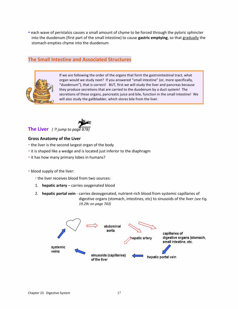
blood supply of the liver:
the liver receives blood from two sources:
1. hepatic artery – carries oxygenated blood
2. hepatic portal vein - carries deoxygenated, nutrient-rich blood from systemic capillaries of digestive organs (stomach, intestines, etc) to sinusoids of the liver (see Fig.
19.29c on page 743)
If we are following the order of the organs that form the gastrointestinal tract, what organ would we study next? If you answered “small intestine” (or, more specifically, “duodenum”), that is correct! BUT, first we will study the liver and pancreas because they produce secretions that are carried to the duodenum by a duct system! The secretions of these organs, pancreatic juice and bile, function in the small intestine! We will also study the gallbladder, which stores bile from the liver.
Chapter 23 Digestive System 18
Microscopic Anatomy of the Liver (see Fig. 23.25)
each lobe of the liver is formed by numerous microscopic lobules; a lobule consists of:
special epithelial cells called hepatocytes
heavily fenestrated sinusoids alternate with the rows of hepatocytes and radiate from the central vein in the center of a lobule
blood flows through the sinusoids from branches of the hepatic artery and hepatic portal vein
∙ hepatic macrophages (Kupffer cells) occupy the sinusoids
- what do these phagocytic cells remove?
portal triads are located in the CT where three or more lobules meet ∙ list the three basic structures that a portal triad contains:
Composition of Bile
features of bile:
1. what color is bile?
2. is alkaline (pH 7.6 to 8.6)
3. contains water, ions, cholesterol, etc. (NO digestive enzymes!)
4. contains bile salts (which are the only substances in bile with a digestive function)
a. emulsify fat globules into fat droplets
- bile salts disperse (emulsify) large clumps of fat molecules called fat globules into smaller clumps called fat droplets so they can more readily be chemically digested by enzymes
b. plays a role in the absorption of long chain fatty acids, glycerol, cholesterol, and fat soluble vitamins by forming micelles
5. contains bile pigments, such as bilirubin, which is a normal product of RBC destruction and is excreted into bile by hepatocytes

Chapter 23 Digestive System 19
duct system that carries bile ( see Fig. 23.21):
∙ bile secreted by hepatocytes enters into bile canaliculi, which are tiny intercellular canals formed by the cell membranes of adjacent hepatocytes
bile canaliculi lead into bile ducts at the periphery of lobules
bile ducts merge and eventually form the right and left hepatic ducts
the right and left hepatic ducts unite and exit the liver as the common hepatic duct
joins with the cystic duct from the gallbladder to form the common bile duct
leads into the hepatopancreatic ampulla that opens into the duodenum
The Function of the Liver: ( return to the right column on the bottom of page 878!)
hepatocytes not only can secrete or generate 900 ml of bile daily, but they can also do what?
(1)
(2)
(3)
hepatocytes also produce most plasma proteins (albumin, clotting factors, complement, etc.)
Duct System of the Liver, Gallbladder, and Pancreas
1.c. right and left hepatic ducts
2. common hepatic duct
3. cystic duct
4. common bile duct
5. opening of accessory pancreatic duct
6. pancreatic duct
7. hepatopancreatic ampulla
8. duodenal papilla

Chapter 23 Digestive System 20
The Gallbladder
• describe the appearance, size and location of the gallbladder:
• bile is carried by the duct system to the gallbladder
the gallbladder stores and concentrates bile until it is needed in the small intestine
when the small intestine is empty, a valve around the common bile duct closes and bile backs up into the cystic duct to the gallbladder for storage
The Pancreas
• exocrine secretions in pancreatic juice are carried by the pancreatic duct and the accessory pancreatic duct from the pancreas to what specific organ (see Fig. 23.21)?
∙ the exocrine part of the pancreas is formed by clusters of epithelial cells called pancreatic acini that secrete pancreatic juice (see Fig. 23.26)
• the endocrine tissues, called pancreatic islets, are scattered among the acini and contain cells that release what two important hormones?
Composition of Pancreatic Juice
consists of:
∙ water
∙ bicarbonate ions (sodium bicarbonate), which makes pancreatic juice slightly alkaline - the slightly alkaline secretion is important because the enzymes that function in the small intestine do not function in an acid environment
∙ digestive enzymes:
pancreatic amylase – digests carbohydrates (specifically starch)
pancreatic lipase – digests fats (specifically triglycerides)
nucleases - digest nucleic acids (DNA and RNA)
trypsinogen, chymotrypsinogen, procarboxypeptidase - these are inactive enzymes that, when converted to the active forms (trypsin; chymotrypsin; carboxypeptidase), they digest proteins - the protein-digesting enzymes produced in an inactive form, which protects the cells of the pancreas from digesting themselves
Chapter 23 Digestive System 21
We will write some equations for the activation of the protein-digesting enzymes in class:
The Small Intestine (back to page 875!)
Gross Anatomy
most chemical digestion and absorption of nutrients takes place in the small intestine! it has structural adaptations for these functions:
1. length - what is its length in the living person?
2. circular folds
3. villi
4. microvilli
list the three subdivisions of the small intestine:
- the ileocecal valve (sphincter) is between the ileum and the cecum of the large intestine
Microscopic Anatomy
Modifications for Absorption
most chemical digestion and absorption of nutrients takes place in the small intestine, so there are special features of the small intestine that facilitate chemical digestion and absorption of nutrients: its great length, circular folds, villi, and microvilli
• has permanent folds of the mucosa and submucosa called circular folds (Fig. 23.22 a)
- these circular folds greatly increase surface area for chemical digestion and absorption
- they cause chyme to spiral slowly through the small intestine, to increase time for digestion and absorption
• villi (singular is “villus”)
- finger-like projections of the mucosa (Fig. 23.22)
- how high are the villi?
- formed by simple columnar epithelium with goblet cells covering a core of lamina propria - the lamina propria contains blood capillaries and a lacteal (a lymphatic capillary) for absorption of
nutrients
Chapter 23 Digestive System 22
Is the pH of the chyme in the stomach acidic or alkaline? You may have been noticing that various secretions that are produced by the small intestine or that empty into the small intestine (bile and pancreatic juice) are ALKALINE. This is important to neutralize the acidic chyme that empties from the stomach into the small intestine to create a proper pH environment for the function of enzymes that act in the small intestine. They do not function in an acidic environment.
• simple columnar epithelium of the mucosa of the small intestine:
- contains simple columnar absorptive epithelial cells that have microvilli that form the brush border:
- microvilli are fingerlike projections of the cell membrane of the epithelial cells - functions of microvilli: ∙ they contain digestive enzymes called brush border enzymes produced by the epithelial cells and they increase surface area for chemical digestion
∙ they also increase surface area for absorption of nutrients into the epithelial cells
Histology of the Small Intestine Wall
- goblet cells are also present in the epithelium
- what do goblet cells produce?
• intestinal crypts (intestinal glands; crypts of Lieberkűhn) of the mucosa
- deep crevices lined with glandular epithelial cells that secrete intestinal juice
∙ intestinal juice is slightly alkaline; it is a watery mixture that contains what?
∙ intestinal juice serves as what?
- scattered through the crypt epithelium are enteroendocrine cells that secrete hormones such as
secretin and cholecystokinin
- continuously dividing stem cells at the bases of the crypts replenish the epithelium!
- intestinal glands contain Paneth cells, which fortify the small intestine’s defenses by releasing what?
• what do duodenal glands in the submucosa of the duodenum produce and what does the secretion
help to do (page 877)?
Chapter 23 Digestive System 23
Intestinal Juice: Composition and Control
the intestinal glands normally secrete what?
is intestinal juice normally slightly alkaline?
why is intestinal juice enzyme-poor?
Motility of the Small Intestine (page 886)
intestinal smooth muscle mixes chyme thoroughly with what and moves food residues through what?
in contrast to the peristaltic waves of the stomach, what is the most common motion of the small
intestine?
segmentation (see Fig. 23.3b)
- localized mixing contractions that mix chyme with the digestive juices and bring the particles of food into contact with the mucosa for absorption
peristalsis - weak, wave-like contractions that propel chyme toward the large intestine
Chemical Digestion in the Small Intestine
First, a little review of chemical digestion so far…
in the mouth - salivary amylase begins digestion of carbohydrates
in the stomach – pepsin begins digestion of proteins ; lingual lipase and gastric lipases begin digestion of lipids
so, chyme entering the small intestine contains partially digested carbohydrates, proteins and lipids

Chapter 23 Digestive System 24
chemical digestion in the small intestine is a collective effort of enzymes in pancreatic juice produced by the pancreas and brush border enzymes on the microvilli produced by the epithelial cells in the small intestine:
Digestion of Carbohydrates
pancreatic amylase - in pancreatic juice
small intestine produces: maltase, sucrase, lactase Digestion of Proteins
the pancreas produces trypsin, chymotrypsin and carboxypeptidase
small intestine produces: intestinal peptidases Digestion of Lipids
before lipids are chemically digested, bile salts emulsify fat globules into fat droplets
the pancreas produces pancreatic lipase
the small intestine produces intestinal lipase Digestion of Nucleic Acids
the pancreas produces ribonuclease and deoxyribonuclease
the small intestine produces other enzymes that also play a role in digesting DNA & RNA
A SUMMARY OF THE END PRODUCTS OF CHEMICAL DIGESTION:
carbohydrates monosaccharides
proteins amino acids and peptides
lipids fatty acids and monoglycerides
DNA and RNA sugars and nucleotides
Chapter 23 Digestive System 25
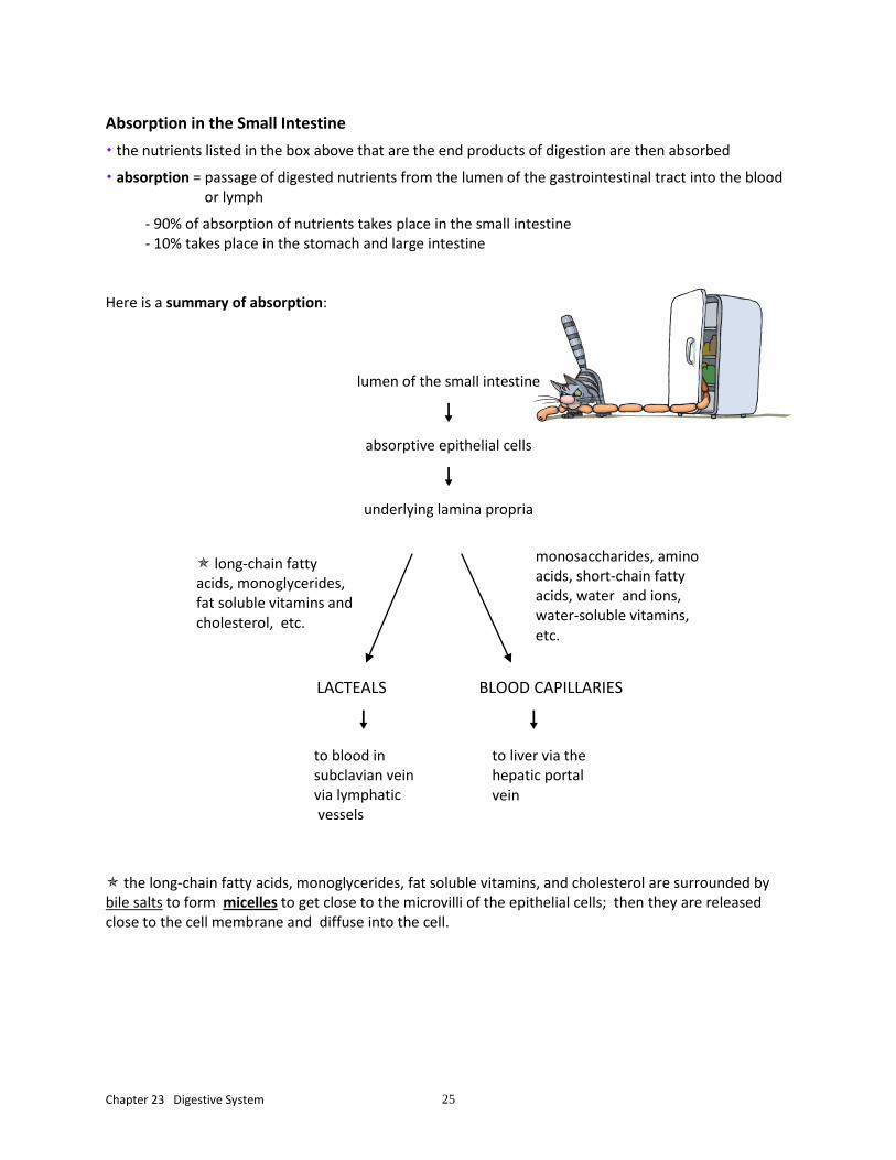
Absorption in the Small Intestine
the nutrients listed in the box above that are the end products of digestion are then absorbed
absorption = passage of digested nutrients from the lumen of the gastrointestinal tract into the blood or lymph
- 90% of absorption of nutrients takes place in the small intestine - 10% takes place in the stomach and large intestine
Here is a summary of absorption:
lumen of the small intestine
absorptive epithelial cells
underlying lamina propria
LACTEALS BLOOD CAPILLARIES
the long-chain fatty acids, monoglycerides, fat soluble vitamins, and cholesterol are surrounded by bile salts to form micelles to get close to the microvilli of the epithelial cells; then they are released close to the cell membrane and diffuse into the cell.
monosaccharides, amino acids, short-chain fatty acids, water and ions, water-soluble vitamins, etc.
long-chain fatty acids, monoglycerides, fat soluble vitamins and cholesterol, etc.
to blood in subclavian vein via lymphatic vessels
to liver via the hepatic portal vein

Chapter 23 Digestive System 26
The Large Intestine (page 887!)
the large intestine extends from what to what?
~5 ft. long, 2.5 in. in diameter contrast its length and diameter with the small intestine:
which is longer?
which is larger in diameter?
so, why do you suppose the small intestine is called “small” and the large intestine is called “large”?

Chapter 23 Digestive System 27
what are the major digestive functions of the large intestine?
Gross Anatomy
the colon has three bands of smooth muscle called what?
their tone puckers the wall of the large intestine into what?
Subdivisions of the Large Intestine
list subdivisions of the large intestine (and locate them in Fig. 23.29):
- which part is saclike?
- what does the appendix contain and what is its role?
- additionally, the appendix serves as what?
the ileocecal valve controls the passage of chyme from the ileum to the large intestine
list the four distinct regions of the colon (not counting the flexures) and note their locations in Fig. 23.29
in the pelvis, and the level of the third sacral vertebra, the sigmoid colon joints what?
define anal canal:
the anal canal opens to the body exterior at what opening?

Chapter 23 Digestive System 28
list the two sphincters of the anal canal and the type of muscles that form each, and whether each is voluntary or involuntary:
Microscopic Anatomy
the epithelium forms intestinal crypts, as in the small intestine, but there are not villi and no circular folds
does the mucosa of the large intestine produce enzymes?
the simple columnar epithelium contains numerous goblet cells that produce what?
- what are the functions of the mucus produced by the goblet cells?
Bacterial Flora
about how many discrete types of bacterial form the bacterial flora of the large intestine?!
the last stage of digestion occurs in the colon through the activity of bacteria in the colon
the bacteria in the large intestine:
∙ ferment any remaining carbohydrates and release H+ ions, CO2 and methane gas
∙ convert remaining protein to amino acid and break down amino acids
∙ convert bilirubin to stercobilin, which gives feces their brown color
∙ synthesize B vitamins and vitamin K
Digestive Processes Occurring in the Large Intestine
are digestion and absorption major functions of the large intestine?
∙ some absorption occurs in the large intestine: some water, electrolytes, some vitamins (B and K)
∙ as the chyme remains in the large intestine for 12 to 24 hours, water absorption converts it to a semisolid form called feces

Chapter 23 Digestive System 29
from Table 23.2, indicate the major functions of the large intestine?
Motility of the Large Intestine
haustral contractions:
∙ describe haustral contractions:
∙ as a haustrum fills with food residue, what happens?
∙ these movements also do what?
mass movements (mass peristalsis)
= long slow-moving, powerful waves of contraction that begin in the transverse colon and force the feces in the colon into the rectum
The Defecation Reflex
Mass movement, a strong peristaltic wave that begins in the transverse colon, drives the feces in the colon into the rectum distention of the rectum initiates the urge to defecate internal sphincter of the anus relaxes involuntarily; external sphincter of the anus relaxes voluntarily defecation
The End!