Embed Size (px)
Citation preview

Sec. F.1 - F.2 SSRCR Volume I - April 2015
F1
PART F
MEDICAL DIAGNOSTIC AND INTERVENTIONAL X-RAY AND IMAGING SYSTEMS
Sec. F.1 - Purpose and Scope. This Part establishes requirements, for which a registrant [licensee]
is responsible, for use of diagnostic and interventional x-ray equipment and imaging systems by, or
under the supervision of, an individual authorized by and licensed in accordance with State statutes
to engage in the healing arts or veterinary medicine. The provisions of this Part are in addition to,
and not in substitution for, other applicable provisions of Parts A, B, D, G J, I, X, and Z of these
regulations.
Sec. F.2 - Definitions. As used in this Part, the following definitions apply:
"Accessible surface" means the external surface of the enclosure or housing of the radiation
producing machine as provided by the manufacturer.
"Air kerma" means kerma in air (see definition of Kerma).
"Air kerma rate (AKR)" means the air kerma per unit time.
"Alert value" means a dose index (e.g., of CTDIvol(mGy) or DLP(mGy-cm)) that is set by the
registrant [licensee] to trigger an alert to the CT operator prior to scanning within an ongoing
examination. The Alert value represents a universal dose index value well above the registrant
[licensee]'s established range for the examination that warrants more stringent review and
consideration before proceeding.
"Aluminum equivalent" means the thickness of type 1100 aluminum alloy1/ affording the same
attenuation, under specified conditions, as the material in question.*/
"Articulated joint" means a joint between two separate sections of a tabletop which joint provides the
capacity of one of the sections to pivot on the line segment along which the sections join.*/
"Attenuation block" means a block or stack of type 1100 aluminum alloy, or aluminum alloy having
equivalent attenuation, with dimensions 20 centimeters (cm) or larger by 20 cm or larger by 3.8 cm,
that is large enough to intercept the entire x-ray beam.*/
"Automatic exposure control (AEC)" means a device which automatically controls one or more
technique factors in order to obtain at a preselected location(s) a required quantity of radiation.*/
"Automatic exposure rate control (AERC)" means a device which automatically controls one or
more technique factors in order to obtain, at a preselected location(s), a required quantity of radiation
per unit time.*/
1/ The nominal chemical composition of type 1100 aluminum is 99.00 percent minimum aluminum, 0.12 percent copper. */Based on Current FDA standards.

SSRCR Volume I - April 2015 Sec. F.2
F2
"Barrier" (See "Protective barrier").
"Beam axis" means a line from the source through the centers of the x-ray fields.
"Beam-limiting device" means a device which provides a means to restrict the dimensions of the x-
ray field.*/
"Bone densitometry" means a noninvasive measurement of certain physical characteristics of bone
that reflect bone strength. Test results are typically reported as bone mineral content or density and
are used for diagnosing osteoporosis, estimating fracture risk, and monitoring changes in bone
mineral content.
"Bone densitometer" means a device intended for medical purposes to measure bone density and
mineral content by x-ray or gamma ray transmission measurements through the bone and adjacent
tissues. This generic type of device may include signal analysis and display equipment, patient and
equipment supports, component parts, and accessories.*/
"C-arm fluoroscope" means a fluoroscopic x-ray system in which the image receptor and the x-ray
tube housing assembly are connected or coordinated to maintain a spatial relationship. Such a
system allows a change in the direction of the beam axis with respect to the patient without moving
the patient.*/
"Cantilevered tabletop" means a tabletop designed such that the unsupported portion can be extended
at least 100 cm beyond the support.*/
"Cassette holder" means a device, other than a spot-film device, that supports and/or fixes the
position of the image receptor during a radiographic exposure.
"Coefficient of variation (C)" means the ratio of the standard deviation to the mean value of a
population of observations. It is estimated using the following equation:
1-n
)x - x(
x
1 =
x
s = C
2
in
1=i
2/1
where:
s = Estimated standard deviation of the population.
x = Mean value of observations in sample;
xi = ith observation in sample;
n = Number of observations sampled.*/
"Computed radiography (CR; also see DR)" means a digital x-ray imaging method in which a photo-
stimulable phosphor is used to capture and store a latent image. The latent image is read out by
stimulating the phosphor with a laser. Computed radiography systems may use cassettes to house
*/ Based on Current FDA standards.

Sec. F.2 SSRCR Volume I - April 2015
F3
the phosphor, or it may be integrated into a digital radiography system.
"Computed tomography (CT)" means the production of a tomogram by the acquisition and computer
processing of x-ray transmission data.*/
"Computed tomography dose index" (CTDI) means the average absorbed dose, along the z-axis,
from a series of contiguous irradiations. It is measured from one axial CT scan (one rotation of the
x-ray tube), and is calculated by dividing the integrated absorbed dose by the nominal total beam
collimation. The scattering media for CTDI consist of two (16 and 32 cm in diameter)
polymethylmethacrylate (PMMA, e.g., acrylic or Lucite) cylinders of 14 cm length. The equation is:
Where: D(z) = the radiation dose profile along the z-axis,
N = the number of tomographic sections imaged in a single axial scan. This is
equal to the number of data channels used in a particular scan. The value of N
may be less than or equal to the maximum number of data channels available
on the system, and
T = the width of the tomographic section along the z-axis imaged by one data
channel. In multiple-detector-row (multislice) CT scanners, several detector
elements may be grouped together to form one data channel. In single-
detector-row (single-slice) CT, the z-axis collimation (T) is the nominal scan
width.
"CTDI100" means the accumulated multiple scan dose at the center of a 100-mm scan and
underestimates the accumulated dose for longer scan lengths. It is thus smaller than the equilibrium
dose. The CTDI100, requires integration of the radiation dose profile from a single axial scan over
specific integration limits. In the case of CTDI100, the integration limits are +50 mm, which
corresponds to the 100-mm length of the commercially available “pencil” ionization chamber.
CTDI100 is acquired using a 100-mm long, 3-cc active volume CT “pencil” ionization chamber and
one of the two standard CTDI acrylic phantoms (16 and 32 cm diameter) and a stationary patient
table. The equation is:
"CTDIvol "see "Volume Computed Tomography Dose Index (CTDIvol) "
"CTDIw" see "Weighted Computed Tomography Dose Index (CTDIw) "
"Cone Beam Computed Tomography (CBCT)" is a volumetric imaging modality. Volumetric data
are acquired using two dimensional digital detector arrays, and a cone-shaped x-ray beam (instead of
*/
Based on Current FDA standards.

SSRCR Volume I - April 2015 Sec. F.2
F4
fan-shaped) that rotates around the patient. Reconstruction algorithms can be used to generate
images of any desired plane.
"Control panel" means that part of the x-ray control upon which are mounted the switches, knobs,
pushbuttons, keypads, touchscreens, and other hardware necessary for manually setting the
technique factors.
"Cradle" means:
(1) A removable device which supports and may restrain a patient above an x-ray table;
or
(2) A device;
(i) Whose patient support structure is interposed between the patient and the
image receptor during normal use;
(ii) Which is equipped with means for patient restraint; and
(iii) Which is capable of rotation about its long (longitudinal) axis.*/
"CT" (See "Computed tomography").
"CT conditions of operation" means all selectable parameters governing the operation of a CT x-ray
system including nominal tomographic section thickness, filtration, and the technique factors as
defined in F.2.*/
"CT gantry" means tube housing assemblies, beam-limiting devices, detectors, and the supporting
structures, frames, and covers which hold and/or enclose these components within a computed
tomography system.*/
"CT number" means the number used to represent the x-ray attenuation associated with each
elemental area of the CT image:
where:
k = A constant, a normal value of 1,000 when the Houndsfield scale of CT
number is used;
x = Linear attenuation coefficient of the material of interest;
w = Linear attenuation coefficient of water.
"Cumulative air kerma" means the total air kerma accrued from the beginning of an examination or
procedure and includes all contributions from fluoroscopic and radiographic irradiation.
*/ Based on Current FDA standards.
CTN = k ( - )x w
w

Sec. F.2 SSRCR Volume I - April 2015
F5
"Detector" (See "Radiation detector")
"Diagnostic reference level" (DRL) is an investigational level used to identify unusually high
radiation doses or dose rates for common medical X-ray imaging procedures. DRLs are suggested
action levels above which a facility should review its methods and determine if acceptable image
quality can be achieved at lower doses. DRLs should not be applied to an individual patient.
"Diagnostic source assembly" means the tube housing assembly with a beam-limiting device
attached.*/
"Diagnostic x-ray system" means an x-ray system designed for irradiation of any part of the human
[or animal] body for the purpose of diagnosis or visualization.*/
"Digital radiography (DR)" means an x-ray imaging method (or radiography) which produces a
digital rather than analog image. DR includes both computed radiography and direct digital
radiography.
"Direct digital radiography (DDR; also see CR and DR)" means an x-ray imaging method in which a
digital sensor, usually incorporating a thin-film transistor, is used to capture an x-ray image. Some
DDR systems use a scintillator to convert x-rays to light and a photodiode array to convert light to
charge, while others use a photoconductor to convert x-rays directly to charge, which is stored on the
thin-film transistor.
"Direct scattered radiation" means that scattered radiation which has been deviated in direction only
by materials irradiated by the useful beam (See "Scattered radiation").
"Direct supervision" means a qualified practitioner must exercise general supervision and be present
in the facility and immediately available to furnish assistance and direction throughout the
performance of the procedure. It does not mean that the licensed practitioner must be present in the
room when the procedure is being performed.
"Dose" means the absorbed dose as defined by the International Commission on Radiation Units and
Measurements. The absorbed dose, D, is the quotient of de by dm, where de is the mean energy
imparted to matter of mass dm; thus D=de/dm, in units of J/kg, where the special name of the unit of
absorbed dose is gray (Gy).*/
"Dose area product (DAP) (aka kerma-area product (KAP) )" means the product of the air kerma and
the area of the irradiated field and is typically expressed in Gy-cm2, so it does not change with
distance from the x-ray tube.
"Dose length product (DLP)" means the indicator of the integrated radiation dose from a complete
CT examination. It addresses the total scan length by the formula:
DLP (mGy-cm) = CTDIvol (mGy) x scan length (cm)
"Dose profile" means the dose as a function of position along a line.*/
*/ Based on Current FDA standards.

SSRCR Volume I - April 2015 Sec. F.2
F6
"Effective dose (E)" means the sum of the tissue-weighted equivalent doses for the radiosensitive
tissues and organs of the body. It is given by the expression E = ΣT (wT HT), in which HT is the
equivalent dose in tissue or organ T and wT is the tissue weighting factor for tissue or organ T. The
unit of E and HT is joule per kilogram (J·kg–1
), with the special name sievert (Sv).
"Equipment" (See "X-ray equipment") means x-ray equipment.*/
"Exposure (X)" means the quotient of dQ by dm where dQ is the absolute value of the total charge of
the ions of one sign produced in air when all the electrons and positrons liberated or created by
photons in air of mass dm are completely stopped in air; thus X=dQ/dm, in units of C/kg. A second
meaning of exposure is the process or condition during which the x-ray tube produces x-ray
radiation.*/
"Field emission equipment" means equipment which uses an x-ray tube in which electron emission
from the cathode is due solely to the action of an electric field.*/
"Filter" means material placed in the useful beam to preferentially absorb selected radiations.
"Fluoroscopic imaging assembly" means a subsystem in which x-ray photons produce a set of
fluoroscopic images or radiographic images recorded from the fluoroscopic image receptor. It
includes the image receptor(s), electrical interlocks, if any, and structural material providing linkage
between the image receptor and diagnostic source assembly.*/
"Fluoroscopic irradiation time" means the cumulative duration during an examination or procedure
of operator-applied continuous pressure to the device, enabling x-ray tube activation in any
fluoroscopic mode of operation.*/
"Fluoroscopically-Guided Interventional (FGI) Procedures" means an interventional diagnostic or
therapeutic procedure performed via percutaneous or other access routes, usually with local
anesthesia or intravenous sedation, which uses external ionizing radiation in the form of fluoroscopy
to localize or characterize a lesion, diagnostic site, or treatment site, to monitor the procedure, and to
control and document therapy.
"Fluoroscopy" means a technique for generating x-ray images and presenting them simultaneously
and continuously as visible images. This term has the same meaning as the term “radioscopy” in the
standards of the International Electrotechnical Commission.*/
"Focal spot (actual)" means the area projected on the anode of the x-ray tube bombarded by the
electrons accelerated from the cathode and from which the useful beam originates.
"General purpose radiographic x-ray system" means any radiographic x-ray system which, by
design, is not limited to radiographic examination of specific anatomical regions.*/
"General supervision" means the procedure is performed under the overall direction and control of
the qualified practitioner but who is not required to be physically present during the performance of
the procedure.
*/ Based on Current FDA standards.

Sec. F.2 SSRCR Volume I - April 2015
F7
"Half-value layer (HVL)" means the thickness of specified material which attenuates the beam of
radiation to an extent such that the AKR is reduced by one-half of its original value. In this
definition, the contribution of all scattered radiation, other than any which might be present initially
in the beam concerned, is deemed to be excluded.*/
"Hand-held x-ray equipment" means x-ray equipment that is designed to be hand-held during
operation.
"Healing arts screening" means the testing of human beings using x-ray machines for the detection
or evaluation of health indications when such tests are not specifically and individually ordered by a
licensed practitioner of the healing arts legally authorized to prescribe such x-ray tests for the
purpose of diagnosis or treatment.
"Heat unit" means a unit of energy equal to the product of the peak kilovoltage, milliamperes, and
seconds, i.e., kVp x mA x second.
"HVL" (See "Half-value layer").
"Image intensifier" means a device, installed in its housing, which instantaneously converts an x-ray
pattern into a corresponding light image of higher intensity.*/
"Image receptor" means any device, such as a fluorescent screen, radiographic film, x-ray image
intensifier tube, solid-state detector, or gaseous detector which transforms incident x-ray photons
either into a visible image or into another form which can be made into a visible image by further
transformations. In those cases where means are provided to preselect a portion of the image
receptor, the term “image receptor” shall mean the preselected portion of the device.*/
"Irradiation" means the exposure of matter to ionizing radiation.
"Isocenter" means the center of the smallest sphere through which the beam axis passes when the
equipment moves through a full range of rotations about its common center.*/
"Kerma" means the quantity defined by the International Commission on Radiation Units and
Measurements. The kerma, K, is the quotient of dEtr by dm, where dEtr is the sum of the initial
kinetic energies of all the charged particles liberated by uncharged particles in a mass dm of
material; thus K=dEtr/dm, in units of J/kg, where the special name for the unit of kerma is gray (Gy).
When the material is air, the quantity is referred to as "air kerma." */
"Kerma-area product (KAP) " (See "dose area product")
"Kilovolts peak" (See "Peak tube potential").
"kV" means kilovolts.
"kVp" (See "Peak tube potential").
"kWs" means kilowatt second.
*/ Based on Current FDA standards.

SSRCR Volume I - April 2015 Sec. F.2
F8
"Last-image hold (LIH) radiograph" means an image obtained either by retaining one or more
fluoroscopic images, which may be temporarily integrated, at the end of a fluoroscopic exposure or
by initiating a separate and distinct radiographic exposure automatically and immediately in
conjunction with termination of the fluoroscopic exposure.*/
"Lead equivalent" means the thickness of lead affording the same attenuation, under specified
conditions, as the material in question.
"Leakage radiation" means radiation emanating from the diagnostic source assembly except for:
(1) The useful beam; and
(2) Radiation produced when the exposure switch or timer is not activated.*/
"Leakage technique factors" means the technique factors associated with the diagnostic source
assembly which are used in measuring leakage radiation. They are defined as follows:
(1) For diagnostic source assemblies intended for capacitor energy storage equipment,
the maximum-rated peak tube potential and the maximum-rated number of exposures
in an hour for operation at the maximum-rated peak tube potential with the quantity
of charge per exposure being 10 millicoulombs (or 10 mAs) or the minimum
obtainable from the unit, whichever is larger;
(2) For diagnostic source assemblies intended for field emission equipment rated for
pulsed operation, the maximum-rated peak tube potential and the maximum-rated
number of x-ray pulses in an hour for operation at the maximum-rated peak tube
potential; and
(3) For all other diagnostic source assemblies, the maximum-rated peak tube potential
and the maximum-rated continuous tube current for the maximum-rated peak tube
potential.*/
"Light field" means that area of the intersection of the light beam from the beam-limiting device and
one of the set of planes parallel to and including the plane of the image receptor, whose perimeter is
the locus of points at which the illumination is one-fourth of the maximum in the intersection.*/
"Line-voltage regulation" means the difference between the no-load and the load line potentials
expressed as a percent of the load line potential; that is,
Percent line-voltage regulation = 100 (Vn-Vl)/Vl
where:
Vn = No-load line potential; and
Vl = Load line potential.*/
"mA" means milliampere.
*/ Based on Current FDA standards.

Sec. F.2 SSRCR Volume I - April 2015
F9
"mAs" means milliampere second.
"Medical event" means one or more of the following criteria have occurred:
a. Unintended skin dose to the same area in a single procedure greater than 2 Gy (200 rad);
b. Unintended dose other than skin dose in a single procedure greater than:
i. 5 times the facility’s established protocol, and > 0.5 Gy (50 rad) to any organ, or
ii. 5 times the facility’s established protocol, and > 0.05 Sv (5 rem) effective dose;
c. Wrong patient or wrong site for entire procedure when the resultant dose is:
i. Dose > 0.5 Gy (50 rad) to any organ or,
ii Effective dose ≥ 0.05 Sv (5 rem).
"Mobile x-ray equipment" (See "X-ray equipment").
"Mode of operation" means, for fluoroscopic systems, a distinct method of fluoroscopy or
radiography provided by the manufacturer and selected with a set of several technique factors or
other control settings uniquely associated with the mode. The set of distinct technique factors and
control settings for the mode may be selected by the operation of a single control. Examples of
distinct modes of operation include normal fluoroscopy (analog or digital), high-level control
fluoroscopy, cineradiography (analog and digital), digital subtraction angiography, electronic
radiography using the fluoroscopic image receptor, and photospot recording. In a specific mode of
operation, certain system variables affecting kerma, AKR, or image quality, such as image
magnification, x-ray field size, pulse rate, pulse duration, number of pulses, source-image receptor
distance (SID), or optical aperture, may be adjustable or may vary; their variation per se does not
comprise a mode of operation different from the one that has been selected.*/
"Multiple tomogram system" means a computed tomography x-ray system which obtains x-ray
transmission data simultaneously during a single scan to produce more than one tomogram.*/
"Noise" in CT means the standard deviation of the fluctuations in CT number expressed as a
percentage of the attenuation coefficient of water. Its estimate (Sn) is calculated using the following
expression:
n
w
S = 100 CS s
where:
CS = Linear attenuation coefficient of the material of interest.
w = Linear attenuation coefficient of water.
s = Estimated [S]standard deviation of the CT numbers of picture elements in a specified
area of the CT image.*/
*/ Based on Current FDA standards.
SSRCR Volume I - April 2015 Sec. F.2
F10
"Nominal tomographic section thickness" means the full width at half-maximum of the sensitivity
profile taken at the center of the cross-sectional volume over which x-ray transmission data are
collected.*/
"Notification value" means a protocol-specific dose index (e.g. CTDIvol(mGy) or of DLP(mGy-cm))
that is set by the registrant [licensee] to trigger a notification to the CT operator prior to scanning
when the dose index exceeds the established range for the examination.
"Patient" means an individual or animal subjected to healing arts examination, diagnosis or
treatment.
"Picture element" means an elemental area of a tomogram.*/
"PBL" See "Positive beam limitation."
"Peak tube potential" means the maximum value of the potential difference across the x-ray tube
during an exposure.*/
"Personal supervision" means a qualified practitioner must exercise General Supervision and be
present in the room or adjacent control area during the performance of the procedure.
"Phantom" means a volume of material behaving in a manner similar to tissue with respect to the
attenuation and scattering of radiation. This requires that both the atomic number (Z) and the
density of the material be similar to that of tissue.
"Photostimulable storage phosphor (PSP)" means a material used to capture and store radiographic
images in computed radiography systems.
"PID" (See "Position indicating device").
"Pitch" means the table incrementation, in CT, per x-ray tube rotation, divided by the nominal x-ray
beam width at isocenter.
"Portable x-ray equipment" (See "X-ray equipment").
"Position indicating device (PID)" means a device on dental x-ray equipment used to indicate the
beam position and to establish a definite source-surface (skin) distance. It may or may not
incorporate or serve as a beam-limiting device.
"Positive beam limitation" means the automatic or semi-automatic adjustment of an x-ray beam to
the size of the selected image receptor, whereby exposures cannot be made without such adjustment.
"Primary protective barrier" means the material, excluding filters, placed in the useful beam to
reduce the radiation exposure [beyond the patient and cassette holder] for protection purposes.*/
*/ Based on Current FDA standards.

Sec. F.2 SSRCR Volume I - April 2015
F11
"Protective apron" means an apron made of radiation absorbing materials used to reduce radiation
exposure.
"Protocol" means a collection of settings and parameters that fully describe an examination.
"Pulsed mode" means operation of the x-ray system such that the x-ray tube current is pulsed by the
x-ray control to produce one or more exposure intervals of duration less than one-half second.*/
"Qualified Expert (QE)" means an individual who is granted professional privileges based on
education and experience to provide clinical services in diagnostic medical physics by the Agency.
"Quality Assurance" means a program providing for verification by written procedures such as
testing, auditing, and inspection to ensure that deficiencies, deviations, defective equipment, or
unsafe practices, or a combination thereof, relating to the use, disposal, management, or manufacture
of radiation devices are identified, promptly corrected, and reported to the appropriate regulatory
authorities as required.
"Qualified medical physicist (QMP) " means an individual who meets each of the following
credentials:
1. Has earned a master's and/or doctoral degree in physics, medical physics, biophysics,
radiological physics, medical health physics, or equivalent disciplines from an accredited
college or university; and
2. Has been granted certification in the specific subfield(s) of medical physics with its
associated medical health physics aspects by an appropriate national certifying body and
abides by the certifying body's requirements for continuing education;
"Radiation detector" means a device which in the presence of radiation provides a signal or other
indication suitable for use in measuring one or more quantities of incident radiation.
"Radiation Protocol Committee (RPC)" means the representative group of qualified individuals in a
CT or FGI facility responsible for the ongoing review and management of CT or FGI protocols to
ensure that exams being performed achieve the desired diagnostic image quality at the lowest
radiation dose possible while properly exploiting the capabilities of the equipment being used.
"Radiation therapy simulation system" means a radiographic or fluoroscopic x-ray system intended
for localizing the volume to be exposed during radiation therapy and confirming the position and
size of the therapeutic irradiation field.*/
"Radiograph" means an image receptor on which the image is created directly or indirectly by an x-
ray pattern and results in a permanent record.
"Radiography" means a technique for generating and recording an x-ray pattern for the purpose of
providing the user with an image(s) after termination of the exposure.*/
"Recording" means producing a retrievable form of an image resulting from x-ray photons.*/

SSRCR Volume I - April 2015 Sec. F.2
F12
"Reference plane" means a plane which parallel to and which can be offset (as specified in
manufacturer information provided to users) from the location of the tomographic plane(s).
"Scan" means the complete process of collecting x-ray transmission data for the production of a
tomogram. Data may be collected simultaneously during a single scan for the production of one or
more tomograms.*/
"Scan increment" means the amount of relative displacement of the patient with respect to the CT x-
ray system between successive scans measured along the direction of such displacement.*/
"Scan sequence" means a pre-selected set of two or more scans performed consecutively under pre-
selected CT conditions of operation.*/
"Scan time" means the time elapsed during the accumlation of x-ray transmission data for a single
scan.*/
"Scattered radiation" means radiation that, during passage through matter, has been deviated in
direction (See "Direct scattered radiation").
"Sensitivity profile" means the relative response of the CT x-ray system as a function of position
along a line perpendicular to the tomographic plane.*/
"Single tomogram system" means a CT x-ray system which obtains x-ray transmission data during a
scan to produce a single tomogram.*/
"Shutter" means a device attached to the tube housing assembly which can intercept the entire cross
sectional area of the useful beam and which has a lead equivalency not less than that of the tube
housing assembly.
"SID" (See "Source-image receptor distance").
"Size-specific dose estimate (SSDE)" means a patient dose estimate which takes into consideration
corrections based on the size of the patient, using linear dimensions measured on the patient or
patient images.
"Source" means the focal spot of the x-ray tube.*/
"Source-image receptor distance" means the distance from the source to the center of the input
surface of the image receptor.
"Source-skin distance (SSD)" means the distance from the source to the center of the entrant x-ray
field in the plane tangent to the patient skin surface.*/
"Spot-film"2/ means a radiograph which is made during a fluoroscopic examination to permanently
record conditions which exist during that fluoroscopic procedure.*/
*/ Based on Current FDA standards. 2/ Digital image receptors used in place of film with spot-film devices should be considered "spot-film".

Sec. F.2 SSRCR Volume I - April 2015
F13
"Spot-film device" means a device intended to transport and/or position a radiographic image
receptor between the x-ray source and fluoroscopic image receptor. It includes a device intended to
hold a cassette over the input end of the fluoroscopic image receptor for the purpose of producing a
radiograph.*/
"Stationary x-ray equipment" (See "X-ray equipment").
"Stray radiation" means the sum of leakage and scattered radiation.
"Substantial radiation dose level" (SRDL) means an appropriately-selected dose used to trigger
additional dose-management actions during a procedure and medical follow-up for a radiation level
that might produce a clinically-relevant injury in an average patient.
"Technique factors" means the following conditions of operation:
(1) For capacitor energy storage equipment, peak tube potential in kilovolts (kV) and
quantity of charge in milliampere-seconds (mAs);
(2) For field emission equipment rated for pulsed operation, peak tube potential in kV,
and number of x-ray pulses;
(3) For CT equipment designed for pulsed operation, peak tube potential in kV, scan time
in seconds, and either tube current in milliamperes (mA), x-ray pulse width in
seconds, and the number of x-ray pulses per scan, or the product of tube current, x-ray
pulse width, and the number of x-ray pulses in mAs;
(4) For CT equipment not designed for pulsed operation, peak tube potential in kV, and
either tube current in mA and scan time in seconds, or the product of tube current and
exposure time in mAs and the scan time when the scan time and exposure time are
equivalent; and
(5) For all other equipment, peak tube potential in kV, and either tube current in mA and
exposure time in seconds, or the product of tube current and exposure time in mAs.*/
"Tomogram" means the depiction of the x-ray attenuation properties of a section through the body.*/
"Tomographic plane" means that geometric plane which the manufacturer identified as
corresponding to the output tomogram.*/
"Tomographic section" means the volume of an object whose x-ray attenuation properties are
imaged in a tomogram.
"Tube" means an x-ray tube, unless otherwise specified.*/
*/ Based on Current FDA standards.

SSRCR Volume I - April 2015 Sec. F.2
F14
"Tube housing assembly" means the tube housing with tube installed. It includes high-voltage
and/or filament transformers and other appropriate elements when such are contained within the tube
housing.*/
"Unintended" radiation dose in diagnostic or interventional x-ray means a patient radiation dose
resulting from a human error or equipment malfunction during the procedure.
"Useful beam" means the radiation which passes through the tube housing port and the aperture of
the beam limiting device when the exposure switch or timer is activated.*/
"Visible area" means that portion of the input surface of the image receptor over which incident x-
ray photons are producing a visible image.*/
"Volume Computed Tomography Dose Index (CTDIvol)" means a radiation dose parameter derived
from the CTDIw (weighted or average CTDI given across the field of view). The formula is:
CTDIvol = (N)(T)(CTDIw)/I, where
N = number of simultaneous axial scans per x-ray source rotation,
T = thickness of one axial scan (mm), and
I = table increment per axial scan (mm).
Thus,
CTDIvol = CTDIw / pitch
"Weighted Computed Tomography Dose Index (CTDIw)" means the estimated average CTDI100
across the field of view (FOV). The equation is:
Where 1/3 and 2/3 approximate the relative areas represented by the center and edge
values derived using the 16 or 32 cm acrylic phantom. CTDIw uses CTDI100 and an f-
factor for air (0.87 rad/R or 1.0 mGy/mGy).
"X-ray control" means a device which controls input power to the x-ray high-voltage generator
and/or the x-ray tube. It includes equipment such as timers, phototimers, automatic brightness
stabilizers, and similar devices, which control the technique factors of an x-ray exposure.*/
"X-ray exposure control" means a device, switch, button or other similar means by which an
operator initiates and/or terminates the radiation exposure. The x-ray exposure control may include
such associated equipment as timers and back-up timers.
"X-ray equipment" means an x-ray system, subsystem, or component thereof. Types of x-ray
equipment are as follows:
(1) "Mobile x-ray equipment" means x-ray equipment mounted on a permanent base with

Sec. F.2 – F.3 SSRCR Volume I - April 2015
F15
wheels and/or casters for moving while completely assembled;
(2) "Portable x-ray equipment" means x-ray equipment designed to be hand-carried; and
(3) "Stationary x-ray equipment" means x-ray equipment which is installed in a fixed
location.
(4) "Hand-held x-ray equipment" means x-ray equipment that is designed to be hand-held
during operation.
"X-ray field" means that area of the intersection of the useful beam and any one of a set of planes
parallel to and including the plane of the image receptor, whose perimeter is the locus of points at
which the AKR is one-fourth of the maximum in the intersection.
"X-ray high-voltage generator" means a device which transforms electrical energy from the potential
supplied by the x-ray control to the tube operating potential. The device may also include means for
transforming alternating current to direct current, filament transformers for the x-ray tube(s), high-
voltage switches, electrical protective devices, and other appropriate elements.*/
"X-ray system" means an assemblage of components for the controlled production of x-rays. It
includes minimally an x-ray high-voltage generator, an x-ray control, a tube housing assembly, a
beam-limiting device, and the necessary supporting structures. Additional components which
function with the system are considered integral parts of the system.*/
"X-ray table" means a patient support device with its patient support structure (tabletop) interposed
between the patient and the image receptor during radiography and/or fluoroscopy. This includes,
but is not limited to, any stretcher equipped with a radiolucent panel and any table equipped with a
cassette tray (or bucky), cassette tunnel, fluoroscopic image receptor, or spot-film device beneath the
tabletop.
"X-ray tube" means any electron tube which is designed for the conversion of electrical energy into
x-ray energy.*/
Sec. F.3 - General and Administrative Requirements.
a. Radiation Safety Requirements. The registrant [licensee] shall be responsible for directing
the operation of the x-ray system(s) under his or her administrative control and shall assure
that the requirements of these regulations are met in the operation of the x-ray system(s).
i. The registrant [licensee] shall have a radiation safety program. The radiation safety
program shall include but not be limited to the following:
(1) The use of ionizing radiation within its purview is performed in accordance
with existing laws and regulations.
(2) All persons are protected as required by Part D, Standards for Protection
Against Radiation, of these regulations.
*/ Based on Current FDA standards.

SSRCR Volume I - April 2015 Sec. F.3
F16
(3) Upon discovery of a medical event, the registrant [licensee] shall:
(i) Contact the Agency regarding the medical event within one
business day;
(ii) Provide a written report, including the analysis of the medical
event, by a QMP [QE] to the Agency within 15 business days;
(iii) Provide a clinical summary to the prescribing physician and
patient within 15 business days; and
(iv) Maintain record of the medical event as part of the patient's
permanent medical record.
ii. An x-ray system which does not meet the provisions of these regulations shall not be
operated for diagnostic purposes unless the Agency or a QMP [QE] determines that
the non-compliance shall not pose a significant radiation risk or significantly affect
image quality, and arrangements have been made to correct the non-compliance
within 30 days.
iii. The QMP [QE], if required in this Part, shall complete initial and routine compliance
evaluations following nationally recognized procedures or those recognized by the
Agency. These evaluations shall include a review of the required QC tests.
iv. All x-ray equipment shall be installed and used in accordance with the equipment
manufacturer’s specifications.
v. Individuals operating the x-ray systems shall meet the qualifications required by the
Agency.
vi. A sufficient number of protective apparel (e.g., aprons, gloves, collars) and shields
shall be available to provide the necessary radiation protection for all patients and
personnel who are involved with x-ray operations.
vii. All protective apparel and auxiliary shields shall be evaluated annually for integrity
and clearly labeled with their lead equivalence.
viii. Each registrant [licensee] shall have a mechanism in place for the referring physician
to access information on selecting the most appropriate diagnostic procedure to
answer the clinical question.
ix. Nationally recognized diagnostic reference levels (DRLs) shall be utilized when
applicable.
x. The registrant [licensee] shall use auxiliary equipment designed to minimize patient
and personnel exposure commensurate with the needed diagnostic information.

Sec. F.3 SSRCR Volume I - April 2015
F17
xi. Portable or mobile x-ray equipment shall be used only for examinations where it is
impractical to transfer the patient to a stationary x-ray installation.
xii. Neither the x-ray tube housing nor the collimating device shall be held during an
exposure. Exceptions are allowed for [Agency approved] devices specifically
designed to be hand-held.
xiii. The useful x-ray beam shall be limited to the area of clinical interest.
xiv. Consideration shall be given to selecting the appropriate technique and employing
available dose reduction methods and technologies across all patient sizes and clinical
indications.
xv. A facility shall have a documented procedure in place for verification of patient
identity and exam to be performed, including identification of the appropriate body
part.
xvi. For general radiographic systems not equipped with an operational anatomic
programming option, protocols shall be documented and readily available to the
operator. At a minimum, these protocols shall include:
(1) Patient's (adult and pediatric, if appropriate) body part and anatomical size
(2) Technique factors
(3) Type of image receptor used
(4) Source to image receptor distance used (except for dental intraoral
radiography)
(5) Type of grid, if any.
xvii. The registrant [licensee] shall create and make available to x-ray operators written
safety procedures, including instructions for patient holding and any restrictions of
the operating technique required for the safe operation of the particular x-ray system.
The operator shall be able to demonstrate familiarity with these procedures.
xviii. The registrant [licensee] shall restrict the presence of individuals in the immediate
area of the patient being examined to those required or in training for the medical
procedure, or the parent or guardian of a patient while the x-ray tube is energized.
The following applies to all individuals, other than the patient being examined:
(1) All persons shall be positioned such that no part of the body will be struck by
the useful beam unless protected by not less than 0.5 millimeter lead
equivalent material;
(2) All persons shall be protected from the secondary radiation by protective
garments or whole body protective barriers of not less than 0.25 millimeter
lead equivalent material;

SSRCR Volume I - April 2015 Sec. F.3
F18
(3) Instances may warrant having human patients other than the one being
examined in the room during the exam. If the procedure results in scatter
radiation in excess of 0.02 mSv (2 mR) in any one hour at the position of
these patients, they shall be protected from the direct scatter radiation by
whole body protective barriers of not less than 0.25 millimeter lead equivalent
material or shall be positioned so that the 0.02 mSv (2 mR) in any one hour
limit is met.
xix. Individuals shall not be exposed to the useful beam except for healing arts purposes
and unless such exposure has been authorized by a licensed practitioner of the healing
arts. This provision specifically prohibits deliberate exposure for the following
purposes:
(1) Exposure of an individual for training, demonstration, or other non-healing
arts purposes; and
(2) Exposure of an individual for the purpose of healing arts screening except as
authorized by the Agency.
xx. In cases where a patient or image receptor must be provided with auxiliary support,
mechanical support devices shall be used whenever possible. If a patient or image
receptor must be provided with auxiliary support during a radiation exposure:
(1) Written safety procedures, as required by F.3a.xv., shall indicate the
requirements for selecting a holder and the procedure the holder shall follow;
(2) The human holder shall be instructed in personal radiation safety and
protected as required by F.3a.xvi.;
(3) No individual shall be used routinely to hold the image receptor or patient
during a radiation exposure;
(4) In those cases where the patient must hold the image receptor, except during
intraoral examinations, any portion of the body other than the area of clinical
interest struck by the useful beam shall be protected by not less than
0.5 millimeter lead equivalent material.
xxi. All individuals who are associated with the operation of an x-ray system are subject
to the requirements of Part D of these regulations.
xxii. Healing Arts Screening. Any person proposing to conduct a healing arts screening
program shall not initiate such a program without prior approval of the Agency.
When requesting such approval, that person shall submit the information outlined in
Appendix A of this Part. If any information submitted to the Agency becomes invalid
or outdated, the Agency shall be immediately notified. FDA/MQSA-certified
facilities are registered with the Agency for the use of dedicated mammographic
equipment to conduct mammography screening.

Sec. F.3 SSRCR Volume I – April 2015
F19
xxiiii. Maintenance of Records. The registrant [licensee] shall maintain the following
information on each x-ray system for inspection by the Agency for a minimum of 5
years or as noted below:
(1) Model and serial numbers of all major components, and user's manuals for
those components, including software, shall be maintained for the life of the
system.
(2) Records of surveys, calibrations, maintenance, and modifications (e.g., major
software and hardware upgrades) performed on the x-ray system(s); and
(3) A copy of all correspondence with the Agency regarding the x-ray system.
xiv. X-Ray Utilization Record. Each facility shall maintain a record containing the
patient's name, the type of examinations, and the dates the examinations were
performed.
b. Quality Assurance.
i. The registrant [licensee] shall establish and maintain a quality assurance (QA)
program. In addition to the standards in the modality specific sections, the registrant
[licensee] shall:
(1) Maintain documentation of minimum qualifications for practitioners, medical
physicists, and x-ray equipment operators.
(2) Designate an appropriately trained individual to manage the QA program.
(3) Establish and maintain written QA and quality control (QC) procedures,
including evaluation frequencies and tolerances.
(4) Check each study for artifacts. If an artifact is present, the source shall be
identified and appropriate action taken.
(5) Perform repeat / reject analysis of radiographic images at least quarterly
following specifications of a nationally recognized organization.
(6) Complete preventative maintenance on the x-ray systems in accordance with
manufacturer specifications at intervals not to exceed 12 months.
(7) Maintain documentation showing the testing instruments used in determining
compliance with the provisions of this section are properly calibrated and
maintained in accordance with the Agency minimum standard or accepted
professional standards when no Agency minimum is defined.
(8) Complete and document an annual review of the QA program.

SSRCR Volume I - April 2015 Sec. F.3
F20
(9) Retain QA/QC records of evaluations and reviews in accordance with state
statutes, regulations, but in no case less than three years.
ii. X-Ray Film Processing Facilities. A registrant [licensee] using analog image
receptors (e.g. radiographic film) shall have available suitable equipment for handling
and processing radiographic film in accordance with the following provisions:
(1) Manually developed film:
(a) Processing tanks shall be constructed of mechanically rigid, corrosion
resistant material; and
(b) Developing solutions shall be prepared, replenished, and replaced
following manufacturer recommendations.
(c) The temperature of solutions in the tanks shall be maintained within
the range of 60o
F to 80o
F (16o
C to 27o
C). Film shall be developed in
accordance with the time-temperature relationships recommended by
the film manufacturer, or, in the absence of such recommendations,
with the following time-temperature chart:
Manual Film Developing Technique Chart
Developer
Temperature
°C / °F
Developing
Time
(Minutes)
Developer
Temperature
°C / °F
Developing
Time
(Minutes)
26.7 / 80 2.0 20.6 / 69 4.5
26.1 / 79 2.0 20.0 / 68 5.0
25.6 / 78 2.5 19.4 / 67 5.5
25.0 / 77 2.5 18.9 / 66 5.5
24.4 / 76 3.0 18.3 / 65 6.0
23.9 / 75 3.0 17.8 / 64 6.5
23.3 / 74 3.5 17.2 / 63 7.0
22.8 / 73 3.5 16.7 / 62 8.0
22.2 / 72 4.0 16.1 / 61 8.5
21.7 / 71 4.0 15.6 / 60 9.5
21.1 / 70 4.5
(d) Devices shall be utilized which will indicate the actual temperature of
the developer solution and signal the passage of a preset time.
(2) Automatic processors and other closed processing systems:
(a) Automatic processors shall be operated and maintained following
Sec. F.3 SSRCR Volume I – April 2015
F21
manufacturer specifications.
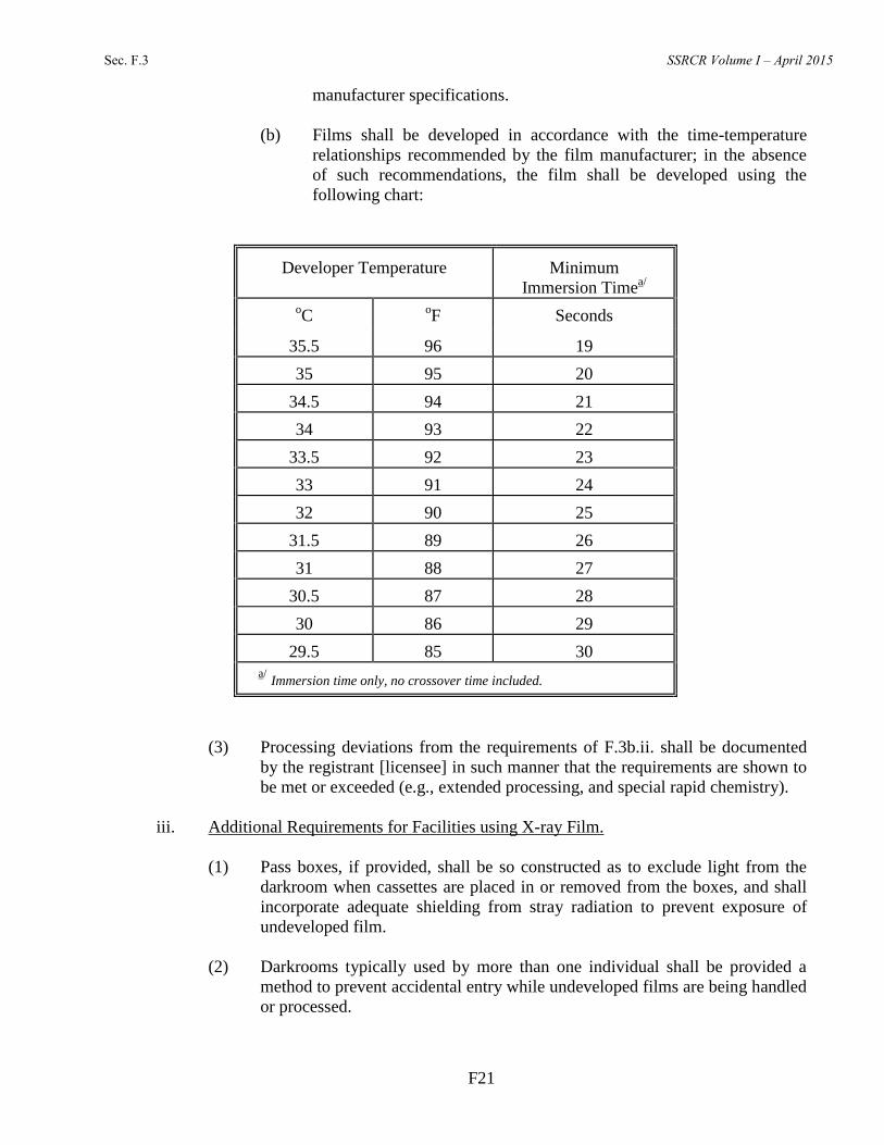
(b) Films shall be developed in accordance with the time-temperature
relationships recommended by the film manufacturer; in the absence
of such recommendations, the film shall be developed using the
following chart:
Developer Temperature
Minimum
Immersion Timea/
oC
oF
Seconds
35.5
96
19
35
95
20
34.5
94
21
34
93
22
33.5
92
23
33
91
24
32
90
25
31.5
89
26
31
88
27
30.5
87
28
30
86
29
29.5
85
30
a/ Immersion time only, no crossover time included.
(3) Processing deviations from the requirements of F.3b.ii. shall be documented
by the registrant [licensee] in such manner that the requirements are shown to
be met or exceeded (e.g., extended processing, and special rapid chemistry).
iii. Additional Requirements for Facilities using X-ray Film.
(1) Pass boxes, if provided, shall be so constructed as to exclude light from the
darkroom when cassettes are placed in or removed from the boxes, and shall
incorporate adequate shielding from stray radiation to prevent exposure of
undeveloped film.
(2) Darkrooms typically used by more than one individual shall be provided a
method to prevent accidental entry while undeveloped films are being handled
or processed.

SSRCR Volume I - April 2015 Sec. F.3
F22
(3) Film shall be stored in a cool, dry place and shall be protected from exposure
to stray radiation. Film in open packages shall be stored in a light tight
container.
(4) Film cassettes and intensifying screens shall be inspected periodically and
shall be cleaned and replaced as necessary.
(5) Outdated x-ray film shall not be used for diagnostic radiographs.
(6) The film and intensifying screen shall be spectrally compatible.
(7) Facilities shall maintain a light-tight darkroom, use proper safelighting and
safeguards, and evaluate darkroom integrity and daylight loading systems for
film fog every six months and after a change that may impact film fog.
(8) Facilities other than dental, podiatry, and veterinary shall:
(a) Have a continuous and documented sensitometric quality control
program, including quality control tests for speed, contrast and fog,
These tests shall be performed according to specifications of the
manufacturer, a QMP [QE], or a nationally recognized organization.
(b) Maintain a light-tight darkroom and use proper safelighting and
safeguards such that any film type in use exposed in a cassette to x-
radiation sufficient to produce an optical density from 1 to 2 when
processed shall not suffer an increase in optical density greater than
0.1 when exposed in the darkroom for 2 minutes with all safelights on.
If used, daylight film handling boxes shall preclude fogging of the
film.
(c) Limit the base plus fog of unexposed film to an optical density less
than 0.25 when developed by the routine procedure used by the
facility.
iv. Facilities Using Computed Radiography (CR) or Direct Digital Radiography (DDR).
(1) When exposure indicators are available, the facility shall establish and
document an acceptable range for the exposure values for examinations
routinely performed at the facility. The indicated exposure values for each
image shall be compared to the established range. Consistent deviations from
established ranges shall be investigated, corrective actions taken as necessary,
and results documented.
(2) Facilities shall establish and follow an image quality control program in
accord with the recommendations of a QMP [QE], the system manufacturer,
or a nationally recognized organization.

Sec. F.3 – F.4 SSRCR Volume I – April 2015
F23
(3) Facilities other than dental, podiatric and veterinary, shall quarterly complete
phantom image evaluation using a phantom approved by a QMP [QE], system
manufacturer, or the Agency. The analysis at a minimum shall include:
artifacts, spatial resolution, contrast/noise, workstation monitors, and exposure
indicator constancy.
(4) In addition to F.3b.iv.(1) through (3), CR facilities shall perform erasure of all
CR cassettes, at least on a weekly basis.
c. Exemptions.
i. Dental facilities. Dental facilities performing only intra-oral, panoramic,
cephalometric or volumetric dental imaging are exempt from the following provisions
of this Section: Sec.F.3a.viii (information available to referring physician) and
Sec.F.3b.i.(5) (repeat analysis).
ii. Podiatry facilities. Podiatry facilities are exempt from the following provisions of
this Section: Sec.F.3a.vii. (information available to referring physician) and
Sec.F.3b.i.(5) (repeat analysis).
iii. Veterinary facilities. Veterinary facilities are exempt from the following provisions
of this Section: Sec.F.3a.viii. (information available to referring physician), Sec.F.3a.
ix. (use of reference levels), Sec.F.3a.xii. (use of dose reduction techniques),
Sec.F.3a.xiii. (patient identification), Sec.F.3a.xiv. (protocol control),
Sec.F.3a.xviii.(3) (routine holding of patient), Sec.F.3a.xx. (healing arts screening),
Sec.F.3b.i.(5) (repeat analysis), and Sec.F.3b.iii.(8)(a) through (c) (use of
sensitometric equipment).
Sec. F.4 - General Requirements for All Diagnostic and Interventional X-Ray Systems. In addition
to other requirements of this Part, all diagnostic and interventional x-ray systems shall meet the
following requirements. Requirements specific to dental intra-oral, panoramic, cephalometric,
volumetric dental imaging equipment are included in Sec.F.7.
a. Warning Label.
i. On systems manufactured on or before June 10, 2006, the control panel containing
the main power switch shall bear the warning statement, or the warning statement in
F.4a.ii., legible and accessible to view: "WARNING: This x-ray unit may be
dangerous to patient and operator unless safe exposure factors, operating instructions
are observed."
ii. On systems manufactured after June 10, 2006, the control panel containing the main
power switch shall bear the warning statement, legible and accessible to view:
"WARNING: This x-ray unit may be dangerous to patient and operator unless safe
exposure factors, operating instructions and maintenance schedules are observed."
b. Leakage Radiation from the Diagnostic Source Assembly. The leakage radiation from the
diagnostic source assembly measured at a distance of 1 meter in any direction from the

SSRCR Volume I – April 2015 Sec. F.4
F24
source shall not exceed 0.88 milligray (mGy) air kerma (vice 100 milliroentgen (mR)
exposure) in 1 hour when the x-ray tube is operated at its leakage technique factors. If the
maximum rated peak tube potential of the tube housing assembly is greater than the
maximum rated peak tube potential for the diagnostic source assembly, positive means shall
be provided to limit the maximum x-ray tube potential to that of the diagnostic source
assembly. Compliance shall be determined by measurements averaged over an area of 100
square centimeters with no linear dimension greater than 20 centimeters (21CFR1020.30(k)).
c. Radiation from Components Other Than the Diagnostic Source Assembly. The radiation
emitted by a component other than the diagnostic source assembly shall not exceed an air
kerma of 18 microgray (vice 2 milliroentgens exposure) in 1 hour at 5 centimeters from any
accessible surface of the component when it is operated in an assembled x-ray system under
any conditions for which it was designed. Compliance shall be determined by measurements
averaged over an area of 100 square centimeters with no linear dimension greater than 20
centimeters. (21CFR1020.30(l))
d. Technique Indicators.
i. For x-ray equipment capable of displaying technique factors, the technique factors to
be used during an exposure shall be indicated before the exposure begins. If
automatic exposure controls are used, the technique factors which are set prior to the
exposure shall be indicated. (21CFR1020.31(a)(1))
ii. The requirement of F.4d.i. may be met by permanent markings on equipment having
fixed technique factors. Indication of technique factors shall be visible from the
operator's position except in the case of spot films made by the fluoroscopist.
(21CFR1020.31(a)(1))
iii. The accuracy of the indicated kilovoltage peak (kVp) shall meet manufacturer
specifications. In the absence of a manufacturer specification, kVp accuracy shall be
within +10 percent.
e. Beam Quality.
i. The half value layer (HVL) of the useful beam for a given x-ray tube potential
shall not be less than the values shown in Table 1. If it is necessary to
determine such half-value layer at an x-ray tube potential which is not listed in
Table 1 of this section, linear interpolation or extrapolation may be made.
Positive means shall be provided to ensure that at least the minimum filtration
needed to achieve beam quality requirements is in the useful beam during
each exposure. (21CFR1020.30(m)) In the case of a system, which is to be
operated with more than one thickness of filtration, this requirement can be
met by a filter interlocked with the kilovoltage selector which will prevent x-
ray emissions if the minimum required filtration is not in place. (21 CFR
1020.30)

Sec. F.4 SSRCR Volume I - April 2015
F25
TABLE 1
(21CFR1020.30(m))
X-Ray Tube Voltage (kilovolt peak)
Design
Operating
Range
Measured
Operating
Potential
Minimum HVL (mm in Aluminum)
Specified
Dental
Systems \1\
Other X-Ray
Systems\2\
Other X-Ray
Systems\3\
Below 51
30 1.5 0.3 0.3
40 1.5 0.4 0.4
50 1.5 0.5 0.5
51 to 70
51 1.5 1.2 1.3
60 1.5 1.3 1.5
70 1.5 1.5 1.8
Above 70
71 2.1 2.1 2.5
80 2.3 2.3 2.9
90 2.5 2.5 3.2
100 2.7 2.7 3.6
110 3.0 3.0 3.9
120 3.2 3.2 4.3
130 3.5 3.5 4.7
140 3.8 3.8 5.0
150 4.1 4.1 5.4 \1\ Dental x-ray systems designed for use with intraoral image receptors and manufactured after December
1, 1980.
\2\ Dental x-ray systems designed for use with intraoral image receptors and manufactured before or on
December 1, 1980, and all other x-ray systems subject to this section and manufactured before June 10,
2006.
\3\ All x-ray systems, except dental x-ray systems designed for use with intraoral image receptors, subject
to this section and manufactured on or after June 10, 2006.
ii. Optional filtration on fluoroscopic systems. Fluoroscopic systems manufactured on
or after June 10, 2006, incorporating an x-ray tube(s) with a continuous output of 1
kilowatt or more and an anode heat storage capacity of 1 million heat units or more
shall provide the option of adding x-ray filtration to the diagnostic source assembly in
addition to the amount needed to meet the half-value layer provisions of this
subsection. The selection of this additional x-ray filtration shall be either at the
option of the user or automatic as part of the selected mode of operation. A means of
indicating which combination of additional filtration is in the x-ray beam shall be
provided. (21CFR1020.30(m)(2))
SSRCR Volume I – April 2015 Sec. F.4
F26
iii. Measuring compliance. For capacitor energy storage equipment, compliance shall be
determined with the maximum selectable quantity of charge per exposure.
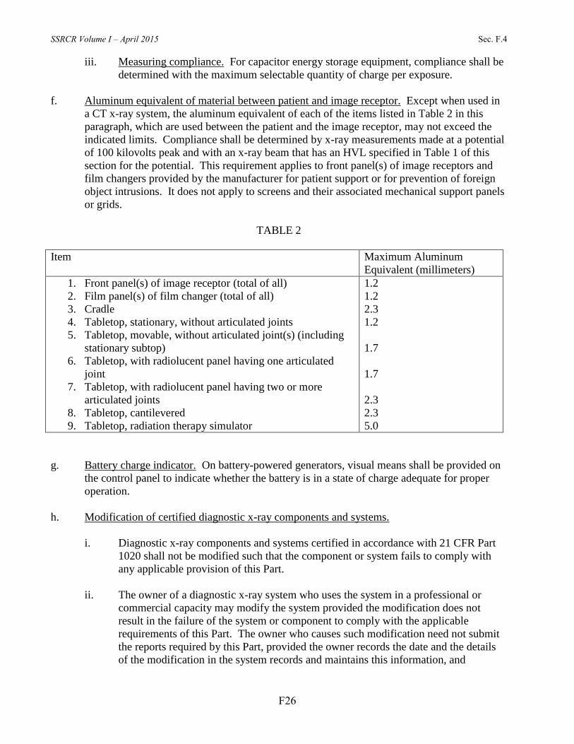
f. Aluminum equivalent of material between patient and image receptor. Except when used in
a CT x-ray system, the aluminum equivalent of each of the items listed in Table 2 in this
paragraph, which are used between the patient and the image receptor, may not exceed the
indicated limits. Compliance shall be determined by x-ray measurements made at a potential
of 100 kilovolts peak and with an x-ray beam that has an HVL specified in Table 1 of this
section for the potential. This requirement applies to front panel(s) of image receptors and
film changers provided by the manufacturer for patient support or for prevention of foreign
object intrusions. It does not apply to screens and their associated mechanical support panels
or grids.
TABLE 2
Item Maximum Aluminum
Equivalent (millimeters)
1. Front panel(s) of image receptor (total of all)
2. Film panel(s) of film changer (total of all)
3. Cradle
4. Tabletop, stationary, without articulated joints
5. Tabletop, movable, without articulated joint(s) (including
stationary subtop)
6. Tabletop, with radiolucent panel having one articulated
joint
7. Tabletop, with radiolucent panel having two or more
articulated joints
8. Tabletop, cantilevered
9. Tabletop, radiation therapy simulator
1.2
1.2
2.3
1.2
1.7
1.7
2.3
2.3
5.0
g. Battery charge indicator. On battery-powered generators, visual means shall be provided on
the control panel to indicate whether the battery is in a state of charge adequate for proper
operation.
h. Modification of certified diagnostic x-ray components and systems.
i. Diagnostic x-ray components and systems certified in accordance with 21 CFR Part
1020 shall not be modified such that the component or system fails to comply with
any applicable provision of this Part.
ii. The owner of a diagnostic x-ray system who uses the system in a professional or
commercial capacity may modify the system provided the modification does not
result in the failure of the system or component to comply with the applicable
requirements of this Part. The owner who causes such modification need not submit
the reports required by this Part, provided the owner records the date and the details
of the modification in the system records and maintains this information, and

Sec. F.4 – F.5 SSRCR Volume I - April 2015
F27
provided the modification of the x-ray system does not result in a failure to comply
with this Part.
i. Multiple Tubes. Where two or more radiographic tubes are controlled by one exposure
switch, the tube which has been selected shall be clearly indicated prior to initiation of the
exposure. Only the selected tube can be energized. This indication shall be both on the x-ray
control panel and at or near the tube housing assembly which has been selected.
j. Mechanical Support of Tube Head. The tube housing assembly supports shall be adjusted
such that the tube housing assembly will remain stable during an exposure unless tube
housing movement is a designed function of the x-ray system.
k. Locks. All position locking, holding, and centering devices on x-ray system components and
systems shall function as intended.
l. Maintaining Compliance. Diagnostic x-ray systems and their associated components used on
humans and certified pursuant to the Federal X-Ray Equipment Performance Standard (21
CFR Part 1020) shall be maintained in compliance with applicable requirements of that
standard.
Sec. F.5 - Fluoroscopic Equipment. The provisions of this Part apply to equipment for fluoroscopic
imaging or for recording images from the fluoroscopic image receptor. (21CFR1020.32)
a. Only image-intensified or direct-digital receptor fluoroscopic equipment shall be used for
fluoroscopy.
b. Primary Protective Barrier.
i. Limitation of useful beam. The fluoroscopic imaging assembly shall be provided
with a primary protective barrier which intercepts the entire cross section of the
useful beam at any SID. The x-ray tube used for fluoroscopy shall not produce x-rays
unless the barrier is in position to intercept the entire useful beam. The AKR due to
transmission through the barrier with the attenuation block in the useful beam
combined with radiation from the fluoroscopic imaging receptor shall not exceed
3.34x10⁻³
percent of the entrance AKR, at a distance of 10 cm from any accessible
surface of the fluoroscopic imaging assembly beyond the plane of the image receptor.
Radiation therapy simulation systems shall be exempt from this requirement provided
the systems are intended only for remote control operation. (21CFR 1020.32(a)(1))
ii. Measuring compliance. The AKR shall be measured in accordance with F.5e. The
AKR due to transmission through the primary barrier combined with radiation from
the fluoroscopic image receptor shall be determined by measurements averaged over
an area of 100 square cm with no linear dimension greater than 20 cm. If the source
is below the tabletop, the measurement shall be made with the input surface of the
fluoroscopic imaging assembly positioned 30 cm above the tabletop. If the source is
above the tabletop and the SID is variable, the measurement shall be made with the
end of the beam-limiting device or spacer as close to the tabletop as it can be placed,
provided that it shall not be closer than 30 cm. Movable grids and compression

SSRCR Volume I – April 2015 Sec. F.5
F28
devices shall be removed from the useful beam during the measurement. For all
measurements, the attenuation block shall be positioned in the useful beam 10 cm
from the point of measurement of entrance AKR and between this point and the input
surface of the fluoroscopic imaging assembly. (21CFR 1020.32(a)(2))
c. Field Limitation.
i. Angulation. For fluoroscopic equipment manufactured after February 25, 1978, when
the angle between the image receptor and the beam axis of the x-ray beam is variable,
means shall be provided to indicate when the axis of the x-ray beam is perpendicular
to the plane of the image receptor. Compliance with F.5c.v. and F.5c.vi. shall be
determined with the beam axis indicated to be perpendicular to the plane of the image
receptor. (21 CFR 1020.32(b)(1))
ii. Further means for limitation. Means shall be provided to permit further limitation of
the x-ray field to sizes smaller than the limits of F.5c.v. and F.5c.vi. Beam-limiting
devices manufactured after May 22, 1979, and incorporated in equipment with a
variable SID and/or capability of a visible area of greater than 300 cm2, shall be
provided with means for stepless adjustment of the x-ray field. Equipment with a
fixed SID and the capability of a visible area of no greater than 300 cm2 shall be
provided with either stepless adjustment of the x-ray field or with a means to further
limit the x-ray field size at the plane of the image receptor to 125 cm2 or less.
Stepless adjustment shall, at the greatest SID, provide continuous field sizes from the
maximum obtainable to a field size containable in a square of 5 cm by 5 cm. (21CFR
1020.32(b)(2))
iii. Spot-film devices. In addition to applicable regulations in F.6 (Radiographic
Equipment), the following requirements shall apply to spot-film devices, except when
the spot-film device is provided for use with a radiation therapy simulation system:
(21CFR1020.31(h))
(1) Means shall be provided between the source and the patient for adjustment of
the x-ray field size in the plane of the image receptor to the size of that portion
of the image receptor which has been selected on the spot-film selector. Such
adjustment shall be accomplished automatically when the x-ray field size in
the plane of the image receptor is greater than the selected portion of the
image receptor. If the x-ray field size is less than the size of the selected
portion of the image receptor, the field size shall not open automatically to the
size of the selected portion of the image receptor unless the operator has
selected that mode of operation. (21CFR1020.31(h)(1))
(2) Neither the length nor width of the x-ray field in the plane of the image
receptor shall differ from the corresponding dimensions of the selected portion
of the image receptor by more than 3 percent of the SID when adjusted for full
coverage of the selected portion of the image receptor. The sum, without
regard to sign, of the length and width differences shall not exceed 4 percent
of the SID. On spot-film devices manufactured after February 25, 1978, if the
angle between the plane of the image receptor and beam axis is variable,

Sec. F.5 SSRCR Volume I - April 2015
F29
means shall be provided to indicate when the axis of the x-ray beam is
perpendicular to the plane of the image receptor, and compliance shall be
determined with the beam axis indicated to be perpendicular to the plane of
the image receptor. (21CFR1020.31(h)(2))
(3) The center of the x-ray field in the plane of the image receptor shall be aligned
with the center of the selected portion of the image receptor to within 2
percent of the SID. (21CFR1020.31(h)(3))
(4) Means shall be provided to reduce the x-ray field size in the plane of the
image receptor to a size smaller than the selected portion of the image
receptor such that: (21CFR1020.31(h)(4))
(a) For spot-film devices used on fixed-SID fluoroscopic systems which
are not required to, and do not provide stepless adjustment of the x-ray
field, the minimum field size, at the greatest SID, does not exceed 125
square cm; or (21CFR1020.31(h)(4)(i))
(b) For spot-film devices used on fluoroscopic systems that have a
variable SID and/or stepless adjustment of the field size, the minimum
field size, at the greatest SID, shall be containable in a square of 5 cm
by 5 cm. (21CFR1020.31(h)(4)(ii))
iv. A capability may be provided for overriding the automatic x-ray field size adjustment
in case of system failure. If it is so provided, a signal visible at the fluoroscopist’s
position shall indicate whenever the automatic x-ray field size adjustment override is
engaged. Each such system failure override switch shall be clearly labeled as
follows:
For X-ray Field Limitation System Failure
(21CFR1020.31(h)(5))
v. Fluoroscopy and radiography using the fluoroscopic imaging assembly with
inherently circular image receptors.
(1) For fluoroscopic equipment manufactured before June 10, 2006, other than
radiation therapy simulation systems, the following applies: (21CFR
1020.32(b)(4)(i)
(a) Neither the length nor width of the x-ray field in the plane of the image
receptor shall exceed that of the visible area of the image receptor by
more than 3 percent of the SID. The sum of the excess length and the
excess width shall be no greater than 4 percent of the SID.
(21CFR 1020.32(b)(4)(i)(A))
(b) For rectangular x-ray fields used with circular image receptors, the
error in alignment shall be determined along the length and width

SSRCR Volume I – April 2015 Sec. F.5
F30
dimensions of the x-ray field which pass through the center of the
visible area of the image receptor. (21CFR 1020.32(b)(4)(i)(B))
(2) For fluoroscopic equipment manufactured on or after June 10, 2006, other
than radiation simulation systems, the maximum area of the x-ray field in the
plane of the image receptor shall conform with one of the following
requirements: (21CFR 1020.32(b)(4)(ii))
(a) When any linear dimension of the visible area of the image receptor
measured through the center of the visible area is less than or equal to
34 cm in any direction, at least 80 percent of the area of the x-ray field
overlaps the visible area of the image receptor, or (21CFR
1020.32(b)(4)(ii)(A))
(b) When any linear dimension of the visible area of the image receptor
measured through the center of the visible area is greater than 34 cm in
any direction, the x-ray field measured along the direction of greatest
misalignment with the visible area of the image receptor does not
extend beyond the edge of the visible area of the image receptor by
more than 2 cm. (21CFR 1020.32(b)(4)(ii)(B))
vi. Fluoroscopy and radiography using fluoroscopic imaging assembly with inherently
rectangular image receptors. For x-ray systems manufactured on or after June 10,
2006, the following applies: (21CFR1020.32(b)(5))
(1) Neither the length nor width of the x-ray field in the plane of the image
receptor shall exceed that of the visible area of the image receptor by more
than 3 percent of the SID. The sum of the excess length and the excess width
shall be no greater than 4 percent of the SID. (21CFR1020.32(b)(5)(i))
(2) The error in alignment shall be determined along the length and width
dimensions of the x-ray field which pass through the center of the visible area
of the image receptor. (21cfr1020.32(b)(5)(ii))
vii. Override capability. If the fluoroscopic x-ray field size is adjusted automatically as
the SID or image receptor size is changed, a capability may be provided for
overriding the automatic adjustment in case of system failure. If it is so provided, a
signal visible at the fluoroscopist’s position shall indicate whenever the automatic
field adjustment is overridden. Each such system failure override switch shall be
clearly labeled as follows:
FOR X-RAY FIELD
LIMITATION SYSTEM FAILURE
(21CFR 1020.32(b)(6))
d. Activation of Tube. X-ray production in the fluoroscopic mode shall be controlled by a
device which requires continuous pressure by the operator for the entire time of any
exposure. When recording serial radiographic images from the fluoroscopic image receptor,

Sec. F.5 SSRCR Volume I - April 2015
F31
the operator shall be able to terminate the x-ray exposure(s) at any time, but means may be
provided to permit completion of any single exposure of the series in process. (21CFR
1020.32(c))
e. Air Kerma Rates. For fluoroscopic equipment, the following requirements apply:
i. Fluoroscopic equipment manufactured before May 19, 1995.
(1) Equipment provided with automatic exposure rate control (AERC) shall not
be operable at any combination of tube potential and current that will result in
an AKR in excess of 88 mGy per minute (vice 10 R/min exposure rate) at the
measurement point specified in F.5e.iv., except as specified in F.5e.i.(5).
(21CFR 1020.32(d)(1)(i))
(2) Equipment provided without AERC shall not be operable at any combination
of tube potential and current that will result in an AKR in excess of 44 mGy
per minute (vice 5 R/min exposure rate) at the measurement point specified in
F.5e.iv., except as specified in F.5e.i.(5). (21CFR 1020.32(d)(1)(ii))
(3) Equipment provided with both an AERC mode and a manual mode shall not
be operable at any combination of tube potential and current that will result in
an AKR in excess of 88 mGy per minute (vice 10 R/min exposure rate) in
either mode at the measurement point specified in F.5e.iv., except as specified
in F.5e.i.(5). (21CFR 1020.32(d)(1)(iii))
(4) Equipment may be modified in accordance with this Part to comply with
F.5e.ii. When the equipment is modified, it shall bear a label indicating the
date of the modification and the statement:
MODIFIED TO COMPLY WITH 21 CFR 1020.32(H)(2)
(21CFR 1020.32(d)(1)(iv))
(5) Exceptions: During recording of fluoroscopic images.
ii. Fluoroscopic equipment manufactured on or after May 19, 1995.
(1) Shall be equipped with AERC if operable at any combination of tube potential
and current that results in an AKR greater than 44 mGy per minute (vice 5
R/min exposure rate) at the measurement point specified in F.5e.iv. Provision
for manual selection of technique factors may be provided. (21CFR
1020.32(d)(2)(i))
(2) Shall not be operable at any combination of tube potential and current that
will result in an AKR in excess of 88 mGy per minute (vice 10 R/min
exposure rate) at the measurement point specified in F.5e.iv., except as
specified in F.5e.ii.(3). (21CFR 1020.32(d)(2)(ii))
(3) Exceptions:

SSRCR Volume I – April 2015 Sec. F.5
F32
(a) For equipment manufactured prior to June 10, 2006, during the
recording of images from a fluoroscopic image receptor using
photographic film or a video camera when the x-ray source is operated
in a pulsed mode. (21CFR 1020.32(d)(2)(iii)(A))
(b) For equipment manufactured on or after June 10, 2006, during the
recording of images from the fluoroscopic image receptor for the
purpose of providing the user with a recorded image(s) after
termination of the exposure. Such recording does not include images
resulting from a last-image-hold feature that are not recorded. (21CFR
1020.32(d)(2)(iii)(B))
iii. Fluoroscopy equipment with optional high-level control
(1) When high-level control is selected and the control is activated, in which case
the equipment shall not be operable at any combination of tube potential and
current that will result in an AKR in excess of 176 mGy per minute (vice 20
R/min exposure rate) at the measurement point specified in F.5e.iv. Special
means of activation of high-level controls shall be required. The high-level
control shall be operable only when continuous manual activation is provided
by the operator. A continuous signal audible to the fluoroscopist shall
indicate that the high-level control is employed.
iv. Measuring compliance. Compliance with this subsection shall be determined as
follows:
(1) If the source is below the x-ray table, the AKR shall be measured at 1 cm
above the tabletop or cradle. (21CFR 1020.32(d)(3)(i))
(2) If the source is above the x-ray table, the AKR shall be measured at 30 cm
above the tabletop with the end of the beam-limiting device or spacer
positioned as closely as possible to the point of measurement. (21CFR
1020.32(d)(3)(ii))
(3) In a C-arm type of fluoroscope, the AKR shall be measured at 30 cm from the
input surface of the fluoroscopic imaging assembly, with the source
positioned at any available SID, provided that the end of the beam-limiting
device or spacer is no closer than 30 cm from the input surface of the
fluoroscopic imaging assembly. (21CFR 1020.32(d)(3)(iii))
(4) In a C-arm type of fluoroscope having an SID less than 45 cm, the AKR shall
be measured at the minimum SSD. (21CFR 1020.32(d)(3)(iv))
(5) In a lateral type of fluoroscope, the air kerma rate shall be measured at a point
15 cm from the centerline of the x-ray table and in the direction of the x-ray
source with the end of the beam-limiting device or spacer positioned as
closely as possible to the point of measurement. If the tabletop is movable, it

Sec. F.5 SSRCR Volume I - April 2015
F33
shall be positioned as closely as possible to the lateral x-ray source, with the
end of the beam-limiting device or spacer no closer than 15 cm to the
centerline of the x-ray table. (21CFR 1020.32(d)(3)(v))
v. Exemptions. Fluoroscopic radiation therapy simulation systems are exempt from the
requirements set forth in F.5e. when used for therapy simulation purposes. (21CFR
1020.32(d)(4))
f. Indication of potential and current. During fluoroscopy and cinefluorography, x-ray tube
potential and current shall be continuously indicated. Deviation of x-ray tube potential and
current from the indicated value shall not exceed the maximum deviation as stated by the
manufacturer. (21CFR 1020.32(f))
g. Source-skin distance.
i. Means shall be provided to limit the source-skin distance to not less than 38 cm on
stationary fluoroscopes and to not less than 30 cm on mobile and portable
fluoroscopes. In addition, for fluoroscopes intended for specific surgical or
interventional applications that would be prohibited at the source-skin distances
specified in this paragraph, provisions may be made for operating at shorter source-
skin distances but in no case less than 20 cm.
ii. For stationary, mobile, or portable C-arm fluoroscopic systems manufactured on or
after June 10, 2006, having a maximum source-image receptor distance of less than
45 cm, means shall be provided to limit the source-skin distance to not less than 19
cm. Such systems shall be labeled for extremity use only. In addition, for those
systems intended for specific surgical that would be prohibited at the
source-skin distance specified in this paragraph, provisions may be made for
operation at shorter source-skin distances but in no case less than 10 cm.
h. Fluoroscopic irradiation time, display, and signal.
i. Fluoroscopic equipment manufactured before June 10, 2006:
(1) Shall be provided with means to preset the cumulative irradiation time of the
fluoroscopic tube. The maximum cumulative time of the timing device shall
not exceed 5 minutes without resetting. A signal audible to the fluoroscopist
shall indicate the completion of any preset cumulative irradiation time. Such
signal shall continue to sound while x-rays are produced until the timing
device is reset. Fluoroscopic equipment may be modified in accordance with
21 CFR 1020.30(q) to comply with the requirements of this paragraph. When
the equipment is modified, it shall bear a label indicating the statement:
Modified to comply with 21 CFR 1020.32(h)(2)
(21CFR 1020.32(h)(1)(i))

SSRCR Volume I – April 2015 Sec. F.5
F34
(2) As an alternative to the requirements of this paragraph, radiation therapy
simulation systems may be provided with a means to indicate the total
cumulative exposure time during which x-rays were produced, and which is
capable of being reset between x-ray examinations. (21CFR 1020.32(h)(1)(ii))
ii. For x-ray controls manufactured on or after June 10, 2006, there shall be provided for
each fluoroscopic tube:
(1) A display of the fluoroscopic irradiation time at the fluoroscopist’s working
position. This display shall function independently of the audible signal
described in this subsection. The following requirements apply: (variation of
21CFR 1020.32(h)(2)(i))
(a) When the x-ray tube is activated, the fluoroscopic irradiation time in
minutes and tenths of minutes shall be continuously displayed and
updated at least once every 6 seconds. (21CFR 1020.32(h)(2)(i)(A))
(b) The fluoroscopic irradiation time shall also be displayed within 6
seconds of termination of an exposure and remain displayed until
reset. (21CFR 1020.32(h)(2)(i)(B))
(c) Means shall be provided to reset the display to zero prior to the
beginning of a new examination or procedure. (21CFR
1020.32(h)(2)(i)(C))
(2) A signal audible to the fluoroscopist shall sound for each passage of 5 minutes
of fluoroscopic irradiation time during an examination or procedure. The
signal shall sound until manually reset or, if automatically reset, for at least 2
seconds. (21CFR 1020.32(h)(2)(ii))
i. Display of last-image-hold (LIH). Fluoroscopic equipment manufactured on or after June 10,
2006, shall be equipped with means to display LIH image following termination of the
fluoroscopic exposure. (21CFR 1020.32(j))
i. For an LIH image obtained by retaining pretermination fluoroscopic images, if the
number of images and method of combining images are selectable by the user, the
selection shall be indicated prior to initiation of the fluoroscopic exposure. (21CFR
1020.32(j)(1))
ii. For an LIH image obtained by initiating a separate radiographic-like exposure at the
termination of fluoroscopic imaging, the technique factors for the LIH image shall be
selectable prior to the fluoroscopic exposure, and the combination selected shall be
indicated prior to initiation of the fluoroscopic exposure. (21CFR 1020.32(j)(2))
iii. Means shall be provided to clearly indicate to the user whether a displayed image is
the LIH radiograph or fluoroscopy. Display of the LIH radiograph shall be replaced
by the fluoroscopic image concurrently with re-initiation of fluoroscopic exposure,

Sec. F.5 SSRCR Volume I - April 2015
F35
unless separate displays are provided for the LIH radiograph and fluoroscopic images.
(21CFR 1020.32(j)(3))
j. Displays of values of AKR and cumulative air kerma. Fluoroscopic equipment manufactured
on or after June 10, 2006, shall display at the fluoroscopist’s working position the AKR and
cumulative air kerma. The following requirements apply for each x-ray tube used during an
examination or procedure: (21CFR 1020.32(k))
i. When the x-ray tube is activated and the number of images produced per unit time is
greater than six images per second, the AKR in mGy/min shall be continuously
displayed and updated at least once every second. (21CFR 1020.32(k)(1))
ii. The cumulative air kerma in units of mGy shall be displayed either within 5 seconds
of termination of an exposure or displayed continuously and updated at least once
every 5 seconds. (21CFR 1020.32(k)(2))
iii. The display of the AKR shall be clearly distinguishable from the display of the
cumulative air kerma. (21CFR 1020.32(k)(3))
iv. The AKR and cumulative air kerma shall represent the value for conditions of free-in-
air irradiation at one of the following reference locations specified according to the
type of fluoroscope. (21CFR 1020.32(k)(4))
(1) For fluoroscopes with x-ray source below the x-ray table, x-ray source above
the table, or of lateral type, the reference location shall be the respective
locations specified in F.5e.iv(1), F.5e.iv(2) or F.5e.iv(5) (21CFR
1020.32(k)(4)(i))
(2) For C-arm fluoroscopes, the reference location shall be 15 cm from the
isocenter toward the x-ray source along the beam axis. Alternatively, the
reference location shall be at a point specified by the manufacturer to
represent the location of the intersection of the x-ray beam with the patient’s
skin. (21CFR 1020.32(k)(4)(ii))
v. Means shall be provided to reset to zero the display of cumulative air kerma prior to
the commencement of a new examination or procedure. (21CFR 1020.32(k)(5))
vi. The displayed AKR and cumulative air kerma shall not deviate from the actual values
by more than ±35 percent over the range of 6 mGy/min and 100 mGy to the
maximum indication of AKR and cumulative air kerma, respectively. Compliance
shall be determined with an irradiation time greater than 3 seconds. (21CFR
1020.32(k)(6))
k. Protection From Scatter Radiation.
i. For stationary fluoroscopic systems, ancillary shielding, such as drapes, self-
supporting curtains, or viewing shields, shall be available and used as supplemental

SSRCR Volume I – April 2015 Sec. F.5
F36
protection for all individuals other than the patient in the room during a fluoroscopy
procedure.
ii. Where sterile fields or special procedures prohibit the use of normal protective
barriers or drapes, all of the following conditions shall be met.
(1) Shielding required under F5.k.i shall be maintained to the degree possible
under the clinical conditions.
(2) All persons, except the patient, in the room where fluoroscopy is performed
shall wear protective aprons that provide a lead equivalent shielding of at least
0.25 mm.
(3) The fluoroscopic field size shall be reduced to the minimum required for the
procedure being performed (area of clinical interest).
(4) Operating and safety procedures shall reflect the above conditions, and
fluoroscopy personnel shall exhibit awareness of situations requiring the use
and/or non-use of the protective drapes.
l. Operator Qualifications.
i. In addition to the applicable sections of these regulations, the operation of a
fluoroscopic x-ray system for clinical purposes shall be limited to:
(1) A licensed practitioner working within his or her scope of practice;
(2) A Radiologist Assistant (RA) (if recognized by the Agency) working within
his or her scope of practice and under the direct supervision of a licensed
practitioner meeting the conditions of F.5l.i.(1);
(3) An individual who passed the American Registry of Radiologic Technologists
(ARRT) Fluoroscopy Exam (or equivalent) and holds a valid certification, and
only under the personal supervision of the licensed practitioner meeting the
conditions of F.5l.i.(1);
(4) A medical resident or radiologic technology student, in training, and only
under the personal supervision of the licensed practitioner meeting the
conditions of F.5l.i.(1).
ii. All persons operating, or supervising the operation of, fluoroscopy systems shall have
completed a minimum of 4 hours training that includes but is not limited to the
following:
(1) Basic properties of radiation;
(2) Biological effects of x-ray;

Sec. F.5 SSRCR Volume I - April 2015
F37
(3) Radiation protection methods for patients and staff;
(4) Units of measurement and dose, including DAP (dose-area product) values &
air kerma;
(5) Factors affecting fluoroscopic outputs;
(6) High level control options;
(7) Dose management including dose reduction techniques, monitoring, and
recording;
(8) Principles and operation of the specific fluoroscopic x-ray system(s) to be
used;
(9) Fluoroscopic and fluorographic outputs of each mode of operation on the
system(s) to be used clinically; and
(10) Applicable requirements of these regulations.
iii. All persons operating, or supervising the operation of, fluoroscopy systems during
FGI procedures shall have completed a minimum of 8 hours of training approved by
the Agency. The topics shall include:
(1) The topics provided in F.5l.ii.;
(2) Methods to reduce patient dose using advanced imaging and recording
features;
(3) Procedures for recording pertinent data specified in F.5o.
(4) Minimum of one hour of hands-on fluoroscopic machine training
demonstrating application of topics required in this subsection.
iv. The training required in this subsection shall be provided by a QMP [QE] or another
individual approved by the Agency.
v. Two years after the effective date of this rule, the registrant [licensee] shall ensure
that prior to performing fluoroscopy procedures each person operating, or supervising
the operation of, fluoroscopy systems completed the training required in this
subsection.
vi. The registrant [licensee] shall either provide a minimum of 2 hours in-service training
every 2 years for all individuals operating or supervising the operation of fluoroscopy
systems used or require evidence of continuing medical education meeting the
conditions of this subsection.

SSRCR Volume I – April 2015 Sec. F.5
F38
vii. Documentation pertaining to the requirements of F.5 shall be maintained for review
for three years.
m. Equipment Operation.
i. All fluoroscopic images shall be viewed, directly or indirectly, and interpreted by a
licensed practitioner of the healing arts.
ii. Overhead fluoroscopy shall not be used as a positioning tool for general purpose radiographic
examinations.
iii. Operators shall be competent in the standard operating procedures of the unit in use,
including the use of available dose-saving features, and the relative radiation output
rates of the various modes of operation.
iv. Procedure planning for fluoroscopic procedures on pregnant patients shall include
feasible modifications to minimize the dose to the conceptus.
v. Procedure planning for fluoroscopic procedures on pediatric patients shall include
feasible modifications to minimize dose.
vi. The registrant [licensee] shall use all methods available on the fluoroscopy system to
monitor dose during a fluoroscopic procedure.
vii. The facility shall establish a written policy regarding patient dose management in
fluoroscopically guided procedures in conformance with the ACR-AAPM Technical
Standard for Management of the Use of Radiation in Fluoroscopic Procedures (ACR
Resolution 44 – 2013), NCRP Report 168, or equivalent.
n. Qualified Medical Physicist Evaluations.
i. Fluoroscopic equipment shall be evaluated by a QMP [QE] within 30 days of
installation and of any maintenance of the system that may affect the exposure rate.
Thereafter, the measurements shall be made annually or at intervals not to exceed 12
months from the date of the prior measurement by or under the direction of a QMP
[QE]. At a minimum these evaluations shall include:
(1) A measurement of entrance exposure rates that covers the full range of patient
thicknesses, including those that are expected to drive the system to maximum
output in all modes clinically used, including fluoroscopy, high-level control,
acquisition, digital subtraction and CINE, when available. These
measurements shall:
(a) For systems without automatic exposure control, be made utilizing a
milliamperage and kVp typical of the clinical use of the fluoroscopic
system;

Sec. F.5 SSRCR Volume I - April 2015
F39
(b) For systems with automatic exposure control, be made utilizing
sufficient attenuating material in the useful beam to produce a
milliamperage and kVp typical of the clinical use of the fluoroscopic
system;
(2) A measurement and verification of compliance of maximum AKR for
fluoroscopy and high-level control, if available. Measurements shall be made
in accordance with Sec F.5e.iv.
(3) An evaluation of high contrast resolution and low contrast resolution in both
fluoroscopic and spot-film modes
(4) An evaluation of the operation of the 5-minute timer, warning lights,
interlocks, and collision sensors.
(5) An evaluation of the beam quality and collimation in the fluoroscopy and
spot-film modes.
(6) An evaluation of the availability and accuracy of technique indicators and
integrated radiation dose displays.
(7) An evaluation of any changes that may impact patient and personnel
protection devices.
ii. Measurements required in Sec F.5n.i. shall be performed with a calibrated dosimetry
system per manufacturer recommendations not to exceed 2 years and records
maintained for 5 years for inspection by the Agency.
o. Additional requirements for facilities performing fluoroscopically-guided interventional
(FGI) procedures.
i. A registrant [licensee] utilizing FGI procedures shall establish a Radiation Protocol
Committee (RPC) in accordance with the following.
(1) The registrant [licensee] may establish a system-wide committee if the
registrant has more than one site.
(2) Two or more registrants [licensees] may form a cooperative RPC as long as
each facility has a representative on the committee.
(3) If the registrant [licensee] has already established a radiation safety
committee, the requirements of this subsection may be delegated to that
committee if the members meet the requirements of F.5o.iv.
ii. A quorum of the RPC shall meet as often as necessary, but at intervals not to exceed
12 months.

SSRCR Volume I – April 2015 Sec. F.5
F40
iii. Record of RPC. A record of each RPC meeting shall include the date, names of
individuals in attendance, minutes of the meeting, and any actions taken. The
registrant [licensee] shall maintain the record for inspection by the Agency.
iv. Provide an annual report to the radiation safety committee or radiation safety officer,
in the absence of a radiation safety committee,
v. RPC Members. Members shall include but not be limited to the following
individuals:
(1) A supervising physician of the healing arts who meets the requirements in
Sec. F.5l.;
(2) A QMP [QE];
(3) The lead technologist; and
(4) Other individuals as deemed necessary by the registrant [licensee].
vi. Establish and implement FGI procedure protocols.
(1) The RPC shall establish and implement written protocols, or protocols
documented in an electronic report system, that include but are not limited to
the following.
(a) Identification of individuals who are authorized to use fluoroscopic
systems for interventional purposes.
(b) A method to be used to monitor patient radiation dose during FGI.
(c) Dose notification levels, as appropriate, at which the physician is
notified and appropriate actions are taken for patient safety.
(d) SRDL values following nationally recognized standards,
(e) Actions to be taken for cases when a SRDL is exceeded which may
include patient follow-up.
(f) A review of the established protocols at an interval not to exceed 12
months.
(2) A record of each RPC protocol shall be maintained for inspection by the
Agency. If the RPC revises a protocol, documentation shall be maintained that
includes the justification for the revision and the previous protocol for
inspection by the Agency.
vii. Procedures for maintaining records.

Sec. F.5 – F.6 SSRCR Volume I - April 2015
F41
(1) A record of radiation output information shall be maintained so the radiation
dose to the skin may be estimated in accordance with established protocols.
The record shall include the following:
(a) Patient identification;
(b) Type and date of examination;
(c) Identification of the fluoroscopic system used; and
(d) Peak skin dose, cumulative air kerma or dose area product used if the
information is available on the fluoroscopic system.
(e) If the peak skin dose, cumulative air kerma or dose area product are
not displayed on the fluoroscopic system, records shall include other
information necessary to estimate the radiation dose to the skin in
accordance with established protocol or the following as necessary:
(i) Fluoroscopic mode, such as, high-level or pulsed mode of
operation;
(ii) Cumulative fluoroscopic exposure time; and
(iii) Number of films or recorded exposures.
(2) The registrant [licensee] shall maintain records required by this subparagraph
for inspection by the Agency.
Sec. F.6 - Radiographic Equipment. The following regulations apply to all non-dental registrants
[licensees] using diagnostic x-ray equipment. Requirements specific to using dental intra-oral, hand
held, panoramic, and cephalometric equipment are in Sec.F.7.
a. Digital radiographic systems shall be evaluated by a QMP [QE] within 30 days of clinical
use and by or under the direction of a QMP [QE] at intervals not to exceed 12 months unless
otherwise determined by the Agency. The evaluation shall follow nationally recognized
procedures or those recognized by the Agency. Unless otherwise specified in this Part,
dental, podiatric, and veterinary systems are exempt from this requirement.
b. Control and indication of technique factors.
i. Timers. Means shall be provided to terminate the exposure at a preset time interval, a
preset product of current and time, a preset number of pulses, or a preset radiation
exposure to the image receptor. (21CFR1020.31(a)(2))
(1) Except during serial radiography, the operator shall be able to terminate the
exposure at any time during an exposure of greater than one-half second.
Except during panoramic dental radiography, termination of exposure shall
cause automatic resetting of the timer to its initial setting or to zero. It shall

SSRCR Volume I - January 2015 Sec. F.6
F42
not be possible to make an exposure when the timer is set to a zero or off
position if either position is provided. (21CFR1020.31(a)(2)(i))
(2) During serial radiography, the operator shall be able to terminate the x-ray
exposure(s) at any time, but means may be provided to permit completion of
any single exposure of the series in process. (21CFR1020.31(a)(2)(ii))
ii. Automatic exposure controls. When an automatic exposure control is provided:
(1) Indication shall be made on the control panel when this mode of operation is
selected; (21CFR1020.31(a)(3)(i))
(2) When the x-ray tube potential is equal to or greater than 51 kilovolts peak
(kVp), the minimum exposure time for field emission equipment rated for
pulse operation shall be equal to or less than a time interval equivalent to two
pulses and the minimum exposure time for all other equipment shall be equal
to or less than 1/60 second or a time interval required to deliver 5
milliampere-seconds (mAs), whichever is greater; (21CFR1020.31(a)(3)(ii))
(3) Either the product of peak x-ray tube potential, current, and exposure time
shall be limited to not more than 60 kilowatt-seconds (kWs) per exposure or
the product of x-ray tube current and exposure time shall be limited to not
more than 600 mAs per exposure, except when the x-ray tube potential is less
than 51 kVp, in which case the product of x-ray tube current and exposure
time shall be limited to not more than 2,000 mAs per exposure; and
(21CFR1020.31(a)(3)(iii))
(4) A visible signal shall indicate when an exposure has been terminated at the
limits described in F.6b.ii.(3), and manual resetting shall be required before
further automatically timed exposures can be made. (21CFR1020.31(a)(3)(iv))
iii. Accuracy. Deviation of technique factors under Sec.F.6b. from indicated values shall
not exceed the limits given by the manufacturer. (variation of 21CFR1020.31(a)(4))
c. Reproducibility.
i. Coefficient of variation. For any specific combination of selected technique factors,
the estimated coefficient of variation of the air kerma shall be no greater than 0.05.
(21CFR1020.31(b)(1)
ii. Measuring compliance. Determination of compliance shall be based on 10
consecutive measurements taken within a time period of 1 hour. Equipment
manufactured after September 5, 1978, shall be subject to the additional requirement
that all variable controls for technique factors shall be adjusted to alternate settings
and reset to the test setting after each measurement. The percent line-voltage
regulation shall be within ±1 of the mean value for all measurements. For equipment
having automatic exposure controls, compliance shall be determined with a sufficient
thickness of attenuating material in the useful beam such that the technique factors

Sec. F.6 SSRCR Volume I - April 2015
F43
can be adjusted to provide individual exposures of a minimum of 12 pulses on field
emission equipment rated for pulsed operation or no less than one-tenth second per
exposure on all other equipment. (21CFR1020.31(b)(2))
d. Linearity. The following requirements apply for any fixed x-ray tube potential within the
range of 40 percent to 100 percent of the maximum rated. (variation of 21CFR1020.31(c))
i. Equipment having independent selection of x-ray tube current (mA). The average
ratios of air kerma to the indicated milliampere-seconds product (mGy/mAs) obtained
at any two consecutive tube current settings shall not differ by more than 0.10 times
their sum. This is: ׀X1 – X20.10 ≥ ׀(X1 + X2); where X1 and X2 are the average
mGy/mAs values obtained at each of two consecutive mAs selector settings or at two
settings differing by no more than a factor of 2 where the mAs selector provides
continuous selection. (21CFR1020.31(c)(1))
ii. Equipment having selection of x-ray tube current-exposure time product (mAs). For
equipment manufactured after May 3, 1994, the average ratios of air kerma to the
indicated milliampere-seconds product (mGy/mAs) obtained at any two consecutive
mAs selector settings shall not differ by more than 0.10 times their sum. This is: ׀X1
– X20.10 ≥ ׀(X1 + X2); where X1 and X2 are the average mGy/mAs values obtained at
each of two consecutive mAs selector settings or at two settings differing by no more
than a factor of 2 where the mAs selector provides continuous selection.
(21CFR1020.31(c)(2))
iii. Measuring compliance. Determination of compliance will be based on 10 exposures,
made within 1 hour, at each of the two settings. These two settings may include any
two focal spot sizes except where one is equal to or less than 0.45 mm and the other is
greater than 0.45 mm. For purposes of this requirement, focal spot size is the focal
spot size specified by the x-ray tube manufacturer. The percent line-voltage
regulation shall be determined for each measurement. All values for percent line-
voltage regulation at any one combination of technique factors shall be within ±1 of
the mean value for all measurements at these technique factors.
(21CFR1020.31(c)(3))
e. Field limitation and alignment for mobile, portable, and stationary general purpose x-ray
systems. Except when spot-film devices are in service, mobile, portable, and stationary
general purpose radiographic x-ray systems shall meet the following requirements:
(21CFR1020.31(d))
i. Variable x-ray field limitation. A means for stepless adjustment of the size of the x-
ray field shall be provided. Each dimension of the minimum field size at an SID of
100 cm shall be equal to or less than 5 cm. (21CFR1020.31(d)(1))
ii. Visual definition.
(1) Means for visually defining the perimeter of the x-ray field shall be provided.
The total misalignment of the edges of the visually defined field with the
respective edges of the x-ray field along either the length or width of the

SSRCR Volume I - April 2015 Sec. F.6
F44
visually defined field shall not exceed 2 percent of the distance from the
source to the center of the visually defined field when the surface upon which
it appears is perpendicular to the axis of the x-ray beam.
(21CFR1020.31(d)(2)(i))
(2) When a light localizer is used to define the x-ray field, it shall provide an
average illuminance of not less than 160 lux (15 footcandles) at 100 cm or at
the maximum SID, whichever is less. The average illuminance shall be based
on measurements made in the approximate center of each quadrant of the light
field. Radiation therapy simulation systems are exempt from this requirement.
(21CFR1020.31(d)(2)(ii))
(3) The edge of the light field at 100 cm or at the maximum SID, whichever is
less, shall have a contrast ratio, corrected for ambient lighting, of not less than
4 in the case of beam-limiting devices designed for use on stationary
equipment, and a contrast ratio of not less than 3 in the case of beam-limiting
devices designed for use on mobile and portable equipment. The contrast
ratio is defined as I1/I2, where I1 is the illuminance 3 mm from the edge of the
light field toward the center of the field; and I2 is the illuminance 3 mm from
the edge of the light field away from the center of the field. Compliance shall
be determined with a measuring aperture of 1 mm. (21CFR1020.31(d)(2)(iii))
f. Field indication and alignment on stationary general purpose x-ray equipment. Except when
spot-film devices are in service, stationary general purpose x-ray systems shall meet the
following requirements in addition to those prescribed in F.6e.: (21CFR1020.31(e))
i. Means shall be provided to indicate when the axis of the x-ray beam is perpendicular
to the plane of the image receptor, to align the center of the x-ray field with respect to
the center of the image receptor to within 2 percent of the SID, and to indicate the
SID to within 2 percent; (21CFR1020.31(e)(1))
ii. The beam-limiting device shall numerically indicate the field size in the plane of the
image receptor to which it is adjusted; (21CFR1020.31(e)(2))
iii. Indication of field size dimensions and SIDs shall be specified in centimeters and/or
inches and shall be such that aperture adjustments result in x-ray field dimensions in
the plane of the image receptor which correspond to those indicated by the beam-
limiting device to within 2 percent of the SID when the beam axis is indicated to be
perpendicular to the plane of the image receptor; and (21CFR1020.31(e)(3))
iv. Compliance measurements will be made at discrete SIDs and image receptor
dimensions in common clinical use (such as SIDs of 100, 150, and 200 cm and/or 36,
40, 48, 72 inches and nominal image receptor dimensions of 13, 18, 24, 30, 35, 40,
and 43 cm and/or 5, 7, 8, 9, 10, 11, 12, 14, and 17 inches) or at any other specific
dimensions at which the beam-limiting device or its associated diagnostic x-ray
system is uniquely designed to operate. (21CFR1020.31(e)(4))
g. Field limitation on x-ray equipment other than general purpose radiographic systems.

Sec. F.6 SSRCR Volume I - April 2015
F45
i. X-ray systems designed for one image receptor size. Radiographic equipment
designed for only one image receptor size at a fixed SID shall be provided with
means to limit the field at the plane of the image receptor to dimensions no greater
than those of the image receptor, and to align the center of the x-ray field with the
center of image receptor to within 2 percent of the SID, or shall be provided with
means to both size and align the x-ray field such that the x-ray field at the plane of the
image receptor does not extend beyond the edge of the image receptor.
ii. Other x-ray systems. Radiographic systems not specifically covered in F.6e., F.6f.,
F.6g.ii., F.6g.iii., and systems covered in F.6g.i., which are also designed for use with
extraoral image receptors and when used with an extraoral image receptor, shall be
provided with means to limit the x-ray field in the plane of the image receptor so that
such field does not exceed each dimension of the image receptor by more than 2
percent of the SID, when the axis of the x-ray beam is perpendicular to the plane of
the image receptor. In addition, means shall be provided to align the center of the x-
ray field with the center of the image receptor to within 2 percent of the SID, or
means shall be provided to both size and alignment the x-ray field such that the x-ray
field at the plane of the image receptor does not extend beyond any edge of the image
receptor. These requirements may be met with: (21CFR1020.31(f)(4))
(1) A system which performs in accordance with F.6e. and F.6f.; or when
alignment means are also provided, may be met with either;
(21CFR1020.31(f)(4)(i))
(2) An assortment of removable, fixed-aperture, beam-limiting devices sufficient
to meet the requirement for each combination of image receptor size and SID
for which the unit is designed. Each such device shall have clear and
permanent markings to indicate the image receptor size and SID for which it
is designed; or (21CFR1020.31(f)(4)(ii))
(3) A beam-limiting device having multiple fixed apertures sufficient to meet the
requirement for each combination of image receptor size and SID for which
the unit is designed. Permanent, clearly legible markings shall indicate the
image receptor size and SID for which each aperture is designed and shall
indicate which aperture is in position for use. (21CFR1020.31(f)(4)(iii))
h. Positive beam limitation (PBL). The requirements of this subsection shall apply to
radiographic systems which contain PBL. (21CFR1020.31(g))
i. Field size. When a PBL system is provided, it shall prevent x-ray production when:
(21CFR1020.31(g)(1))
(1) Either the length or width of the x-ray field in the plane of the image receptor
differs from the corresponding image receptor dimension by more than 3
percent of the SID; or (21CFR1020.31(g)(1)(i))

SSRCR Volume I - April 2015 Sec. F.6
F46
(2) The sum of the length and width differences stated in F.6h.i.(1) without regard
to sign exceeds 4 percent of the SID. (21CFR1020.31(g)(1)(ii))
(3) The beam-limiting device is at an SID for which PBL is not designed for
sizing. (21CFR1020.31(g)(1)(iii))
ii. Conditions for PBL. When provided, the PBL system shall function as described in
F.6h.i. whenever all the following conditions are met: (21CFR1020.31(g)(2))
(1) The image receptor is inserted into a permanently mounted cassette holder;
(21CFR1020.31(g)(2)(i))
(2) The image receptor length and width are less than 50 cm;
(21CFR1020.31(g)(2)(ii))
(3) The x-ray beam axis is within ±3 degrees of vertical and the SID is 90 cm to
130 cm inclusive; or the x-ray beam axis is within ±3 degrees of horizontal
and the SID is 90 cm to 205 cm inclusive; (21CFR1020.31(g)(2)(iii))
(4) The x-ray beam axis is perpendicular to the plane of the image receptor to
within ±3 degrees; and (21CFR1020.31(g)(2)(iv))
(5) Neither tomographic nor stereoscopic radiography is being performed.
(21CFR1020.31(g)(2)(v))
iii. Measuring compliance. Compliance with the requirements of F.6h.i. shall be
determined when the equipment indicates that the beam axis is perpendicular to the
plane of the image receptor and the provisions of F.6h.ii. are met. Compliance shall
be determined no sooner than 5 second after insertion of the image receptor.
(21CFR1020.31(g)(3))
iv. Operator initiated undersizing. The PBL system shall be capable of operating such
that, at the discretion of the operator, the size of the field may be made smaller than
the size of the image receptor through stepless adjustment of the field size. Each
dimension of the minimum field size at an SID of 100 cm shall be equal to or less
than 5 cm. Return to PBL function as described in F.6h.i. shall occur automatically
upon any change of image receptor size or SID. (21CFR1020.31(g)(4))
v. Override of PBL. A capability may be provided for overriding PBL in case of system
failure and for servicing the system. This override may be for all SIDs and image
receptor sizes. A key shall be required for any override capability that is accessible to
the operator. It shall not be possible to remove the key while PBL is overridden.
Each such key switch or key shall be clearly and durably labeled as follows:
For X-Ray Field Limitation System Failure
The override capability is considered accessible to the operator if it is referenced in
the operator’s manual or in other material intended for the operator or if its location is

Sec. F.6 SSRCR Volume I - April 2015
F47
such that the operator would consider it part of the operational controls.
(21CFR1020.31(g)(5))
vi. Disabling of PBL. A facility has the option to permanently functionally disable a
PBL system. When this option is chosen, the standards for manual collimation apply.
i. Source-skin distance. The minimum source-skin distance shall not be less than 30 cm,
except intraoral dental equipment covered under F.6i.ii. and veterinary equipment.
j Radiation from capacitor energy storage equipment. Radiation emitted from the x-ray tube
shall not exceed: (21CFR1020.31(l))
i. An air kerma of 0.26 microGy (vice 0.03 mR exposure) in 1 minute at 5 cm from any
accessible surface of the diagnostic source assembly, with the beam-limiting device
fully open, the system fully charged, and the exposure switch, timer, or any discharge
mechanism not activated. Compliance shall be determined by measurements
averaged over an area of 100 square cm, with no linear dimensions greater than 20
cm; and(21CFR1020.31(l)(1))
ii. An air kerma of 0.88 mGy (vice 100 mR exposure) in one hour at 100 cm from the x-
ray source, with beam-limiting device fully open, when the system is discharged
through the x-ray tube either manually or automatically by use of a discharge switch
or deactivation of the input power. Compliance shall be determined by measurements
of the maximum air kerma per discharge multiplied by the total number of discharges
in 1 hour (duty cycle). The measurements shall be averaged over an area of 100
square cm with no linear dimension greater than 20 cm. (21CFR1020.31(l)(2))
k. Radiation Exposure Control.
i. Exposure Initiation. Means shall be provided to initiate the radiation exposure by a
deliberate action on the part of the operator, such as the depression of a switch.
Radiation exposure shall not be initiated without such an action. In addition, it shall
not be possible to initiate an exposure when the timer is set to a "zero" or "off"
position if either position is provided.
ii. Exposure Indication. Means shall be provided for visual indication observable at or
from the operator's protected position whenever x-rays are produced. In addition, a
signal audible to the operator shall indicate that the exposure has terminated.
iii. Operator Protection, Except Veterinary Systems.
(1) Stationary Radiographic Systems. Stationary radiographic systems shall be
required to have the x-ray control, including the exposure switch, permanently
mounted in a protected area so that the operator is required to remain in that
protected area during the entire exposure.
(2) Mobile and Portable Systems. Mobile and portable x-ray systems which are:

SSRCR Volume I - April 2015 Sec. F.6 – F.7
F48
(a) Used continuously for greater than one week in the same location, i.e.,
a room or suite, shall meet the requirements of F.6k.iii.(1);
(b) Used for less than one week at the same location shall be provided
with either a protective barrier at least 2 meters (6.5 feet) high for
operator protection during exposures, or means shall be provided to
allow the operator to be at least 2.7 meters (9 feet) from the tube
housing assembly during the exposure.
(3) Podiatry Systems. Podiatry facilities shall meet the protection requirements in
F.6k.iii.(2)(b).
iv. Operator and Ancillary Personnel Protection for Veterinary Systems.
All stationary, mobile or portable x-ray systems used for veterinary work shall be
provided with either a 2 meter (6.5 feet) high protective barrier for operator protection
during exposures, or shall be provided with means to allow the operator to be at least
2 meters (6.5 feet) from the tube housing assembly during exposures. Otherwise, in
cases where animals are held, the operator and ancillary personnel shall be protected
by a minimum of 0.25 mm lead equivalent from scatter radiation and 0.5 mm from
the useful beam. Refer to F.7 for hand-held intraoral dental x-ray units used in
veterinary practice.
l. Tube Stands for Portable X-Ray Systems. Except during veterinary field operations where it
is impractical to do so, a tube stand or other mechanical support shall be used for portable x-
ray systems, so that the x-ray tube housing assembly need not be hand-held during an
exposure.
m. Systems designed for mammography. All systems designed for mammography shall comply
with Mammography Quality Standards Act of 1998.
n. Prohibitions. Capacity energy storage equipment shall not be used to image humans 2 years
after the effective date of this Part.
Sec.F.7 Dental Facilities. In addition to the applicable provisions of Sec.F.3, the requirements of
Sec.F.7 apply to dental facilities using intraoral, panoramic, and cephalometric x-ray equipment.
Dental facilities using cone beam computed tomography (CBCT) technology shall follow applicable
provisions of Sec.F.11h.
a. Quality Assurance. In addition to the general quality assurance provisions in Sec.F.3, the
following requirements apply to a dental facility:
i. If using film, maintain a light-tight darkroom, use proper safelighting and safeguards,
and evaluate darkroom integrity and daylight loading systems for film fog every six
months and after a change that may impact film fog.
ii. If using a filmless system, maintain and operate PSP and DDR systems according to
manufacturer specifications.

Sec. F.7 SSRCR Volume I - April 2015
F49
iii. Registrant [licensee] shall provide initial training and annual evaluations of x-ray
operators to include but not limited to: positioning of the x-ray tube, image
processing, operator location during x-ray exposure, source to skin distance, radiation
protection, appropriate radiographic protocol, and applicable regulatory requirements.
Records of training and annual evaluations shall be maintained for inspection by the
Agency.
b. Warning Label.
i. On systems manufactured on or before June 10, 2006, the control panel containing
the main power switch shall bear the warning statement or the warning statement in
F.7b.ii, legible and accessible to view: "WARNING: This x-ray unit may be
dangerous to patient and operator unless safe exposure factors, operating instructions
are observed."
ii. On systems manufactured after June 10, 2006, the control panel containing the main
power switch shall bear the warning statement, legible and accessible to view:
"WARNING: This x-ray unit may be dangerous to patient and operator unless safe
exposure factors, operating instructions and maintenance schedules are observed."
c. Radiation Exposure Control. Means shall be provided to initiate the radiation exposure by a
deliberate action on the part of the operator, such as the depression of a switch. Radiation
exposure shall not be initiated without such an action.
d. Exposure Control Location and Operator Protection. Except for units designed to be hand-
held, the exposure control shall allow the operator to be:
i. Behind a protective barrier at least 2 meters (6.5 feet) tall or
ii. At least 2 meters (6.5 feet) from the tube housing assembly, outside the path of the
useful x-ray beam, while making exposures.
e. Administrative Controls.
i. Patient and image receptor holding devices shall be used when the techniques permit.
ii. Except for units designed to be hand-held, the tube housing and position indicating
device (PID) shall not be hand-held during an exposure.
iii. Dental fluoroscopy without image intensification shall not be used.
f. Hand-Held Intraoral Equipment. In addition to the standards in this chapter, the following
applies specifically to hand-held devices:
i. The hand-held x-ray system shall be equipped with a backscatter shield of not less
than 0.25 mm lead equivalent and 15.2 cm (6 inches) in diameter that is positioned as
close as practicable to the distal end of the position indication device.
SSRCR Volume I – January 2015 Sec. F.7
F50
ii. The facility shall maintain documentation that each operator has completed training
as specified by the manufacturer, and approved by the Agency.
iii. The facility shall adopt and follow protocols provided by the manufacturer, and
approved by the agency, regarding the safe operation of the device.
iv. When operating a hand-held intraoral dental radiographic unit, operators shall wear a
0.25 mm lead equivalent apron, unless otherwise authorized by the Agency or a
certified health or qualified medical physicist.
v. If the operator has difficulty in holding the device stationary during the exposure, the
operator shall use a stand to immobilize the device.
vi. The registrant [licensee] shall secure the hand-held device from unauthorized removal
or use.
g. Beam-on indicators. The x-ray control shall provide visual indication whenever x-rays are
produced. In addition, a signal audible to the operator shall indicate that the exposure has
terminated. (21CFR1020.31(j))
h. Multiple Tubes. Where two or more radiographic tubes are controlled by one exposure
switch, the tube which has been selected shall be clearly indicated prior to initiation of the
exposure. Only the selected tube can be energized. This indication shall be both on the x-ray
control panel and at or near the tube housing assembly which has been selected.
(21CFR1020.31(k))
i. Mechanical Support of Tube Head. The tube housing assembly supports shall be adjusted
such that the tube housing assembly will remain stable during an exposure unless tube
housing movement is a designed function of the x-ray system.
j. Battery charge indicator. On battery-powered generators, visual means shall be provided on
the control panel to indicate whether the battery is in a state of charge adequate for proper
operation. (21CFR1020.30(o))
k. Locks. All position locking, holding, and centering devices on x-ray system components and
systems shall function as intended.
l. Technique Indicators.
i. For x-ray equipment capable of displaying technique factors, the technique factors to
be used during an exposure shall be indicated before the exposure begins. If
automatic exposure controls are used, the technique factors which are set prior to the
exposure shall be indicated. (21CFR1020.31(a)(1))
ii. The requirement of F.7l.i. may be met by permanent markings on equipment having
fixed technique factors. (21CFR1020.31(a)(1))

Sec. F.7 SSRCR Volume I - April 2015
F51
m. Exposure Reproducibility. For any specific combination of selected technique factors, the
estimated coefficient of variation of the air kerma shall be no greater than 0.05.
(21CFR1020.31(b)(1))
n. Timers. Means shall be provided to terminate the exposure at a preset time interval, a preset
product of current and time, a preset number of pulses, or a preset radiation exposure to the
image receptor. (21CFR1020.31(a)(2))
o. Kilovolt Peak. Deviation of technique factors from indicated values shall not exceed the
limits provided by the manufacturer. (variation of 21CFR1020.31(a)(4)) At a minimum,
the kVp on variable kVp units shall be accurate to within 10 percent and within 20 percent on
fixed kVp units.
p. X-ray Beam Alignment.
i. The useful x-ray beam shall be limited to the area of clinical interest.
ii. Intraoral Dental Units
(1) X-ray systems designed for use with an intraoral image receptor shall be
provided with means to limit the source-to-skin distance (SSD) to not less
than18 cm (21CFR1020.31(i)(1))
(2) The x-ray field at the minimum SSD shall be containable in a circle having a
diameter of no more than 7 cm. (21CFR1020.31(f)(1)(i))
iii. Extraoral, Panoramic and Cephalometric Units
(1) X-ray systems designed for use with extraoral image receptors and when used
with an extraoral image receptor, shall be provided with means to limit the x-
ray field in the plane of the image receptor so that such field does not exceed
each dimension of the image receptor by more than 2 percent of the SID,
when the axis of the x-ray beam is perpendicular to the plane of the image
receptor. In addition, means shall be provided to align the center of the x-ray
field with the center of the image receptor to within 2 percent of the SID, or
means shall be provided to both size and alignment the x-ray field such that
the x-ray field at the plane of the image receptor does not extend beyond any
edge of the image receptor. These requirements may be met with:
(21CFR1020.31(f)(4))
(a) An assortment of removable, fixed-aperture, beam-limiting devices
sufficient to meet the requirement for each combination of image
receptor size and SID for which the unit is designed. Each such device
shall have clear and permanent markings to indicate the image receptor
size and SID for which it is designed; or (21CFR1020.31(f)(4)(ii))
(b) A beam-limiting device having multiple fixed apertures sufficient to
meet the requirement for each combination of image receptor size and

SSRCR Volume I – January 2015 Sec. F.7
F52
SID for which the unit is designed. Permanent, clearly legible
markings shall indicate the image receptor size and SID for which
each aperture is designed and shall indicate which aperture is in
position for use. (21CFR1020.31(f)(4)(iii))
q. Beam Quality. The Half Value Layer (HVL) of the useful beam for a given x-ray tube
potential shall not be less than the values shown in Table 1. If it is necessary to determine
such half-value layer at an x-ray tube potential which is not listed in Table 1 of this section,
linear interpolation or extrapolation may be made. Positive means shall be provided to
ensure that at least the minimum filtration needed to achieve beam quality requirements is in
the useful beam during each exposure. In the case of a system, which is to be operated with
more than one thickness of filtration, this requirement can be met by a filter interlocked with
the kilovoltage selector which will prevent x-ray emissions if the minimum required filtration
is not in place. (21 CFR 1020.30)
Table 1
(21CFR1020.30(m))
X-Ray Tube Voltage (kilovolt peak)
Design
Operating Range
Measured
Operating
Potential
Minimum HVL (mm in Aluminum)
Specified Dental
Systems \1\
Other X-Ray
Systems\2\
Other X-Ray
Systems\3\
Below 51
30 1.5 0.3 0.3
40 1.5 0.4 0.4
50 1.5 0.5 0.5
51 to 70
51 1.5 1.2 1.3
60 1.5 1.3 1.5
70 1.5 1.5 1.8
Above 70
71 2.1 2.1 2.5
80 2.3 2.3 2.9
90 2.5 2.5 3.2
100 2.7 2.7 3.6
110 3.0 3.0 3.9
120 3.2 3.2 4.3
130 3.5 3.5 4.7
140 3.8 3.8 5.0
150 4.1 4.1 5.4 \1\ Dental x-ray systems designed for use with intraoral image receptors and manufactured after December 1, 1980. \2\ Dental x-ray systems designed for use with intraoral image receptors and manufactured before or on December 1, 1980, and all other x-ray
systems subject to this section and manufactured before June 10, 2006.
\3\ All x-ray systems, except dental x-ray systems designed for use with intraoral image receptors, subject to this section and manufactured on or after June 10, 2006.
r. Intraoral dental x-ray machines shall not be operated at less than a measured 51 kVp
effective 2 years after the publication of this rule.
s. Modification of certified diagnostic x-ray components and systems.

Sec. F.7, F.11 SSRCR Volume I - April 2015
F53
i. Diagnostic x-ray components and systems certified in accordance with 21 CFR Part
1020 shall not be modified such that the component or system fails to comply with
any applicable provision of this Part. (21CFR1020.30(q) but doesn’t mention variance
option)
ii. The owner of a diagnostic x-ray system who uses the system in a professional or
commercial capacity may modify the system provided the modification does not
result in the failure of the system or component to comply with the applicable
requirements of this Part. The owner who causes such modification need not submit
the reports required by this Part, provided the owner records the date and the details
of the modification in the system records and maintains this information, and
provided the modification of the x-ray system does not result in a failure to comply
with this Part. (21CFR1020.30(q)(2))
t. Leakage Radiation from the Diagnostic Source Assembly. The leakage radiation from the
diagnostic source assembly measured at a distance of 1 meter in any direction from the
source shall not exceed 0.88 milligray (mGy) air kerma (vice 100 milliroentgen (mR)
exposure) in 1 hour when the x-ray tube is operated at its leakage technique factors. If the
maximum rated peak tube potential of the tube housing assembly is greater than the
maximum rated peak tube potential for the diagnostic source assembly, positive means shall
be provided to limit the maximum x-ray tube potential to that of the diagnostic source
assembly. Compliance shall be determined by measurements averaged over an area of 100
square centimeters with no linear dimension greater than 20 centimeters. (21CFR1020.30(k))
u. Radiation from Components Other Than the Diagnostic Source Assembly. The radiation
emitted by a component other than the diagnostic source assembly shall not exceed an air
kerma of 18 microgray (vice 2 milliroentgens exposure) in 1 hour at 5 centimeters from any
accessible surface of the component when it is operated in an assembled x-ray system under
any conditions for which it was designed. Compliance shall be determined by measurements
averaged over an area of 100 square centimeters with no linear dimension greater than 20
centimeters. (21CFR1020.30(l))
v. Maintaining Compliance. Diagnostic x-ray systems and their associated components used on
humans and certified pursuant to the Federal X-Ray Equipment Performance Standard (21
CFR Part 1020) shall be maintained in compliance with applicable requirements of that
standard.
Sec. F.11 - Computed Tomography Equipment.
a. Requirements for CT Equipment.
i. Accreditation. All diagnostic CT x-ray systems for human use shall be accredited by
an accrediting organization recognized by the Agency unless otherwise authorized by
the Agency.

SSRCR Volume I – April 2015 Sec. F.11
F54
ii. Technical and Safety Information. The technical and safety information relating to
the conditions of operation, dose information and imaging performance provided by
the CT manufacturer shall be maintained by the facility.
iii. Termination of Exposure.
(1) Means shall be provided to terminate the x-ray exposure automatically by
either de-energizing the x-ray source or shuttering the x-ray beam in the event
of equipment failure affecting data collection. Such termination shall occur
within an interval that limits the total scan time to no more than 110 percent of
its preset value through the use of either a backup timer or devices which
monitor equipment function. (21cfr1020.33(f)(2)(i))
(2) A visible signal shall indicate when the x-ray exposure has been terminated
through the means required by Subsection F.11a.iii.(1). (21cfr1020.33(f)(2)(i))
(3) The operator shall be able to terminate the x-ray exposure at any time during a
scan, or series of scans under CT x-ray system control, of greater than one-
half second duration. (first part of 21cfr1020.33(f)(2)(ii))
iv. Tomographic Plane Indication and Alignment.
(1) For any single tomogram system, means shall be provided to permit visual
determination of the tomographic plane or a reference plane offset from the
tomographic plane. (21cfr1020.33(g)(1))
(2) For any multiple tomogram system, means shall be provided to permit visual
determination of the location of a reference plane. This reference plane can be
offset from the location of the tomographic planes. (version of
21cfr1020.33(g)(2))
(3) If a mechanism using a light source is used to satisfy the requirements of
Subsections F.11a.iv.(1) or F.11a.iv.(2), the light source shall allow visual
determination of the location of the tomographic plane or reference plane
under ambient light conditions of up to 500 lux. (21cfr1020.33(g)(5))
v. Beam-On and Shutter Status Indicators and Control Switches.
(1) The CT x-ray control and gantry shall provide visual indication whenever x-
rays are produced and, if applicable, whether the shutter is open or closed.
(First part of 21cfr1020.33(h)(1))
(2) Each emergency button or switch shall be clearly labeled as to its function.
vi. Indication of CT Conditions of Operation.
(1) The CT x-ray system shall be designed such that the CT conditions of
operation to be used during a scan or a scan sequence shall be indicated prior

Sec. F.11 SSRCR Volume I - April 2015
F55
to the initiation of a scan or a scan sequence. On equipment having all or
some of these conditions of operation at fixed values, this requirement may be
met by permanent markings. Indication of CT conditions of operation shall be
visible from any position from which scan initiation is possible.
(21cfr1020.33(f))
vii. Additional Requirements Applicable to CT X-Ray Systems Containing a Gantry
Manufactured After September 3, 1985.
(1) The total error in the indicated location of the tomographic plane or reference
plane shall not exceed 5 millimeters. (21cfr1020.33(g)(3))
(2) If the x-ray production period is less than one-half second, the indication of x-
ray production shall be actuated for at least one-half second. Indicators at or
near the gantry shall be discernible from any point external to the patient
opening where insertion of any part of the human body into the primary beam
is possible. (second part of 21cfr1020.33(h)(1))
(3) The deviation of indicated scan increment versus actual increment shall not
exceed plus or minus 1 millimeter with any mass from 0 to 100 kilograms
resting on the support device. The patient support device shall be incremented
from a typical starting position to the maximum incremented distance or 30
centimeters, whichever is less, and then returned to the starting position.
Measurement of actual versus indicated scan increment may be taken
anywhere along this travel. (21cfr1020.33(i))
(4) Premature termination of the x-ray exposure by the operator shall necessitate
resetting of the CT conditions of operation prior to the initiation of another
scan. (second part of 21cfr1020.33(f)(2)(ii)
b. CT Facility Design Requirements.
i. Aural Communication. Provision shall be made for two-way aural communication
between the patient and the operator at the control panel.
ii. Viewing Systems.
(1) Windows, mirrors, closed-circuit television, or an equivalent shall be provided
to permit continuous observation of the patient during irradiation and shall be
so located that the operator can observe the patient from the control panel.
(2) When the primary viewing system is by electronic means, an alternate
viewing system (which may be electronic) shall be available for use in the
event of failure of the primary viewing system.
c. CT Surveys, Performance Evaluations, Routine QC, and Operating Procedures.
i. Radiation Protection Surveys.

SSRCR Volume I – April 2015 Sec. F.11
F56
(1) All CT x-ray systems installed after [insert the effective date of the
regulations] shall have a radiation protection survey completed by, or under
the direct supervision of, the QMP [QE] within 30 days of installation.
Existing systems not previously surveyed shall have a survey made by, or
under the direct supervision of, a QMP [QE] within 12 months of the effective
date. In addition, such surveys shall be done after any change in the facility or
equipment which might cause a significant increase in radiation hazard.
(2) The registrant [licensee] shall obtain a written report of the survey from the
QMP [QE], and a copy of the report shall be made available to the Agency
upon request.
ii. System Performance Evaluations.
(1) The annual testing of the CT x-ray system shall be performed by, or under the
personal supervision of, a QMP [QE] who assumes the responsibility and
signs the final performance evaluation report.
(2) Evaluation standards and tolerances shall be established by the QMP [QE] and
maintained by the facility. These standards and tolerances shall meet
nationally recognized standards and tolerances for the CT x-ray system.
(3) The evaluation of a CT x-ray system shall be performed after initial
installation and before use on human patients, and at intervals not to exceed
12 months. In addition, the QMP [QE] shall complete an evaluation of the CT
system within 30 days or after any change or replacement of components
which, in the opinion of the QMP [QE], could cause a change in the radiation
output or image quality.
(4) The evaluation shall include but not be limited to:
(a) Geometric factors and alignment including:
(i) Alignment light accuracy;
(ii) Table increment accuracy.
(b) Image localization from scanned projection radiograph (localization
image);
(c) Radiation beam width;
(d) Image quality including:
(i) High-contrast (spatial) resolution;
(ii) Low-contrast resolution;

Sec. F.11 SSRCR Volume I - April 2015
F57
(iii) Image uniformity;
(iv) Noise;
(v) Artifact evaluation.
(e) CT number accuracy;
(f) Image quality for acquisition workstation display devices;
(g) A review of the results of the routine QC required under F.11a.iii.;
(h) A safety evaluation of audible and visual signals, posting
requirements;
(i) Dosimetry.
(5) The measurement of the radiation output of a CT x-ray system shall be
performed with a calibrated dosimetry system. The calibration of such system
shall be traceable to a national standard. The dosimetry system shall have
been calibrated within the preceding 2 years.
iii. Routine Quality Control. A routine QC program on the CT system shall:
(1) Be developed by a QMP [QE] and include acceptable tolerances for points
evaluated;
(2) Incorporate the use of a water equivalent phantom. At a minimum, noise, CT
number, and artifacts shall be evaluated.
(3) Be completed at time intervals and under system conditions specified by the
QMP [QE]. The interval shall not to exceed 1 week.
(4) Be documented and maintained for inspection by the Agency.
iv. Operating Procedures.
(1) The operator of the CT x-ray system shall meet the minimum operator
requirements of these regulations and be specifically trained on the
operational features of the unit by a manufacturer's applications specialist,
QMP [QE], or someone deemed qualified by the Agency.
(2) The following information shall be readily available to the CT operator:
(a) Instructions on performing routine QC, including the use of the CT
phantom(s), a schedule of routine QC appropriate for the system,
allowable variations set by the QMP [QE] for the indicated

SSRCR Volume I – April 2015 Sec. F.11
F58
parameters, and the results of at least the most recent routine QC
completed on the system; and
(b) Scanning protocols established by the RPC, including instructions on
reporting deviations .
(3) If the QMP [QE] evaluation or routine QC of the CT x-ray system identifies
that a system operating parameter has exceeded a tolerance established by the
QMP [QE], use of the CT x-ray system on patients shall be limited to those
uses permitted by established written instructions of the QMP [QE].
d. CT Radiation Protocol Committee (RPC). The registrant [licensee] shall develop and
maintain an RPC in accordance with the following:
i. Members of the RPC.
(1) Members of the RPC shall include but not be limited to the:
(a) Lead CT radiologist;
(b) Lead CT technologist;
(c) QMP [QE]; and
(d) Other individuals as deemed necessary by the registrant [licensee]
(e.g., Radiation Safety Officer, Chief Medical or Administrative
Officer, Radiology Department Administrator/Manager).
(2) If the registrant [licensee] has more than one site with CT, they may establish
a system-wide RPC.
(3) Two or more registrants [licensees] may form a cooperative RPC as long as
each facility has a representative on the committee.
(4) If the registrant [licensee] has already established a radiation safety
committee, the requirements of this subsection may be delegated to that
committee if the members meet the requirements of F.11d.i.
ii. Responsibilities of the RPC. The RPC shall:
(1) Review existing CT protocols along with the evaluation and implementation
of new and innovative technologies that can improve image quality and/or
lower patient dose in comparison with the older protocol.
(2) Review the capabilities of the individual CT scanner to ensure maximum
performance is achieved.
Sec. F.11 SSRCR Volume I – April 2015
F59
(3) Determine and review the protocols used frequently or could result in
significant doses. This review shall include acquisition and reconstruction
parameters, image quality, and radiation dose. At a minimum, the facility
shall review the following clinical protocols, if performed, at intervals not to
exceed 12 months:
(a) Pediatric Head;
(b) Pediatric Abdomen;
(c) Adult Head;
(d) Adult Abdomen;
(e) Adult Chest;
(f) Brain Perfusion.
(4) Establish and implement written protocols, or protocols documented in an
electronic reporting system, that include but are not limited to the following:
(a) A method to be used to monitor the CT radiation output.
(b) A standardized protocol naming policy.
(c) A DRL, notification value, and alert value for CT procedures reviewed
in F.11d.ii.3. Notification and alert values may be applied by using
trigger values in conformance with NEMA XR-29 or facility-
established values and procedures as defined by the QMP [QE].
(d) Actions to be taken for cases when the dose alert value was exceeded
which may include patient follow-up.
(e) A process determining who has access and authority to make changes
to the protocol management systems, including a method to prevent
inadvertent or unauthorized modifications to a CT protocol.
(5) If CT fluoroscopy is performed, the RPC shall establish and implement
operating procedures and training designed to minimize patient and
occupational radiation exposure.
(6) Provide an annual report to the radiation safety committee or radiation safety
officer, in the absence of a radiation safety committee,
(7) At a minimum the RPC members in F.11d.i.(1)(a) through (c) shall meet as
often as necessary to conduct business but at intervals not to exceed 12
months.

SSRCR Volume I – April 2015 Sec F.11
F60
iii. Records.
(1) A record of each RPC meeting shall be maintained. The record shall include
the date, names of individuals in attendance, minutes of the meeting, and any
action taken.
(2) The registrant [licensee] shall maintain a record of RPC policies and
procedures.
(3) The registrant [licensee] shall maintain a record of radiation output
information so the radiation dose may be estimated in accordance with
established protocols (e.g., SSDE). The record shall include:
(a) Patient identification;
(b) Type and date of examination;
(c) Identification of the CT system used; and
(d) The dose values the CT system provides (e.g., CTDIvol, DLP, SSDE).
e. CT systems used in treatment planning. CT systems solely used for treatment planning in
radiation oncology shall meet the requirements in Part X.10 of these regulations.
f. PET CT and SPECT CT Systems. CT systems solely used to calculate attenuation
coefficients in nuclear medicine studies shall meet the requirements in Sections F.11.a.
through F.11d. unless otherwise exempted below:
i. F.11a.i. (Accreditation)
ii. In lieu of F.11c.ii., a QMP [QE] shall complete a performance evaluation on the CT
system following nationally recognized guidelines or those approved by the Agency
at intervals not to exceed 12 months.
iii. In lieu of F.11c.iii., routine QC checks shall be completed at intervals not to exceed 1
week. These checks shall be established and documented by a QMP [QE] following
nationally recognized guidelines or those approved by the Agency.
iv. F.11c.iv.(2)(b) (RPC)
g. Veterinary CT Systems. CT systems, including CBCT systems, solely used in non-human
imaging shall meet the requirements of F.11c.i. (radiaiton protection surveys) and are
otherwise exempt from the standards of Section F.11.
h. Cone Beam Computed Tomography Systems.
i. CBCT facilities shall meet F.4., F.6i. and k., and F.11a.ii through F.11a.vii., as
applicable.

Sec. F.11 SSRCR Volume I - April 2015
F61
ii. Beam alignment. The x-ray field in the plane of the image receptor shall not exceed
beyond the edge of the image receptor by more than 2 percent of the SID, when the
axis of the x-ray beam is perpendicular to the plane of the image receptor. In
addition, the center of the x-ray field shall be aligned with the center of the image
receptor to within 2 percent of the SID.
iii. A performance evaluation shall be performed by, or under the direct supervision of, a
QMP [QE]. The evaluation shall follow nationally recognized standards and
tolerances or those recognized by the Agency. The evaluation shall be performed
within 30 days of initial installation, at intervals not to exceed 12 months, and within
30 days after any change or replacement of components which, in the opinion of the
QMP [QE], could cause a change in the radiation output or image quality. The
facility shall maintain documentation of the established standards and tolerances and
testing results.
iv. The registrant [licensee] shall follow the QC recommendations provided by the
CBCT manufacturer. In the absence of manufacturer provided QC recommendations,
the registrant [licensee] shall implement and document QC guidelines established by
a QMP [QE] in accordance to nationally recognized guidelines or those recognized by
the Agency.
v. The registrant [licensee] or RPC, if established, shall implement and document
a policy addressing deviations from established protocols.
vi. The CBCT x-ray system shall only be operated by an individual who has been
specifically trained in its operation.
vii. The following information shall be readily available to the CBCT operator:
(1) Instructions on performing routine QC, including the use of the CBCT
phantom(s), a schedule of routine QC appropriate for the system, allowable
variations set by the QMP [QE], if required, for the indicated parameters, and
the results of at least the most recent routine QC completed on the system.
viii. Exemption. A QMP [QE] performance evaluation on CBCT systems capable of
operating at no greater than 100 kV or 20 mA shall be performed at intervals not to
exceed 24 months, or an interval approved by the Agency.
ix. Exemption. The registrant [licensee] using fluoroscopy systems capable of CBCT
shall meet F.11.h, except F.11.a.ii through F.11.a.vii in F.11.h.i.
Sec. F.15 - Dual-Energy X-ray Absorptiometry (DXA) (Bone Densitometry).
a. DXA systems shall be:
i. Certified by the manufacturer pursuant to the Medical Device Act and Subchapter C –
Electronic Product Radiation Control (EPRC) of Chapter V of the Federal Food, Drug

SSRCR Volume I - April 2015 Sec. F.11
F62
and Cosmetic Act.;
ii. Registered [licensed] in accordance with Part B of these regulations; and
iii. At a minimum, maintained and operated in accordance with the manufacturer’s
specifications.
b. Operator Requirements. In addition to the minimum qualifications outlined in Part Z of these
regulations, operators shall complete training specific to patient positioning and the operation
of the DXA system.
c. During the operation of any DXA system:
i. In the absence of a survey performed by or under the supervision of a QMP [QE]
determining the minimum distance the operator may be from the patient and radiation
source, the operator, ancillary personnel, and members of the general public shall be
positioned at least two meters from the patient and DXA system during the
examination.
e. Quality Assurance. In addition to the applicable requirements in Part F.3b.i, a facility
performing DXA shall:
i. Conform to the DXA system manufacturer recommendations and recommendations
of recognized professional societies such as the International Society for Clinical
Densitometry or the American College of Radiology;
f. Records. The registrant [licensee] shall keep the following records for a minimum of 3
years:
i. The maintenance and QC tests as prescribed by F.15a.iii. and F.15e.

Appendix A SSRCR Volume I - April 2015
F63
PART F
APPENDIX A
INFORMATION TO BE SUBMITTED BY PERSONS
PROPOSING TO CONDUCT HEALING ARTS SCREENING
Persons requesting that the Agency approve a healing arts screening program shall submit the
following information for evaluation and approval:
a. Name and address of the applicant and, where applicable, the names and addresses of
agents within this State;
b. Diseases or conditions for which the x-ray examinations are to be used in diagnoses;
c. A description of the x-ray examinations proposed in the screening program i.e., type
and number of views;
d. Description of the population to be examined in the screening program, i.e., age
range, sex, physical condition, and other appropriate information;
e. An evaluation of any known alternate methods not involving ionizing radiation that
could achieve the goals of the screening program and why these methods are not used
instead of the x-ray examinations;
f. An evaluation by a QMP [QE] of the x-ray system(s) to be used in the screening
program. The evaluation shall include the following:
1. Documentation that such system(s) satisfy all applicable requirements
of these regulations;
2. Measurement of appropriate patient exposures from the x-ray
examinations to be performed;
g. A description of the x-ray quality control program;
h. A copy of the protocol informaiton for the x-ray examination procedures to be used;
i. The qualifications of each individual who will be operating the x-ray system(s);
j. The qualifications of the individual who will be supervising the operators of the x-ray
system(s). The extent of supervision and the method of work performance evaluation
shall be specified;
k. The name and address of the practitioner licensed in the state who will interpret the
radiograph(s);

SSRCR Volume I – April 2015 Appendix A
F64
l. Procedures to be used in advising the individuals screened and their practitioner of
the healing arts or health care provider of the results of the screening procedure and
any further medical needs indicated;
m. Procedures for the retention or disposition of the radiographs and other records
pertaining to the x-ray examinations;
n. Frequency of screening of individuals; and
o. The duration of the screening program.