Embed Size (px)
Citation preview
1
Computed Radiography dose optimization in pediatric patients B. Justea, R. Tortosab, J.I. Villaescusab, G. Verdúa aPolytechnic University of Valencia, Chemical and Nuclear Engineering Department, Camí de Vera, s/n. 46022, Valencia, Spain. bUniversitary Hospital La Fe de Valencia. Avda. Campanar, 21- 46009, Valencia. Spain
Abstract. Radiation dose reduction in pediatric X-ray imaging is especially important because of children radiation sensitivity. For any radiographic examination performed at a fixed radiographic tube potential, the patient absorbed dose is directly proportional to the value of milliampere-seconds (mAs) selected by the operator. Nevertheless, reducing X-ray exposure has the unavoidable disadvantage of increasing the quantum noise in the resultant image. The objective of this work is to identify the minimum tube current setting required for maintaining accurate examinations, to modify, if required, the daily protocols applied at La Fe de Valencia Universitary Hospital. To accomplish this goal, a noise addition software has been developed in order to study the diagnostic accuracy as a function of reducing dose by artificially increasing the image noise. The noise addition tool has been applied to several thorax images acquired from pediatric unit to simulate new lower dose radiographies and allow medical researchers to study how lower dose affects the patient pneumonia diagnosis. KEYWORDS: AGFA, Computed Radiography, dose reduction, pediatrics, noise addition, imaging. 1. Introduction It is well known that children are more sensitive to radiation than adults and they are thought to have a significantly increased risk of radiation related serious after-effects [1]. Despite developments in imaging techniques for diagnostic radiology, the problem of dealing with image quality against health risk due to radiation exposure still persists. Recently, traditional Screen-Film (SF) radiography used at the pediatric unit of La Fe de Valencia University Hospital has been replaced by Computed AGFA Radiographic systems [2]. In conventional radiography, the amount of radiation needed to produce an acceptable image is specific to the SF system and chemical processing conditions. Nevertheless, the acquisition process in Computed Radiography (CR) is independent from the display process, and allows these systems to produce acceptable images over a wide range of radiographic exposures. Unfortunately, this fact introduces the risk of systematic overexposure [3]. The installation of the new AGFA Computed Radiography system has been followed by a patient dose survey. It has been found that children doses had increased in comparison with doses measured for conventional radiography in the same hospital. According to the International Commission on Radiological Protection (ICRP) [4], patient doses in CR should always be kept as low as reasonably achievable (ALARA). Usually established protocols for pediatric CR examinations are designed to produce images with low noise, but some diagnosis may not require the best image quality. Since less radiation making the image means more quantum noise in the image, and the acceptability of digital images depends on the noise content, we have investigated the required techniques to obtain the necessary image quality to make a confident diagnosis for any given clinical application in order to show the feasibility of reducing CR doses to children while maintaining diagnostic integrity. Concretely, this paper investigates the possibility of evaluating the diagnostic accuracy as a function of reducing dose by artificially increasing the image noise. To address this issue, a noise addition tool has been developed to get the simulated images using pediatric original Dicom images.
2
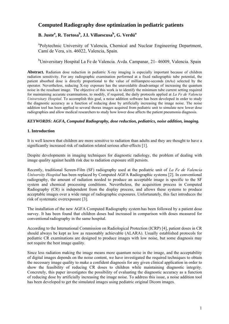
We have analyzed several CR images in a range of different image qualities, where statistical noise is the relevant quality parameter to be varied through mAs reduction. The purpose of this study is to determine a better image quality/patient radiation exposure optimization. 2. Methodology Given the important impact that delivered overdose could have on the children health, limiting CR radiation dose is especially important to avoid overdose that could cause them health problems. Nevertheless, reducing tube current increases the noise content of the images [5], [6]. The main goal of this study is to determine how variation in mAs tube current impacts in the image quality of pediatric explorations. We describe an application of the method centered on thorax CR, which evaluates the effect of noise in children pneumonia diagnostic [7]. 2.1. Experimental Procedure Dose reduction radiographic techniques were first determined as a function of exposure variation using the chest of the RANDO® female Phantom (Alderson). This phantom is constructed with a natural human skeleton which is cast inside soft tissue-simulating material. Tissues in the phantom are designed to have the same absorption as human tissue at the normal radiotherapy exposure levels. Figure 1: Chest Rando phantom.
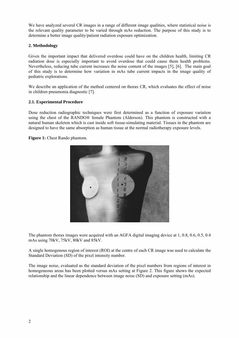
The phantom thorax images were acquired with an AGFA digital imaging device at 1, 0.8, 0.6, 0.5, 0.4 mAs using 70kV, 75kV, 80kV and 85kV. A single homogenous region of interest (ROI) at the centre of each CR image was used to calculate the Standard Deviation (SD) of the pixel intensity number. The image noise, evaluated as the standard deviation of the pixel numbers from regions of interest in homogeneous areas has been plotted versus mAs setting at Figure 2. This figure shows the expected relationship and the linear dependence between image noise (SD) and exposure setting (mAs).
3
Figure 2: Relationship between image noise (SD) and exposure setting (mAs) at different kVp.
0.2 0.3 0.4 0.5 0.6 0.7 0.8 0.9 1 1.1 1.220
25
30
35
40
45
50
55
60
Image tube current (mAs)
Sta
ndar
d D
evia
tion
of a
n ho
mog
eneo
us re
gion
of i
nter
est (
SD
)
70 KVP
y = -41.194*x+69.291 r2 = 0.98
y = -22.407*x+55.464r2 = 0.90
75 KVP
85 KVP
80 KVPy = -25.2*x+52.016r2 = 0.83
y = -15.193*x+40.052r2 = 0.89
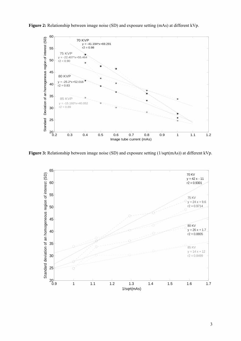
Figure 3: Relationship between image noise (SD) and exposure setting (1/sqrt(mAs)) at different kVp.
0.9 1 1.1 1.2 1.3 1.4 1.5 1.6 1.720
25
30
35
40
45
50
55
60
65
1/sqrt(mAs)
Sta
ndar
d de
viat
ion
of a
n ho
mog
eneo
us re
gion
of i
nter
est (
SD
)
70 KV y = 42 x - 11r2 = 0.9301
75 KV y = 24 x + 9.6r2 = 0.9714
80 KV y = 26 x + 1.7r2 = 0.8805
85 KV y = 14 x + 12r2 = 0.8499
4
2.2. Simulation techniques Reduction in tube current (mAs) image simulation is obtained by adding to the original radiography a Gaussian distributed random noise without altering the mean signal level. The simulation model assumes that we can add some amount of Gaussian distributed random noise σ(add) to a CR image obtained at tube current A1 mAs in order to modify the original image data set as if it had been collected with a lower tube current A2 mAs. Quantum mottle noise (X-ray photon statistics) obeys Poisson statistics which states that the noise is proportional to the square root of the number of photons. Since each CR is the number of detected photons times a gain factor multiplier, the additional random noise σ(add) can be expressed using the original image data and the desired new mAs value. The standard deviation of pixels value from a region of interest (ROI), σ(A1), is the noise corresponding to the exposure setting A1 mAs. We want to add statistical noise by altering the pixels numbers to give a standard deviation σ(A2) corresponding to a lower exposure setting A2 mAs. The main noise source comes from X-ray detection, giving a ratio of the noise levels at the two selected exposures:
A2/A1 = (A1)(A2)/σσ (1) from this equation σ(A2) can be calculated. The noise distributions are independent, so the standard deviation of the noise distribution to be added, σ (add), can be obtained as follows:
(add) (A1)(A2) 222 σσσ += (2) This equation is used to estimate the amount of noise to be added to a Dicom Agfa computed radiography in order to obtain the same image as it had been taken with lower tube current. 2.2.1. Software validation adjustment The expected relationship between image noise and tube current has been checked by imaging the Rando female phantom at a range of different mAs values. Starting with a phantom image at a known tube current, statistical noise (the standard deviation of pixels number within a region) in the image was measured and the expected increase in noise at a reduced exposure was calculated using equation 2 as well as data from figure 2 and 3 and added to the initial image. Standard deviation differences obtained from figures 2 and 3 were always below the 7% for this range of current, being those obtained from figure 3 slightly larger.
According to this, lower dose images of a CR phantom were created using data from figure 3 and simulated using the developed software. Distributed noises from each image were measured and compared with those obtained with CR taken with the real lower dose.
Each original CR phantom obtained from the experimental procedure have been subjected under different simulations in order to obtain a range of several images as if they had been taken with lower exposures. The ratios between the noise measurement of simulated images and the real CR with the same tube current setting, are showed in Table I.
5
Table I: Ratios of the simulated noise measured from real CR.
Rando Phantom 75 KV
Original
Simulated
0.5 mAs 0.6 mAs 0.8 mAs 1 mAs 0.4 mAs 1.10 0.99 1.10 1.01 0.5 mAs 0.96 1.09 0.96 0.6 mAs 1.08 1.1 0.8 mAs 1.05
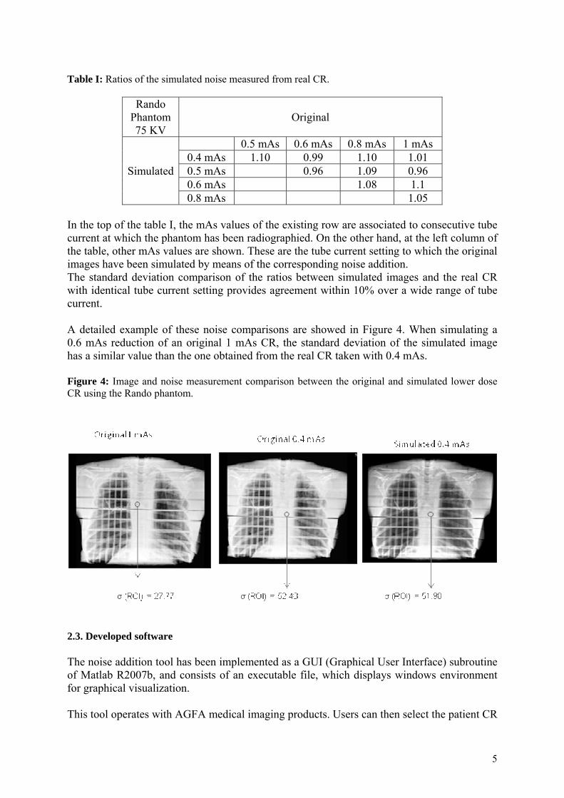
In the top of the table I, the mAs values of the existing row are associated to consecutive tube current at which the phantom has been radiographied. On the other hand, at the left column of the table, other mAs values are shown. These are the tube current setting to which the original images have been simulated by means of the corresponding noise addition. The standard deviation comparison of the ratios between simulated images and the real CR with identical tube current setting provides agreement within 10% over a wide range of tube current. A detailed example of these noise comparisons are showed in Figure 4. When simulating a 0.6 mAs reduction of an original 1 mAs CR, the standard deviation of the simulated image has a similar value than the one obtained from the real CR taken with 0.4 mAs. Figure 4: Image and noise measurement comparison between the original and simulated lower dose CR using the Rando phantom.
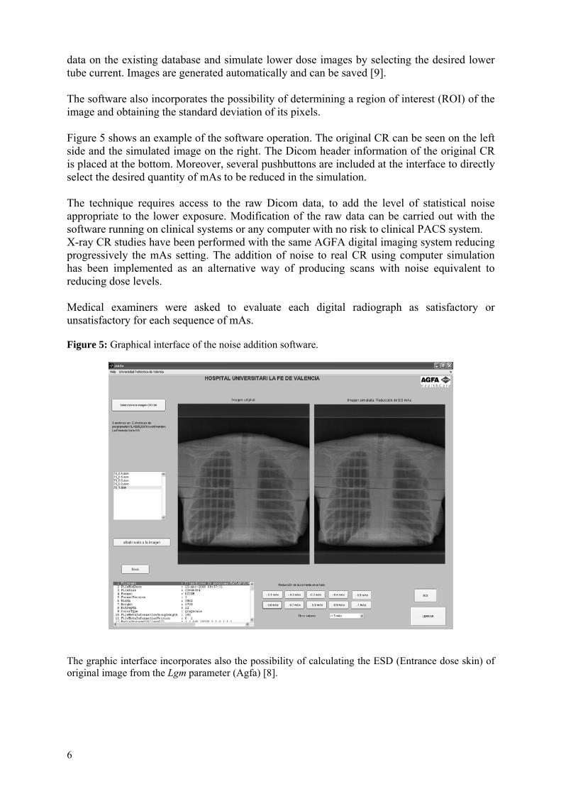
2.3. Developed software The noise addition tool has been implemented as a GUI (Graphical User Interface) subroutine of Matlab R2007b, and consists of an executable file, which displays windows environment for graphical visualization. This tool operates with AGFA medical imaging products. Users can then select the patient CR
6
data on the existing database and simulate lower dose images by selecting the desired lower tube current. Images are generated automatically and can be saved [9]. The software also incorporates the possibility of determining a region of interest (ROI) of the image and obtaining the standard deviation of its pixels. Figure 5 shows an example of the software operation. The original CR can be seen on the left side and the simulated image on the right. The Dicom header information of the original CR is placed at the bottom. Moreover, several pushbuttons are included at the interface to directly select the desired quantity of mAs to be reduced in the simulation. The technique requires access to the raw Dicom data, to add the level of statistical noise appropriate to the lower exposure. Modification of the raw data can be carried out with the software running on clinical systems or any computer with no risk to clinical PACS system. X-ray CR studies have been performed with the same AGFA digital imaging system reducing progressively the mAs setting. The addition of noise to real CR using computer simulation has been implemented as an alternative way of producing scans with noise equivalent to reducing dose levels. Medical examiners were asked to evaluate each digital radiograph as satisfactory or unsatisfactory for each sequence of mAs. Figure 5: Graphical interface of the noise addition software.
The graphic interface incorporates also the possibility of calculating the ESD (Entrance dose skin) of original image from the Lgm parameter (Agfa) [8].
7
3. Results and validation. Thorax image evaluation Once the software was validated, some real images concerning pneumonia cases have been tested with the aid of pediatric doctors. Patients (age range, 1 month to 15 years) were chosen from among children who were undergoing medical pneumonia evaluation and those being examined during routine visits to the pediatric clinic. After revising the case of 10 different cases, it has been established that pursuit radiographies of this kind of disease can be taken reducing 0.5 mAs the original used current.
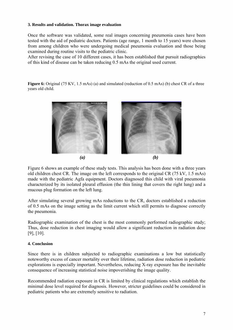
Figure 6: Original (75 KV, 1.5 mAs) (a) and simulated (reduction of 0.5 mAs) (b) chest CR of a three years old child.
(a) (b)
Figure 6 shows an example of these study tests. This analysis has been done with a three years old children chest CR. The image on the left corresponds to the original CR (75 kV, 1.5 mAs) made with the pediatric Agfa equipment. Doctors diagnosed this child with viral pneumonia characterized by its isolated pleural effusion (the thin lining that covers the right lung) and a mucous plug formation on the left lung. After simulating several growing mAs reductions to the CR, doctors established a reduction of 0.5 mAs on the image setting as the limit current which still permits to diagnose correctly the pneumonia. Radiographic examination of the chest is the most commonly performed radiographic study; Thus, dose reduction in chest imaging would allow a significant reduction in radiation dose [9], [10].
4. Conclusion Since there is in children subjected to radiographic examinations a low but statistically noteworthy excess of cancer mortality over their lifetime, radiation dose reduction in pediatric explorations is especially important. Nevertheless, reducing X-ray exposure has the inevitable consequence of increasing statistical noise impoverishing the image quality. Recommended radiation exposure in CR is limited by clinical regulations which establish the minimal dose level required for diagnosis. However, stricter guidelines could be considered in pediatric patients who are extremely sensitive to radiation.
8
This work allows medical physicists and radiologists to determine the dose reduction that could maintain an accurate diagnostic in pediatric CR. The connection between CR image noise and tube current (mAs) setting has been studied using an AGFA CR noise addition tool. This software simulates lower dose images from existing patient CR data and allows studying the effect of lower exposure on the diagnostic ability. The software has been validated using the RANDO® female Phantom (Alderson) obtaining 10% agreement in terms of noise measurement over a wide range of tube current. The computer noise simulation tool is being now applied at La Fe de Valencia University Hospital to study the possibility of reducing image dose at children. Concretely thorax explorations are being analyzed. Results indicate that substantial reductions in the radiation dose associated with ordinary radiographic exams using CR are possible. Since different medical imaging tasks require different levels of image quality, we expect to avoid unnecessary children doses, which have no additional benefit for the clinical purpose intended. To that, the radiologist and medical physicist must also cooperate to assure that doses are maintained at the lowest level consistent with diagnostic quality. Acknowledgements The authors wish to thank The Hospital Universitary la Fe de Valencia for its collaboration. REFERENCES [1] BRENNER, D.J., ELLISTON, C.D., HALL, E.J., BERDON, W.E., “Estimated risks of
radiation-induced fatal cancer from pediatric CT,” American Journal of Roentgenology, pp. 289-296, (2001).
[2] AGFA healthcare. Available: http://www.agfa.com/healthcare [3] GUR, D, FUHMAN, CR, FEIST, JH, et al (1993) “Natural migration to a higher dose in CR
imaging”. Proc 8th European Congress of Radiology, Vienna, Sept 12–17, p 154. [4] ICRP Publication 93: Managing patient dose in digital radiology, 93 [5] WILLIS, C. E., “Strategies for dose reduction in ordinary radiographic examinations using CR
and DR.” Pediatric Radiology 34, (2004) pp.196-200. [6] HUFTON, A. P., DOYLE, S. M., AND CARTY, H. M. L., “Digital radiography in paediatrics:
radiation dose considerations and magnitude of possible dose reduction,” The British Journal of Radiology. 71, pp. 186–199, (1998).
[7] LI, J., TOTH, T., MCOLASH, S., HSIEH, J. AND BROMBERG N., “Simulating Low Dose CT Scans by Noise Addition,” GE Medical Systems. Nuclear Science Symposium Conference Record, (2002) IEEE.
[8] JUSTE, B., VILLAESCUSA, J. I., GRANERO, D., VERDÚ G., “Improving pediatric radiation dose management using Agfa digital radiography DICOM header information” EMBC 07 Proceedings of the 29th Annual International Conference of the IEEE EMBS Cité Internationale, Lyon, France August 23-26, (2007).
[9] COHEN MD, LONG B, CORY DA, et al “Digital imaging of the newborn chest”. Clinic Radiol 40:365–368 13, (1989).
[10] BARNES GT, SONES RA, TESIC MM. “Digital chest radiography: performance of a prototype unit”. Radiology (1985) ;154:801-806