Embed Size (px)
Citation preview

6/21/2011
1
Break Time
Fluid Exchanges
1
Pneumonia
• General term for several types of inflammation
• Unilateral/Bilateral/portion of lung
• Inflammation WITH infection– Most common cause of infectious death in US
– 5th leading cause of death in US
• Bacterial
• Viral
• Other– Aspiration
2
Pneumonia S&S
• Vary
• Cough, fever, SOB, chills, sweating
• Chest pain, cyanosis
• Blood in sputum
3
6/21/2011
2
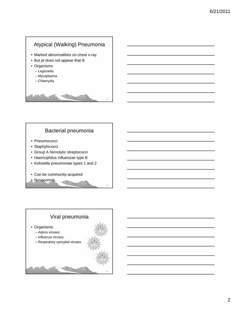
Atypical (Walking) Pneumonia
• Marked abnormalities on chest x-ray
• But pt does not appear that ill
• Organisms– Legionella
– Mycoplasma
– Chlamydia
4
Bacterial pneumonia
• Pneumococci
• Staphylococci
• Group A hemolytic streptococci
• Haemophilus influenzae type B
• Kebsiella pneumoniae types 1 and 2
• Can be community-acquired
• Nosocomial5
Viral pneumonia
• Organisms– Adeno viruses
– Influenza viruses
– Respiratory syncytial viruses
6
6/21/2011
3
Other Pneumonia Etiology
• Aspiration – Liquids or other material into trachea
– Serious swallowing problems
– Elderly
– Weak
– Neurologic problems
7
Pneumonia Dx & Tx
• Diagnosis– H&P
– Chest x-ray
– ABGs
– Bronchoscopy
– Sputum/Blood cultures
• Treatment– Underlying cause
– Antibiotics (bacterial)
– Penicillin (pneumococcal)
– Tetracycline
– Erythromycin
– Sulfonamides
– Analgisics
– O2
– Bed rest, inc. fluids, high-calorie diet, postural drainage
8
Pneumonitis
• Lung Inflammation WITHOUT infection
• S&S– SOB
– Dry cough
– Chronic pneumonitis• Fatigue
• Loss of appetite
• Weight loss (unintentional)
• Scarred tissue (fibrosis)
• Etiologies– Hypersensitivity/Chemical
– Irritating substances inflame alveoli
• Bird feathers/excrement
• Dusts
• Molds
– Hot Tub lung, Farmer’s lung
• Chemicals
• Chemotherapy/Radiotherapy
• Medications – aspirin overdose
• Humidifiers9

6/21/2011
4
Pneumonitis Dx & Tx
• Diagnosis
– Imaging (x-ray/CT)• Through all 5 lobes
– PFTs
– Bronchoscopy w/lavage, bx
• Treatment
– Avoid irritants• Mask/respiratory
• Stopping hobby
– Corticosteroids
– O2
– Antibiotics• IF infection present
10
Pneumothorax
• Collection of air in pleural cavity (between two layers)– May cause
collapsed lung
• Spontaneous– Air leaks from lung
• Pulmonary disease
• Tumor
• Pulmonary tissue tear
• Traumatic– Air enters from outside body
• GSW
• Stabbing
• Crushing injury
• Rib fx11
Tension pneumothorax
• Particularly dangerous form
• Occurs when air escapes into the pleural cavity from a bronchus
• Air cannot regain entry into the bronchus
• Continuously increasing air pressure in the pleural cavity
• Causes progressive collapse of the lung tissue
• Mediastinal shift
12
6/21/2011
5
Pneumothorax S&S
• Depend on severity of lung collapse
• Complete collapse– Sudden, severe chest pain
– Severe dyspnea
– Symptoms of shock
• Weak, shallow respirations
• Sucking sounds at trauma site
• Mediastinal shift to unaffected side (emergency)
13
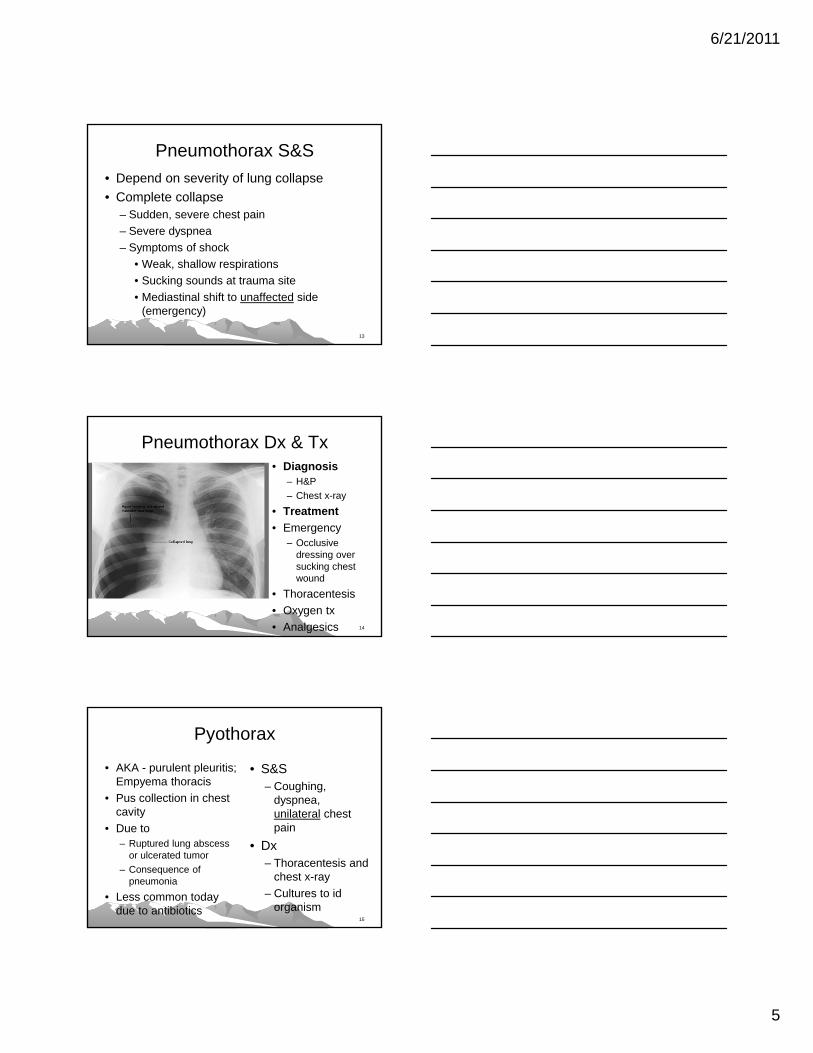
Pneumothorax Dx & Tx• Diagnosis
– H&P
– Chest x-ray
• Treatment
• Emergency– Occlusive
dressing over sucking chest wound
• Thoracentesis
• Oxygen tx
• Analgesics 14
Pyothorax
• AKA - purulent pleuritis; Empyema thoracis
• Pus collection in chest cavity
• Due to – Ruptured lung abscess
or ulcerated tumor
– Consequence of pneumonia
• Less common today due to antibiotics
• S&S– Coughing,
dyspnea, unilateral chest pain
• Dx– Thoracentesis and
chest x-ray
– Cultures to id organism
15

6/21/2011
6
16http://radiology.med.sc.edu/empyema.htm
Elliptical fluid collection in major fissureFissure location supports empyema versus lung abscess.
Respiratory Failure
• Impaired Gas Exchange
– Too little O2 in blood (Hypoxemic)
– Too much CO2 in blood (Hypercapnic)
– OR both at same time
17http://www.bbc.co.uk/scotland/learning/bitesize/standard/biology/investigating_cells/cells_and_diffusion_rev3.shtml
Respiratory Failure
• Acute– Short term
– Can develop quickly
– May require emergency tx
• Chronic– On-going
– Develops more slowly
– Lasts longer
18

6/21/2011
7
Respiratory Failure S&S
• Shortness of breath (hypoxemia)
• Tachypnea (hypercapnea)
• Air hunger (hypoxemia)– feeling that can't breathe in enough air
• Cyanosis (hypoxemia)
• Confusion (hypercapnia)
• Sleepiness (hypoxemia)
• Arrhythmias (hypoxemia)19
Respiratory Failure Dx & Tx
• Diagnosis
• H&P
• Pulse oximetry
• ABGs
• Chest x-ray (underlying condition)
• EKG
• Treatment– Underlying condition
• Acute– ICU
– O2
• Tracheostomy
• Ventilator
• Chronic– Home/LTC
– Portable O2
– CPAP/Rocking bed 20
SARS
• Severe Acute Respiratory Syndrome
• 2003 –first outbreak in China
• Corona virus mutation
• 8,098 people worldwide; 774 died
• US – 8 people w/infection– All had traveled outside US
• Airborne droplets; touching contaminated surfaces and then face/eyes
21
http://udel.edu/~cmcneil/coronavirus.html

6/21/2011
8
Tuberculosis (A15-18)• Acute, subacute, or chronic inflammation
• Mycobacterium tuberculosis infection– Human-ONLY infection – 5,000+ years
– Tubercle bacillus; Spread by inhaling droplets
– Can remain suspended in air for many hours
– Dry form can survive for months (if no sunlight)
– Primary lesion in lung; Any body tissue can be affected
• M. bovis in cattle/buffalo/deer – we can get
• M. avium in birds – we can get22
Pulmonary TB
• Tubercle– any small, rounded mass produced by
infection with Mycobacterium tuberculosis
• TB can go to other/multiple body areas– almost any part of the body
– bone marrow, bones, CNS, joints, muscles, lymphatic system, urinary tract
• 1/3 of humans have TB infection!
• 2 million deaths every year
• 23
TB Stages and S&S
• Latent stage– Macrophages wall-off
– Positive PPD test
– No S&S
– Can’t transmit to others
– 10% will progress to active stage
– More if immuno-compromised
• HIV
• Elderly, infants
– Meds can prevent active stage - isoniazid (INH)
• Active stage– Can transmit
– Expelled saliva• cough, sneeze, talk, spit
• S&S– Bad cough 3+ weeks
– Weight loss
– Coughing up blood/mucus
– Weakness or fatigue
– Night sweats
– Fever and chills
24

6/21/2011
9
TB Diagnosis
• H&P
• Chest x-ray
• Mantoux (PPD) test– False positive
– False negative
• Acid-fast bacilli (AFB) smear
• QuantiFERON-TB Gold – blood test (2005)
• + M. tuberculosis culture– confirms TB Dx
25
http://www.med.cmu.ac.th/dept/pediatrics/04-divisions_home_thai/09-id-home/int-cases/ic-id61/page1.htm
Active TB Medications
• 4 medications at same time– Nydrazid® or INH (isoniazid)
– Rifadin® (rifampin)
– Myambutol® (ethambutol)
– pyrazinamide
• Many other meds/combinations due to multidrug-resistant TB (MDR-TB) strains
26
TB in Montana
• 6 new cases in MT in 2010
• State Tuberculosis Sanitarium at Galen– 1913-1993
– Relatively close to Butte• Death rate from TB in Butte was 2x national average
27http://www.montana.edu/cpa/news/nwiprint.php?article=609

6/21/2011
10
URIs• Univ. of Maryland Medical Center website
• Common cold– http://www.umm.edu/ency/article/000678.htm
• Rhinitis– http://www.umm.edu/allergies/rhinitis.htm
• Sinusitis – http://www.umm.edu/patiented/articles/sinusitis_000062.htm
• Pharyngitis– http://www.umm.edu/ency/article/000655.htm
• Laryngitis– http://www.umm.edu/altmed/articles/laryngitis-000099.htm28
Aging and Respiratory System
• Increased risks in elderly
• Less effective immune system
• Loss of elasticity & weaker muscles
• Less efficient
• Less reserve
• Changes in posture
• = Lower tolerance for exercise
29
Review of ICD-10-CM Ch10
• Organization (acute vs chronic; upper/lower)
• Guidelines (minimal)– COPD and Asthma
– Acute Respiratory Failure
– Influenza
– Ventilator associated pneumonia (VAP)
• Excludes1 and Excludes2 notes
• Combination and Multiple coding (External causes)
30

6/21/2011
11
ICD-10-CM Organization• Chapter
• Blocks – (J00-J06 Acute URIs)
• Category –– 3 alphanumeric
characters (J01)
• Subcategory – 4 or 5 alphanumeric characters– Decimal AFTER 1st 3
characters
• Codes – 3-7 characters
• X = 5th/6th digit placeholders for future expansion– T36.0x1 = Poisoning by,
adverse effect of and underdosing of penicillins
• 7th character – A initial encounter
– D subsequent encounter
– S sequela
31
Chapter 10 Blocks
• J00-J06 Acute URIs
• J09-J18 Flu and pneumonia
• J20-22 Other acute lower resp. inf.
• J30-J39 Other diseases of URT
• J40-47 Chronic LR diseases
• J60-J70 Lung diseases due to external agents
• J80-84 Other resp. dis. princ. Affecting interstitium
• J85-J86 Suppurative/ necrotic conditions of LRT
• J90-J94 Other disease of pleura
• J95 Intraoperative / postprocedural complications NEC
• J96-99 Other respiratory diseases
32
Chapter 10 Guidelines• COPD and Asthma
– With and without acute exacerbation
– Definition of acute exacerbation
• Acute Respiratory Failure– Principal dx sequencing, As 2ndary dx
– Sequencing of ARF & another acute condition
• Influenza – avian/H1N1 code only confirmed cases (exc. Inpatient guideline)
• VAP (2011) -– Dr. documentation, sequencing 33

6/21/2011
12
Acute Exacerbation (ICD-10-CM)
• A worsening or a decompenstation of a chronic condition
• NOT equivalent to an infection superimposed on a chronic condition
• Exacerbation may be triggered by an infection
• Increase in seriousness of a disease or disorder, marked by greater intensity in S&S – Mosby's Medical
Dictionary, 8th ed.
34
Chapter 10 Notes
• Location of notes
• Beginning of chapter– Multiple sites not specifically indexed, code to
lower site
– Use add’l code for exposure to/use of tobacco smoke
• Excludes2 note (multiple codes)– Perinatal, infectious disease, injury, etc.
35
ICD-10- CM Excludes Notes
• Two types
• Excludes1 – “pure” - NOT coded here– Indicates MUTUALLY EXCLUSIVE codes
– One code OR the other, never both
• Excludes2 – Not included here– Permitted assignment of two+ codes
– Assign more than one code IF documentation
36

6/21/2011
13
Ch 10 combination codes
• Many very specific codes that include– Location
• J01.00 Acute maxillary sinusitis, unspec.
• J43.1 Panlobar emphysema
– Etiology• J03.011 Acute recurrent strep tonsillitis
• J61 Pneumoconiosis due to asbestos …
– Stages• J45.31 Mild persistent asthma with (acute)
exacerabtion
– W/Wo• J04.10/11 Acute tracheitis w/o, w obstruction 37
Ch 10 multiple coding
• Code also any assoc. FB in respiratory tract
• Code also type of asthma (COPD)
• Code first any associated
– Lung abscess (J85.1)
– Therapy (T45.1x- Y84.2))
38
Chapter 10 Multiple coding• Code first any underlying disease
– Rheumatic fever (I00)
– Underlying neoplasm
– Appropriate code from T41
• Code first – (T51-T65) to identify cause
– (T36-T50 w 7th char. S) to id drug
– (T51-T65) to identify external agent
– (T51-T65) to identify substance
39
6/21/2011
14
Chapter 10 Multiple coding
• Use additional code to id
– Drug (T36-T50)
– External cause (W88-W90, X39.0-)
– Infectious agent (B95-B97)
– Virus (flu) (B97)
– Type of pneumonia (B95-97)
– Disorder
40
ICD-10-CM Coding Example• D/C Dx: Moderate persistent asthma with status
asthmaticus, COPD, acute bronchitis
• Pt w/gradual increase in SOB, unresponsive to home nebulizer. Pt had malaise, coughing, fever. In ER, he rec. respiratory tx, but did not improve. At admission, theophlline level was 5.9. Chest x-ray - no infiltrates. Bolused with IV steriods and rec. frequent RT. IV antibiotics for acute bronchitis.
• IV aminophylline boluses & drip increased theophylline to therapeutic range.
• Ventolin tx decreased to q 4 hr. and steroids rapidly tapered back to 10 mg of Prednisone as he improved
41
Coding Example, cont.• J44.0 Disease, lung, obstructive (chronic),
with acute, bronchitis– Excludes1 chronic bronchitis
– Code also type of asthma, if applicable
• J45.42 Asthma, moderate persistent, with, status asthmaticus– Excludes2 asthma with COPD
• J20.9 Bronchitis, acute or subactute– Excludes2 acute bronchitis with COPD
42

6/21/2011
15
Homework Answers
43
1. Which feature is found only in the left lung?
Cardiac notchHorizontal fissureOblique fissureSuperior lobar bronchusThree lobes
44
2. Which part of the left lung might partially fill the costomediastinal recess in full inspiration?
ApexCupolaHilumLingulaMiddle lobe
45
6/21/2011
16
3. The oblique fissure of the right lung separates which structures?
Lower lobe from lingulaLower lobe from upper lobe onlyLower lobe from both upper &
middle lobesLower lobe from middle lobe onlyUpper from middle lobe
•46
4. A 4-year-old girl is coughing, and mother states she was playing with some beads and had apparently aspirated one. Where would it most likely be?
Apicoposterior segmental bronchus of L lung
Left main bronchusLingular segment of left lungRight main bronchusTerminal bronchiole of right lung, lower
lobe47
5. Which statement is true about the right lung? It is slightly smaller than the left lungIt has a lingular segmental bronchusIt occupies the rightmost portion of
mediastinumIts upper lobar bronchus lies behind and
above the right pulmonary arteryIt has the right phrenic nerve passing
posterior to the lung root
48

6/21/2011
17
6. A 10-y-o boy had a tonsillectomy. At home he lay supine in bed for 2 weeks, developing a fever, chest pain, & cough. He returned to the hospital and was diagnosed w/R lung pneumonia due to aspiration during tonsillectomy. In which broncho-pulmonary segment of the lung would fluid (pus) most likely accumulate by the force of gravity?
Anterior basal segment--inferior lobeAnterior segment--superior lobeLateral segment--middle lobeSuperior segment--inferior lobeSuperior lingual segment--lingula
49
7. You are observing a doctor perform a bronchoscopy. As he passes the scope down the trachea, a cartilagenous structure is seen separating the R and L main stem bronchi. He asks what it is called. You reply that it really does look like a ship's keel and that it is called the
CarinaCricoid cartilageCostal cartilagePulmonary ligamentTracheal ring
50
8. Because of its angle with the trachea and size of the main bronchus, a bronchoscope would pass more readily into which lung?
LeftRight
51
6/21/2011
18
9. How do the lungs stay inflated?
– Each lung is ribbed with cartilage to prevent collapse on exhalation
– The lungs are tethered to the ribcage with a network of connective tissue
– The lungs rely on a vacuum within the chest, maintained by the diaphragm
52
10. Our breathing is controlled by the part of the brain called thecerebrummedulla oblongataspinal cord
53
11. Which of these structures has NO cartilage around it? – primary bronchus
– secondary bronchus
– terminal bronchiole
– larynx
– trachea
54

6/21/2011
19
12. If a person's vital capacity is 4000mL, and her expiratory reserve volume is 1000mL and her inspiratory reserve volume is 2500mL, then her tidal volume is– 3500mL
– 3000mL
– 1500mL
– 1000mL
– 500mL
55
13. A segment of lung tissue that is bounded by connective tissue partitions and supplied by a single bronchiole is called– a lobe.
– an alveolar sac.
– an alveolar duct.
– a lobule.
56
14. The mucous membrane lining the nasal cavity and upper pharynx has a protective function that relates to which of the following organ systems?– nervous system
– endocrine system
– lymphoid system
– integumentary system
57

6/21/2011
20
15. Activity of which of the following organ systems generates carbon dioxide?– muscular system
– nervous system
– skeletal system
– endocrine system
58
General Resources• Frazier, M. S. & Drzymkowski, J. W. Essentials of
Human Diseases and Conditions, 4th ed., Saunders, 2009
• Gray, H. Anatomy of the Human Body. 1918.
– http://www.bartleby.com/107/
• Neighbors, M. & Tennehill-Jones, R. Human Diseases, Thomson Learning, 2000.
• Scott, A. S. & Fong, E. Body Structures and Functions, 11th ed., Delmar, 2009
ICD 10 CM
• 2011 release of ICD 10 CM– http://www.cdc.gov/nchs/icd/icd10cm.htm
– Preface [PDF - 93 KB]
– ICD-10-CM Guidelines [PDF - 494 KB]
– ICD-10-CM PDF Format
– ICD-10-CM XML Format
– ICD-10-CM 2010 to 2011 Addenda
– ICD-10-CM List of codes and Descriptions
– General Equivalence Mapping Files60

6/21/2011
21
Asthma Resources
• Asthma. Interactive tutorial. MedlinePlus. http://www.nlm.nih.gov/medlineplus/tutorials/asthma/
htm/index.htm
• Asthma. CDC.
– http://www.cdc.gov/asthma/default.htm
• Subbarao, P. et.al. Asthma: epidemiology, etiology and risk factors. CMAJ.
– http://www.cmaj.ca/cgi/content/full/181/9/E181
• S. Agarwal, MD, S. Kache, MD. Status Asthmaticus.– http://peds.stanford.edu/Rotations/picu/pdfs/14_status_asthmaticus.pdf
61
Resources• Bronchiectasis. National Heart Lung and
Blood Institute. – http://www.nhlbi.nih.gov/health/dci/Diseases/b
rn/brn_signsandsymptoms.html
• Coughing. MedlinePlus. Video.– http://www.nlm.nih.gov/medlineplus/ency/anat
omyvideos/000039.htm
• Evaluation of the Patient with Chronic Cough - May 1, 2004 – http://www.aafp.org/afp/2004/0501/afp200405
01p2159-f1.gif 62
Resources
• Breath sounds. MedlinePlus– http://www.nlm.nih.gov/medlineplus/ency/articl
e/003323.htm
• Costochondritis. Mayo Clinic– http://www.mayoclinic.com/health/costochondritis/DS
00626
• LungLab Tour. Lawrence Berkeley National Laboratory.– http://imglib.lbl.gov/ImgLib/COLLECTIONS/LUNG_ST
RUCTURE/.tour/page1.html63

6/21/2011
22
Resources
• Pneumonitis. Mayo Clinic.– http://www.mayoclinic.com/health/pneumonitis
/DS00962
• Respiratory Failure. Nat’l Heart Blood & Lung Institute.– http://www.nhlbi.nih.gov/health/dci/Diseases/rf
/rf_whatis.html
• Severe Acute Respiratory Syndrome (SARS). Fact Sheet. US CDC.– http://www.cdc.gov/ncidod/sars/factsheet.htm64
Hanta Resources
• Hanta virus (ppt). Center for Food Security and Public Health. Iowa State University.– http://www.authorstream.com/Presentation/Tirone-
58187-Hantavirus-Overview-Organism-Hantaviruses-Old-World-New-History-Past-Hemorrhagic-Feve-as-Travel-Places-Nature-ppt-powerpoint/
• Hanta virus Fact Sheet. Washington State Dept. of Health.– http://www.doh.wa.gov/ehsphl/factsheet/hanta.htm
65
TB Resource
• Tuberculosis. Natural Standard Research Collaboration. 2009?– http://www.righthealth.com/topic/Tuberculosis/overvie
w/NaturalStandard20?fdid=NaturalStandard_9963952962731a41652573f3dcb08d64§ion=Full_Article
66