Embed Size (px)
Citation preview

NOTE
PARP Inhibitors as P-glyoprotein Substrates
DENISE LAWLOR,1 PATRICIA MARTIN,1 STEVEN BUSSCHOTS,1 JULIEN THERY,1 JOHN J. O’LEARY,1
BRYAN T. HENNESSY,2 BRITTA STORDAL1
1Department of Histopathology, St James’ Hospital and Trinity College Dublin, Dublin 8, Ireland2Department of Medical Oncology, Beaumont Hospital and Royal College of Surgeons Ireland, Dublin 9, Ireland
Received 6 August 2013; revised 27 February 2014; accepted 28 February 2014
Published online in Wiley Online Library (wileyonlinelibrary.com). DOI 10.1002/jps.23952
ABSTRACT: The cytotoxicity of PARP inhibitors olaparib, veliparib, and CEP-8983 were investigated in two P-glycoprotein (P-gp) over-expressing drug-resistant cell models (IGROVCDDP and KB-8-5-11). IGROVCDDP and KB-8-5-11 were both resistant to olaparib andresistance was reversible with the P-gp inhibitors elacridar, zosuquidar, and valspodar. In contrast, the P-gp overexpressing models werenot resistant to veliparib or CEP-8983. Olaparib and veliparib did not induce protein expression of P-gp in IGROVCDDP or KB-8-5-11at doses that successfully inhibit PARP. Olaparib therefore appears to be a P-gp substrate. Veliparib and CEP-8983 do not appear to besubstrates. Veliparib and CEP-8983 may therefore be more useful in combined chemotherapy regimens with P-gp substrates and may beactive in platinum and taxane-resistant ovarian cancer. C© 2014 Wiley Periodicals, Inc. and the American Pharmacists Association J PharmSciKeywords: olaparib; veliparib; CEP-8983; PARP inhibitor; drug Resistance; cell lines; P-glycoprotein; cancer chemotherapy; toxicity
INTRODUCTION
Parp inhibitors are a new class of chemotherapy agents that tar-get the cell’s DNA damage repair pathways. PARP inhibitorsare potentially very useful for treating BRCA1/2-dysfunctionalcancers, as in these cancers the DNA repair machinery is al-ready impaired. The results of proof of concept clinical trialsof the PARP inhibitor olaparib in breast and ovarian can-cer patients with germline BRCA1/2 mutations have beenencouraging.1,2
For any new chemotherapy agents, it is important to estab-lish if they are substrates of the classical ABC transporters,such as P-glycoprotein (P-gp). Agents that are not P-gp sub-strates may be more useful clinically, as if transporter-drivendrug resistance develops the cells are unlikely to be resistantto the wide range of chemotherapy drugs that are also P-gp
substrates. P-gp mRNA has been detected in primary ovar-ian tumors,3 and its expression has been associated with pooroverall survival.3 Between 16-25% of primary ovarian tumorsare highly positive for P-gp by immunohistochemistry (IHC).4–6
There is limited clinical data to support the induction of P-gp inthe clinic, unlike in cancer cell lines treated with chemotherapy.Despite this, some studies have shown P-gp staining to increasein ovarian tumors after chemotherapy.6 P-gp has been shownto be an independent prognostic factor in some ovarian cancerstudies4 but not in others.5,6 Similarly, between 44% and 66%7,8
of breast cancers stain positive for P-gp by IHC, some studiesfound it to be an independent prognostic factor7 and othersdid not.8 The induction of P-gp in response to doxorubicin andepirubicin treatment was found to be predictive of survival in
Correspondence to: Britta Stordal (Telephone: +353-1-8962876; Fax: +353-1-8963285; E-mail: [email protected])
Denise Lawlor and Patricia Martin contributed equally to this work.This article contains supplementary material available from the authors upon
request or via the Internet at http://onlinelibrary.wiley.com/.
Journal of Pharmaceutical SciencesC© 2014 Wiley Periodicals, Inc. and the American Pharmacists Association
one breast cancer study.9 The role of P-gp in BRCA1-mutatedclinical breast or ovarian cancer has not been studied in detail.However, a study that examined the gene expression profiles ofBRCA1/2 tumors (n = 34) versus sporadic ovarian cancer (n =27) in an Ashkenazi Jewish population did not find P-gp to besignificantly differentially expressed.10
There is currently limited data on the P-gp substrate statusof PARP inhibitors. Olaparib has been shown to induce P-gpgene expression in an animal tumor model.11 Veliparib has beendescribed as a weak P-gp substrate in a study using a P-gptransfected cell line.12 In contrast, the novel PARP inhibitorCEP-8983 has not been examined for its P-gp substrate status.There has also been no work to date examining PARP inhibitorsusing cell models of acquired drug resistance overexpressingP-gp. This study will examine the PARP inhibitors olaparib,veliparib, and CEP-8983 in two cell models of acquired drugresistance where the major mechanism of drug resistance isoverexpression of P-gp: IGROVCDDP ovarian cells13 and KB-8-5-11 cervical cells.14,15
MATERIALS AND METHODS
Cell Culture and Cytotoxicity Assays
IGROV-1 and IGROVCDDP ovarian cancer cells16,17 were ob-tained from Prof. Jan Schellens (Netherlands Cancer Institute)and grown as previously described.13 KB-3-1 and KB-8-5-11 cer-vical cancer cells14,15 were obtained from Prof. Michael Gottes-man (National Cancer Institute) and grown in Dulbecco’s mod-ified Eagle’s medium (Sigma, Dublin, Ireland), 1% Pen strep,2% L-glutamine, and 1% Na Pyruvate with 10% FCS (Lonza,Verviers, Belgium). KB-8-5-11 cells were routinely grown inwith colchicine; the drug was removed 3 days before the startof all experiments. All cell lines were maintained in a humidi-fied atmosphere with 5% CO2 at 37◦C. All cultures were testedroutinely and were mycoplasma free. All cell lines were STRfingerprinted to confirm identity.
Lawlor et al., JOURNAL OF PHARMACEUTICAL SCIENCES 1

2 NOTE
PARP inhibitors olaparib and veliparib and zosuquidar wereobtained from Selleck chemicals (Houston, TX). CEP-8983 wasobtained from Cephalon Inc (Now part of Teva Pharmaceu-tical industries, Tikva, Isreal). Elacridar was obtained fromSanta Cruz Biotechnology (Heidelberg, Germany). Valspodarwas obtained from Sigma. To determine the cytotoxicity of thechemotherapy drugs, cells were plated into flat-bottomed, 96-well plates at a cell density of 2 × 10⁴ cells/well and allowed toattach overnight. Twenty-four hours later, wells were treatedin triplicate with serial dilutions of drug in a final volume of200 :L. The concentration ranges of chemotherapy drugs andP-gp inhibitors used for the cytotoxicity assays used on eachcell line is given in Table S1. Drug-free controls were includedin each assay. Plates were incubated for a further 5 days at37◦C in a humidified atmosphere with 5% CO2 and cell vi-ability was determined using an acid phosphatase assay forIGROV-1, IGROVCDDP and an MTT assay for KB-3-1 and KB-8-5-11.18 The MTT assay was used for KB-3-1 and KB-8-5-11as these cell lines have a low level of acid phosphatase yield-ing a low absorbance with confluent cells. Similarly, the acidphosphatase assay was used for IGROV-1 and IGROVCDDP aslow absorbances were obtained on confluent cells with the MTTassay.
Western Blots
The Western blots were performed as previously described.13
Primary and secondary antibodies used are listed in Table S2.
Taqman Low-Density Arrays
The Taqman low-density arrays were performed as previouslydescribed.13
Statistical Analysis
All experiments were performed at minimum in biological trip-licate. Two-sample, two-tailed Student’s t-tests were used todetermine significant differences using p ≤ 0.05 as a cut off.
RESULTS
IGROVCDDP and KB-8-5-11 Are Resistant to Known P-gpSubstrates
IGROVCDDP and KB-8-5-11 cells were resistant to knownP-gp substrates doxorubicin and vincristine (Tables 1 and2).19 The resistance to these agents was reversed in both celllines by treatment with P-gp inhibitors elacridar,20 zosuquidar,and valspodar21 (p < 0.05). The dose of 0.25 :M elacridarhas been previously shown to prevent P-gp transport activ-ity in IGROVCDDP13 and KB-8-5-11 cells22 and has a minimalgrowth inhibitory effect. The doses of zosuquidar (1.5 :M) andvalspodar (0.25 :M IGROV-1 and IGROVCDDP; 31.25 nM KB-3-1 and KB-8-5-11) were optimized to have a minimal growthinhibitory effect on the cell lines while reversing the knownP-gp substrate doxorubicin. Zosuquidar used at 1–3 :M hasbeen previously shown in the literature to specifically reverseP-gp transport activity in a variety of cell models.23,24 Similarly,
Table 1. Resistance Profile of IGROVCDDP Examining P-gp Substrates
IGROV-1 IC50 IGROVCDDP IC50
Resistant VersusSensitive
IGROV-1+/− Inhibitor
IGROVCDDP+/− Inhibitor
Drug (Units) Mean ± SD n Mean ± SD n Fold p Value p Value p Value
Known P-gp SubstratesDoxorubicin (nM) 21.81 ± 3.73 4 86.04 ± 16.18 4 3.94 2.45E-04
+ Elacridar 0.25 :M 12.80 ± 0.77 3 12.97 ± 0.92 4 1.01 0.81 0.01 6.07E-04+ Zosuquidar 1.5 :M 12.52 ± 2.20 3 8.00 ± 1.27 4 0.64 0.02 0.01 7.24E-05+ Valspodar 0.25 :M 13.92 ± 2.67 5 13.49 ± 1.18 4 0.97 0.80 7.71E-03 1.09E-04
Vincristine (nM) 8.30 ± 1.50 4 26.76 ± 4.24 4 3.22 1.76E-04+ Elacridar 0.25 :M 1.69 ± 0.14 4 0.26 ± 0.04 5 0.16 1.14E-07 1.21E-04 1.60E-05+ Zosuquidar 1.5 :M 1.35 ± 0.12 4 0.27 ± 0.04 5 0.20 2.63E-06 9.03E-05 1.60E-05+ Valspodar 0.25 :M 1.48 ± 0.20 4 0.55 ± 0.09 3 0.37 6.59E-04 1.04E-04 1.38E-04
Parp InhibitorsOlaparib (:M) 1.25 ± 0.11 7 11.17 ± 1.98 8 8.96 6.88E-09
+ Elacridar 0.25 :M 1.17 ± 0.11 4 4.65 ± 0.49 5 3.99 2.40E-06 0.27 2.30E-04+ Zosuquidar 1.5 :M 1.90 ± 0.31 5 4.63 ± 0.52 5 2.43 7.81E-06 1.47E-03 6.33E-05+ Valspodar 0.25 :M 1.45 ± 0.22 5 8.06 ± 1.66 4 5.56 2.14E-05 0.06 0.02
Veliparib (:M) 54.23 ± 5.38 7 50.55 ± 8.33 9 0.93 0.328+ Elacridar 0.25 :M 45.88 ± 4.14 7 46.19 ± 7.83 10 1.01 0.926 6.92E-03 6.33E-02+ Zosuquidar 1.5 :M 44.34 ± 1.60 5 48.77 ± 3.42 8 1.10 0.021 2.78E-03 0.58+ Valspodar 0.25 :M 38.38 ± 3.41 4 47.13 ± 3.16 7 1.23 0.002 5.25E-04 0.32
CEP-8983 (:M) 5.69 ± 0.75 8 5.35 ± 0.75 8 0.94 0.372+ Elacridar 0.25 :M 5.97 ± 1.07 9 5.48 ± 0.78 8 0.92 0.306 0.55 0.73+ Zosuquidar 1.5 :M 5.14 ± 0.81 6 4.09 ± 0.60 4 0.80 0.134 0.21 0.01+ Valspodar 0.25 :M 4.45 ± 0.42 5 4.31 ± 0.61 5 0.97 0.692 0.01 0.03
P-gp InhibitorsElacridar (:M) 3.17 ± 0.12 4 1.62 ± 0.03 4 0.51 1.97E-06Zosuquidar (:M) 5.81 ± 0.64 4 5.72 ± 1.31 6 0.98 0.90Valspodar (:M) 4.15 ± 1.01 4 2.77 ± 0.71 6 0.67 0.03
Lawlor et al., JOURNAL OF PHARMACEUTICAL SCIENCES DOI 10.1002/jps.23952
NOTE 3
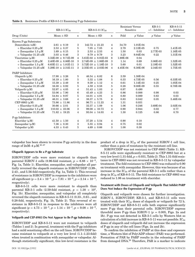
Table 2. Resistance Profile of KB-8-5-11 Examining P-gp Substrates
KB-3-1 IC50 KB-8-5-11 IC50
Resistant VersusSensitive
KB-3-1+/− Inhibitor
KB-8-5-11+/− Inhibitor
Drug (Units) Mean ± SD n Mean ± SD n Fold p Value p Value p Value
Known P-gp SubstratesDoxorubicin (nM) 2.61 ± 0.19 3 142.72 ± 21.23 4 54.70 1.01E-04
+ Elacridar 0.25 :M 2.53 ± 0.37 5 7.05 ± 7.05 4 2.79 2.13E-05 0.75 1.41E-05+ Zosuquidar 1.5 :M 1.69 ± 0.15 3 2.53 ± 2.53 4 1.50 0.03 2.77E-03 1.16E-05+ Valspodar 31.25 nM 3.03 ± 0.46 3 9.79 ± 9.79 3 3.23 9.84E-04 0.22 1.31E-04
Vincristine (:M) 3.32E-08 ± 9.71E-09 4 5.61E-02 ± 8.91E-03 6 1.69E-06 0.04+ Elacridar 0.25 :M 2.40E-09 ± 8.86E-10 3 2.74E-09 ± 1.06E-09 3 1.14 0.69 3.06E-03 1.52E-05+ Zosuquidar 1.5 :M 6.65E-11 ± 1.81E-11 3 3.72E-10 ± 1.18E-10 3 5.60 0.01 2.19E-03 1.52E-05+ Valspodar 31.25 nM 5.29E-10 ± 2.31E-11 3 2.35E-05 ± 2.03E-06 4 4.45E-04 6.38E-06 2.34E-03 1.75E-05
PARP InhibitorsOlaparib (:M) 17.98 ± 3.26 5 46.54 ± 6.82 6 2.59 1.38E-04
+ Elacridar 0.25 :M 16.19 ± 1.80 5 5.32 ± 1.08 5 0.33 4.73E-05 0.56 6.33E-05+ Zosuquidar 1.5 :M 15.39 ± 2.49 8 9.39 ± 1.12 4 0.61 1.14E-03 0.25 1.05E-04+ Valspodar 31.25 nM 16.38 ± 0.92 5 4.59 ± 0.58 5 0.28 9.08E-09 0.55 6.78E-06
Veliparib (:M) 52.97 ± 4.05 4 51.43 ± 1.03 4 0.97 0.490+ Elacridar 0.25 :M 53.06 ± 7.90 6 45.49 ± 4.23 5 0.86 0.088 0.98 0.03+ Zosuquidar 1.5 :M 54.84 ± 9.97 5 44.01 ± 4.91 4 0.80 0.089 0.83 0.03+ Valspodar 31.25 nM 46.31 ± 3.68 4 38.37 ± 5.75 6 0.83 0.042 0.05 5.46E-04
CEP-8983 (:M) 73.86 ± 11.86 4 96.71 ± 11.23 4 1.31 0.031+ Elacridar 0.25 :M 30.66 ± 2.01 3 32.37 ± 1.09 4 1.06 0.246 3.69E-04 2.02E-04+ Zosuquidar 1.5 :M 110.02 ± 23.06 3 99.08 ± 10.73 4 0.90 0.944 0.02 0.77+ Valspodar 31.25 nM 71.82 ± 19.25 4 93.04 ± 14.03 4 1.30 0.125 0.86 0.70
P-gp InhibitorsElacridar (:M) 41.50 ± 1.50 4 37.20 ± 5.54 4 0.90 0.18Zosuquidar (:M) 7.36 ± 0.30 3 5.55 ± 0.94 4 0.75 0.02Valspodar (:M) 4.33 ± 0.43 5 4.69 ± 0.66 3 1.08 0.38
valspodar has been shown to reverse P-gp activity in the doserange of 2nM–4 :M.25–27
Olaparib Appears to Be a P-gp Substrate
IGROVCDDP cells were more resistant to olaparib thanparental IGROV-1 cells (8.96-fold resistant, p = 6.88 × 10⁻⁹,Fig. 1a, Table 1). Elacridar, zosuquidar, and valspodar all par-tially reversed the olaparib resistance in IGROVCDDP (3.99-,2.43-, and 5.56-fold respectively, Fig. 1a, Table 1). This reversalof resistance in IGROVCDDP in response to the inhibitors wereall significant (p = 2.4 × 10⁻⁶, p = 7.81 × 10⁻⁶, p = 2.14 × 10⁻⁵,respectively).
KB-8-5-11 cells were more resistant to olaparib thanparental KB-3-1 cells (2.59-fold resistant, p = 1.38 × 10⁴,Fig. 1b). Elacridar, zosuquidar, and valspodar all completelyreversed the olaparib resistance in KB-8-5-11 (0.33-, 0.61-, and0.28-fold, respectively, Fig. 1b, Table 2). This reversal of re-sistance in KB-8-5-11 in response to the inhibitors were allsignificant (p = 4.73 × 10⁻⁵, p = 1.14 × 10⁻³, p = 9.08 × 10⁻⁹,respectively).
Veliparib and CEP-8983 Do Not Appear to Be P-gp Substrates
IGROVCDDP and KB-8-5-11 were not resistant to veliparib(Tables 1 and 2). In general, treatment with the P-gp inhibitorshad a mild sensitizing effect on the cell lines. IGROVCDDP be-came resistant to veliparib at a very low level (1.1–1.23-fold)when treated in combination with zosuquidar or valspodar. Al-though statistically significant, this low-level resistance is the
product of a drop in IC50 of the parental IGROV-1 cell line,rather than a gain of resistance by the resistant cell line.
IGROVCDDP was not resistant to CEP-8983 (Table 1). KB-8-5-11 cells were significantly resistant to CEP-8983 but at avery low level (1.31-fold, p = 0.031, Table 2). This 1.31-fold resis-tance to CEP-8983 was not reversed in KB-8-5-11 by valspodartreatment. The fold resistance to CEP-8983 was reduced to 0.90on treatment with zosuquidar. However, this was because of anincrease in the IC50 of the parental KB-3-1 cells rather than adrop in IC50 of KB-8-5-11. The fold resistance to CEP-8983 wasreduced to 1.06 on treatment with elacridar.
Treatment with Doses of Olaparib and Veliparib That Inhibit PARPDoes Not Induce the Expression of P-gp
Olaparib and veliparib were chosen for further investigation.IGROV-1, IGROVCDDP, KB-3-1, and KB-8-5-11 cells weretreated with their IC50 doses of olaparib or veliparib for 72 h.IGROVCDDP and KB-8-5-11 cells both express significantlymore P-gp than their parental cells. IGROVCDDP expressthreefold more P-gp than IGROV-1 (p = 0.005, Figs. 2a and2b). P-gp was not detected in KB-3-1 cells by Western blot socalculation of a fold increase in KB-8-5-11 was not possible. IC50
doses of olaparib and veliparib did not increase the expressionof P-gp in any of the cell lines (Figs. 2a and 2b).
To confirm the inhibition of PARP at this dose and exposuretime, a Western blot of Poly(ADP-ribose) (PAR) was performed.Formation of PAR by PARP results in the releasing of PARPfrom damaged DNA.28 Therefore, PAR is a marker to indicate
DOI 10.1002/jps.23952 Lawlor et al., JOURNAL OF PHARMACEUTICAL SCIENCES

4 NOTE
Figure 1. Cytotoxicity of olaparib. (a) IGROV-1 and IGROVCDDP. (b)KB-3-1 and KB-8-5-11. Open bars indicate parental cell lines; gray barsindicate resistant cell lines. Diagonally striped bars indicate treatmentwith 0.25 :M elacridar. Vertically striped bars indicate treatment with1.5 :M zosuquidar. Checked bars indicate treatment with 0.25 :Mor 31.25 nM valspodar (IGROVCDDP and KB-8-5-11, respectively).Graphs show means and standard deviation of a minimum of n = 3biological repeats. *Indicates a significant difference of the resistantcell line from the parent cell line p ≤ 0.05 Student’s t-test. #Indicatesa significant difference on the addition of a P-gp inhibitor; p ≤ 0.05Student’s t-test.
whether PARP inhibitors have successfully inhibited PARP’sactivity. PAR has been used for this purpose in several clin-ical trials.29,30 PAR expression was significantly decreased inIGROV-1 and KB-3-1 in response to both olaparib and veliparib.Reductions in the range of 14–29-fold were observed (p < 1.0 ×10⁻⁵, Figs. 2c and 2d). Olaparib and veliparib decreased PARexpression in IGROVCDDP and KB-8-5-11 but these changeswere only significant in response to veliparib in both cases. Thisindicates that the doses of parp inhibitors chosen for treatmentwere successful at inhibiting PARP.
DISCUSSION
Olaparib Appears to Be a P-gp Substrate
IGROVCDDP and KB-8-5-11 are suitable cell models for study-ing P-gp transport, as they both overexpress P-gp and no otherABC transporters such as MRP1-6 and BCRP (Table S3). P-gphas been previously shown to be functionally active by accumu-lation assays in both IGROVCDDP and KB-8-5-11 cells.13,31
Olaparib was the only examined PARP inhibitor to whichIGROVCDDP and KB-8-5-11 were both resistant (8.96- and2.59-fold, respectively). This resistance was also significantlyreversed by elacridar, zosuquidar, and valspodar in both celllines (Figs. 1a and 1b). Very low-level resistance to CEP-8983was observed in KB-8-5-11 (1.3-fold, p = 0.031). However, thisresistance was not reversed by zosuquidar or valspodar treat-ment. Drug resistance when it occurs in the clinical treatmentof cancer is typically in the range of 2–12-fold.32–38 Therefore,we are regarding the statistically significant 1.3-fold resis-tance to CEP-8983 in KB-8-5-11 as below the level of biologicalsignificance. Therefore, olaparib appears to be a P-gp substrate,whereas veliparib and CEP-8983 appear not to be substrates(Tables 1 and 2).
Resistance to olaparib has been previously shown to be as-sociated with increased gene expression of P-gp in a mousetumor model.11 In contrast, we do not see any induction of P-gpprotein expression in response to a 72-h olaparib treatment inany of the cell lines examined (Figs. 2a and 2b). These samedoses of drug were shown to decrease PAR, a marker of PARPinhibition (Figs. 2c and 2d). However, it should be noted that itoften takes a long-term exposure to a P-gp substrate, such as indrug-resistant cell line development to induce the expression ofP-gp. We are currently developing parp inhibitor resistant celllines to address this issue. Veliparib was previously found tobe a weak substrate for P-gp in transfected cells.12 Our resultsshow that veliparib is not a substrate for P-gp in IGROVCDDPand KB-8-5-11, which are also consistent with these findings(Tables 1 and 2).
The reversal of olaparib resistance by elacridar inIGROVCDDP was only partial compared with that seen inKB-8-5-11 (Fig. 1). IGROVCDDP was 8.96-fold resistant toolaparib, whereas KB-8-5-11 was only 2.59-fold resistant.Therefore, complete reversal may have been easier to achievein KB-8-5-11. The partial reversal in IGROVCDDP may alsobe because of other non-P-gp mechanisms of drug resistancethat cause resistance to olaparib. IGROVCDDP cells are resis-tant to cisplatin. As platinums and olaparib both affect DNAdamage and repair pathways, there may be an overlap in themechanisms of resistance between these agents.
P-glycoprotein has a very broad substrate specificity and isbelieved to have multiple binding sites. Most of the classic P-gpsubstrates are natural products that cannot be unambiguouslyaligned with each other because of a lack of similar orientationpoints or chemical domains.39 Therefore, the presence or ab-sence of a particular chemical domain cannot predict whethera compound is a P-gp substrate. However, several factors re-lating to the structure of a compound can suggest whether itis a P-gp substrate. The chemical structures of olaparib, veli-parib, and CEP-8983 are given in Figures 3a–3c. A molecularweight of more than 400 Da is typical of P-gp substrates39; outof the drugs we investigated, Olaparib is the only one exceeding400 (MW 434.46 Da), and veliparib and CEP-8983 are smaller
Lawlor et al., JOURNAL OF PHARMACEUTICAL SCIENCES DOI 10.1002/jps.23952

NOTE 5
Figure 2. P-glycoprotein and PAR protein expression in response to treatment with olaparib or veliparib. IGROV-1, IGROVCDDP, KB-3-1, andKB-8-5-11 cells were treated for 72 h with an IC50 dose of olaparib or veliparib and compared with a drug-free control. Western blots are shownfor (a and b) P-gp and (c and d) PAR. Representative images of n = 4 biological replicates are shown.
Figure 3. Molecular structure of PARP inhibitors used in the study.(a) Olaparib, (b) veliparib, and (c) CEP-8983.
(MW 244.29 Da and MW 306.31 Da, respectively). Compoundswith a combined number of oxygen and nitrogen atoms ≥8 areoften P-gp substrates, and ≤4 nonsubstrates.39 None of the parpinhibitors we have examined are easily defined by this rule.Olaparib has a combined number of 7 (N4O3) and veliparib andCEP-8983 both have a combined number of 5 (N4O1 and N2O3,respectively). However, olaparib is higher toward the criteria ofP-gp substrate and veliparib and CEP-8983 are lower towardthe criteria of nonsubstrate. This is consistent with our data(Tables 1 and 2).
There are a variety of online in silico tools that can predictthe P-gp substrate status of a compound based on its molecularstructure. Using the tool developed by Wang et al.,40 doxoru-bicin is predicted to be a P-gp substrate with a probability of0.74. Olaparib and veliparib are both predicted to be substrateswith probabilities of 0.87 and 0.77, respectively. CEP-8983 hada 0.55 probability of being a substrate. In contrast, anotheronline tool that makes a binary substrate/nonsubstrate classi-fication categorized olaparib as a substrate and veliparib as anonsubstrate.41 This suggests that these tools are valuable forscreening large numbers of compounds, but that there is stillvalue in in vitro conformation of P-gp substrate status.
Clinical Implications
P-glycoprotein in the intestine may become saturated withrapidly absorbed drugs because of the large concentration ofdrug present. Olaparib is rapidly absorbed in the intestine withpeak plasma levels occurring 1–3 h after dosing.42,43 This sug-gests that olaparib’s P-gp substrate status is not having a sig-nificant impact on intestinal absorption. However, veliparib isabsorbed faster than olaparib, peak plasma levels occurring0.5–1.5 h after dosing.44 One factor in this faster absorptionmay be that veliparib is not a P-gp substrate.
P-glycoprotein has a greater impact at the individual tissuelevel where the concentration of xenobiotic is lower comparedwith the intestine.45 The role of P-gp in clinical drug resis-tance is controversial, as outlined in the introduction with somestudies finding it a prognostic marker4,7 and others not.5,6,8 As
DOI 10.1002/jps.23952 Lawlor et al., JOURNAL OF PHARMACEUTICAL SCIENCES
6 NOTE
personalized biomarker panels are developed for ovarian andbreast cancer treatment, it is potentially relevant to includeP-gp, and to use this to guide the choice of PARP inhibitor foran individual patient.
The IGROVCDDP cisplatin-resistant ovarian cell line isan unusual model, as it is also cross-resistant to paclitaxel,which is mediated by P-gp.13 IGROVCDDP models the resis-tance phenotype of ovarian cancer patients who have failedstandard frontline combination platinum/taxane chemother-apy. IGROVCDDP is not resistant to veliparib or CEP-8983.Therefore, these agents could be useful for the second-line treat-ment of platinum/taxane resistant ovarian cancer.
The response rates of single-agent olaparib in relapsed plat-inum/taxane pretreated ovarian cancer range from 12% to40%.1,46–49 Response rates are higher in platinum-sensitiveBRCA1/2-mutated ovarian cancer patients and range from 41%to 62%.1,48,49 Platinum sensitivity (relapse >6 months afterchemotherapy) is the most consistent predictive factor of re-sponse among salvage chemotherapy regimens in a pretreatedovarian cancer.50–52 Therefore, platinum-sensitive patients whoare also BRCA1/2 mutation carriers, have the best possiblechance of responding to parp inhibitors. Conversely, pretreatedpatients who are platinum resistant (relapse ≤6 months afterchemotherapy) and BRCA1/2 wild-type patients have a muchlower response rate to olaparib, 3.9%.48
Only one study has been published using veliparib for thetreatment of relapsed platinum/taxane pretreated ovarian can-cer, which reported a response rate of 45% (n = 11). How-ever, this study used veliparib in combination with cyclophos-phamide and the small cohorts were all BRCA2 mutation car-riers that could contribute to the higher response rate.30 It maybe that there is a limited difference in response rate betweenolaparib and veliparib in the clinical treatment of relapsed ovar-ian cancer, which suggests that the impact of P-gp is limited inthis setting. Unfortunately, P-gp was not examined as a markerin any of the olaparib and veliparib clinical studies.
The maximal tolerated doses and the peak plasma levelsof olaparib are higher than veliparib in cancer patients.43,44
Olaparib is also a more potent drug in vitro, the average IC50
in a panel of 17 BRCA1/2 wild-type ovarian cancer cell lineswas 4.05 :M; compared with veliparib, average IC50 was 44.64:M.53 Olaparib may therefore be more successful in the clinicaltreatment of cancer than veliparib by being a more potent drugthat has a higher maximal tolerated dose in patients regard-less of its P-gp substrate status. However, the combination ofagents with differing mechanisms of cytotoxic action is routinein clinical cancer therapy. If a PARP inhibitor is to be combinedwith another class of agent that is a P-gp substrate, with otherfactors such as toxicity being equal, then veliparib or CEP-8983could be superior to olaparib.
CONCLUSIONS
Olaparib appears to be a P-gp substrate. In contrast, veli-parib and CEP-8983 do not appear to be substrates. Veli-parib and CEP-8983 may therefore be more useful in combinedchemotherapy regimens with other P-gp substrates or as sal-vage chemotherapy after exposure to P-gp substrates. Velipariband CEP-8983 may be useful in the treatment of platinum andtaxane-resistant ovarian cancer.
ACKNOWLEDGMENTS
This research was funded by the following grants: MarieCurie Reintegration Grant from the European Union FP7 Pro-gramme (B.S.), Irish Cancer Society Postdoctoral Fellowship(B.S.), Translational Research Award from the Health ResearchBoard and Science Foundation Ireland (B.H.), and a CareerDevelopment Award from the Conquer Cancer Foundation ofthe American Society of Clinical Oncology (B.H.). The authorsthank Prof. Michael Gottesman and Dr. Jean-Pierre Gillet fromthe National Cancer Institute for comments on the manuscriptand collaborating on the TLDA array. We would also like tothank Dr. Sandra Roche from Dublin City University for sup-plying the Elacridar.
REFERENCES
1. Audeh MW, Carmichael J, Penson RT, Friedlander M, Powell B, Bell-McGuinn KM, Scott C, Weitzel JN, Oaknin A, Loman N, Lu K, Schmut-zler RK, Matulonis U, Wickens M, Tutt A. 2010. Oral poly (ADP-ribose)polymerase inhibitor olaparib in patients with BRCA1 or BRCA2 mu-tations and recurrent ovarian cancer: A proof-of-concept trial. Lancet376:245–251.2. Tutt A, Robson M, Garber JE, Domchek SM, Audeh MW, WeitzelJN, Friedlander M, Arun B, Loman N, Schmutzler RK, Wardley A,Mitchell G, Earl H, Wickens M, Carmichael J. 2010. Oral poly(ADP-ribose) polymerase inhibitor olaparib in patients with BRCA1 orBRCA2 mutations and advanced breast cancer: A proof-of-concept trial.Lancet 376:235–244.3. Materna V, Pleger J, Hoffmann U, Lage H. 2004. RNA expression ofMDR1/P-glycoprotein, DNA-topoisomerase I, and MRP2 in ovarian car-cinoma patients: Correlation with chemotherapeutic response. GynecolOncol 94:152–160.4. Penson RT, Oliva E, Skates SJ, Glyptis T, Fuller AF, Jr., AF, Jr., Good-man A, Seiden MV. 2004. Expression of multidrug resistance-1 proteininversely correlates with paclitaxel response and survival in ovariancancer patients: A study in serial samples. Gynecol Oncol 93:98–106.5. Arts HJG, Katsaros D, de Vries EGE, Massobrio M, Genta F, DaneseS, Arisio R, Scheper RJ, Kool M, Scheffer GL, Willemse PHB, van derZee AGJ, Suurmeijer AJH. 1999. Drug resistance-associated markersP-glycoprotein, multidrug resistance-associated protein 1, multidrugresistance-associated protein 2, and lung resistance protein as prog-nostic factors in ovarian carcinoma. Clin Cancer Res 5:2798–2805.6. Ozalp SS, Yalcin OT, Tanir M, Kabukcuoglu S, Etiz E. 2002. Mul-tidrug resistance gene-1 (Pgp) expression in epithelial ovarian malig-nancies. Eur J Gynaecol Oncol 23:337–340.7. Gregorcyk S, Kang Y, Brandt D, Kolm P, Singer G, Perry RR. 1996.p-Glycoprotein expression as a predictor of breast cancer recurrence.Ann Surg Oncol 3(1):8–14.8. Larkin A, O’Driscoll L, Kennedy S, Purcell R, Moran E, CrownJ, Parkinson M, Clynes M. 2004. Investigation of MRP-1 protein andMDR-1 P-glycoprotein expression in invasive breast cancer: A prognos-tic study. Int J Cancer 112(2):286–294.9. Atalay C, Demirkazik A, Gunduz U. 2008. Role of ABCB1 andABCC1 gene induction on survival in locally advanced breast cancer. JChemother 20(6):734–739.10. Jazaeri AA, Yee CJ, Sotiriou C, Brantley KR, Boyd J, Liu ET. 2002.Gene expression profiles of BRCA1-linked, BRCA2-linked, and sporadicovarian cancers. J Natl Cancer Inst 94:990–1000.11. Rottenberg S, Jaspers JE, Kersbergen A, van der BE, Nygren AO,Zander SA, Derksen PW, de Bruin M, Zevenhoven J, Lau A, Boulter R,Cranston A, O’Connor MJ, Martin NM, Borst P, Jonkers J. 2008. Highsensitivity of BRCA1-deficient mammary tumors to the PARP inhibitorAZD2281 alone and in combination with platinum drugs. Proc NatlAcad Sci USA 105:17079–17084.
Lawlor et al., JOURNAL OF PHARMACEUTICAL SCIENCES DOI 10.1002/jps.23952

NOTE 7
12. Li X, Delzer J, Voorman R, de Morais SM, Lao Y. 2011. Dispositionand drug–drug interaction potential of veliparib (ABT-888), a novel andpotent inhibitor of poly(ADP-ribose) polymerase. Drug Metab Dispos39:1161–1169.13. Stordal B, Hamon M, McEneaney V, Roche S, Gillet JP, O’LearyJJ, Gottesman M, Clynes M. 2012. Resistance to paclitaxel ina cisplatin-resistant ovarian cancer cell line is mediated by P-glycoprotein. PLoS One 7:e40717.14. Richert N, Akiyama S, Shen D, Gottesman MM, Pastan I. 1985.Multiply drug-resistant human KB carcinoma cells have decreasedamounts of a 75-kDa and a 72-kDa glycoprotein. Proc Natl Acad SciUSA 82:2330–2333.15. Richert ND, Aldwin L, Nitecki D, Gottesman MM, Pastan I. 1988.Stability and covalent modification of P-glycoprotein in multidrug-resistant KB cells. Biochemistry 27:7607–7613.16. Ma J, Maliepaard M, Kolker HJ, Verweij J, Schellens JH. 1998. Ab-rogated energy-dependent uptake of cisplatin in a cisplatin-resistantsubline of the human ovarian cancer cell line IGROV-1. CancerChemother Pharmacol 41:186–192.17. Ma J, Maliepaard M, Nooter K, Boersma AW, Verweij J, StoterG, Schellens JH. 1998. Synergistic cytotoxicity of cisplatin and topote-can or SN-38 in a panel of eight solid-tumor cell lines in vitro. CancerChemother Pharmacol 41:307–316.18. Martin A, Clynes M. 1993. Comparison of 5 microplate colorimetricassays for in vitro cytotoxicity testing and cell proliferation assays.Cytotechnology 11:49–58.19. Choudhuri S, Klaassen CD. 2006. Structure, function, expression,genomic organization, and single-nucleotide polymorphisms of humanABCB1 (MDR1), ABCC (MRP), and ABCG2 (BCRP) efflux transporters.Int J Toxicol 25:231–259.20. Hyafil F, Vergely C, Du Vignaud P, Grand-Perret T. 1993. Invitro and in vivo reversal of multidrug resistance by GF120918, anacridonecarboxamide derivative. Cancer Res 53:4595–4602.21. Thomas H, Coley HM. 2003. Overcoming multidrug resistance incancer: An update on the clinical strategy of inhibiting p-glycoprotein.Cancer Control 10:159–165.22. Pichler A, Prior JL, Piwnica-Worms D. 2004. Imaging reversalof multidrug resistance in living mice with bioluminescence: MDR1P-glycoprotein transports coelenterazine. Proc Natl Acad Sci USA101:1702–1707.23. Abu Ajaj K, Graeser R, Kratz F. 2012. Zosuquidar and an albumin-binding prodrug of zosuquidar reverse multidrug resistance in breastcancer cells of doxorubicin and an albumin-binding prodrug of doxoru-bicin. Breast Cancer Res Treat 134:117–129.24. Mease K, Sane R, Podila L, Taub ME. 2012. Differential selectivityof efflux transporter inhibitors in Caco-2 and MDCK–MDR1 monolay-ers: A strategy to assess the interaction of a new chemical entity withP-gp, BCRP, and MRP2. J Pharm Sci 101:1888–1897.25. Mistry P, Stewart AJ, Dangerfield W, Okiji S, Liddle C, BootleD, Plumb JA, Templeton D, Charlton P. 2001. In vitro and in vivoreversal of P-glycoprotein-mediated multidrug resistance by a novelpotent modulator, XR9576. Cancer Res 61:749–758.26. Crowe A. 2002. The influence of P-glycoprotein on morphine trans-port in Caco-2 cells. Comparison with paclitaxel. Eur J Pharmacol440:7–16.27. Westers TM, Houtenbos I, Schuurhuis GJ, Ossenkoppele GJ, vande Loosdrecht AA. 2005. Quantification of T-cell mediated apoptosis inheterogeneous leukemia populations using four-color multiparameterflow cytometry. Cytometry 66A:71–77.28. Wang X, Weaver DT. 2011. The ups and downs of DNA repairbiomarkers for PARP inhibitor therapies. Am J Cancer Res 1:301–327.29. Rajan A, Carter CA, Kelly RJ, Gutierrez M, Kummar S, SzaboE, Yancey MA, Ji J, Mannargudi B, Woo S, Spencer S, Figg WD, Giac-cone G. 2012. A phase I combination study of olaparib with cisplatinand gemcitabine in adults with solid tumors. Clin Cancer Res 18:2344–2351.30. Kummar S, Ji J, Morgan R, Lenz HJ, Puhalla SL, Belani CP, Gan-dara DR, Allen D, Kiesel B, Beumer JH, Newman EM, Rubinstein L,
Chen A, Zhang Y, Wang L, Kinders RJ, Parchment RE, TomaszewskiJE, Doroshow JH 2012. A phase I study of veliparib in combination withmetronomic cyclophosphamide in adults with refractory solid tumorsand lymphomas. Clin Cancer Res 18:1726–1734.31. Fojo A, Akiyama Si, Gottesman MM, Pastan I. 1985. Reduced drugaccumulation in multiply drug-resistant human KB carcinoma celllines. Cancer Res 45:3002–3007.32. Kawai H, Kiura K, Tabata M, Yoshino T, Takata I, HirakiA, Chikamori K, Ueoka H, Tanimoto M, Harada M. 2002. Character-ization of non-small-cell lung cancer cell lines established before andafter chemotherapy. Lung Cancer 35:305–314.33. Kuroda H, Sugimoto T, Ueda K, Tsuchida S, Horii Y, InazawaJ, Sato K, Sawada T. 1991. Different drug sensitivity in two neurob-lastoma cell lines established from the same patient before and afterchemotherapy. Int J Cancer 47:732–737.34. Hida T, Ueda R, Takahashi T, Watanabe H, Kato T, Suyama M,Sugiura T, Ariyoshi Y, Takahashi T. 1989. Chemosensitivity and ra-diosensitivity of small cell lung cancer cell lines studied by a newly de-veloped 3-(4,5-Dimethylthiazol-2-yl)-2,5-diphenyltetrazolium bromide(MTT) hybrid assay. Cancer Res 49:4785–4790.35. Berendsen HH, de Leij L, de Vries EGE, Mesander G, Mulder NH,de Jong B, Buys CHCM, Postmus PE, Poppema S, Sluiter HJ, TheHT 1988. Characterization of three small cell lung cancer cell linesestablished from one patient during longitudinal follow-up. Cancer Res48:6891–6899.36. Sakai W, Swisher EM, Jacquemont C, Chandramohan KV, CouchFJ, Langdon SP, Wurz K, Higgins J, Villegas E, Taniguchi T. 2009.Functional restoration of BRCA2 protein by secondary BRCA2 muta-tions in BRCA2-mutated ovarian carcinoma. Cancer Res 69:6381–6386.37. Stronach EA, Chen M, Maginn EN, Agarwal R, Mills GB, WasanH, Gabra H. 2011. DNA-PK mediates AKT activation and apoptosis in-hibition in clinically acquired platinum resistance. Neoplasia 13:1069–1080.38. Langdon SP, Lawrie SS, Hay FG, Hawkes MM, McDonald A, Hay-ward IP, Schol DJ, Hilgers J, Leonard RCF, Smyth JF. 1988. Charac-terization and properties of nine human ovarian adenocarcinoma celllines. Cancer Res 48:6166–6172.39. Didziapetris R, Japertas P, Avdeef A, Petrauskas A. 2003. Classi-fication analysis of P-glycoprotein substrate specificity. J Drug Target11:391–406.40. Wang Z, Chen Y, Liang H, Bender A, Glen RC, Yan A. 2011. P-glycoprotein substrate models using support vector machines based ona comprehensive data set. J Chem Inf Model 51:1447–1456.41. Bikadi Z, Hazai I, Malik D, Jemnitz K, Veres Z, Hari P, Ni Z, Loo TW,Clarke DM, Hazai E, Mao Q. 2011. Predicting P-glycoprotein-mediateddrug transport based on support vector machine and three-dimensionalcrystal structure of P-glycoprotein. PLoS One 6:e25815.42. Fong PC, Boss DS, Yap TA, Tutt A, Wu P, Mergui-Roelvink M, Mor-timer P, Swaisland H, Lau A, O’Connor MJ, Ashworth A, CarmichaelJ, Kaye SB, Schellens JH, de Bono JS. 2009. Inhibition of poly(ADP-ribose) polymerase in tumors from BRCA mutation carriers. N Engl JMed 361:123–134.43. Yamamoto N, Nokihara H, Yamada Y, Goto Y, Tanioka M, ShibataT, Yamada K, Asahina H, Kawata T, Shi X, Tamura T. 2012. A phaseI, dose-finding and pharmacokinetic study of olaparib (AZD2281) inJapanese patients with advanced solid tumors. Cancer Sci 103:504–509.44. Kummar S, Kinders R, Gutierrez ME, Rubinstein L, Parchment RE,Phillips LR, Ji J, Monks A, Low JA, Chen A, Murgo AJ, Collins J, Stein-berg SM, Eliopoulos H, Giranda VL, Gordon G, Helman L, Wiltrout R,Tomaszewski JE, Doroshow JH. 2009. Phase 0 clinical trial of the poly(ADP-ribose) polymerase inhibitor ABT-888 in patients with advancedmalignancies. J Clin Oncol 27(16):2705–2711.45. Hamidovic A, Hahn K, Kolesar J. 2010. Clinical significance ofABCB1 genotyping in oncology. J Oncol Pharm Pract 16(1):39–44.46. Kaye SB, Lubinski J, Matulonis U, Ang JE, Gourley C, KarlanBY, Amnon A, Bell-McGuinn KM, Chen LM, Friedlander M, SafraT, Vergote I, Wickens M, Lowe ES, Carmichael J, Kaufman B. 2012.
DOI 10.1002/jps.23952 Lawlor et al., JOURNAL OF PHARMACEUTICAL SCIENCES

8 NOTE
Phase II, open-label, randomized, multicenter study comparing theefficacy and safety of olaparib, a Poly (ADP-ribose) polymerase in-hibitor, and pegylated liposomal doxorubicin in patients with BRCA1 orBRCA2 mutations and recurrent ovarian cancer. J Clin Oncol 30:372–379.47. Ledermann J, Harter P, Gourley C, Friedlander M, Vergote I,Rustin G, Scott C, Meier W, Shapira-Frommer R, Safra T, Matei D,Macpherson E, Watkins C, Carmichael J, Matulonis U. 2012. Olaparibmaintenance therapy in platinum-sensitive relapsed ovarian cancer.New Engl J Med 366:1382–1392.48. Gelmon KA, Tischkowitz M, Mackay H, Swenerton K, Robidoux A,Tonkin K, Hirte H, Huntsman D, Clemons M, Gilks B, Yerushalmi R,Macpherson E, Carmichael J, Oza A. 2011. Olaparib in patients withrecurrent high-grade serous or poorly differentiated ovarian carcinomaor triple-negative breast cancer: A phase 2, multicentre, open-label,non-randomised study. Lancet Oncol 12:852–861.49. Fong PC, Yap TA, Boss DS, Carden CP, Mergui-Roelvink M, GourleyC, De Greve J, Lubinski J, Shanley S, Messiou C, A’Hern R, Tutt A,
Ashworth A, Stone J, Carmichael J, Schellens JHM, de Bono JS, KayeSB. 2010. Poly(ADP)-ribose polymerase inhibition: frequent durableresponses in BRCA carrier ovarian cancer correlating with platinum-free interval. J Clin Oncol 28:2512–2519.50. Murphy M, Stordal B. Erlotinib or gefitinib for the treatment ofrelapsed platinum pretreated non-small cell lung cancer and ovariancancer: A systematic review. Drug Resist Updat 14(3):177–190.51. Stordal B, Pavlakis N, Davey R. 2007. Oxaliplatin for the treatmentof cisplatin-resistant cancer: A systematic review. Cancer Treat Rev33:347–357.52. Stordal B, Pavlakis N, Davey R. 2007. A systematic review of plat-inum and taxane resistance from bench to clinic: An inverse relation-ship. Cancer Treat Rev 33:688–703.53. Stordal B, Timms K, Farrelly A, Gallagher D, Busschots S, RenaudMl, Thery J, Williams D, Potter J, Tran T, Korpanty G, Cremona M,Carey M, Li J, Li Y, Aslan O, O’Leary JJ, Mills GB, Hennessy BT. 2013.BRCA1/2 mutation analysis in 41 ovarian cell lines reveals only onefunctionally deleterious BRCA1 mutation. Mol Oncol 7:567–579.
Lawlor et al., JOURNAL OF PHARMACEUTICAL SCIENCES DOI 10.1002/jps.23952


![PARP Inhibitors for Ovarian Cancer · PDF fileAstra Zeneca Pharmaceuticals LP;2017. Rucaparib (Rubraca®)[package insert].Boulder, CO. Clovis ... Dyspnea Photosensitivity ≥ 20% Hypertension](https://img.pdfslide.us/doc/110x75/5a9d60ef7f8b9abd058c7da5/parp-inhibitors-for-ovarian-cancer-zeneca-pharmaceuticals-lp2017-rucaparib-rubracapackage.jpg)