Embed Size (px)
Citation preview
Parasite Biology 3 Course Guide
2019/2020
Course Organiser: Professor Alex Rowe Course Email: [email protected]
Course code: BILG09003 Level 9 course (20 credits)
Student name:…………………...…/Matriculation No:……………
Trypanosomes
2
In-course assessment deadlines and feedback Assessment Deadline Return date Feedback
Course Essay Thursday 31st October 2019 at 3.30pm
Thursday 21st November 2019
Thursday 28th November 2019 11.35am – 12.15pm
Practical Assessment
Thursday 28th November 2019 at 10.00am
Thursday 5th December 2019
Thursday 28th November 2019 11.10 – 11.30am
If you require this document in an alternative format, such as large print or a coloured background, please contact the course secretary by email: [email protected] or phone the BTO on: 0131 650 8649.
3
Contents
Section 1. Introduction
1.1 Background. 1.2 Aims and content of Parasite Biology 3. 1.3 Problems: attendance and completion of assignments. 1.4 Graduate attributes you should develop during this course.
Section 2. Course Components
2.1 Lectures and practical sessions. 2.2 In-Course Assessment. 2.3 Written examination. 2.4 Use of Learn by the course 2.5 Lecture recordings
Section 3. Books, journals and libraries
Section 4. Student representatives, course auditing and development
Section 5. Timetable
Section 6. Formal assessment, submission dates of in-course assessment and detection of plagiarism
6.1 Passing the course. 6.2 Marking scheme. 6.3 Written examination and weighting of marks. 6.4 In-Course Assessment, weighting of marks, submission deadlines and
detection of plagiarism.
Section 7. Penalties for late submission of assessed coursework
Section 8. Plagiarism
Section 9. Information relating to examinations, year weightings, progression and complaints
Section 10. Safety procedures
Appendices
Appendix 1. Guidance on the in-course assessments Appendix 1.1 Instructions for the in-course essay Appendix 1.2 Essay feedback sheet Appendix 1.3 Instructions for the Practical in-course assessment
Appendix 2. General reading list
Appendix 3: Format of written examination Appendix 4: Practical Manual
4
SECTION 1. Introduction
1.1 Background.
The study of parasites continues to expand, since these organisms cause many
intractable diseases of economic importance. This course concentrates on some of
the parasites that cause important medical and veterinary diseases. The information
and practical work contained in the course have been chosen to appeal not only to
students interested in applied parasitology, but also to those with a general interest
in the epidemiology, ecology and evolution, immunology, molecular and cellular
biology of parasites. The course provides a good foundation for students aiming for
parasitology-based fourth year courses and careers, and is a useful adjunct to
courses on cell biology, genetics, zoology, evolutionary biology and immunology.
Parasite Biology is one of the third year courses contributing to the Junior Honours
Year of the B.Sc. (Biological Sciences) organised by the Biology Teaching
Organisation. Performance in the course will therefore count towards Honours
Degree classification.
The course is taught in the Ashworth Laboratories, School of Biological Sciences, at
King's Buildings. The Ashworth Laboratories are the traditional seat of parasitology
in the University and house the University's extensive collection of parasitological
specimens. The Course Organiser is Professor Alex Rowe, Ashworth Laboratories,
King's Buildings. The email address for the Course Secretary is: [email protected].
The core teaching staff and their research expertise
DB: Dr Dave Bartley (Veterinary helminth drug resistance and vaccine development)
TL: Prof Tom Little (Genetics and evolution of parasitic interactions)
KM: Prof Keith Matthews (Trypanosome development)
FM: Prof Francisca Mutapi (Parasite immuno-epidemiology)
NP: Dr Nisha Philip (Parasite transmission)
AR: Prof Alex Rowe (Malaria host-parasite interactions)
PSch: Dr Petra Schneider (Evolution of parasite life history strategies)
PS: Dr Phil Spence (Immunity to malaria)
AS: Prof Achim Schnaufer (Parasite genomes)
JT: Dr Joanne Thompson (Intracellular parasites)
5
1.2 Aims and content of Parasite Biology 3.
The overall aim of the course is to provide a basic knowledge of parasites, including
their epidemiology, evolution, immunology, genetics, molecular and cell biology. The
lectures show how the major groups of parasites have solved the special problems
faced by species living on or in other animals. Further, the course will illustrate how
new developments in the study of parasitic organisms are highlighting their unique,
and often elegant, biology and how this may be exploited to combat them. In
particular, the course will take a multidisciplinary view of parasite biology, with
particular focus on recent findings in parasitology research to show how modern
research methods are being directed towards understanding the parasitic lifestyle.
The course also looks at how knowledge of parasite biology is used in designing and
implementing control programmes, and the problems, such as drug resistance, that
can emerge. Dedicated practical sessions will be associated with each taught theme
in order to highlight particular points and to provide further understanding of parasite
biology in its many forms.
1.3 Problems: attendance and completion of assignments.
Any student with a query or problem should contact the Course Secretary
([email protected]) in the first instance. Any student who finds they are unable to
attend classes for any reason is requested to let the Course Secretary know as soon
as possible. It is essential that any student having problems with attendance or
completion of assignments inform the Course Organiser as well as their Personal
Tutor without delay.
1.4 Graduate attributes you should develop during this course.
The University has identified six groups of abilities (see headings below) that should
be developed as part of the University of Edinburgh training experience, and to
enhance your employability as a graduate for the 21st Century. These abilities take
your skill-base beyond simply academic knowledge and are enhanced at each stage
6
of your degree. They relate to this course in a number of specific ways, as outlined
below.
Knowledge and Understanding: All components of the course provide this to some
degree but your lectures, in particular, provide an important framework upon which
you can build these attributes. This University considers itself to be a research-led
institution and all of the lecturing staff on Parasite Biology 3 run successful and
active research programmes. The material you will be exposed to will often be
based on lecturers’ research activities, providing you with cutting edge information
and ideas. In this course you will develop a comprehensive knowledge of core
concepts in modern parasitology and in the approaches to investigate the biology of
parasites, the spread of human and veterinary parasites in the field and approaches
that are being used to develop vaccines and therapies against these pathogens.
Research and Enquiry: These skills are enhanced by encouraging further reading
of books, research papers and electronic materials, to embellish your lecture and
practical material. They underpin your ICA material (Essay and Practical
Assessment). These aspects are particularly enhanced through the investigations
you will carry out to prepare for the in-course essay, the subject for which you will
choose yourself, providing real experience in researching a particular parasite-
related topic. Combined, the essay and practical assessment provide a route to
surveying current and past scientific arguments in an appropriate context, and
provide the foundation for hypothesis-driven analysis.
Personal and Intellectual Autonomy: By reading and preparation of your in-course
essay, based on a topic of your choosing, you will learn to synthesise your own
views, develop reasoned arguments and further refine your scientific judgement.
The practical assessment exercise also provides you with an opportunity to apply the
knowledge you will have acquired in the laboratory to answer questions that are
often faced by health workers and researchers, and will help you to understand how
parasites are diagnosed and controlled. Such skills will enhance your capacity for
life-long and independent learning, as well as providing experience that is highly
relevant for research careers in this field.
7
Communication: This is a key attribute of all scientists and it is therefore important
that you develop skills to interact constructively with others and convey
knowledgeable and balanced scientific views. We provide training in this area
through our in-course work and by providing a portal for interaction between students
via the Learn Discussion Forum.
Personal Effectiveness: The ability to organise and summarise your thoughts and
material in a flexible and accessible way are core features that are required for
personal effectiveness. Planning, time management and reflection are central to
this. Of course, these features also interlink with your personal and intellectual
autonomy. By providing you with a timetable where key submission dates are
highlighted, we are encouraging you to develop your effectiveness throughout this
course. These same skills extend to other courses and to your overall ability to
maximise your achievement whilst studying at this University. However, they also
apply to other aspecst of your current and future life. Many aspects of what you
achieve in life can be significantly influenced by you! For this reason, the in-course
work that forms part of Parasite Biology 3 is specifically geared to self-led learning.
Technical and Practical Skills: for many scientific careers it is important that you
not only understand the conceptual basis of how experiments are designed and
carried out, but also that you have the underpinning practical skills required for
employability. Our course has an important lab component that is designed to
prepare you for this, as well as to assist you in your future Honours course projects.
In particular, the practicals cover a diverse range of methods used in parasitology
research, and provide exposure to modern approaches and concepts in the field.
These classes are led by research active staff and supervised by research active
demonstrators, usually at PhD level. University staff from the Moredun Research
Institute take part, providing a range of exposure to basic and applied research
concepts and methods. The lab skills you develop from your practical sessions, in
critical observation, careful recording, investigation and interpretation, quantification
and analysis, should serve you well in any future employment. They will also
complement your course material, enhancing your understanding of basic concepts
in parasite biology research.
8
Section 2. Course components
2.1 Lectures, tutorial and practical sessions.
Lectures will begin at 9.00 am on Mondays and Thursdays throughout the course.
In addition to lectures there will be an essay tutorial and a series of practical
sessions linked thematically to the lectures, in order to provide training in
parasitological techniques (e.g. handling and identifying parasites) or give
experience of experiments designed to analyse parasite physiology and behaviour.
Laboratory books will not be formally marked but the Course Organiser reserves the
right to inspect laboratory books at any time should this be deemed appropriate. The
Essay tutorial is scheduled in the timetable. Material contained within practical
sessions will also be subject to assessment within the written examination at the
end of the course as well as in a practical test.
Each practical session will be introduced at the end of the associated lectures.
2.2 In-Course Assessment (ICA).
The in-course assessment exercise is included to broaden your experience of the
scientific and parasitological literature and to develop your written communication
and investigative skills. For one part of the in-course assessment you are required
to write a 1500-word essay based on a review of the scientific literature on a
parasite-related topic of your own choice. This exercise is designed to help in writing
introductions and discussions for projects, e.g. fourth year honours project
dissertations. The essay is worth 30% of your final mark for the course.
You are asked to submit the essay by on Thursday 31st October by 3.30pm. This
late submission date is not given with the intention of your leaving the writing of this
essay until the last minute after completing exercises for other courses. This date
has been chosen to give you a chance to read and think about as many aspects of
parasite biology as possible before you chose your topic.
9
Guidance for preparing an essay and notes for scientific writing are given in
Appendix 1.1. Guidance on how to use and acknowledge information from literature
sources and the avoidance of plagiarism is given in Appendix 1.1 and Section 8.
There will be a tutorial to provide guidance for essay writing. This will take place
during week 2 on Monday 23rd September at 10-11am in Ashworth Laboratories,
Lab 1 (Room 80). Some examples of good quality essays will be provided on Learn.
A further in-course assessment will involve a practical test exercise which aims to
consolidate the knowledge gained in your practical sessions. The practical test will
take the form of a Question Mark Perception (QMP) exercise on computer. Self-
study practical revision time before the Practical Test has been scheduled in the time
table. There will be questions from each of the practical session in the course. This
component is worth 20% of your final mark for the course, and will be assessed at
the end of November before the written examinations commence, as detailed in the
time-table. For this assessment, ensure you learn and use the correct spelling e.g.
of parasite scientific names, drugs, vaccines etc.
NOTE: Weighting of marks, deadlines and location for submission, and penalties for
late submission of assessed coursework are given below (Section 6). Written
feedback on the essay and marks will be given out as soon as possible.
2.3 Written examination.
A two-hour written examination will be held in weeks 12 or 13. Students will be
notified of the date and time of the examination via their timetable. The format of the
written exam is shown in Appendix 3. Weighting of marks is given below (Section 6).
10
2.4 Use of Learn by the course.
For information about Learn please refer to the “Essential Guide”.
2.4.1 Plagiarism. As discussed below, this course will use Turnitin to check that
essays are free from plagiarism (see Section 6.4 and Section 8).
2.4.2 Lecture notes. The lecture hand-outs will be put onto the Parasite Biology
Course Learn page at least 24 hours in advance of each lecture. These hand-outs
will not be a substitute for attending the lectures, nor for students taking their own
notes in the lectures. Supplementary and essential material may be given out in the
lectures that will not be mounted on Learn. The notes will be put on Learn for you to
check the accuracy of your own notes. This will be done in recognition of the fact
that the written examination follows very closely on the end of the course and to help
you consolidate your notes as easily and as rapidly as possible during Semester 1.
2.4.3 Discussion forum. The Learn page for the Parasite Biology course will
include a discussion forum as for other courses. In the case of Parasite Biology,
however, the discussion forum is solely for the use of students. Lecturers will not
participate in the discussion forum. They will be present throughout the practical
classes following their lectures, and therefore are available to answer your questions
in person.
Very important note: the BTO have warned that any misuse of discussion forum
areas will result in the immediate removal of the forum areas.
2.4 Lecture recordings. All lectures will be recorded unless the lecturer has a
specific reason to opt out. In such circumstances, the reason for opting out will be
explained to the class. Research has shown that students who attend lectures in
person get better grades than those who rely on recordings. We recommend using
the recordings to revisit areas you don’t understand, rather than watching the full
recording and using it as a substitute for attending in person.
11
Section 3. Books, journals and libraries
There is no set text book for the course. Individual lecturers will provide you with
relevant information on research articles and reviews and web sites that will provide
you with information to support individual lectures.
There are, however, a number of excellent text books that may help you understand
many fundamental aspects of parasite biology, including life cycles and transmission,
evolutionary parasitology, human and veterinary diseases, epidemiology,
immunology, drug and vaccine development. For example, Parasitology: a
conceptual approach. Loker, ES & Hofkin BV (2015) Garland Science, is a highly
readable text with lovely diagrams that gives a broad overview of modern parasite
biology. This and other key texts are shown in the “Essential reading” section of the
reading list in Appendix 2, and many are available online. The “Recommended
reading” section (Appendix 2) also highlights a number of other books that may
prove useful to support lecture-based learning and to research material for the in-
course essay. The Resource list on Learn provides links for further information on
the above texts.
Books and journals on parasitological topics will be found mainly in the Noreen and
Kenneth Murray Library; the major course texts are kept on the Reserve Shelves all
year round. Other texts and journals will be found on the appropriate shelves. The
Erskine Medical Library collection in the Main Library, George Square and The Lady
Smith of Kelvin Veterinary Library at the Royal (Dick) School of Veterinary Studies,
Easter Bush also have relevant books and journals.
Section 4. Student representatives, course auditing and development
Feedback will be obtained verbally and informally throughout the course as well as
by a questionnaire which will be made available on EUCLID. The results will
contribute to course development and the quality assessment of courses. All
students are kindly requested to help with these exercises.
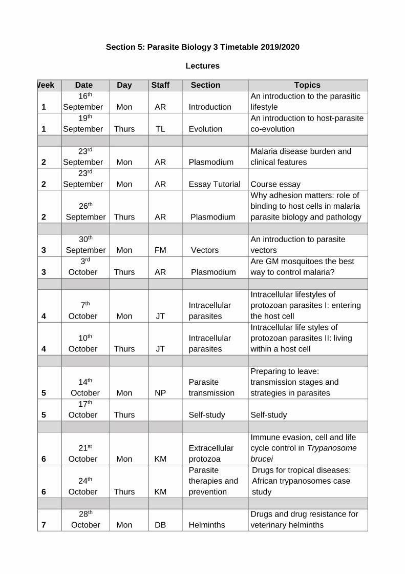
Section 5: Parasite Biology 3 Timetable 2019/2020
Lectures
Week Date Day Staff Section Topics
1
16th
September Mon AR Introduction
An introduction to the parasitic
lifestyle
1
19th
September Thurs TL Evolution
An introduction to host-parasite
co-evolution
2
23rd
September Mon AR Plasmodium
Malaria disease burden and
clinical features
2
23rd
September Mon AR Essay Tutorial Course essay
2
26th
September Thurs AR Plasmodium
Why adhesion matters: role of
binding to host cells in malaria
parasite biology and pathology
3
30th
September Mon FM Vectors
An introduction to parasite
vectors
3
3rd
October Thurs AR Plasmodium
Are GM mosquitoes the best
way to control malaria?
4
7th
October Mon JT
Intracellular
parasites
Intracellular lifestyles of
protozoan parasites I: entering
the host cell
4
10th
October Thurs JT
Intracellular
parasites
Intracellular life styles of
protozoan parasites II: living
within a host cell
5
14th
October Mon NP
Parasite
transmission
Preparing to leave:
transmission stages and
strategies in parasites
5
17th
October Thurs Self-study Self-study
6
21st
October Mon KM
Extracellular
protozoa
Immune evasion, cell and life
cycle control in Trypanosome
brucei
6
24th
October Thurs KM
Parasite
therapies and
prevention
Drugs for tropical diseases:
African trypanosomes case
study
7
28th
October Mon DB Helminths
Drugs and drug resistance for
veterinary helminths
13
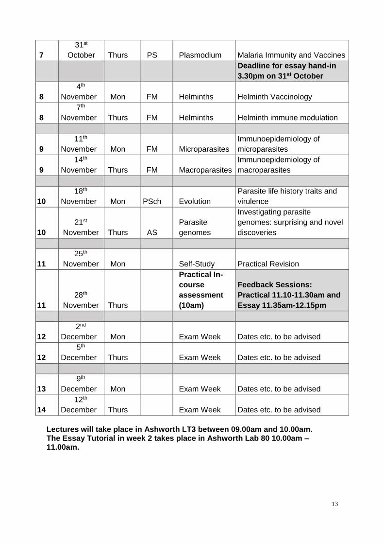
7
31st
October Thurs PS Plasmodium Malaria Immunity and Vaccines
Deadline for essay hand-in
3.30pm on 31st October
8
4th
November Mon FM Helminths Helminth Vaccinology
8
7th
November Thurs FM Helminths Helminth immune modulation
9
11th
November Mon FM Microparasites
Immunoepidemiology of
microparasites
9
14th
November Thurs FM Macroparasites
Immunoepidemiology of
macroparasites
10
18th
November Mon PSch Evolution
Parasite life history traits and
virulence
10
21st
November Thurs AS
Parasite
genomes
Investigating parasite
genomes: surprising and novel
discoveries
11
25th
November Mon Self-Study Practical Revision
11
28th
November Thurs
Practical In-
course
assessment
(10am)
Feedback Sessions:
Practical 11.10-11.30am and
Essay 11.35am-12.15pm
12
2nd
December Mon Exam Week Dates etc. to be advised
12
5th
December Thurs Exam Week Dates etc. to be advised
13
9th
December Mon Exam Week Dates etc. to be advised
14
12th
December Thurs Exam Week Dates etc. to be advised
Lectures will take place in Ashworth LT3 between 09.00am and 10.00am. The Essay Tutorial in week 2 takes place in Ashworth Lab 80 10.00am – 11.00am.
14
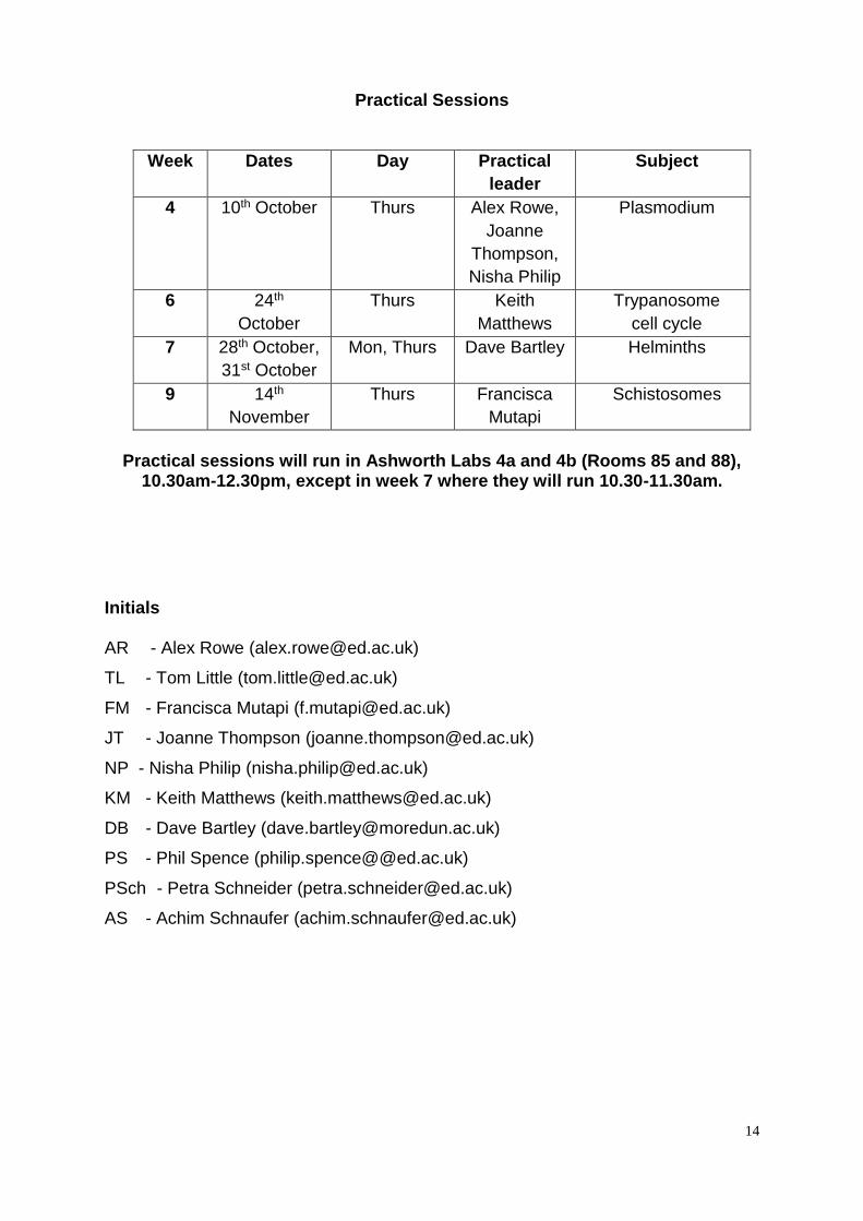
Practical Sessions
Week Dates Day Practical
leader
Subject
4 10th October Thurs Alex Rowe,
Joanne
Thompson,
Nisha Philip
Plasmodium
6 24th
October
Thurs Keith
Matthews
Trypanosome
cell cycle
7 28th October,
31st October
Mon, Thurs Dave Bartley Helminths
9 14th
November
Thurs Francisca
Mutapi
Schistosomes
Practical sessions will run in Ashworth Labs 4a and 4b (Rooms 85 and 88),
10.30am-12.30pm, except in week 7 where they will run 10.30-11.30am.
Initials AR - Alex Rowe ([email protected])
TL - Tom Little ([email protected])
FM - Francisca Mutapi ([email protected])
JT - Joanne Thompson ([email protected])
NP - Nisha Philip ([email protected])
KM - Keith Matthews ([email protected])
DB - Dave Bartley ([email protected])
PS - Phil Spence (philip.spence@@ed.ac.uk)
PSch - Petra Schneider ([email protected])
AS - Achim Schnaufer ([email protected])
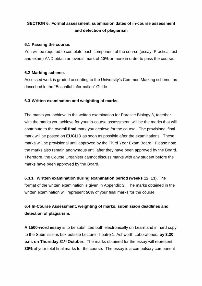
SECTION 6. Formal assessment, submission dates of in-course assessment
and detection of plagiarism
6.1 Passing the course.
You will be required to complete each component of the course (essay, Practical test
and exam) AND obtain an overall mark of 40% or more in order to pass the course.
6.2 Marking scheme.
Assessed work is graded according to the University’s Common Marking scheme, as
described in the “Essential Information” Guide.
6.3 Written examination and weighting of marks.
The marks you achieve in the written examination for Parasite Biology 3, together
with the marks you achieve for your in-course assessment, will be the marks that will
contribute to the overall final mark you achieve for the course. The provisional final
mark will be posted on EUCLID as soon as possible after the examinations. These
marks will be provisional until approved by the Third Year Exam Board. Please note
the marks also remain anonymous until after they have been approved by the Board.
Therefore, the Course Organiser cannot discuss marks with any student before the
marks have been approved by the Board.
6.3.1 Written examination during examination period (weeks 12, 13). The
format of the written examination is given in Appendix 3. The marks obtained in the
written examination will represent 50% of your final marks for the course.
6.4 In-Course Assessment, weighting of marks, submission deadlines and
detection of plagiarism.
A 1500-word essay is to be submitted both electronically on Learn and in hard copy
to the Submissions box outside Lecture Theatre 1, Ashworth Laboratories, by 3.30
p.m. on Thursday 31st October. The marks obtained for the essay will represent
30% of your total final marks for the course. The essay is a compulsory component
16
of this course; the course can only be passed if an essay is handed in by the above
deadline. Marks will be returned by Thursday 21st November.
Please note:
You must place a bar code label with your BTO 2019/20 PIN on the front page of the
hard copy of your work.
The essay will be subjected to the Plagiarism Detection Service by the BTO using
Turnitin in Learn. All students will be required to submit an electronic copy of their
essay for this purpose. The electronic copy MUST be identical to the hard copy.
For instructions on using Learn please refer to the “Essential Guide”. Guidance on
avoiding plagiarism is given in Section 8.
Penalties for late submission of assessed course work and guidance on applying for
extensions to deadlines are described in the essential guide.
The second in-course assessment component is a Question Mark Perception
exercise on computer, based on the practical material and specimens. This Practical
Test will be a “closed book” test. Thus, students are NOT allowed to consult any
books, notes or similar material during the test. Laboratory notebooks should be
used for revision before the test. The Practical Test will contribute 20% of your final
mark for the course. The test will include questions on all of the practicals and
related introductory material. The questions will cover practical procedures, analysis
of results, and discussion topics raised when the experiments are reviewed. Thus, it
will be to your advantage to write up each practical as completely as possible after
the class, to be used later for your revision before the Test.
Any student absent from the Practical Test without a good reason will receive a zero
mark for it. A student who can provide evidence of a valid reason for absence (e.g. a
medical certificate for an illness), will be given an alternative assignment to complete
and may also be asked to present their lab notebook. Students, who require
arrangements for Special Needs, must assure that the relevant documentation
reaches the Secretary of the Course by Friday of week 6 at the latest.
17
SECTION 7. Penalties for late submission of assessed coursework
IMPORTANT: The Parasite Biology 3 course will apply the standard conditions
for submissions of course work in accordance with common practice across
the College of Science and Engineering.
Please refer to the Essential guide for further information on policies and procedure
for the late submission of coursework and extensions.
18
SECTION 8. Plagiarism
Plagiarism is passing off someone else's work, whether intentionally or
unintentionally, as your own for your own benefit.
The University of Edinburgh views cases of plagiarism extremely seriously, and is
committed to ensuring that plagiarism is detected and dealt with appropriately.
The following information on plagiarism comes from the BTO:
Plagiarism can be defined as the deliberate use of another person's work, as if it
were your own, without adequate acknowledgement of the original source. If this is
done in work that you submit for assessment, then you are attempting to deceive the
examiners. In other words, plagiarism is cheating - trying to claim the credit for
something that is not your work. This is a serious offence, because it threatens to
undermine the value of University degrees. We therefore take it very seriously, and
can impose severe penalties on students who are found guilty of plagiarism.
The School of Biological Sciences uses a wide range of methods to detect possible
plagiarism, including electronic methods that detect similarities and frequencies of
words or phrases (see above Section 6.4). Copies of the work of students in
previous years are also kept, to ensure that work is not copied from earlier years.
You will need to read and agree with a Declaration of Own Work on Learn for
each course in Biological Sciences. This document informs you about the
Plagiarism Detection Service used by the University to detect plagiarism and
potential consequences. In brief:
1. All cases of suspected plagiarism will be investigated, and, where the work of one
student clearly has been used by another in an attempt to deceive the examiners,
the case will be referred to the School Academic Misconduct Officer, and both
students may receive a mark reduction of 10%, 30% or the mark reduced to zero.
2. Similarly, any submitted work that contains unacknowledged blocks of text from
published works (including web-based sources) will be referred to the School
19
Academic Misconduct Officer, and the student may receive a mark reduction of
10%, 30% or the mark reduced to zero.
3. In all cases above, the University's disciplinary procedures may be invoked,
which can lead to the plagiarism being recorded permanently on a student's
academic record, and, in extreme cases, to expulsion from the University.
For information on plagiarism and copying please refer to the Essential Guide.
The University guidelines can be found at
https://www.wiki.ed.ac.uk/display/SBSUndergraduateIntranet/Study+skills
There is also further helpful information here:
http://archive.bio.ed.ac.uk/jdeacon/writing/plagiar.htm
The Golden rule is to write every sentence in every piece of submitted work in
YOUR OWN WORDS!
For an excellent 3 minute video (“In your own words”) on avoiding plagiarism made
by UoE staff and students see:
https://www.youtube.com/watch?v=8xxyt26nasA
Why avoid plagiarism? It is a form of cheating and it will not advance your
learning.
Why does it happen? Lack of awareness; poor note taking practises (particularly
direct copy-paste from internet sources); time pressure/disorganisation.
How to avoid plagiarism:
•Time management – prepare in advance, don’t leave work to the last moment
•Record your sources carefully when making notes and NEVER copy and paste into
your notes
•Always use your own words. Don't copy text directly into your essay unless you
indicate this using quotation marks as well as by referencing the source
•Use citation software (Endnote, Reference manager, Zotero)
20
SECTION 9. Information relating to examinations, year weightings, progression
and complaints.
9.1 Examinations
Your attention is drawn to important information and advice in the pre-
registration guide you received last year.
1. The examination period (weeks 12 and 13) will follow immediately after the end
of the course and there will be little opportunity to catch up or revise between the end
of the course and the written examination. You are strongly advised to prepare for
the written examination as you go along and make sure your lecture notes are in a
condition from which you can revise without having to rewrite them or borrow notes
from other students at the last moment. Your in-course assessment is to be handed
in during week 7 (essay) and Week 11 (Practical test) to leave you time for revision
during the course and before the examination period.
2. There will be no re-sit examinations either to improve your mark in the course or
to obtain entry qualifications for a specific honours programme. Re-sit examinations
are only allowed to gain sufficient credit points for graduation with an Ordinary
degree.
3. If for good reasons (such as incapacitating illness) you are unable to sit one or
more degree examinations in the normal examination diet, you will be allowed to
take the written examination at the August diet. These will not be classified as re-sits
and a decision on your progress into Senior Honours Year will necessarily be
delayed until after the postponed examination(s). Consult your PERSONAL TUTOR
or the course secretary.
4. If you need to submit a Special Circumstances application, please see the
BTO Student Support information here: www.ed.ac.uk/biology/current-
students/biology-teaching-organisation/bto-student-support/bto-spec-circ
21
5. Candidates for Ordinary Degrees who are eligible for re-sit examinations will be
required to re-sit only those components they have failed.
6. Any candidate for an Ordinary Degree who did not submit an essay will be
required to write an essay during the summer vacation and to submit this essay
before the beginning of the August 2020 diet of re-sit examinations.
9.2 Year weightings, progression and complaints
Information relating to Year weightings, Progression Rules issued by the BTO, and
complaints procedures is given in the BTO booklet “Essential Information for
Biological Sciences Students”.
22
SECTION 10. Safety procedures
Introduction to safety procedures for working with parasitic infections
The study of parasite biology involves many techniques common to all branches of
biology: microscopy, dissection, growing organisms, to name but a few. The
elementary safety procedures for working in biological laboratories that are described
in the “Essential Guide” therefore apply to this course, including wearing your
laboratory coats at all times. However, the practical classes that make up the large
bulk of Parasite Biology 3 contain EXTRA HAZARDS - the protozoa and worms that
are the subjects of the course. Some particular precautions to take when working with
living parasites are described in this Section.
IMPORTANT NOTE: The stages of the organisms you will handle have been
especially chosen because they are generally accepted as being non-infective
to people and are therefore not recognised as major hazards. But there may be
occasions when inoculation with an organism might cause an infection. An
example would be someone who is immune-suppressed as a result of having
had their spleen removed. Anyone who thinks they might be at risk for any
reason should see the course organiser as soon as possible.
Routes of contamination and precautions against contamination with parasites
Parasitic infections may be accidentally acquired in the biological laboratory and the
following extracts from the University's Booklet on Health and Safety in Biological
Laboratories explain some of the routes by which people can be contaminated and
some essential precautions against picking up parasites in general. These routes of
infection and precautions should be borne in mind by all those working with potentially
infective materials.
23
Accidental Ingestion
Smoking, eating and drinking are prohibited in all laboratory areas. Laboratory coats
must not be worn on visits to areas outside the laboratory, e.g. canteens, shops,
libraries. Care must be taken to avoid accidental ingestion of parasites from
contaminated fingers. Hands should be washed after the class and prior to eating,
drinking or applying cosmetics. The dangers of nail biting, licking fingers and touching
the face and hair with potentially contaminated fingers should be especially noted.
Fluids containing parasites should never be pipetted by mouth.
Infection by inhalation
An important route of infections into the body is via the respiratory tract, by inhalation
of airborne particles generated by aerosols. Aerosols can be generated by many
routine laboratory manipulations, including the use of pipettes to withdraw specimens
from stock cultures in a manner that causes splashing, frothing and spray in liquids.
Avoid over-vigorous pipetting of cultures when withdrawing specimens for examination
on slides; always cover specimens with coverslips.
Infection by inoculation or skin contact
Infection may arise from puncturing or cutting the skin with contaminated instruments
such as scissors or needles or via open wounds. Any exposed cuts/grazes should be
covered with a plaster and gloves must always be worn when handling live parasite
material.
The safety notes for students working in teaching laboratories on courses run
by the BTO are compulsory reading. These notes are to be found in “Essential
Information for Biological Science Students”.
24
Appendices
APPENDIX 1 Guidance on in-course assessments
APPENDIX 1.1 Instructions for Essay.
Write an essay of not more than 1500 words on a parasitological topic that interests
you:
Base your essay on information provided by critically reviewing the scientific
literature on the topic you have chosen.
Essays should be word processed.
You should include at least one relevant diagram that you have generated
yourself to clarify/enhance your essay argument.
All references should be correctly cited and listed in a reference list
The assessment sheet below (Appendix 1.2) will give you an idea of what markers
are looking for.
Beware of plagiarism (Sections 6 and 8 above).
The essay marks will contribute 30% of the marks towards the final
assessment of the course.
Written feedback on the essay and marks will be given out as soon as possible.
Notes on the course essay
The course essay is an opportunity for you to investigate and compose an essay on
a parasite-related topic of your choice, following critical analysis of the literature.
You are given this freedom to allow you to develop your skills at researching the
literature and constructing a coherent essay that deals with some aspect of the
course (or a closely related subject in parasite biology) that particularly interests you.
Although some students feel intimidated by the freedom to choose their own topic,
our experience has been that many enjoy the chance to prepare an essay
25
specifically on something that has grabbed their interest. To give you some
guidance:
Topics: Your essay should be focussed on parasite biology and most often is
aligned with some aspects of the course. Topics which compare and/or contrast an
aspect of parasite biology in different parasites can provide a good topic, for example
“How do different intracellular parasites manipulate their host cell to assist their
survival”. Similarly, topics that discuss counter-intuitive concepts can also provide a
good topic, for example ‘Anti-helminth drugs do short-term good, but long-term
damage to host health’.
Topics should focus on protozoa, helminths or arthropods. Although some
viruses, bacteria, fungi and plants can be considered parasitic, they are beyond the
scope of this course and should NOT be used as essay topics. The journal “Trends
in Parasitology” is an excellent source of up-to-date reviews on assorted Parasite
biology subjects that might help you chose a topic (online access via DiscoverEd).
The essay should involve your own research of the literature and should synthesize
information from multiple sources. Essays that simply rehash the lecture material will
not receive a good mark. Similarly, the essay must not be a reworking of something
you have done, or prepared, on another course.
Diagram: you are required to generate a diagram to illustrate a component of
your essay and this will count for 10% of the essay mark (see Appendix 1.2).
(Thanks to Prof Graham Stone and the Animal Biology course guide for the following
instructions). A diagram is more than just a picture. It should convey an idea, or
summarise information in a novel and visual way. It could do this, for example, by
presenting data or showing how a biological system or structure operates, or a
combination of these. It could also be a flow chart that shows logical links between
ideas. Feel free to use your imagination and think outside the box. The key thing is
that it must represent your own novel drawing together of information.
Marks will not be awarded for reproducing images downloaded from web
pages or scanned from papers or books, or for redrawing an existing diagram.
26
A simple picture of a parasite or a diagram of its life-cycle alone is unlikely be
useful, however a parasite or life cycle that is drawn and annotated to
emphasize and illustrate a particular point or concept in the essay could be
Your diagram must be referred to in your essay at an appropriate point in your
text (e.g. ‘see Figure 1’).
You can use more than one diagram if you wish (an image can save many
words, and add to the general mark for your essay), but be sure to identify the
one that you want to be assessed for the diagram component.
Make sure that your diagrams are directly relevant to your essay, and number
them sequentially.
Your diagram can be hand drawn and then scanned, or created in a graphics
software, as you wish. If you scan a hand drawn diagram, make sure that
lines and text are clearly visible in your scanned version.
Make sure that your diagram is suitably labelled, and really shows what you
mean it to show
Text on your diagram must be at least the same size as the text in your essay,
and easy to read.
Figure legends should be written in your own words and should provide a
clear explanation of the concept being illustrated in the diagram. Sources
used to generate the diagram should be cited.
Essay structure: you should be careful to construct a clear structure for your essay,
and often the inclusion of subheadings helps in this. Rambling essays without a
clear structure, argument or conclusion usually score poorly.
References: there should be a detailed bibliography, providing full citation
information. The bibliography is not included in the word limit. Similarly figure
legends are not included in the word limit.
When writing essays, you should refer to your sources in the text and then in a list
after the text. This is so the reader can follow up the work you quoted. Your sources
should be cited as follows:
27
In the text: 'There is evidence that hardening is under nervous control (Fraenkel
1935) and this has been reported for many insects (for review, see Wigglesworth
1952). Evidence for a hormonal control was provided by Cottrell (1962a; 1962b)
working with species of Calliphora.' If a paper has more than one author, cite only
the first followed by et al., eg (Cottrell et al. 1966). The words “et al.” should be is
italicised (it is Latin: an abbreviation for et alia meaning 'and others'). However, in
the reference list, you must give the names of all authors (unless there are more
than 10 authors, in which case you give the first 10 followed by et al., or in the
cases of papers written by a consortium, you give the consortium name). If you
have only read about somebody's work in someone else's paper, refer to it as
(Jones cited in Smith, 1987) and give Smith's paper in the reference list (BUT it is
always best to read the original paper and cite that). In addition, when using web
sources, be aware that not all webpages are accurate or up to date. Essays
relying heavily on web sources will score poorly.
In the reference list: Arrange the references alphabetically by surname of the first
author as it appears in the heading of the paper. It is easier to give the title of the
Journal in full. The Journal name and volume number should be italicised,
underlined or emboldened as should the Latin name of any organism.
For papers from journals: Give Author (surname then initials), date, title of paper,
title of journal, volume, pages, in that order.
Cottrell, C B (1962a) The control of hardening and darkening in Calliphora.
Journal of Experimental Biology 39, 395-411.
Cottrell, C B (1962b) The hydrostatic mechanisms involved in expansion at
ecdysis in Calliphora. Journal of Experimental Biology 39, 413-420.
For a chapter in a book: Give Author, date, chapter title, editor or editors of book,
book title, publisher, page numbers of chapter.
Richards, A G (1953) Structure and function of the integument. In Reoder K
D (ed) Insect Physiology. John Wilkey and Sons, Inc. New York. pp 1-22.
For a whole book:
Free, J B & Butler, C G (1950) Bumblebees. Collins, London, 4th edition.
28
For web-pages: Give the web-address in the text and give the web-address, the
title of the web-pages and if possible the author of the pages in the reference list.
N.B. You must be consistent in the format throughout your reference list and in citing
your references in the text, or you will lose marks.
A tutorial will be given at the end of Lecture 3 on essay choice and structure.
Examples of Essay topics will be given and you will also have an opportunity
to discuss specific essay topics at this session. Examples of high scoring
essays from previous years will also be available on LEARN.
The essay will be assessed using the criteria in Appendix 1.2
TOP TIPS and some common reasons for loss of marks in the course essay.
1. DON'T make too much use of websites. They often aren’t correct or current.
2. DO use diagrams. You can use more than one (but label the one you want to be
specifically assessed). All diagrams show thought and can save words – both of
which can improve your mark. Always draw your own and label them clearly with
what you want. Labelled diagrams are better than pretty pictures in most cases.
Make your diagrams clear and large enough.
3. DO make sure you do plenty of reading before writing your assay. You should
consult a mixture of recent up to date papers on your chosen topic, along with key
older papers.
4. DON’T rely too much on a single source such as a review paper or text book. This
may be a good starting point, but you should read some primary literature too.
5. DON'T go over the word limit, and incur an automatic penalty. Working within a
word limit is part of the exercise.
6. DO get a friend to read your work - he/she will spot inconsistencies/typos etc. that
always creep in. Most don't matter, but some (such as forgetting to put in the word
'not' where one should be) do, and cost marks. If the essay doesn't make sense to
your friends, it probably doesn't make sense.
29
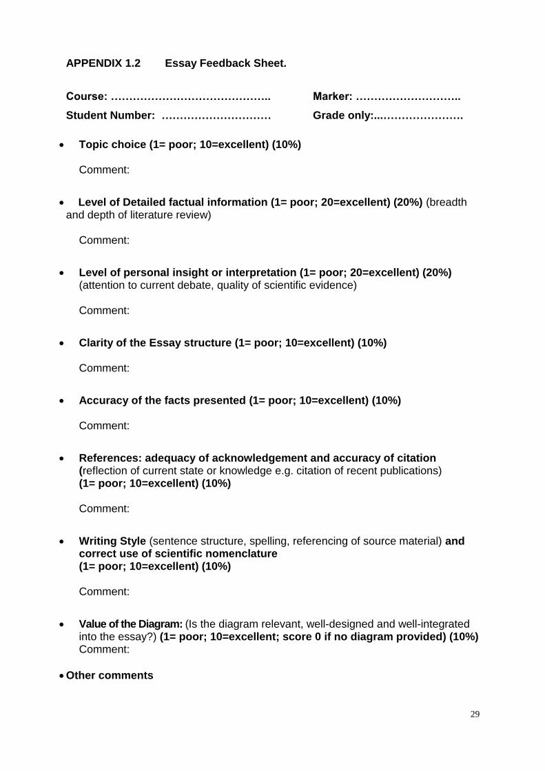
APPENDIX 1.2 Essay Feedback Sheet.
Course: …………………………………….. Marker: ………………………..
Student Number: ………………………… Grade only:...………………….
Topic choice (1= poor; 10=excellent) (10%)
Comment:
Level of Detailed factual information (1= poor; 20=excellent) (20%) (breadth and depth of literature review)
Comment:
Level of personal insight or interpretation (1= poor; 20=excellent) (20%) (attention to current debate, quality of scientific evidence)
Comment:
Clarity of the Essay structure (1= poor; 10=excellent) (10%) Comment:
Accuracy of the facts presented (1= poor; 10=excellent) (10%) Comment:
References: adequacy of acknowledgement and accuracy of citation (reflection of current state or knowledge e.g. citation of recent publications)
(1= poor; 10=excellent) (10%) Comment:
Writing Style (sentence structure, spelling, referencing of source material) and correct use of scientific nomenclature
(1= poor; 10=excellent) (10%) Comment:
Value of the Diagram: (Is the diagram relevant, well-designed and well-integrated into the essay?) (1= poor; 10=excellent; score 0 if no diagram provided) (10%)
Comment:
Other comments
30
APPENDIX 1.3 Instructions for the Practical in-course assessment.
The Practical assessment will be a “closed book” test. Thus, students shall NOT be
allowed to consult any books, notes or similar material during the test. Laboratory
notebooks should be used for revision before the test. The Practical Test will
contribute 20% of your final mark for the course. The test will include questions on all
of the practicals and related introductory material. The questions will cover practical
procedures, analysis of results, and discussion topics raised when the experiments
are reviewed. Thus, it will be to your advantage to write up each practical as
completely as possible after the class, to be used later for your revision before the
Test.
The majority of parasites discussed in this course will be of major human or
veterinary importance, thus issues such as diagnosis and control are central in their
management. It is hoped that by introducing you to these in the practical sessions
and testing your knowledge in this area in the practical test, you will appreciate the
demands of real-life parasitology work.
Sample questions will be provided on Learn.
31
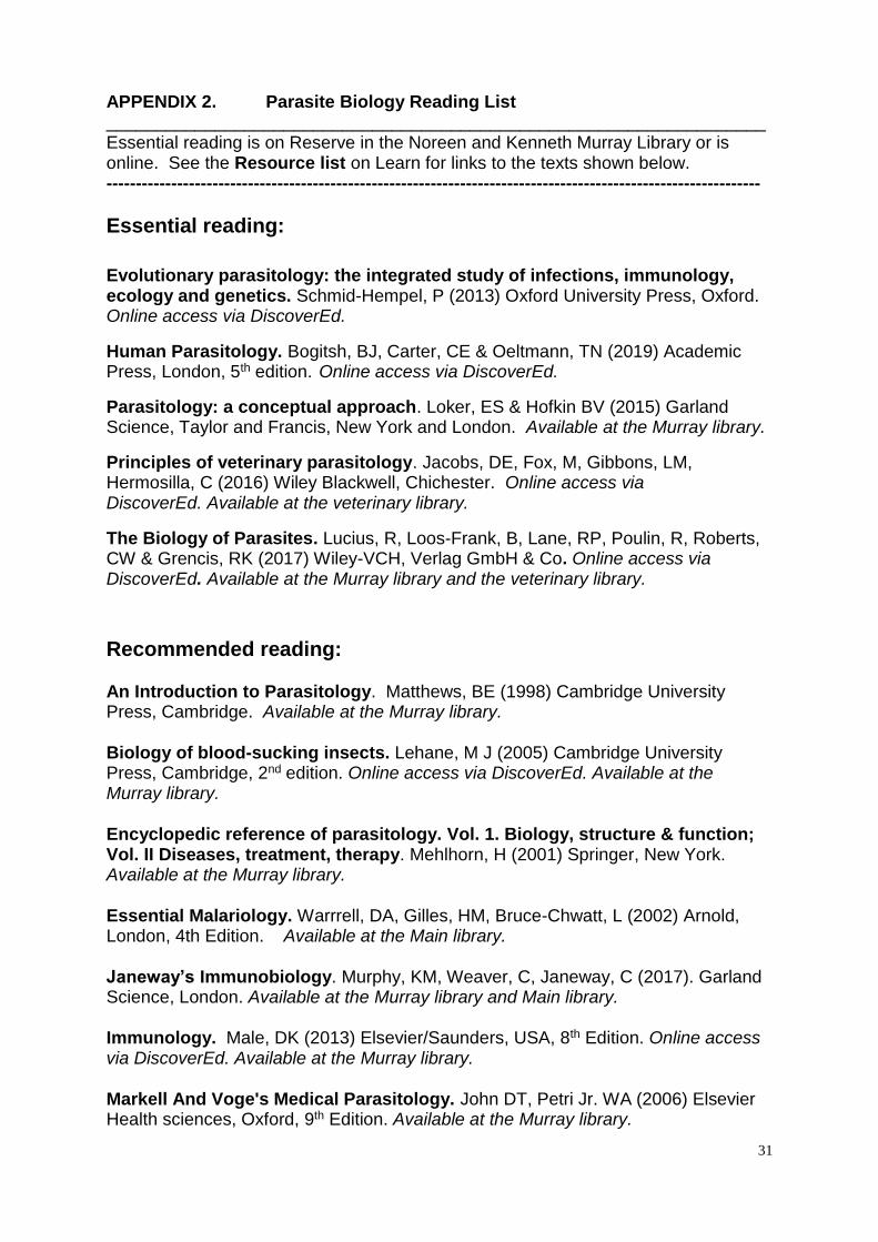
APPENDIX 2. Parasite Biology Reading List ___________________________________________________________________ Essential reading is on Reserve in the Noreen and Kenneth Murray Library or is online. See the Resource list on Learn for links to the texts shown below. ---------------------------------------------------------------------------------------------------------------
Essential reading:
Evolutionary parasitology: the integrated study of infections, immunology, ecology and genetics. Schmid-Hempel, P (2013) Oxford University Press, Oxford. Online access via DiscoverEd.
Human Parasitology. Bogitsh, BJ, Carter, CE & Oeltmann, TN (2019) Academic Press, London, 5th edition. Online access via DiscoverEd.
Parasitology: a conceptual approach. Loker, ES & Hofkin BV (2015) Garland Science, Taylor and Francis, New York and London. Available at the Murray library.
Principles of veterinary parasitology. Jacobs, DE, Fox, M, Gibbons, LM, Hermosilla, C (2016) Wiley Blackwell, Chichester. Online access via DiscoverEd. Available at the veterinary library.
The Biology of Parasites. Lucius, R, Loos-Frank, B, Lane, RP, Poulin, R, Roberts, CW & Grencis, RK (2017) Wiley-VCH, Verlag GmbH & Co. Online access via DiscoverEd. Available at the Murray library and the veterinary library.
Recommended reading: An Introduction to Parasitology. Matthews, BE (1998) Cambridge University Press, Cambridge. Available at the Murray library. Biology of blood-sucking insects. Lehane, M J (2005) Cambridge University Press, Cambridge, 2nd edition. Online access via DiscoverEd. Available at the Murray library. Encyclopedic reference of parasitology. Vol. 1. Biology, structure & function; Vol. II Diseases, treatment, therapy. Mehlhorn, H (2001) Springer, New York. Available at the Murray library. Essential Malariology. Warrrell, DA, Gilles, HM, Bruce-Chwatt, L (2002) Arnold, London, 4th Edition. Available at the Main library. Janeway’s Immunobiology. Murphy, KM, Weaver, C, Janeway, C (2017). Garland Science, London. Available at the Murray library and Main library. Immunology. Male, DK (2013) Elsevier/Saunders, USA, 8th Edition. Online access via DiscoverEd. Available at the Murray library. Markell And Voge's Medical Parasitology. John DT, Petri Jr. WA (2006) Elsevier Health sciences, Oxford, 9th Edition. Available at the Murray library.
32
Modern Parasitology: a textbook of parasitology. Cox, FEG (1993) Blackwell Scientific, Oxford. Online access via DiscoverEd. Available at the Murray library. Parasitic flatworms: molecular biology, biochemistry, immunology and physiology. Maule, AG, Marks NJ (2006) CABI publishing, Wallingford Online access via DiscoverEd. Parasitic nematodes: molecular biology, biochemistry, and immunology. Kennedy MW, Harnett W (Eds) (2001) CABI Publishing, Wallingford. Online access via DiscoverEd. Available at the Murray library. Parasitism: the diversity and ecology of animal parasites. Bush, AO, Fernandez, JC, Esch, GW, Seed, JR (2001) Cambridge University Press, Cambridge. Available at the Murray library. Protozoa and Human Disease. Wiser M F (2011) Garland Scientific, New York. Available at the Murray library. Schmidt & Robert’s Foundations of Parasitology. Roberts, LS & Janovy, J Jnr (2009) McGraw-Hill Higher Education, London, 8th Edition. Available at the Murray library. The encyclopedia of arthropod-transmitted infections of man and domesticated animals. Service, MW (2001) CABI Publishing, Wallingford, Ticks: biology, disease and control. Bowman, AS, Nutall PA (Eds) (2004) Cambridge University Press, Cambridge. Online access via DiscoverEd. Available at the Murray library.
Review articles: “Trends in Parasitology” is an excellent source of up-to-date reviews on assorted Parasite biology topics. Online access via DiscoverEd.
Popular science books: there are some fantastic popular science books about
parasites that you might want to read for fun including: Federal bodysnatchers and the New Guinea virus. Desowitz, R (2004) Norton, New York. Available at the Murray library. New Guinea Tapeworms and Jewish Grandmothers. Desowitz, R (1981) Norton, New York. Available at the Murray library. Parasite Rex. Zimmer, C (2001) Simon and Schuster, New York. Available at the Murray library. Riddled with Life. Zuk, M (2008) Harcourt, Boston. Available at the Murray library. The Malaria Capers. Desowitz, R (1991) Norton, New York. Available at the Murray library.
33
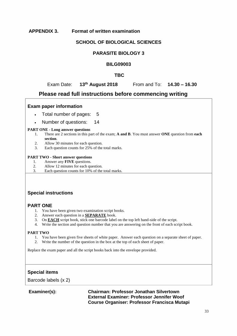
APPENDIX 3. Format of written examination
SCHOOL OF BIOLOGICAL SCIENCES
PARASITE BIOLOGY 3
BILG09003
TBC
Exam Date: 13th August 2018 From and To: 14.30 – 16.30
Please read full instructions before commencing writing
Examiner(s): Chairman: Professor Jonathan Silvertown External Examiner: Professor Jennifer Woof Course Organiser: Professor Francisca Mutapi
Exam paper information
Total number of pages: 5
Number of questions: 14
PART ONE - Long answer questions
1. There are 2 sections in this part of the exam; A and B. You must answer ONE question from each
section.
2. Allow 30 minutes for each question.
3. Each question counts for 25% of the total marks.
PART TWO - Short answer questions
1. Answer any FIVE questions.
2. Allow 12 minutes for each question.
3. Each question counts for 10% of the total marks.
Special instructions
PART ONE 1. You have been given two examination script books.
2. Answer each question in a SEPARATE book.
3. On EACH script book, stick one barcode label on the top left hand-side of the script.
4. Write the section and question number that you are answering on the front of each script book.
PART TWO
1. You have been given five sheets of white paper. Answer each question on a separate sheet of paper.
2. Write the number of the question in the box at the top of each sheet of paper.
Replace the exam paper and all the script books back into the envelope provided.
Special items
Barcode labels (x 2)
34
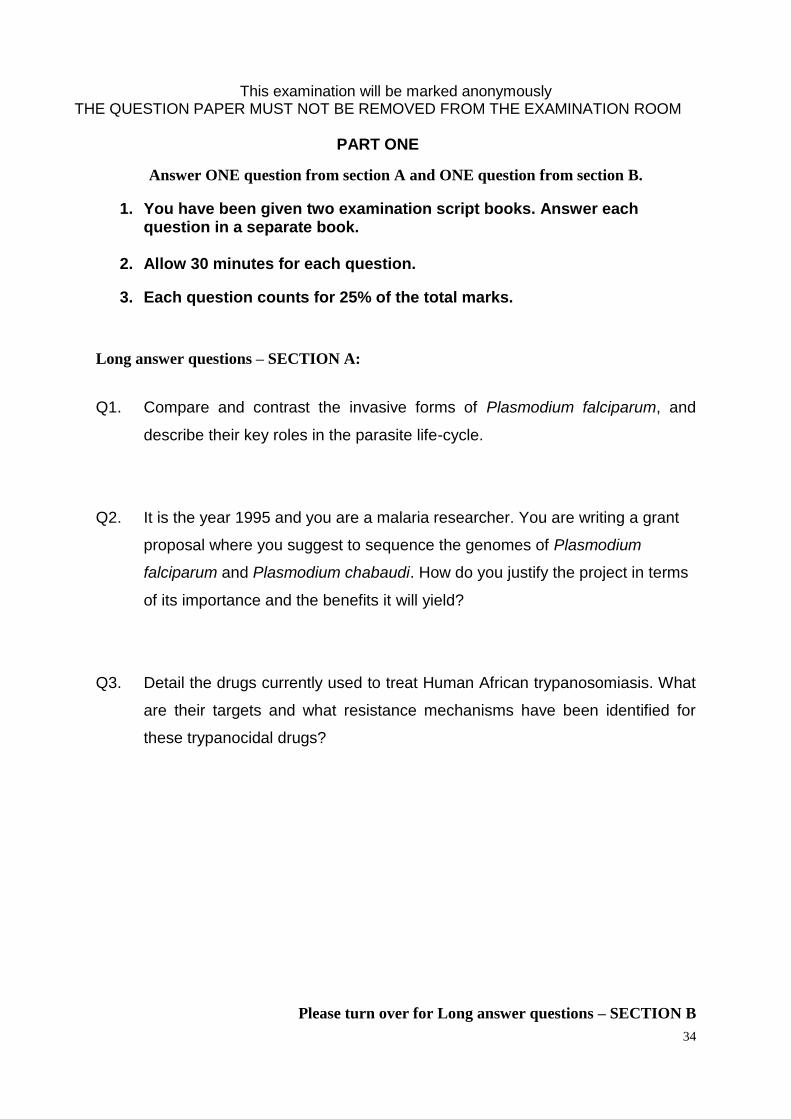
This examination will be marked anonymously THE QUESTION PAPER MUST NOT BE REMOVED FROM THE EXAMINATION ROOM
PART ONE
Answer ONE question from section A and ONE question from section B.
1. You have been given two examination script books. Answer each question in a separate book.
2. Allow 30 minutes for each question.
3. Each question counts for 25% of the total marks.
Long answer questions – SECTION A:
Q1. Compare and contrast the invasive forms of Plasmodium falciparum, and
describe their key roles in the parasite life-cycle.
Q2. It is the year 1995 and you are a malaria researcher. You are writing a grant
proposal where you suggest to sequence the genomes of Plasmodium
falciparum and Plasmodium chabaudi. How do you justify the project in terms
of its importance and the benefits it will yield?
Q3. Detail the drugs currently used to treat Human African trypanosomiasis. What
are their targets and what resistance mechanisms have been identified for
these trypanocidal drugs?
Please turn over for Long answer questions – SECTION B
35
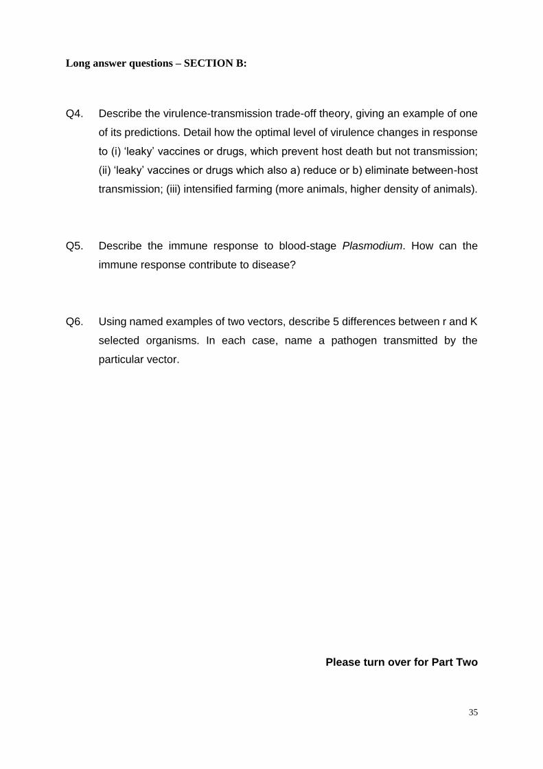
Long answer questions – SECTION B:
Q4. Describe the virulence-transmission trade-off theory, giving an example of one
of its predictions. Detail how the optimal level of virulence changes in response
to (i) ‘leaky’ vaccines or drugs, which prevent host death but not transmission;
(ii) ‘leaky’ vaccines or drugs which also a) reduce or b) eliminate between-host
transmission; (iii) intensified farming (more animals, higher density of animals).
Q5. Describe the immune response to blood-stage Plasmodium. How can the
immune response contribute to disease?
Q6. Using named examples of two vectors, describe 5 differences between r and K
selected organisms. In each case, name a pathogen transmitted by the
particular vector.
Please turn over for Part Two
36
PART TWO
Answer any FIVE questions.
1. You have been given five sheets of white paper. Answer each question on a separate sheet of paper.
2. Write the number of the question in the box at the top of each sheet of paper.
3. Allow 12 minutes for each question.
4. Each question counts for 10% of the marks.
Short answer questions:
Q1. Using the genus Plasmodium as an example, explain the concept of
housekeeping genes, lineage-specific genes and species-specific genes, give
at least one example for each group, and briefly discuss how comparative
genomics can help to identify such genes.
Q2. Describe one long-term form of coevolution. Compare this with a short-term
form of coevolution.
Q3. Draw a diagram of the trypanosome cell cycle labelling each stage.
Q4. How does Trypanosoma cruzi stimulate its uptake into host cells and establish
its intracellular niche?
Q5. In the evolution of Marek’s disease increasing virulence has been observed
since the 1950s. During the lecture we discussed two potential causes that may
have driven this evolution of increased virulence. Name these examples and
explain how they could lead to selection of more virulent Marek’s disease. Is
this higher virulence a problem for vaccinated birds? Explain why. Is it a
problem for birds that have not been vaccinated, and why?
Please turn over for Part Two continued
37
Part Two (continued)
Q6. Name three economically important nematodes of livestock, and draw and/or
describe the direct lifecycle of one of them.
Q7. Define a hemimetabolous life cycle and a holometabolous life cycle. Draw two
diagrams, one to illustrate each of these two life cycles, giving a named
example for each.
Q8. Using named examples, describe three models of inducing protective immunity
against helminth parasites. Explain one advantage and one disadvantage for
each of the 3 models.
End of Paper
38
APPENDIX 4. Practical Manual
PRACTICAL 1: Plasmodium spp The aim of the practical is to think about how the morphology of the parasite that you see down the microscope relates to its biological functions. What do the appearances of the different life cycle stages tell you about how the parasite develops and survives within the human host and how it is transmitted to new hosts? What questions are raised by what you see down the microscope? Three main aims 1. Staining a blood film with Giemsa 2. Identifying life cycle stages of P. falciparum by light microscopy 3. Seeing exflagellation of P. berghei male gametes. Learning objectives: things you should know by the end of the practical! a. How to stain a thin blood film with Giemsa stain. b. How to use a light microscope with oil immersion lens. c. What do malaria parasites look like under the microscope? Be able to identify the major blood stage life cycle stages of Plasmodium falciparum: ring stage trophozoites pigmented trophozoites schizonts gametocytes d. What is exflagellation? NB. The P. falciparum smears you will examine are from in vitro cultures. In P. falciparum infected individuals, you only ever see ring stage parasites in the peripheral blood because the more mature stages are sequestered in microvascular beds. Similarly, for gametocytes, only mature (stage V) gametocytes would be seen in a blood film. Instructions: Section 1 Giemsa staining: work in groups of 2. Wear gloves when handling stain. a) You are provided with air-dried thin blood films of P. falciparum parasites. These have been fixed in methanol to preserve the protein and DNA of the cells. b) write your name in pencil on the frosted end of the slide c) Prepare 20 ml of Giemsa stain by diluting the stock Giemsa stain 1:20 with buffer. Take up 1ml of Giemsa with the plastic pipette. Transfer into the measuring cylinder. Make up to 20ml with buffer from the squeezy bottle. d) pour the stain into the staining dish e) place the blood film in the staining dish (writing and blood film face DOWN). f) leave it for approx 20 minutes. In the meantime, move on to the next section and start to look at the pre-stained slides down the microscope. Don’t forget about your staining slide though…. and after 20minutes:
39
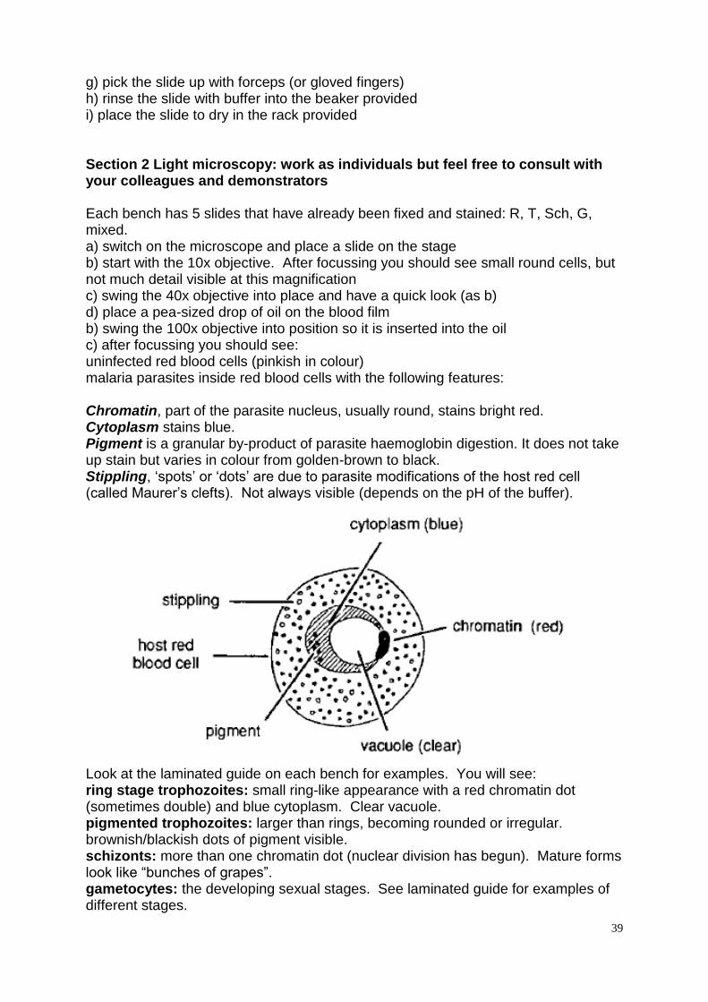
g) pick the slide up with forceps (or gloved fingers) h) rinse the slide with buffer into the beaker provided i) place the slide to dry in the rack provided Section 2 Light microscopy: work as individuals but feel free to consult with your colleagues and demonstrators Each bench has 5 slides that have already been fixed and stained: R, T, Sch, G, mixed. a) switch on the microscope and place a slide on the stage b) start with the 10x objective. After focussing you should see small round cells, but not much detail visible at this magnification c) swing the 40x objective into place and have a quick look (as b) d) place a pea-sized drop of oil on the blood film b) swing the 100x objective into position so it is inserted into the oil c) after focussing you should see: uninfected red blood cells (pinkish in colour) malaria parasites inside red blood cells with the following features: Chromatin, part of the parasite nucleus, usually round, stains bright red. Cytoplasm stains blue. Pigment is a granular by-product of parasite haemoglobin digestion. It does not take up stain but varies in colour from golden-brown to black. Stippling, ‘spots’ or ‘dots’ are due to parasite modifications of the host red cell (called Maurer’s clefts). Not always visible (depends on the pH of the buffer).
Look at the laminated guide on each bench for examples. You will see: ring stage trophozoites: small ring-like appearance with a red chromatin dot (sometimes double) and blue cytoplasm. Clear vacuole. pigmented trophozoites: larger than rings, becoming rounded or irregular. brownish/blackish dots of pigment visible. schizonts: more than one chromatin dot (nuclear division has begun). Mature forms look like “bunches of grapes”. gametocytes: the developing sexual stages. See laminated guide for examples of different stages.
40
For each smear, draw some examples of infected red cells, and label the parts you can identify Slide R Slide T There is a host-parasite interaction that is associated with severe malaria visible on slide T – what is it? Slide Sch Slide G
41
Your own stained slide: What stage(s) of parasites can you see? At the end of the session: Wipe the oil off the 100x objective with microscope lens tissue. Place the slides face down onto a piece of paper towel (to absorb the oil). Put the microscope away. After the practical: See Learn for some revision material including: A copy of the Plasmodium microscopy appearance guides A copy of the quiz A reminder of the main learning points associated with the practical. Section 3: Activation and Exflagellation of Plasmodium berghei gametocytes The aim of this section is to study the activation and exflagellation of gametocytes of the rodent malaria parasite P. berghei, and to understand how this is regulated by the exflagellation factor, Xanthurenic acid. This practical will demonstrate
1. Mouse red blood cells (RBCs) infected with asexual and sexual (gametocytes) stages of P. berghei parasites.
2. Stimulation of gametocytes to undergo activation and exflagellation in conditions that resemble those found in the mosquito midgut.
Introduction Malaria parasites proliferate via asexual replication in the RBCs of their infected mammalian host. In each cycle, a proportion of the parasites withdraw from the asexual cycle and commit to form the sexual stages; the male and female gametocytes. These mature and remain quiescent in the blood of the host until they are taken up by the mosquito within the infected bloodmeal. Within the mosquito midgut, the parasites encounter a reduction in temperature and are exposed to elevated concentrations of a ‘gametocyte exflagellation factor’ that is believed to be Xanthurenic Acid. These conditions stimulate the gametocytes to ‘round-up’ and emerge from RBCs as gametes. Within 10 minutes male gametes undergo three rounds of DNA replication and then begin exflagellation to produce motile and flagellated microgametes which bind RBCs, forming a cluster called an exflagellation
42
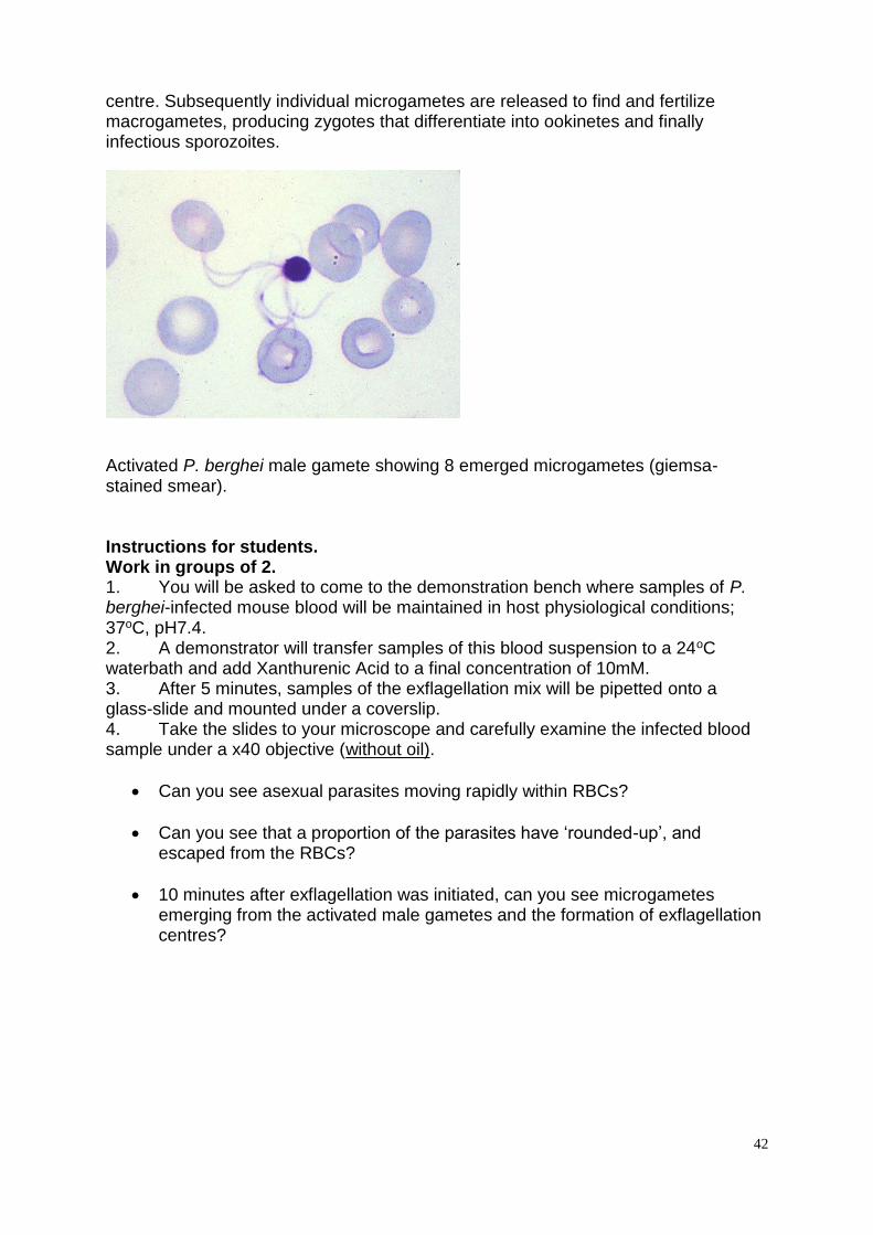
centre. Subsequently individual microgametes are released to find and fertilize macrogametes, producing zygotes that differentiate into ookinetes and finally infectious sporozoites.
Activated P. berghei male gamete showing 8 emerged microgametes (giemsa-stained smear). Instructions for students. Work in groups of 2. 1. You will be asked to come to the demonstration bench where samples of P. berghei-infected mouse blood will be maintained in host physiological conditions; 37oC, pH7.4. 2. A demonstrator will transfer samples of this blood suspension to a 24oC waterbath and add Xanthurenic Acid to a final concentration of 10mM. 3. After 5 minutes, samples of the exflagellation mix will be pipetted onto a glass-slide and mounted under a coverslip. 4. Take the slides to your microscope and carefully examine the infected blood sample under a x40 objective (without oil).
Can you see asexual parasites moving rapidly within RBCs?
Can you see that a proportion of the parasites have ‘rounded-up’, and escaped from the RBCs?
10 minutes after exflagellation was initiated, can you see microgametes emerging from the activated male gametes and the formation of exflagellation centres?
43
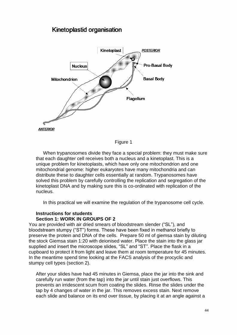
Practical 2: Trypanosoma brucei cell cycle Aims and Objectives The aim of this practical is to study the cell-cycle of the protozoan parasite Trypanosoma brucei, and understand how this is controlled during different stages of their life cycle. Also, we will examine how the function of a structural component in the trypanosome cell can be determined using genetic manipulation of the parasite. The objectives of this practical are to demonstrate: 1. The cell cycle of the tsetse fly midgut form of T.brucei, 2. Regulation of the cell cycle of T.brucei in different life-cycle stages Introduction African trypanosomes are parasitic protozoa responsible for ‘sleeping sickness’ in sub Saharan Africa. They infect humans, game animals and domesticated livestock, and are transmitted by the bite of the tsetse fly. Trypanosomes are extremely ancient organisms: evolutionary analysis has demonstrated that these parasites were among the first organism to branch from the eukaryotic lineage. Their importance as disease causing organisms, and their ancient origins have caused trypanosomes to become a model for understanding (1) the basis of immune evasion and pathogenicity in protozoan parasites and (2) the origins of the basic characteristics of the eukaryotic cell. The basic structure of the trypanosome is shown in Figure 1. It has a nucleus, mitochondrion and flagellum. Parasitologists noticed very early on that trypanosomes have a second DNA containing organelle in addition to the nucleus. This is called the “kinetoplast”, and gives these organisms their Family name, “kinetoplastidae”. Many kinetoplastid organisms are parasites, and these are responsible for several of the major diseases of the developing world (Leishmaniasis, South American trypanosomiasis, in addition to African trypanosomiasis). The amount of DNA in the kinetoplast is quite large, and makes up somewhere between 5-30% of the total DNA of the cell. The kinetoplast is the specialised mitcohondrial genome of trypanosomes and encodes components of the respiratory chain.
44
Figure 1 When trypanosomes divide they face a special problem: they must make sure that each daughter cell receives both a nucleus and a kinetoplast. This is a unique problem for kinetoplasts, which have only one mitochondrion and one mitochondrial genome: higher eukaryotes have many mitochondria and can distribute these to daughter cells essentially at random. Trypanosomes have solved this problem by carefully controlling the replication and segregation of the kinetoplast DNA and by making sure this is co-ordinated with replication of the nucleus. In this practical we will examine the regulation of the trypanosome cell cycle. Instructions for students Section 1: WORK IN GROUPS OF 2
You are provided with air dried smears of bloodstream slender (“SL”), and bloodstream stumpy (“ST”) forms. These have been fixed in methanol briefly to preserve the protein and DNA of the cells. Prepare 50 ml of giemsa stain by diluting the stock Giemsa stain 1:20 with deionised water. Place the stain into the glass jar supplied and insert the microscope slides, “SL” and “ST”. Place the flask in a cupboard to protect it from light and leave them at room temperature for 45 minutes. In the meantime spend time looking at the FACS analysis of the procyclic and stumpy cell types (section 2).
After your slides have had 45 minutes in Giemsa, place the jar into the sink and carefully run water (from the tap) into the jar until stain just overflows. This prevents an irridescent scum from coating the slides. Rinse the slides under the tap by 4 changes of water in the jar. This removes excess stain. Next remove each slide and balance on its end over tissue, by placing it at an angle against a
45
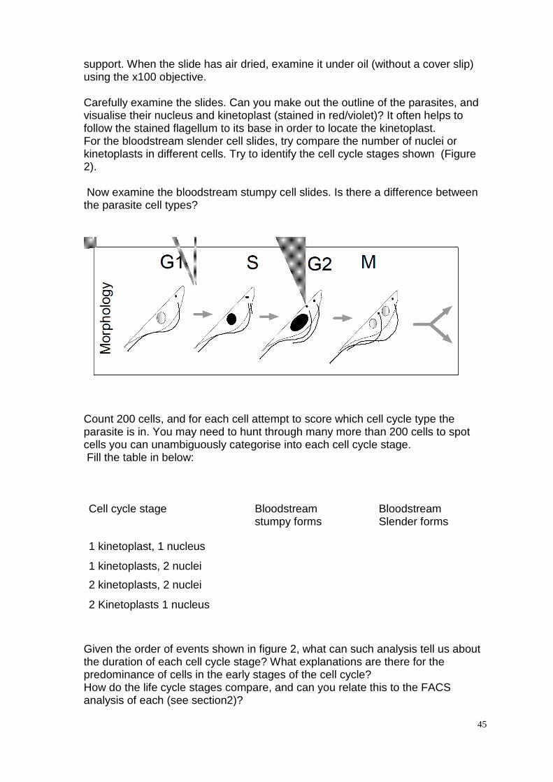
support. When the slide has air dried, examine it under oil (without a cover slip) using the x100 objective. Carefully examine the slides. Can you make out the outline of the parasites, and visualise their nucleus and kinetoplast (stained in red/violet)? It often helps to follow the stained flagellum to its base in order to locate the kinetoplast. For the bloodstream slender cell slides, try compare the number of nuclei or kinetoplasts in different cells. Try to identify the cell cycle stages shown (Figure 2). Now examine the bloodstream stumpy cell slides. Is there a difference between the parasite cell types?
Count 200 cells, and for each cell attempt to score which cell cycle type the parasite is in. You may need to hunt through many more than 200 cells to spot cells you can unambiguously categorise into each cell cycle stage. Fill the table in below: Cell cycle stage Bloodstream
stumpy forms Bloodstream Slender forms
1 kinetoplast, 1 nucleus
1 kinetoplasts, 2 nuclei
2 kinetoplasts, 2 nuclei
2 Kinetoplasts 1 nucleus
Given the order of events shown in figure 2, what can such analysis tell us about the duration of each cell cycle stage? What explanations are there for the predominance of cells in the early stages of the cell cycle? How do the life cycle stages compare, and can you relate this to the FACS analysis of each (see section2)?
46
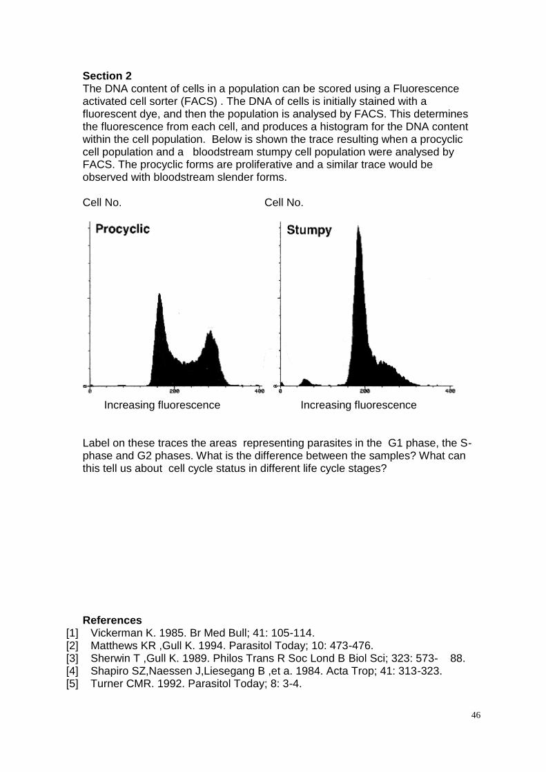
Section 2 The DNA content of cells in a population can be scored using a Fluorescence activated cell sorter (FACS) . The DNA of cells is initially stained with a fluorescent dye, and then the population is analysed by FACS. This determines the fluorescence from each cell, and produces a histogram for the DNA content within the cell population. Below is shown the trace resulting when a procyclic cell population and a bloodstream stumpy cell population were analysed by FACS. The procyclic forms are proliferative and a similar trace would be observed with bloodstream slender forms. Cell No. Cell No.
Increasing fluorescence Increasing fluorescence Label on these traces the areas representing parasites in the G1 phase, the S-phase and G2 phases. What is the difference between the samples? What can this tell us about cell cycle status in different life cycle stages?
References
[1] Vickerman K. 1985. Br Med Bull; 41: 105-114. [2] Matthews KR ,Gull K. 1994. Parasitol Today; 10: 473-476. [3] Sherwin T ,Gull K. 1989. Philos Trans R Soc Lond B Biol Sci; 323: 573- 88. [4] Shapiro SZ,Naessen J,Liesegang B ,et a. 1984. Acta Trop; 41: 313-323. [5] Turner CMR. 1992. Parasitol Today; 8: 3-4.
47
Practical 3: Helminths (A) Simplified Faecal Egg Count Reduction Test using Modified McMaster Egg
Counting Faecal Egg Count Reduction Test (FECRT)
The FECRT is an in vivo assay for anthelmintic resistance that can be used with any drug. The protocol below follows the WAAVP guidelines proposed by Coles et al. (1992 & 2006). Pooled faecal egg counts (FEC) from control (non-treated) animals are compared to egg counts from treated animals. Groups of animals (n=15) are sampled and egg counts are determined 7-14 days post treatment, the reduction in egg count is estimated using the standard formula. Percentage reduction = (Day 0 FEC – Day 14 FEC) x 100
Day 0 FEC
Modern anthelmintics are highly effective generally giving a more than 99%
efficacy. Efficacies of less than 95% suggest that resistance may be present within the parasite population. Background Following the results of the EHA on the two farms- Hermitage and Blacklees, the vet decided to further investigate the resistance status of the farms in order to recommend future treatment strategies. Hermitage was also seeing a small reduction in lamb growth rates, and as they had been using ivermectin this season, the vet suggested testing for ivermectin resistance. Blacklees hadn’t used ivermectin before, as they were in the habit of using benzimidazoles. But before suggesting a switch to ivermectin, the vet wanted to be sure that it would work at Blacklees. He took a further pooled faecal sample from a small number of sheep at each farm on the day of treatment with avermectin and returned two weeks later to take a further sample from the same group. Method 1. Collect 10ml into labelled tubes from the day 0 and day 14 samples from each
farm using the provided syringes. IMPORTANT: the faecal suspension needs to be thoroughly re-suspended by pulling and pushing the syringe plunger several times, avoiding spilling the suspension.
2. Each subsample is placed in a labelled plastic centrifuge tube and centrifuged at 1000 rpm for 2 minutes.
3. The supernatant is carefully poured off leaving the eggs and faecal debris in a
pellet at the bottom of the tube. 4. Carefully add10ml of saturated salt (NaCl) solution to the tube and resuspend the
faecal debris and eggs by gently inverting the tube. IMPORTANT: Do not shake
48
the tube, be patient and gentle, otherwise the sample will become full of air bubbles and very difficult to read.
5. Resuspend again (GENTLY) and pipette approximately 1 ml of the salt
suspension into the first chamber of the McMaster slide. Resuspend the tube and repeat for the process with second chamber. NB IT HELPS IF THE CHAMBERS ARE WET PRIOR TO BEING USED
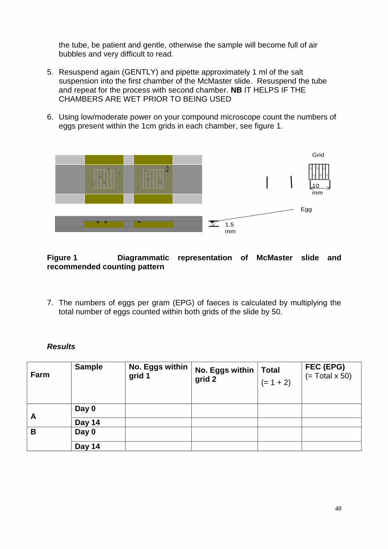
6. Using low/moderate power on your compound microscope count the numbers of
eggs present within the 1cm grids in each chamber, see figure 1.
Figure 1 Diagrammatic representation of McMaster slide and recommended counting pattern 7. The numbers of eggs per gram (EPG) of faeces is calculated by multiplying the
total number of eggs counted within both grids of the slide by 50.
Results
Farm Sample No. Eggs within
grid 1 No. Eggs within grid 2
Total
(= 1 + 2)
FEC (EPG) (= Total x 50)
A Day 0
Day 14
B Day 0
Day 14
10
mm
1.5
mm
Egg
Grid
1.5
mm
49
Practical: Field Screen for benzimidazole resistance Egg hatch Test (EHT)
The EHT is an in vitro test for detection of thiabendazole resistance. Thiabendazole (TBZ) was the first broad spectrum anthelmintic introduced onto the market in 1961 and resistance to this family of anthelmintic is widespread throughout the world. Benzimidazoles work by binding to the microtubule subunit protein, tubulin, and disrupt the microtubule structure and function.
Background
Two farms are to be tested, each of which sent a pooled sample of faeces from 10 sheep to the lab. Hermitage Farm is a large farm in Perthshire run by the Mackinnon family. They operate in close cooperation with their local vet and use monitoring to decide when and how to treat parasite infections. Nearby Blacklees Farm is much smaller and run by two brothers. They use a benzimidazole drug, the same as they have used for years and see no reason to change as it seems to work and they are familiar with it. This year though, their lambs don’t seem to be thriving as they should so they called in the vet who suggested testing for BZ resistance.
Method
The hatching of TBZ susceptible eggs is inhibited in relatively low concentrations
of benzimidazole. In this assay eggs are incubated in a range of concentrations of the drug to calculate the ED50 estimate (concentration of drug at which 50% of eggs are prevented from hatching) which gives an indication of the resistance status of the parasite population. This is one of several bioassays used to measure resistance; each is designed to measure the phenotypic response to anti-parasitic chemicals.
The method requires fresh eggs (within 4 hours of being passed) or eggs
stored anaerobically. The eggs have been extracted from the faeces and incubated in a range of TBZ concentrations for 48 hours which allows sufficient time for the eggs to develop and hatch. Samples from each farm have been incubated in 0, 0.05, 0.1 and 0.3 µg/ml TBZ. Each sample was then fixed using Helminthological Iodine solution.
1. Wash the contents of a sample pot onto a small petri dish, rinse the pot and dilute with tap water to form a uniform layer across the dish.
2. Using an inverted stereo microscope count the numbers of eggs and larvae at
each concentration from farms A and B and record on the table overleaf.
3. Calculate the percentage hatch at each concentration using the standard formula.
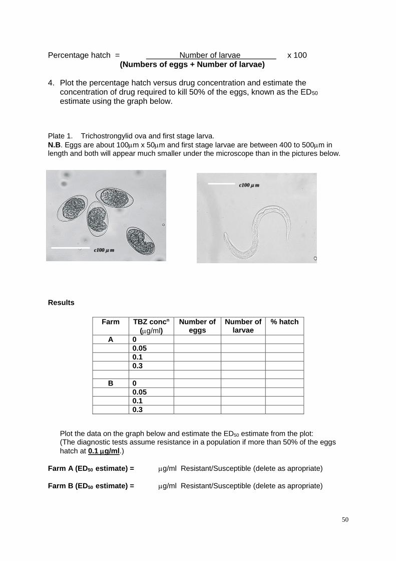
50
Percentage hatch = Number of larvae x 100
(Numbers of eggs + Number of larvae)
4. Plot the percentage hatch versus drug concentration and estimate the concentration of drug required to kill 50% of the eggs, known as the ED50 estimate using the graph below.
Plate 1. Trichostrongylid ova and first stage larva.
N.B. Eggs are about 100m x 50m and first stage larvae are between 400 to 500m in length and both will appear much smaller under the microscope than in the pictures below.
Results
Farm TBZ concn
(g/ml)
Number of eggs
Number of larvae
% hatch
A 0
0.05
0.1
0.3
B 0
0.05
0.1
0.3
Plot the data on the graph below and estimate the ED50 estimate from the plot: (The diagnostic tests assume resistance in a population if more than 50% of the eggs
hatch at 0.1 g/ml.)
Farm A (ED50 estimate) = g/ml Resistant/Susceptible (delete as apropriate)
Farm B (ED50 estimate) = g/ml Resistant/Susceptible (delete as apropriate)
c100 m
c100 m
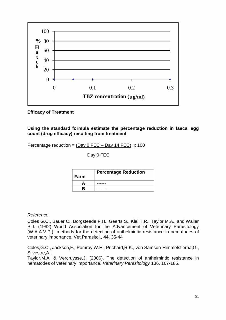
51
Efficacy of Treatment
Using the standard formula estimate the percentage reduction in faecal egg count (drug efficacy) resulting from treatment
Percentage reduction = (Day 0 FEC – Day 14 FEC) x 100
Day 0 FEC
Farm Percentage Reduction
A ------
B ------
Reference
Coles G.C., Bauer C., Borgsteede F.H., Geerts S., Klei T.R., Taylor M.A., and Waller P.J. (1992) World Association for the Advancement of Veterinary Parasitology (W.A.A.V.P.) methods for the detection of anthelmintic resistance in nematodes of veterinary importance. Vet.Parasitol., 44, 35-44 Coles,G.C., Jackson,F., Pomroy,W.E., Prichard,R.K., von Samson-Himmelstjerna,G., Silvestre,A., Taylor,M.A. & Vercruysse,J. (2006). The detection of anthelmintic resistance in nematodes of veterinary importance. Veterinary Parasitology 136, 167-185.
0
20
40
60
80
100
0 0.1 0.2 0.3
%
H
a
t
c
h
TBZ concentration ( g/ml)
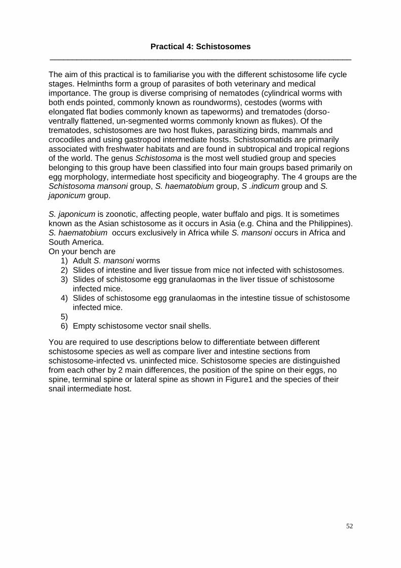
52
Practical 4: Schistosomes ___________________________________________________________________ The aim of this practical is to familiarise you with the different schistosome life cycle stages. Helminths form a group of parasites of both veterinary and medical importance. The group is diverse comprising of nematodes (cylindrical worms with both ends pointed, commonly known as roundworms), cestodes (worms with elongated flat bodies commonly known as tapeworms) and trematodes (dorso-ventrally flattened, un-segmented worms commonly known as flukes). Of the trematodes, schistosomes are two host flukes, parasitizing birds, mammals and crocodiles and using gastropod intermediate hosts. Schistosomatids are primarily associated with freshwater habitats and are found in subtropical and tropical regions of the world. The genus Schistosoma is the most well studied group and species belonging to this group have been classified into four main groups based primarily on egg morphology, intermediate host specificity and biogeography. The 4 groups are the Schistosoma mansoni group, S. haematobium group, S .indicum group and S. japonicum group. S. japonicum is zoonotic, affecting people, water buffalo and pigs. It is sometimes known as the Asian schistosome as it occurs in Asia (e.g. China and the Philippines). S. haematobium occurs exclusively in Africa while S. mansoni occurs in Africa and South America. On your bench are
1) Adult S. mansoni worms 2) Slides of intestine and liver tissue from mice not infected with schistosomes. 3) Slides of schistosome egg granulaomas in the liver tissue of schistosome
infected mice. 4) Slides of schistosome egg granulaomas in the intestine tissue of schistosome
infected mice. 5) 6) Empty schistosome vector snail shells.
You are required to use descriptions below to differentiate between different schistosome species as well as compare liver and intestine sections from schistosome-infected vs. uninfected mice. Schistosome species are distinguished from each other by 2 main differences, the position of the spine on their eggs, no spine, terminal spine or lateral spine as shown in Figure1 and the species of their snail intermediate host.
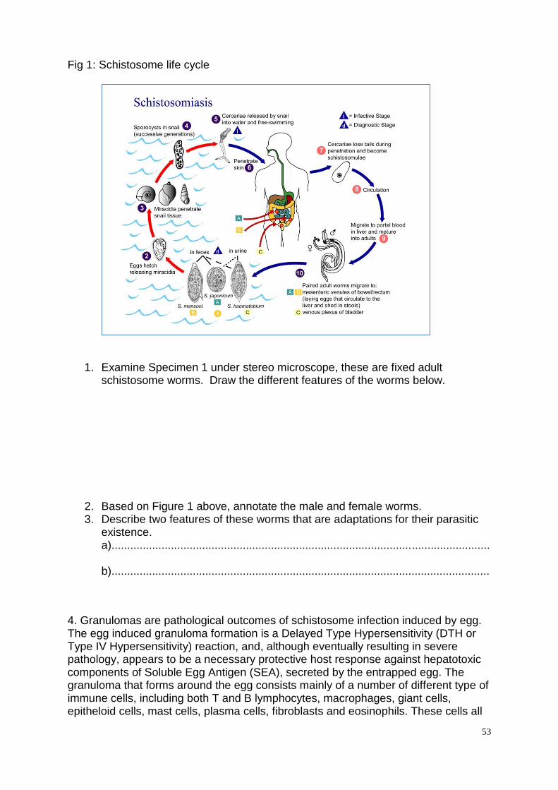
53
Fig 1: Schistosome life cycle
1. Examine Specimen 1 under stereo microscope, these are fixed adult schistosome worms. Draw the different features of the worms below.
2. Based on Figure 1 above, annotate the male and female worms. 3. Describe two features of these worms that are adaptations for their parasitic
existence. a)......................................................................................................................... b).........................................................................................................................
4. Granulomas are pathological outcomes of schistosome infection induced by egg. The egg induced granuloma formation is a Delayed Type Hypersensitivity (DTH or Type IV Hypersensitivity) reaction, and, although eventually resulting in severe pathology, appears to be a necessary protective host response against hepatotoxic components of Soluble Egg Antigen (SEA), secreted by the entrapped egg. The granuloma that forms around the egg consists mainly of a number of different type of immune cells, including both T and B lymphocytes, macrophages, giant cells, epitheloid cells, mast cells, plasma cells, fibroblasts and eosinophils. These cells all
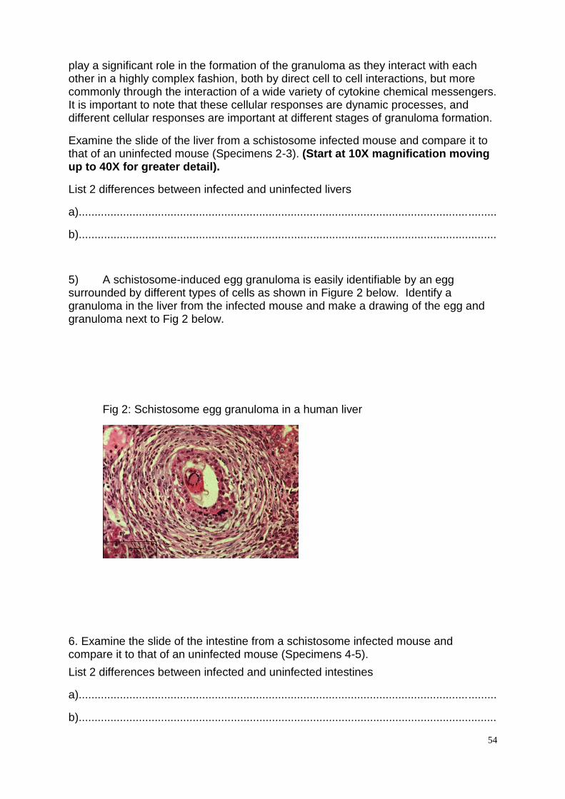
54
play a significant role in the formation of the granuloma as they interact with each other in a highly complex fashion, both by direct cell to cell interactions, but more commonly through the interaction of a wide variety of cytokine chemical messengers. It is important to note that these cellular responses are dynamic processes, and different cellular responses are important at different stages of granuloma formation.
Examine the slide of the liver from a schistosome infected mouse and compare it to that of an uninfected mouse (Specimens 2-3). (Start at 10X magnification moving up to 40X for greater detail).
List 2 differences between infected and uninfected livers
a)....................................................................................................................................
b)....................................................................................................................................
5) A schistosome-induced egg granuloma is easily identifiable by an egg surrounded by different types of cells as shown in Figure 2 below. Identify a granuloma in the liver from the infected mouse and make a drawing of the egg and granuloma next to Fig 2 below.
Fig 2: Schistosome egg granuloma in a human liver
6. Examine the slide of the intestine from a schistosome infected mouse and compare it to that of an uninfected mouse (Specimens 4-5).
List 2 differences between infected and uninfected intestines
a)....................................................................................................................................
b)....................................................................................................................................
55
7) Based on the egg morphology in figure 1 above, identify the schistosome species causing pathology in the mouse liver and intestine.
....................................................................................................................................... 8) Find a blood vessel in any of the tissue sections you have been given and
draw the blood vessel and its contents in the space below.
9. You have been given 3 snail shells, Specimens 6-8. These are shells from the schistosome intermediate host snails. Schistosoma mansoni- Biomphalaria glabrata snails- shell is spiral and more dorso-ventraly flatterned. Schistosoma haematobium -Bulinus truncatus truncatus snails- shells are more globular with a large opening and Schistosoma japonicum - Oncomelania hupensis quadrasi snails- shells are the smallest shells and they are long and thin. Based on this description identify the shells for the intermediate host for each of the 3 species above. Once your identification has been confirmed, make an annotated drawing of each shell below.
56
Materials required for the Practical 4
For each bench please lay out the following on stereo or compound microscopes and put a card with the specimen number next to microscope
Practical Requirements
Equipment
1. Each student will require access to a compound and stereo microscope to examine the slides
2. A pair of sharp seekers without burs; 3. Some tissues to mop up accidents.
Specimens
Each student is supplied with:
1. Specimen 1: A pair of adult S. mansoni worms (1 male, longer and clear, 1 female, shorter, thinner and darker). To be viewed on stereo microscope. Please put these in a watch glass with 70% ethanol. These have been provided by Andrew MacDonald and can be kept for future practicals.
2. Specimens 2/3: Two slides of schistosome egg granulaomas in the liver, 1 from an infected mouse and one from an uninfected mouse. To be viewed on compound microscope. These have been provided by Andrew MacDonald and must be returned to him after the practicals.
3. Specimens 4/5: Two slides of schistosome egg granulaomas in the intestine, 1 from an infected mouse and one from an uninfected mouse. To be viewed on compound microscope. These have been provided by Andrew MacDonald and must be returned to him after the practicals.
4. Specimens 6-8: Three empty schistosome vector snail shells. To be viewed
on stereo microscope. Please put these in a dry watch glass. These can be
kept for future practicals.
If there is not enough material for each student to have their own set of samples (e.g.
the snail shells), specimens can be shared between students.
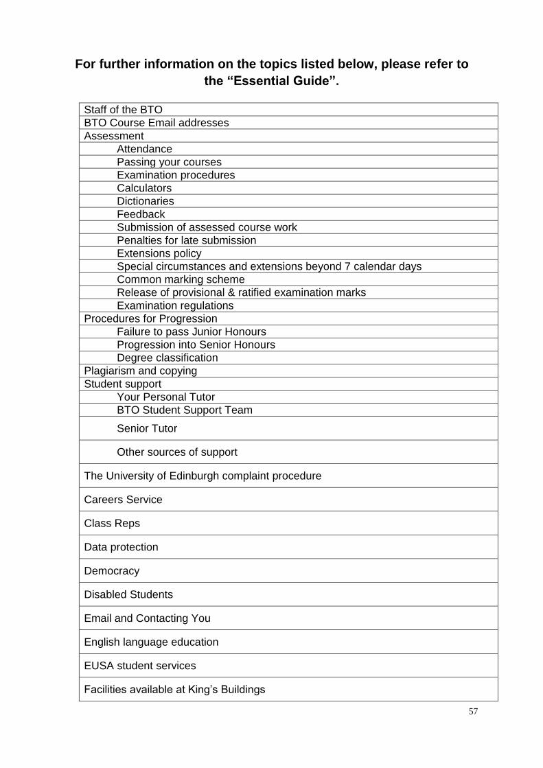
57
For further information on the topics listed below, please refer to
the “Essential Guide”.
Staff of the BTO
BTO Course Email addresses
Assessment
Attendance
Passing your courses
Examination procedures
Calculators
Dictionaries
Feedback
Submission of assessed course work
Penalties for late submission
Extensions policy
Special circumstances and extensions beyond 7 calendar days
Common marking scheme
Release of provisional & ratified examination marks
Examination regulations
Procedures for Progression
Failure to pass Junior Honours
Progression into Senior Honours
Degree classification
Plagiarism and copying
Student support
Your Personal Tutor
BTO Student Support Team
Senior Tutor
Other sources of support
The University of Edinburgh complaint procedure
Careers Service
Class Reps
Data protection
Democracy
Disabled Students
Email and Contacting You
English language education
EUSA student services
Facilities available at King’s Buildings
58
Institute for Academic Development
Lab Coats
Learn
Maps and Transport
Safety in the laboratory
Safety Notes–Teaching Laboratories (King’s Buildings)
Social Media guidance for students
Student Administration
Students on a Tier 4 Visa
Study Skills
Use of Animals in Teaching
Use of barcode labels
Useful contacts
Telephone directory