Embed Size (px)
Citation preview
i Clin Pathol: Mol Pathol 1995;48:M241-M250
Papers
Isolation and characterisation of antibodieswhich specifically recognise the peptide encodedby exon 7 (v2) of the human CD44 gene
A Borgya, A Woodman, M Sugiyama, F Donie, E Kopetzki, Y Matsumura, D Tarin
AbstractAims-Exon 7 of the human CD44 gene isoverexpressed in many commonly oc-curring carcinomas. The aim of the studywas to explore the diagnostic and thera-peutic potential of this frequent ab-normality.Methods-A new monoclonal antibody(mAb, M-23.6. 1) and a polyclonal antibody(pAb,S-6127) to the corresponding anti-gen were raised by inumunising mice andsheep, respectively, with a specially con-structed fusion protein HIV2 (gp32)-CD44exon 7.Results-Characterisation of mAb,M-23.6.1 by ELISA, western blotting, im-munocytochemistry, and FACS analysisconfirmed that it specifically recognisesan epitope in the region between aminoacids 19 and 33 of the peptide encoded bythis exon. Western blotting experimentswith two cell lines, RT112 and ZR75-1,known from RT-PCR data to be over-transcribing the exon, yielded a mono-specific band of approximately 220kkDa,and immunocytochemistry showed dis-crete membrane staining on the same celllines. Fluorescent antibody cell sorting(FACS) revealed binding to greater than90% of the cells of each of these lines.Specificity of recognition of the antigenwas shown by inhibition of the preciseimmunoreactivity typically seen in ELISAand Western blots, by pre-incubation withsynthetic exon 7 peptide or fragments ofit.Conclusions-The new antibodies will beuseful tools for the further analysis of ab-normal CD44 isoforms and their clinicalimplications.(7 Clin Pathol: Mol Pathol 1995;48:M241-M250)
Keywords: CD44 antibodies, tumour marker.
There is now much evidence, reviewed below,that the expression ofthe CD44 gene is severelydisturbed in several common types of humancancer. The repertoire of this gene includes awidely distributed family' ofhighly glycosylatedtransmembranous glycoproteins showing a di-
verse range offunctions. The genomic structureof the human CD44 locus shows a 60 kilobase(kb) sequence composed of at least 20 exons.2Nine of these encode the almost ubiquitouslyexpressed "standard" form (exons 1-5 and 16-19), while in humans at least a further 10 exons,numbered 7-15 and 20, are subject to inclusionthrough alternative splicing. This results ina large array of protein isoforms which aredifferentially expressed in various tissues andat various stages in development.CD44 was originally described as an antigen
on red blood cells and platelets and later workidentified it as a lymphocyte homing receptorresponsible for facilitating migration throughlymph nodes.34 However, it is now clear thatthe various isoforms produced by this gene areinvolved in a wide variety of cell-cell inter-actions, which include acting as receptors forhyaluronic acid,56 collagen, and fibronectin,7'and more recent work has implicated them intumour development and metastasis.i'" How-ever, the marked increase in the level of activityof the gene in cancer cells and the disorder,including the retention of introns, in the as-sembly of transcripts'4 make it difficult toidentify which, if any, of the inappropriatelyexpressed elements are the key ones specificallyassociated with the neoplastic process. Cer-tainly, expression of individual variant exons isaltered in several malignancies; for example,that of exon 11 (v6) is increased in colon andbreast carcinomas,"1'3 of exon 7 in bladder,breast, and colon,"5 and of exons 12/13 incervix." Despite this evidence of grossly dis-turbed CD44 gene function, there is in-sufficient information at present to knowwhether imbalanced expression ofan individualexon or of a particular combination of exonsis specifically linked with induction and pro-gression of malignancy. While much of thiswork has relied upon the use of reverse tran-scription-polymerase chain reaction (RT-PCR)to visualise abnormal gene transcription, sev-eral studies investigating CD44 protein ex-pression have reported similar abnormalfindings'7 18 in tumours compared with normaltissues.On the basis of such new observations the
notion that abnormalities of CD44 expressioncould be useful as a diagnostic or prognostic
Boehringer MannheimGmbH,Nonnewald,D-82372 Penzberg,GermanyA BorgyaF DonieE Kopetzki
Nuffield Departmentof Pathology andBacteriology,University of Oxford,John RadcliffeHospital,Oxford,OX3 9DU, UKA WoodmanM SugiyamaY MatsumuraD D Tarin
Correspondence to:Dr Tarin.
Accepted for publication20 June 1995
M241
on 20 August 2018 by guest. P
rotected by copyright.http://m
p.bmj.com
/C
lin Mol P
athol: first published as 10.1136/mp.48.5.M
241 on 1 October 1995. D
ownloaded from

Bo?gya, Woodman, Sugiyama, Donie, Kopetzki, Matsumura, et al
marker for malignancy is attracting much clin-ical interest. As with all new markers, severalimportant issues, such as the availability ofappropriate reagents and the reproducibility ofthe results must be examined and evaluatedbefore they can be introduced into routinepractice. Additionally in the case of CD44,which produces many isoforms, the most in-formative isoforms or component exons mustbe determined. Currently, it is believed thatprotein based assays such as enzyme linkedimmunosorbent assay (ELISA) and immuno-histochemistry are the simplest and most flex-ible for clinical use. Thus the development ofspecific antibodies to CD44 gene products isvital for the evaluation of the potential roleof this protein in routine tumour diagnosis.Various antibodies are available, either com-mercially or by donation, to the products ofseveral ofthe standard form exons (for example,F10-44-2, exon 1 (Boehringer Mannheim);Hermes 3, exon 5 (Dr EC Butcher)), as wellas to variant exons (for example, 3G5, exon 8(R and D Systems); VFF8, exon 10 (BenderMedisystems); 2F10, exon 11 (R and D Sys-tems); 2G1, exon 15 (Dr DL Cooper)). How-ever, there was until recently no antibodyavailable to the peptide encoded by exon 7.Our previous RT-PCR studies had shown
that one of the disturbances characteristicallyseen in many commonly occurring types ofcarcinoma (for example, breast, colon, andbladder) is the assembly of large transcriptscontaining the products of all exons, includingthe recently identified exon 7.1115 The for-mulation ofmethods for simple rapid detectionof large CD44 protein isoforms containing asegment encoded by this exon was thereforeidentified as an important priority. To satisfythis need, we constructed a fusion protein(HIV2gp32-CD44 exon 7) which was suc-cessfully used to raise monoclonal and poly-clonal antibodies specifically recognising thiscomponent of human CD44, as describedbelow.
MethodsPREPARATION OF SYNTHETIC PEPTIDES ANDFUSION PROTEINS FOR CD44 EXON 7Peptide synthesisThe sequence of CD44 exon 7 was obtainedfrom Matsumura et al.'5 Synthetic peptidescorresponding to amino acids 1-43, 1-13, 9-23, 19-33, and 29-43 encoded by this sequencewere synthesised to use in screening mono-
clonal antibodies against fusion proteins (seebelow). This was accomplished by 9-fluoro-nylmethyloxycarbonyl (Fmoc) chemistry solidphase peptide synthesis on an Applied Bio-systems model 431A peptide synthesizer, usingthe 0-25 mmol scale. Briefly, a 4 molar excessof amino acid derivatives was dissolved in di-methylformamide (DMF) and activated by theaddition ofN-hydroxybenzotriazole (HOBt) inN-methylpyrrolidinone (NMP) and N,N'-di-cyclocarbodiimide (DCC) in NMP. Twentyminute couplings in DMF were followed bydeprotection of the N-termini by 3 and 10 mintreatments with 20% piperidine in DMF. Thepeptide chain was extended through repetitionof the activation/coupling/deprotection cycles.The N-terminus contained three y-amino-butyric acid moieties, lysine, and biotin (at-tached to e-amino group of lysine). Thepeptides were removed from the resin usingtrifluoroacetic acid (TFA) and scavengers.They were then precipitated in di-isopropyl-ether, filtered, washed in di-isopropylether dis-solved in 50% acetic acid, and freeze dried.Peptide purity was determined by reverse phaseHPLC and identity verified by plasma de-sorption mass spectrometry. The sequences ofthe five synthetic peptides are shown in fig 1.
Fusion protein synthesisFor immunisation, biologically synthesisedexon 7 proteins were used in an attempt toproduce an antigen corresponding as closely aspossible to the natural configuration in cells.As CD44 exon 7 encodes a peptide of only 43amino acids, it is not suitable for cytoplasmicexpression in a micro-organism. Therefore aduplicated fusion gene expressible in E coli,comprising of a part of the envelope proteingp32 of the HIV2 retrovirus ligated to twocopies of the CD44 exon 7-DNA sequence inhead to tail tandem repeat orientation, wasconstructed.
Construction of HIV2 (gp32)-partial gene(plasmid pUC18-HIV2-gp32) and E coli ex-pression vector pDS56-6HIS-HIV2-gp32The coding section of amino acids 48-162of the HIV2-gp32 gene'9 was synthesised byoverlapping chemical gene synthesis and sub-sequent subcloning in the pUC18 plasmid.Then a fusion gene was constructed whichcodes for the N-terminus of the amino acidsequence MRGSHHHHHHTDPEF (poly-
Figure 1 Sequences of CD44 exon 7 synthetic peptides. Standard one letter code except J7= norleucine.
CD44 exon 7 (1-13aa)-TLJSTSATATETA-NH2
CD44 exon 7 (9-23aa)-ATETATKRQETWDWF-NH2CD44 exon 7 (19-33aa)-TWDWFSWLFLPSESK-NH2CD44 exon 7 (29-43aa)-PSESKNHLHTTTQJA-NH2CD44 exon 7 (1-43aa) TLJSTSATATETAKRQETWDWFSWLFLPSESKNHLHTTTQJA-NH2
M242
on 20 August 2018 by guest. P
rotected by copyright.http://m
p.bmj.com
/C
lin Mol P
athol: first published as 10.1136/mp.48.5.M
241 on 1 October 1995. D
ownloaded from
Antibodies to the peptide encoded by exon 7
His tail) and the selected HIV2-gp32 antigen.For this, the vector pQE-10 (Viagen, Ger-many)20 was digested with the restriction endo-nucleases BamHI and HindIII, and theresultant 3A4 kbp vector fragment isolated byagarose gel electrophoresis. Subsequently, theplasmid pUC18-HIV2-gp32 was digested bythe restriction endonucleases BamHI andHindIII, and the 400 bp fragment isolated andligated into the 3-4 kbp BamHI/HindIII-pQE-10 vector fragment. The desired plasmid wasidentified by restriction mapping and desig-nated pDS56-6HIS-HIV2-gp32.
Construction of E coli expression vector pDS56-HIV2-CD44 exon 7A fusion gene was constructed which encodesthe N-terminus for the amino acid sequenceMRGSHHHHHHTDPEF (poly-His tail), theselected HIV2-gp32 antigen and two copies ofthe CD44 exon 7 antigen. Two CD44 exon 7gene fragments were produced by two poly-merase chain reactions (PCR).21 In the firstPCR, CD44 exon 7 DNA sequence was amp-lified from a template of subcloned CD44cDNA (exon 5 to 11) incorporating the singularrestriction endonuclease cleavage sites BamHIand HaeIII at the 5' and 3' end respectively.The PCR product was digested with Bam HIand HaeIII and the 150 bp BamHI/HaeIII-CD44 exon 7 fragment purified by agarose gelelectrophoresis. In the second PCR, the CD44exon 7 DNA sequence was amplified fromsubcloned CD44 (exon 5 to 11) as templateand provided with the singular restriction en-donuclease cleavage sites HaeIII and HindIIIat the 3' and 5' end respectively. After digestionwith HaeIII and HindIII the 140 bp BamHI/HaeIII-CD44 exon 7 fragment was isolated byagarose gel electrophoresis.The BamHI/HaeIII-CD44 exon 7 fragment
from the first PCR reaction and the HaeIIVHindIII-CD44 exon 7 from the second PCRreaction were ligated by three-fragment ligationinto the 3-8 kbp BgI II/HindIII-pDS56-6HIS-HIV2-gp32 vector fragment. The plasmid wasidentified by restriction mapping and the PCRsynthesised DNA regions checked by DNAsequencing.
Expression of HIV2(gp32)-CD44 exon 7 antigenin E coliThe E coli K12 strain RM82 (a methioninerevertant of ED865422) was transformed withthe expression plasmid pDS56-HIV2-CD44exon 7 (ampicillin resistance) described aboveand the IacI repressor plasmid pUHAl (kan-amycin resistance).20 The transformed cellswere cultured in DYT medium (1% (y/Vvol)yeast extract, 1% (wt/vol) Bacto Tryptone, Di-fco, and 0-5% NaCG) +ampicillin and kan-amycin (50 mgIl) up to an absorbance of0 6-0 9 at 550 nm. Cells were then inducedwith IPTG (1-5 mmol/l) for 4-8 h. The cellswere harvested, washed with 10 mmol/l phos-phate buffer (pH 6-8) and stored at - 20°Cuntil required.
Preparation ofHIV2(gp32)-CD44 exon 7 antigenfrom E coliTo extract the expressed proteins from the cells,20 g (wet weight) of RM82/pUHAl/pDS56-HIV2-CD44 exon 7 cells were resuspected in100 ml 0 1 M Tris-HCl, pH 7, at 0WC; 30mglysozyme were added and the mixture wasincubated for 20 min at 0°C, before the cellswere lysed completely by mechanical dispersion(French press) and the DNA digested by theaddition of 2 mmol/l MgCl2 and 1 mg DNAaseat 25°C for 30 min. Then 50 ml of 60 mmol/lEDTA, 6% Triton X100, and 1 5 M NaCl, pH7 0, were added to the digestion mixture andincubated for a further 30 min at 0°C. Theinsoluble components (cell debris and inclusionbodies) were pelleted, resuspended in 100 mlphosphate buffer (0 1 M; pH 8 5), and in-cubated for 30 min at 25°C before the inclusionbody products were isolated by centrifugation.
Purification ofHIV2(gp32)-CD44 exon 7 antigenusing metal chelate affinity chromatographyThe ability of accessible neighbouring histidineresidues (poly-His tail) to form tightly as-sociated complexes with immobilised/chelatednickel ions was used for affinity purification ofthe HIV2(gp32)-CD44 exon 7 protein. Affinitypurification of the HIV2(gp32)-CD44 exon 7antigen was according to the method reviewedby Stueber et al.20 Briefly, the inclusion bodypellet (2-5 g wet weight) was suspended in25 ml 6 M guanidine-HCl, 0 1 M phosphatebuffer, pH 8-5, and stirred for 2 h at 25°C.The insoluble components were separated bycentrifugation and the clear supernatant ap-plied to an Ni-NTA-agarose resin column (li-gand: NTA, nitrilotriacetic acid23) equilibratedwith 6 M guanidine-HCl, 0 1 M phosphatebuffer, pH 8.5 (column volume: 50 ml, Ni-NTA agarose from Diagen). The column waswashed with five volumes of 8 M urea,1O mmol/l Tris-HCL, and 0 1 mmol/l phos-phate buffer pH 8-5. The HIV2(gp32)-CD44exon 7 antigen which had been bound to thenickel by the poly-His tail, was eluted with 8Murea, 0 1 M phosphate buffer, pH 4 0. The fullamino acid sequence of the resultantHIV2(gp32)-CD44 exon 7 fusion protein isshown in fig 2.
PREPARATION AND CHARACTERISATION OFMURINE MONOCLONAL ANTI-CD44 EXON 7ANTIBODIESPathogen-free Balb/c mice, approx 12 weeksold, were primed by intraperitoneal (ip) in-jection of 100 jg of purified CD44 exon 7fusion protein (HIV2(gp32)-CD44 exon 7) inFreund's complete adjuvant. The immune re-sponse was boosted by reinjecting a similardose of antigen in Freund's incomplete ad-juvant every four weeks for 12 weeks. A finalcourse of injections of 100 gig antigen/mousein phosphate buffered saline (PBS) was givenintravenously (iv) 72, 48, and 24 h before theirspleen cells were harvested for fusion with my-eloma cells to generate hybridomas.
Splenocytes were harvested from immunised
M243
on 20 August 2018 by guest. P
rotected by copyright.http://m
p.bmj.com
/C
lin Mol P
athol: first published as 10.1136/mp.48.5.M
241 on 1 October 1995. D
ownloaded from

Borgya, Woodman, Sugiyama, Donie, Kopetzki, Matsumura, et al
One letter code; aa = amino acid. Bold capitals = 11 7aa section of HIV2(gp32). Bold capitals italic = 3aa section of CD44 exon 5. Bold capitalsunderlined = 43aa of full CD44 exon 7.
Figure 2 Protein sequence ofHIV2 (gp32)-CD44 exon 7 fusion protein.
mice and fused with mouse myeloma cells ac-
cording to Kohler and Milstein.24 Briefly, splen-ocytes harvested by physical disruption were
mixed in a 5:1 ratio with P3 x 63Ag8-653mouse myeloma cells. Fusion was induced byincubation of the cell pellet with 50% poly-ethylene glycol 1500 (Boehringer Mannheim)for 1 min at 37°C. The fused cells were platedout in flat bottomed 96-well plates (Nunc) at2 x 1 04 cells per well in RPMI 1640 containing10% fetal calf serum (FCS), azaserine-hypo-xanthine (Sigma), and 100 U/ml interleukin-6(Boehringer Mannheim) as a cytokine to re-
place feeder cells.Supernatants were taken for screening at day
10 from wells with more than 30% confluenthybridoma cells. Reactivity was determinedagainst the CD44 exon 7 fusion protein andagainst the CD44 exon 7 (1-43aa) peptide by anELISA (see below). The positive hybridomas werefurther selected by testing their reactivity to nat-ural CD44 exon 7 on cell lines in a cell ELISA (seebelow) and in western blotting. Further cloningof cells producing antibodies which specificallybind to natural CD44 exon 7 was undertakenusing a fluorescent antibody cell sorter (FACS;Becton Dickinson). Supernatants of growingmonoclonal antibodies were tested in the sameway at the primary supernatants.
Reactive epitopes were mapped by pre-incubation of supernatants with 5 gg/ml of thefive synthetic peptides (described above) (1-43aa, 1-13aa, 9-23aa, 19-33aa, 29-43aa) for1 h at 37°C. Immunoreactivity was then de-termined in the peptide ELISA.Three CD44 exon 7 specific hybridomas
were selected for bulk production and puri-fication. The class of these antibodies was de-termined by an iso-strip antibody isotyping kit(Boehringer Mannheim). Bulk production was
undertaken by two methods: in vitro spinnerflask culture (inoculum of 1 x 10⁵ cells/ml inRPMI 1640+10% FCS; stirred at 37°C for7 d on a Techne biological stirrer) or in vivoin athymic nude mice, primed with pristane(Sigma) 10 d before an ip injection of 0 5 x 10⁶hybridoma cells/mouse; ascites was harvested
after 7-14 d. The resultant IgG obtained byboth methods was purified by absorption toprotein A (using Bio-Rad MAPS II kit).The purified mouse IgG samples thus ob-
tained were characterised by ELISA, FACS scan,
western blotting, and immunocytochemistry as
follows.
ELISA for CD44 exon 7fusion protein andsynthetic peptideFor the fusion protein ELISA, 96-well microtitreplates were coated with 51tg/ml fusion proteinHIV2(gp32)-CD44 (exon 7) in carbonate coat-ing buffer (Boehringer Mannheim) for 1 h atroom temperature. Free binding sites were
blocked for 30 min with blocking buffer con-
taining 1% crotein C.For the synthetic peptide ELISA, 96-well mi-
crotitre plates were coated with 200 pl/wellstreptavidin. After blocking with 1% crotein C,2 5,g/ml biotinylated peptide CD44 exon 7(1-43aa) were bound to the plate in incubationbuffer (sodium phosphate 40 mM, 0-5% cro-tein C) for 1 h at room temperature. Aftercoating, both ELISAS were treated as follows.The culture supernatants to be tested were
added with or without the free peptides de-scribed above (1-43aa)(1-13aa)(9-23aa) (19-33) and (29-43) at 5 pg/ml and incubated for1 h at room temperature. Wells were washedagain before the addition of 20 mU/ml per-oxidase labelled sheep anti-mouse Fc-gammaand Fab fragments (Boehringer Mannheim)and incubation for 1 h at room temperature.Reactivity was determined by incubating thewells with the colour substrate ABTS (Boehr-inger Mannheim) for 30 min at room tem-perature. The absorbance at 450/490 nm was
measured on a Dynatech MR 700 microplatereader. Wells were washed with PBS containing0-05% Tween 20 between each incubation step.
Cell ELISA for CD44 exon 7For the screening by cell ELISA two cell lineswere used: RT1 12, a human bladder carcinoma
MRGSHHHHHHTDPEF QQQQQLLDVVKRQQELLRLTVWGTKNLQARVTAIE(poly-HIS)
KYLQDQARLNSWGCAPRQVCHTTVPWVNDS LAPDWDNMTWQEWEKQHIV2 (gp32)-117aa
VRYLEANISKSLEQAQlQQEKNMYELQKLNSWD IRS PAT TLMSTExon 5;3 aa
SATATETATKROETWDWFSWLFLPSESKNHLHTTTOMA PAT TLMSTSAExon 7;43aa Exon 5;3aa
TATETATKROETWDWFSWLFLPSESKNHLHTTTOMAExon 7;43aa
M244
on 20 August 2018 by guest. P
rotected by copyright.http://m
p.bmj.com
/C
lin Mol P
athol: first published as 10.1136/mp.48.5.M
241 on 1 October 1995. D
ownloaded from
Antibodies to the peptide encoded by exon 7
line (ECACC No. 85061106) showing highexon 7 expression as determined by RT-PCR;and MDA-4A4, a subclone from MDA-MB-435 (donated by Dr Janet Price), a humanbreast carcinoma line, with no detectable exon7 transcription.The cells were grown in 96-well plates in
Dulbecco's modified Eagle's medium(DMEM) + 10% FCS until 70-80% confluent.The culture medium was removed, cellswashed with PBS, and allowed to air dry. Cellswere fixed with cold methanol and stored at-20°C. Before use, plates were given 15 minto return to room temperature and non-specificsites were blocked with blocking buffer con-taining 1% crotein C. Test supernatants/anti-bodies were incubated either for 2-4 h at roomtemperature or overnight at 40C. Wells werethen incubated with 20 mU/ml peroxidaselabelled sheep anti-mouse Fab fragments(Boehringer Mannheim) for 1 h at room tem-perature. Reactivity was determined by the ad-dition of the colour substrate BM-Blue(Boehringer Mannheim), and incubated for 30min at room temperature before the reactionwas stopped with 1M H2SO4. The absorbancewas measured at 450/690 nm in an SLT 340ATTC microplate reader. Wells were washedbetween each step with PBS containing 0-1%Tween 20.
Western blottingRT1 12, MDA 4A4, HT29 (human colon car-cinoma), and ZR75 (human breast carcinoma;ATCC No CRL 1500) cell lines were grownto 70-80% confluence in DMEM+ 10% FBS.The cells were removed from the flask using aflexible cell scrapper (Gibco) and washedin PBS + 0-02 mmol/l amidinophenylmethanesulphonyl fluoride (APMSF) (Sigma) and1 mmol/l EDTA as protease inhibitors. The cellpellet collected by centrifugation at 600 g for15 min was lysed on ice for 15 min with 1:1lysis buffer (composition in mmol/l: tris 20 (pH8), NaCl 150, CHAPS 20, EDTA 2, APMSF0 04, plus 20,g/ml aprotinin). The resultantlysate was clarified by centrifugation (10 000g, 15 min), protein concentration determined(Bio-Rad protein assay kit), adjusted to 1 mg/ml by dilution with PBS, and mixed 1:1 (vol/vol) with gel loading buffer. Lysates and mo-lecular weight standards were boiled for 5 minbefore separation by 6% SDS-PAGE underreduced conditions. The separated proteinswere electroblotted (100 mA, 1 h) to im-mobilon-P membrane (Millipore) using trans-fer buffer 48 mmol/l tris, 39 mmol/l glycine,041% SDS, 20% methanol, pH 9-2). Non-specific reactions were blocked with trisbuffered saline (TBS) containing 5% skimmedmilk before the membrane was incubated over-night at 40C with either mrAb Hermes-3 (anti-standard CD44 exon 5) (1/1000) as a positivecontrol or test supernatants/antibodies. Re-activity was determined by the addition of per-oxidase conjugated anti-mouse IgG (Sigma orBoehringer Mannheim, 1/1000) for 1 h at roomtemperature and the signals visualised using theenhanced chemiluminescence (ECL) detection
system (Amersham or Boehringer Mannheim).Hermes-3, ascites, and purified antibodies werediluted in 5% skimmed milk, the membranewas washed between each incubation step withTBS containing 0-1% Tween 20.
ImmunocytochemistryTo determine the cellular localisation of theepitopes recognised by the test antibodies, theRT1 12, ZR75, MDA 4A4, and HT29 cell lineswere grown to 75% confluence on slide flasks(Gibco) in DMEM containing 10% FBS.These cell lines were chosen for use in screeningthe antibodies because RT1 12 and ZR75-1 hadbeen shown in earlier RT-PCR studies to showhigh levels of CD44 exon 7 transcription, whilein HT29 and MDA 4A4 this activity was un-detectable. It had to be assumed, for the pur-poses of this work, that the mRNA levelsreflected the content of the corresponding pro-tein in these cell lines, which later proved tobe correct.The base of each flask was removed and the
adherent cells washed three times with PBSbefore fixation in cold methanol (10 min, 4C).Non-specific reactions were blocked by pre-incubation with 20% normal rabbit serum di-luted in TBS at 37°C for 45 min beforeincubation with Hermes-3 (1/50) or test su-pernatants/antibodies overnight at 4°C. Anyendogenous peroxidase activity was blockedwith 3% hydrogen peroxide in methanol for 10min before the addition of biotinylated anti-mouse IgG (Dako, 1/400 dilution, room tem-perature, 2 h) and horseradish peroxidaseconjugated ABComplex (Dako, room tem-perature, 1 h). Immunostaining was visualisedusing diaminobenzidine/H202 (Sigma) as sub-strate; cells were counterstained with haema-toxylin. Between all antibody incubations, cellswere washed by 3 x 5 min incubations withTBS on a shaking platform.
Immunofluorescent staining andflowcytometryFor one colour immunofluorescence staining,RT112/84, MDA 4A4, ZR 75-1 cell lines orunstimulated peripheral blood lymphocytes(5 x 10⁵ cells) were incubated with hybridomasupernatant containing 10 pg/ml of mAb (30min, 4°C). Cells were washed once and re-suspended in 100 pl of FITC labelled sheepanti-mouse Ig F(ab)2 fragment (BoehringerMannheim) for 30 min at 4°C. Subsequently,they were washed and resuspended in phos-phate buffer (100ll) before fixing in 1 ml ofmethanol (0 5%) and storage at 4°C. BeforeFACS analysis, the cells were washed again andresuspended in 250 pl phosphate buffer. FACSanalysis was carried out in accordance withmanufacturer's instructions.
Determination ofimmunoglobulin classand light chainThe mouse monoclonal antibody isotyping kit(Iso-Strip; Boehringer) was used. Culture su-pernatants (150 pl) diluted 1:100 (v/v) in PBS
M245
on 20 August 2018 by guest. P
rotected by copyright.http://m
p.bmj.com
/C
lin Mol P
athol: first published as 10.1136/mp.48.5.M
241 on 1 October 1995. D
ownloaded from

Borgya, Woodman, Sugiyama, Donii, Kopetzki, Matsumura, et al
Table 1 Reactivity ofprimary hybridoma supernatants with different CD44 v2 peptide regions shown by inhibitionassay using specific synthetic peptides. Figures denote absorbance values at 450 nm
Primary culture Without peptide v2 (1-43) peptide v2 (1-13) peptide v2 (9-23) peptide v2 (19-33) peptide
23.000.1 1-941 0-066 1-467 0-595 0-59523.000.5 0-763 0-046 0-547 0-729 0-05223.000.6 1-022 0-067 0-825 0-785 0-05723.000.8 0-576 0-050 0-499 0-503 0-05623.000.9 0-400 0-058 0-334 0-382 0-06623.000.10 0-505 0-142 0-187 0-620 0-49723.000.19 1-247 0-060 1-184 1-306 0-09423.000.20 0-828 0-058 0-696 0-840 0-062
were added to the development tube and in-cubated at room temperature for 30s. Thetubes were agitated by a brief vortex to re-
suspend the coloured latex before insertion ofthe iso-typing strip. The results were in-terpreted after 5 min by the appearance of ablue band in either the K or X section of thestrip and in one ofthe class or subclass sections.The positive control bands on either side ofthe isotype strip must also appear indicatingthat the antibody coated latex beads were func-tional and had attached to the strip.
PREPARATION OF SHEEP ANTI-CD44 EXON 7
POLYCLONAL ANTIBODIESTo allow applications to be undertaken wherepolyvalent antibodies may be advantageous,sheep (Merino Suffolk) were immunised andboosted with 1001tg HIV2(gp32)-CD44 (exon7) fusion protein in Freund's adjuvant. After12 weeks, animals were test bled and the serumscreened for immunoreactivity in the CD44exon 7 peptide ELISA and cell ELISAS.
PurificationAmmonium sulphate was added to the serumto a final concentration of 1-7 M. After dialysisthe precipitate was applied to a DEAE-sepharose column (Pharmacia) and eluted byincreasing the NaCl concentration. The CD44exon specific IgG within the total IgG wasprepared by immunosorption. CD44 exon 7(1-43aa) peptide synthesised with an additionalspacer and cystein at the amino terminuswas coupled to aminospherosil (BoehringerMannheim) activated by maleimidohexanol-N-hydroxysuccinimide. The IgG fraction of theDEAE column was applied to the CD44 exon7 column in PBS at pH 7-4. The bound CD44exon 7 specific IgG fraction was then elutedby 3 mmol/I HC1. Approximately 5% of thetotal IgG were specific to CD44 exon 7. Puri-fied IgG was further characterised by westernblotting and immunocytochemistry on RT1 12,MDA 4A4, and HT29 cell lines.
ResultsSCREENING OF PRIMARY CULTURESTen mice immunised with the CD44 exon 7fusion protein showed positive antibody titres(1: 1000-1:2000) against the fusion protein andexon 7 synthetic peptides (1-43aa), (1-13aa),and (19-33aa) (data not shown).To date, five cell fusion experiments have
been performed, each with cells from the spleen
of a separate immunised mouse. The super-natants of all resulting clones were screened byELISA for reactivity against CD44 exon 7 peptide(1-43) and natural CD44 exon 7 encodedepitopes on the RT112 cell line between10-14 d postfusion. One fusion was morepromising than the others and from this 480clones yielded nine positive wells in the peptideELISA (1-43aa) (table 1). Furthermore the im-munoreactivities of seven of these nine primarycultures were inhibited with free (19-33aa)peptide and the remaining two showed in-hibition with free peptide (1-13aa) (table 1).In cell ELISA, six of the nine primary culturesgave significant immunoreactivity againstRT 112, and none ofthem, as expected, showedreactivity to MDA 4A4 (negative control).RPMI 1640 was used as the background con-trol and the monoclonal antibody F10-44-2(anti-standard CD44 exon 1) was included asa positive control (table 2).
SCREENING AND EPITOPE MAPPING OFSUBCLONED CULTURESThe six primary hybridoma cultures producingantibodies reacting with CD44 exon 7 peptide
Table 2 Reactivity of CD44 v2 primary hybridomaculture supernatants with natural CD-44 v2 on cell linesshown by (cell ELISA). The values represent absorbance at405 nm
Primary culture RT112 cells MDA 4A4 cells
23.000-1 0-850 0-23723.000.2 0-733 0-24323.000.6 0-830 0-26223.000.10 0-348 0-25023.000.19 0-689 0-25223.000.20 1-010 0-254RPMI-1640 0-210 0-273MAK<CD44std>F10 1-536 1-357
Table 3 Reactivity of cloned hybridoma supernatantswith natural CD44v2 on cell lines shown by (cell ELISA).Figures denote absorbance at 405 nm
Clone RT112 cels MDA 4A4 cells
23.6.1 0-892 0-26323.9.1 0-878 0-26523.10.1 0-900 0-27523.11.2 0-684 0-26323.12.2 0-929 0-28623.22.6 0-884 0-21723.26.6 0-896 0-21223.32.10 0-372 0-25523.34.10 0-388 0-28623.45.19 0-748 0-25223.46.19 0-749 0-27423.48.19 0-685 0-24223.51.20 1-064 0-27623.57.20 0-999 0-297RPMI-1640 0-210 0-273MAK<CD-44std>F1O 1-536 1-357
M246
on 20 August 2018 by guest. P
rotected by copyright.http://m
p.bmj.com
/C
lin Mol P
athol: first published as 10.1136/mp.48.5.M
241 on 1 October 1995. D
ownloaded from
Antibodies to the peptide encoded by exon 7
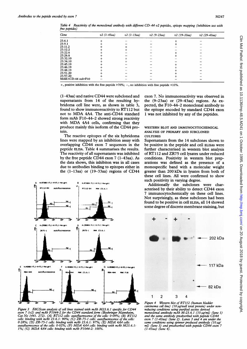
Table 4 Reactivity of the monoclonal antibody with different CD-44 v2 peptides, epitope mapping (inhibition test withfree peptides)
Clone v2 (1-43aa) v2 (1-13aa) v2 (9-23aa) v2 (19-33aa) v2 (29-43aa)
23.6.1 + - - + -23.9.1 + - - + -23.11.2 + + - - -23.12.2 + + - - -23.22.6 + - - + -23.26.6 + - - + -23.32.10 + + - - -23.34.10 + + - - -23.45.19 + - - + -23.46.19 + - - + -23.48.19 + - - + -23.51.20 + - - + -23.57.20 + - - + -MAK<CD-44 std>F1O - - - - -
+, positive inhibition with the free peptide >70%;-, no inhibition with free peptide <10%.
(1-43aa) and native CD44 were subcloned andsupernatants from 14 of the resulting hy-bridoma cell line were, as shown in table 3,found to show immunoreactivity to RT1 12 butnot to MDA 4A4. The anti-CD44 standardform mAb F10-44-2 showed strong reactivitywith MDA 4A4 cells, confirming that theyproduce mainly this isoform of the CD44 pro-tein.The reactive epitopes of the six hybridoma
lines were mapped by an inhibition assay withoverlapping CD44 exon 7 sequences in thepeptide ELISA. Table 4 summarises the results.The reactivity of all supernatants was inhibitedby the free peptide CD44 exon 7 (1-43aa). Asthe data shows, this inhibiton was in all casesdue to antibodies binding to epitopes either inthe (1-13aa) or (19-33aa) regions of CD44
A 4sRTOSI\FLI-HNFLI-H.i9ht B 4aRT0G4'FL1-H'ftL-H*aht
C
exon 7. No immunoreactivity was observed inthe (9-23aa) or (29-43aa) regions. As ex-pected, the F10-44-2 monoclonal antibody tothe epitope encoded by standard CD44 exon1 was not inhibited by any of the peptides.
WESTERN BLOT AND IMMUNOCYTOCHEMICALANALYSIS OF PRIMARY AND SUBCLONEDCULTURESSupernatants from the 14 subclones shown tobe positive in the peptide and cell ELISAS werefurther characterised in western blot analysisof RT1 12 and ZR75 cell lysates under reducedconditions. Positivity in western blot prep-arations was defined as the presence of amonospecific band with a molecular weightgreater than 200 kDa in lysates from both ofthese cell lines. All were confirmed to showsuch positivity in varying degree.
Additionally the subclones were char-acterised by their ability to detect CD44 exon7 immunocytochemically on these cell lines.Not surprisingly, as these subclones had beenfound to be positive in cell ELISA, all 14 showedsome degree ofdiscrete membrane staining, but
+ - + _
4:ZR6S1.FLI-H'FLI-H&lgn% D 4:ZReS4'FLI-H FLI-He9iht
49 ZR?5-1 autofluorescence
I2 193 i11
ZR75-1 23.6.1
N
1 1 1fi -ol
F 4:31D004'FL-H\FLI-Height
0 U .r i _ - 202 kDa
--o- 1 17 kDaG 4t Dee3'FLI-HxFLI-Heiqht64 MAK 23.6.1
It 102 0' 1-'
16-4A4 SIlK FIO
|H7.1.1
I6-h A,.f'U W.R.L
p 1lo 152 16'
Figure 3 FACScan analysis of cell lines stained with mAb M23. 6.1 specific for CD44exon 7 (v2) and mAb Fl 044-2 for the CD44 standard form (Boehringer Mannheim,Cat No 1441. 272). (A) RT112 cells: autofluorescence of the cells: 0-09%; (B) RT112cells: binding with mAb 23.6.1: 90%; (C) ZR-75-1 cels: autofluorescence of the cells:0-28%; (D) ZR-75-1 cells: binding with mAb 23.6.1: 97%; (E) MDA 4A4 cells:autofluorescence of the cells: 0-02%; (F) MDA 4A4 cells: binding with mAb M23. 6.1:1%; (G) MDA 4A4 cells: binding with mAb F1044-2: 100%.
82 kDa
1 2 3 4Figure 4 Western blot ofRT112 (human bladdercarcinoma cell line) (10 ag/weUl total protein) under non-reducing conditions using purified ascites derivedmonoclonal antibody mAb M-23. 6.1 (10 ,uigIml) (lane 1)and the same antibody preabsorbed with peptide CD44exon 7 (1-43aa) (lane 2). Lanes 3 and 4 are under thesame conditions using spinner produced antibody (10 tigIml) (lane 3) and preabsorbed with peptide CD44 exon 7(1-43aa) (lane 4).
E 4stVWfl1FLI-H\FLI-H.lVht
autofluorescenceMIDA-4A4
I
I iDA64
I I-ii IME qii
M247
.",-W
1.I
1l-
JO 'j '"j 2 -f6i 1-Ii 14
on 20 August 2018 by guest. P
rotected by copyright.http://m
p.bmj.com
/C
lin Mol P
athol: first published as 10.1136/mp.48.5.M
241 on 1 October 1995. D
ownloaded from

Borgya, Woodman, Sugiyama, Donii, Kopetzki, Matsumura, et al
Table 5 Immunocytochemical analysis of RT112, ZR75, MDA 4A4, and HT29 cell lines with crude ascites(10 pglml) and spinner produced antibodies (10 pg/ml)
Ascites Spinner
Clone RT112 ZR75 MDA HT29 RT112 MDA
23.6.1 +++I+ +++ +/- - +++ -23.46.19 ++ ++ - - + -23.57.20 + + + + - - + + -
-, No staining; +, weak membrane staining on 0-10% cells; + +, weak membrane staining on 90% + of cells; + + +, moderatemembrane staining on 90% + cells; + + + +, intense membrane staining on 90% + cells.
the intensity of this reaction varied significantlybetween clones.
Consequently, from these data, three CD44exon 7 specific hybridomas were selected forfurther study on the basis that they showedmarked positivity in both western blotting and
< immunocytochemistry on RT1 12 and ZR75-1cell lines but not on HT29 orMDA 4A4. Theywere named mAb<CD44 exon 7>M-23.6.1(IgGl, X), mAb<CD44 exon 7>M-23.46.19(IgGl, K) and mAb<CD44 exon 7>M-
r 23.57.20 (IgGl, k). The data in brackets referto the immunoglobulin class.
FACS analysis ofmonoclonal antibodies23.6.1, 23.46.19, and 23.57.20FACS analysis of these three monoclonal anti-bodies shows positive binding with the CD44exon 7 expressing cells RT112 (mAb 23.6.1. =90%; mAb 23.49.19=72%; mAb 23.57.20.=86%) and with ZR 75-1 (mAb 23.6.1.=97%;mAb 23.49.19. = 97%; mAb 23.57.20. = 96%)(fig 3). With the CD44 exon 7 negative MDA4A4 cells, all three monoclonal antibodiesshowed only about 1% binding. Anti standardfrom F-10-44-2 showed 100% binding withMDA 4A4 (fig 3). Unstimulated peripheralblood lymphocytes were found to be negativewith all three monoclonal antibodies to prod-ucts of exon 7 (data not shown).
Figure 5 Immunocytochemical staining of RT112 human bladder carcinopurified monoclonal antibody M-23.6.1 (10 pglml) obtained from ascites (culture (B), showing strong membrane reactivity. The negative staining obMDA 4A4 is shown in (C). Magnification x 590.
Characterisation and evaluation of4 antibodies secreted by selected subclones
Following bulk production of the antibodiessecreted by these three selected clones, as
C described above, ascites and spinner culturefluids from each were characterised by westernblotting and immunocytochemistry. In westernblots performed under non-reducing con-
ditions, antibodies obtained from all three
* hybridomas by either production method andstandardised to a concentration of 10lg/ml
' defined a single high molecular weight bandcorresponding to a size of just over 200 kDain both RT112 and ZR75-1. However, nobands were observed with MDA 4A4 andHT29.
=Xv Table 5 summarises the immunocyto-;X chemical data which showed differences both
between clones and between production4 * methods. In all cases staining was most intense
on the cell membranes, but some cytoplasmicra
cell line eith reactivity was seen.
(tamend with As all three of these clones had previouslybeen mapped to an epitope in the CD44 exon
........ ......... ....... ..
M248
ma
..'i;.
....
....... ...
on 20 August 2018 by guest. P
rotected by copyright.http://m
p.bmj.com
/C
lin Mol P
athol: first published as 10.1136/mp.48.5.M
241 on 1 October 1995. D
ownloaded from
Antibodies to the peptide encoded by exon 7
7 (19-33aa) region, we decided to purify onlyascites and spinner supernatant of 23.6.1.
CHARACTERISATION OF PROTEIN A PURIFIEDmAb <CD44 EXON 7>M-23.6.1Western blotting with the purified monoclonalantibody identified a monospecific band of ap-proximately 200 kDa in RT1 12 cell lysates, butnot in those from control lines (HT29 andMDA4A4) which are negative for exon 7 expression.Figure 4 shows a western blot on RT1 12 usingpure ascites derived 23.6.1 (10 jg/ml) (lane 1)and the specific inhibition of immunoreactivityby peptide CD44 exon 7 (19-33aa) (lane 2).Similar immunoreactivity was observed withspinner produced antibody (10,ug/ml) (lane 3)and diminished after inhibition with peptideCD44 exon 7 (19-33aa) (lane 4).Immunocytochemical staining ofRT1 12 (fig
5) with pure 23.6.1 (10 pg/ml) obtained fromascites (A) and spinner (B) showed strongmembrane reactivity. As with the crude pre-
parations, more intense staining was observedwith the ascites derived antibody. MDA 4A4cells did not stain (C).
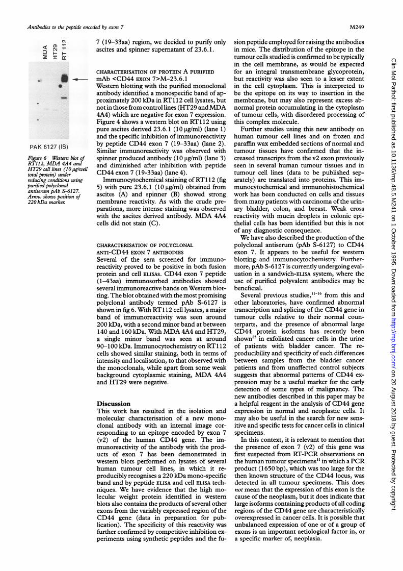
CHARACTERISATION OF POLYCLONALANTI-CD44 EXON 7 ANTIBODIESSeveral of the sera screened for immuno-reactivity proved to be positive in both fusionprotein and cell ELISAS. CD44 exon 7 peptide(1-43aa) immunosorbed antibodies showedseveral immunoreactive bands on Western blot-ting. The blot obtained with the most promisingpolyclonal antibody termed pAb S-6127 isshown in fig 6. With RT1 12 cell lysates, a majorband of immunoreactivity was seen around200 kDa, with a second minor band at between140 and 160 kDa. With MDA 4A4 and HT29,a single minor band was seen at around90-100 kDa. Immunocytochemistry on RT1 12cells showed similar staining, both in terms ofintensity and localisation, to that observed withthe monoclonals, while apart from some weakbackground cytoplasmic staining, MDA 4A4and HT29 were negative.
DiscussionThis work has resulted in the isolation andmolecular characterisation of a new mono-
clonal antibody with an internal image cor-
responding to an epitope encoded by exon 7(v2) of the human CD44 gene. The im-munoreactivity of the antibody with the prod-ucts of exon 7 has been demonstrated inwestern blots performed on lysates of severalhuman tumour cell lines, in which it re-
producibly recognises a 220 kDa mono-specificband and by peptide ELISA and cell ELISA tech-niques. We have evidence that the high mo-
lecular weight protein identified in westernblots also contains the products of several otherexons from the variably expressed region of theCD44 gene (data in preparation for pub-lication). The specificity of this reactivity was
further confirmed by competitive inhibition ex-
periments using synthetic peptides and the fu-
sion peptide employed for raising the antibodiesin mice. The distribution of the epitope in thetumour cells studied is confirmed to be typicallyin the cell membrane, as would be expectedfor an integral transmembrane glycoprotein,but reactivity was also seen to a lesser extentin the cell cytoplasm. This is interpreted tobe the epitope on its way to insertion in themembrane, but may also represent excess ab-normal protein accumulating in the cytoplasmof tumour cells, with disordered processing ofthis complex molecule.
Further studies using this new antibody on
human tumour cell lines and on frozen andparaffin wax embedded sections of normal andtumour tissues have confirmed that the in-creased transcripts from the v2 exon previouslyseen in several human tumour tissues and intumour cell lines (data to be published sep-
arately) are translated into proteins. This im-munocytochemical and immunohistochemicalwork has been conducted on cells and tissuesfrom many patients with carcinoma ofthe urin-ary bladder, colon, and breast. Weak cross
reactivity with mucin droplets in colonic epi-thelial cells has been identified but this is notof any diagnostic consequence.We have also described the production of the
polyclonal antiserum (pAb S-6127) to CD44exon 7. It appears to be useful for westernblotting and immunocytochemistry. Further-more, pAb S-6127 is currently undergoing eval-uation in a sandwich-ELISA system, where theuse of purified polyvalent antibodies may bebeneficial.
Several previous studies,"'6 from this andother laboratories, have confirmed abnormaltranscription and splicing of the CD44 gene intumour cells relative to their normal coun-
terparts, and the presence of abnormal largeCD44 protein isoforms has recently beenshown25 in exfoliated cancer cells in the urineof patients with bladder cancer. The re-
producibility and specificity of such differencesbetween samples from the bladder cancer
patients and from unaffected control subjectssuggests that abnormal patterns of CD44 ex-
pression may be a useful marker for the earlydetection of some types of malignancy. Thenew antibodies described in this paper may bea helpful reagent in the analysis of CD44 geneexpression in normal and neoplastic cells. Itmay also be useful in the search for new sens-
itive and specific tests for cancer cells in clinicalspecimens.
In this context, it is relevant to mention thatthe presence of exon 7 (v2) of this gene was
first suspected from RT-PCR observations on
the human tumour specimens" in which a PCRproduct (1650 bp), which was too large for thethen known structure of the CD44 locus, was
detected in all tumour specimens. This doesnot mean that the expression of this exon is thecause of the neoplasm, but it does indicate thatlarge isoforms containing products of all codingregions of the CD44 gene are characteristicallyoverexpressed in cancer cells. It is possible thatunbalanced expression of one or of a group ofexons is an important aetiological factor in, or
a specific marker of, neoplasia.
CNc OC] 4
PAK 6127 (IS)
Figure 6 Western blot ofRT112, MDA 4A4 andHT29 cell lines (10 pg/welltotal protein) underreducing conditions usingpurified polyclonalantiserum pAb S-6127.Arrow shows position of220 kDa marker.
M249
on 20 August 2018 by guest. P
rotected by copyright.http://m
p.bmj.com
/C
lin Mol P
athol: first published as 10.1136/mp.48.5.M
241 on 1 October 1995. D
ownloaded from

Borgya, Woodman, Sugiyama, Donie, Kopetzki, Matsumura, et al
The new antibody will be a useful tool forthe further analysis of these abnormal CD44isoforms and their clinical implications.
We wish to acknowledge with gratitude the valuable help givenby Dr E Hoess, Dr W Rollinger, Dr S Goodison, Dr H Bod-enmuller, Dr M Kaufmann, Mrs L Summerville, and Miss HMellor. We also thank Dr J E Price for the gift of the MDAMB435 cell line.
1 MacKay CR, Terpe H-J, Stauder R, Marston WL, StarkH, Gunthert U. Expression and modulation of CD44variant isoforms in humans. Cell Biol 1994;124:71-82.
2 Screaton GR, Bell MV, Jackson DG, Comelis, Gerth U, BellJI. Genomic structure of DNA encoding the lymphocytehoming receptor CD44 reveals at least 12 alternativelyspliced exons. Proc Natl Acad Sci USA 1992;89:12160-4.
3 Jalkanen S, Bargatze RF, del los Toyos J, Butcher EC.Lymphocyte recognition of high endothelium: antibodiesto distinctly epitopes of an 85-95kD glycoprotein antigendifferentially inhibit lymphocyte binding to lymph node,mucosal or synovial endothelial cells. Cell Biol 1987;105:983-90.
4 Jalkanen S, Jalkanen M, Bargatze R, Tammi M, ButcherEC. Biochemical properties of glycoproteins involved inlymphocyte recognition of high venules in man. Immunol1987;141:1615-23.
5 Underhill CB, Green SJ, Comoglio PM. Tarone G. Thehyaluronate receptor is identical to a glycoprotein of Mr85 000 (gp85) as shown by a monoclonal antibody thatinterferes with binding activity. Biol Chem 1987;262:13142-5.
6 Aruffo A, Stamenkovic I, Melnick M, Underhill CB, SeedB. CD44 is the principle cell surface receptor for hyal-uronate. Cell 1990;61:1303-13.
7 Carter WG, Wayner EA. Characterisation of the class IIIcollagen receptor, a phosphorylated, transmembrane gly-coprotein expressed in nucleated human cells. JfBiol Chem1989;263:4193-201.
8 Gallatin WM, Wayner EA, Hoffman PA, St John T, ButcherEC, Carter WG. Structural homology between lympho-cyte receptors for high endothelium and class III ex-tracellular matrix receptor. Proc Natl Acad Sci USA 1989;86:4654-8.
9 Stamenkovic I, Amiot M, Pesando JM, Seed B. A lympho-cyte molecule implicated in lymph node homing is amember of the cartilage link protein family. Cell 1989;56:1057-62.
10 Gunthert U, Hofmann M, Rudy W, Reber S, Zoller M,Haussmann I, et al. A new variant of glycoprotein CD44confers metastatic potential to rat carcinoma cells. Cell199 1;65:13-24.
11 Matsumura Y, Tarin D. Significance of CD44 gene products
for cancer diagnosis and disease evaluation. Lancet 1992;340:1053-8.
12 Tanabe KK, Ellis LM, Saya H. Expression of CD44R1adhesion molecule in colon carcinomas and metastases.Lancet 1993;341:725-6.
13 Heider K-H, Dammrich J, Skroch-Azngel P, Muller-Her-melink H-K, Vollmers HP, Herrlich P, et al. Differentialexpression of CD44 splice variants in intestinal- anddiffuse-type gastric carcinomas and normal gastric mu-cosa. Cancer Res 1993;53:4197-203.
14 Matsumura Y, Sugiyama M, Matsumura S, Hayle AJ, Rob-inson P, Smith JC, et al. Unusual retention of introns inCD44 gene transcripts in bladder cancer provides newdiagnostic and clinical oncological opportunities. J Pathol(in press).
15 Matsumura Y, Hanbury D, Smith J, Tarin D. Non-invasivedetection ofmalignancy by identification ofunusual CD44gene activity in exfoliated cancer cells. BMJ 1994;308:619-24.
16 Dall P, Heider K-H, Hekele A, von Minchwitz G,Kauffmann M, Ponta H, et al. Surface glycoprotein ex-pression and messenger RNA-splicing analysis of CD44in uterine cervical cancer and normal cervical epithelium.Cancer Res 1994;54:4754-6.
17 Wielenga VJM, Heider K-H, Offerhaus GJA, Adolf GR,van den Berg FM, Ponta H, et al. Expression of variantCD44 proteins in human colorectal cancer is related totumour progression. Cancer Res 1993;53:4754-6.
18 Kaufmann M, Heider K-H, Sinn H-P, von Minckwitz G,Ponta H, Herrlich P. CD44 variant exon epitopes inprimary breast cancer and length of survival. Lancet 1995;34:615-9.
19 Guyader M, Emerman M, Sonigo P, Clavel F, MontagnierL, Alizon M. Genome organisation and transactivation ofthe human immunodeficiency virus type 2. Nature 1987;326:662-9.
20 Stueber D, Matile H, Garotta G. System for high levelproduction in Escherichia coli and rapid application toepitope mapping, preparation of antibodies and structure-function analysis. I?nmunol Methods 1990;4: 121-52.
21 Mullis KB, Faloona FA. Specific synthesis of DNA invitro via a polymerase-catalyzed chain reaction. MethodsEnzymol 1987;155:335-50.
22 Murray NE, Brammar WJ, Murray K. Lambdoid phagesthat simplify the recovery of in vitro recombinants. MolGen Genet 1977;150:53-61.
23 Hochuli E, Doebeli H, Schacher A. New metal chelateadsorbent selective for proteins and peptides containingneighbouring histidine residues. J Chronzatogr 1987;411:177-84.
24 Kohler G, Milstein C. Continuous cultures of fused cellsscreting antibody of pre-defined specificity. Nature 1975;256:495-97.
25 Sugiyama M, Woodman A, Sugino T, Crowley S, Ho K,Smith J, et al. Non-invasive detection of bladder cancerby identification of abnormal CD44 proteins in exfoliatedcells in urine. _7 Clin Pathot: Mol Pathol 1995;48:M142-7.
M250
on 20 August 2018 by guest. P
rotected by copyright.http://m
p.bmj.com
/C
lin Mol P
athol: first published as 10.1136/mp.48.5.M
241 on 1 October 1995. D
ownloaded from