Embed Size (px)
Citation preview
J7 Clin Pathol: Mol Pathol 1995;48:M83-M87
A comparative study of cell proliferationmarkers in breast carcinomas
A S-Y Leong, S Vinyuvat, C Suthipintawong, J Milios
Division of TissuePathology,Institute of Medical &Veterinary Science,Frome Road,Adelaide,South Australia 5000A S-Y LeongS VmyuvatS SuthipintawongJ Milios
Correspondence to:Professor A S-Y Leong,Division of TissuePathology,Institute of Medical &Veterinary Science,P.O. Box 14,Rundle Mall,Adelaide,South Australia 5000.
Accepted for publication16 January 1995
AbstractAims-To investigate the tumour cell pro-liferative index obtained by immuno-staining of paraffin wax sections of 30cases of breast carcinoma with mono-clonal antibodies MIBI, KiSl and KiS5,and polyclonal Ki67 antisera to the Ki67antigen and 19A2 and PC1O antibodies toproliferating cell nuclear antigen and thepossible correlation between these indicesand that of monoclonal Ki67 antibody infrozen sections of the same tumours.Methods-All tumour samples had beenuniformly fixed and processed and sec-
tions were subjected to microwave antigenretrieval before immunostaining in all in-stances except for monoclonal Ki67 anti-body which was used in cryostat sections.Tumour cell proliferative indices wereevaluated by two independent examiners,each counting 500 tumour cells with theaid of a cross-hatched grid.Results-Proliferative indices obtainedwith MIBI, polyclonal Ki67, KiSi, andKiS5 correlated with those obtained withmonoclonal Ki67 in frozen sections. Pro-liferative indices obtained with mono-
clonal 19A2 and PCIO showed nocorrelation with those of monoclonal Ki67antibody. The staining obtained with MIB1was the most intense and the easiest toread.Conclusions-Monoclonal antibodiesMIBI,KiSl and KiS5 and polyclonal Ki67 anti-serum appear to be suitable substitutesfor monoclonal antibody Ki67 in theassessment of tumour cell proliferativeindex. As these reagents are all immuno-reactive in paraffin wax sections, theyovercome the requirement for frozentissue for immunostaining with mono-clonal Ki67.(J Clin Pathol: Mol Pathol 1995;48:M83-M87)
Keywords: Proliferation markers, Ki67, breast cancer.
Assessment of the tumour growth fractionyields important biological information whichcan be related to the clinical course and thera-peutic response in patients with cancer,besides providing greater insights into tumourgrowth. 16While the counting ofmitotic figures remains
the simplest and most established method ofassessing tumour proliferation, it is not a com-pletely reliable or reproducible procedure, andoften only reflects the mitotic or M phase ofthe cell cycle. Prolongation of the M phase of
the cell cycle may thus result in a deceptivelyhigh rate of proliferation in some tumours suchas basal cell carcinoma of the skin. Better es-tablished techniques for accurate measurementof the growth rate of tumours are the tritiatedthymidine labelling or bromodeoxyuridine in-corporation techniques,78 but both of thesetechniques are laborious, difficult to performand are not appropriate for diagnostic labora-tories. The advent of the flow cytometer pro-vides an automated technique which quantifiescellular DNA content and analyses cell cycledistribution,910 but its application requires theuse of expensive equipment.Gerdes et al" produced the Ki67 monoclonal
antibody which recognises a nuclear antigenpresent only in proliferating cells, permittingan easy immunohistochemical method of as-sessing tumour cell proliferative fraction. Theprecise nature and composition of the antigendetected by the Ki67 antibody is largely un-known but it appears to be expressed at allstages of the cell cycle except GO.'2"3 Moststudies show the Ki67 antigen to increase withprogression of the mitotic cycle, rising duringthe latter half of the S phase and reaching apeak in the G2 and M phases.3 More insightsinto the biology and structure of the Ki67antigen have recently been reported415 and thegene has been partially cloned and sequenced. 16Ki67 immunostaining has been used as a
prognostic indicator in a wide variety oftumours including non-Hodgkin's lymph-omas,'7-'9 gliomas,20 meningiomas, pituitarytumours,22 soft tissue tumours,224 bone tu-mours,25 prostatic carcinoma,26 seminoma,27colorectal carcinoma,28 molar pregnancy,6hepatocellular carcinoma,29 melanoma,30 andlung carcinoma.3' Several studies have es-tablished the relevance of Ki67 analysis infrozen sections as a predictor of behaviour andmetastatic potential in breast cancer,32-39 withsignificant cut off values at 12%4041 and 16%.4°
Proliferating cell nuclear antigen (PCNA) isa 36kDa acidic nuclear protein that has beenhighly conserved throughout evolution and isessential for DNA synthesis.42 PCNA appearsin the nucleus primarily during the syntheticphase of the cell cycle 415 and functions asan auxiliary protein of DNA polymerase-.46Several monoclonal antibodies have been gen-erated to genetically engineered PCNA andhave been used in the demonstration of cellproliferative index in paraffin wax embeddedtissues.47 Many papers describing the ap-plications of these monoclonal antibodies tovarious tumours have been published, claiminga correlation between the proliferative indexassessed by immunostaining of PCNA and the
M83
on 4 August 2018 by guest. P
rotected by copyright.http://m
p.bmj.com
/C
lin Mol P
athol: first published as 10.1136/mp.48.2.M
83 on 1 April 1995. D
ownloaded from

Leong, Vinyuvat, Suthipintawong, Milios
Antibodies to cell proliferation markers
Antibodylclone Class Dilution Microwave retrieval Proteolytic digestion Source
PCNA (19A2) Mouse IgM 50000 Yes No American BiotechPCNA (PC10) Mouse IgG 10000 Yes No DakoKi67 Mouse IgG 100 No No DakoKi67 Rabbit Ig 1 000 Yes No DakoMIB 1 Mouse IgG 1 000 Yes Yes ImmunotechKiS5 Mouse IgG 10 Yes No *KiS 1 Mouse IgG 40 Yes Yes *
* A generous gift from Professor R Parawaresch, Kiel. Source: American Biotech, Plantation, Florida, USA; Dako, Santa Barbara,California, USA; Immunotech, Marseille, France.
values obtained with flow cytometric analysisand Ki67 expression.'8"345-52.The monoclonal Ki67 antibody is immuno-
reactive only in frozen sections and cell pre-
parations. To overcome this handicap, Kreipeet al15354 have generated two monoclonal anti-bodies, KiS 1 and KiS5, directed against a form-alin resistant epitope of the Ki67 antigen.Similarly, Key et al'6 have succeeded in pro-
ducing antibodies against bacterially expressedKi67 fusion proteins which are preserved informalin fixed, paraffin wax embedded tissues.These latter antibodies are effective in tissuestreated by microwave irradiation for retrievalof the Ki67 antigen. 1555 Key et al56 have alsoraised a polyclonal rabbit antibody to a syn-
thetic peptide deduced from a 62 base pairregion encoding the Ki67 epitope.
In this study we compared the proliferationindices obtained with these new antibodies tocell proliferation markers which survive form-alin fixation and embedding in paraffin wax
and correlated them with those of Ki67 im-munostaining in frozen sections in 30 cases ofbreast carcinoma.
MethodsAppropriate tissue blocks from 30 cases ofinfiltrating duct carcinoma of the breast of nospecific type were drawn from the files of theInstitute of Medical and Veterinary Science,Adelaide. All tissues had been fixed in 10%buffered formalin for one hour before threeand a half hours of processing through 95%ethanol, xylene and wax in an automated tissueprocessor as part of the fixation and processingprotocol for oestrogen receptor immunoassayas described previously.57 Details of the anti-bodies used in this study are provided inthe table. Monoclonal Ki67 antibody was
only applied to cryostat sections as describedpreviously.2635 A Streptavidin-biotin peroxidasemethod was used.58 Microwave antigen re-
trieval was used for all other antibodies whichwere applied to paraffin wax sections, using a
modification of the method previously de-scribed.59 Briefly, deparaffinised, rehydratedtissue sections were immersed in a closed plasticcontainer (Kartell, Milan, Italy) filled with10mM citrate buffer, pH 6-0 (2-1 g citric acidin 1 litre and adjusted to pH 6-0 with ap-
proximately 13 ml of 2mM NaOH). The con-
tainer permitted stacking of 20 slides in 250 mlof solution. The immersed sections were ir-radiated in a domestic microwave oven with a
carousel (NEC Model 702, 650W) at max-imum power setting until the buffer solutionboiled. When boiling point was attained, nor-
mally in about five minutes, the power was
adjusted so that the solution simmered. Thissimmering was continued for 10 minutes afterwhich the sections were allowed to remain inthe hot buffer solution for a further 25 minutesbefore immunohistochemical staining. All sec-tions were mounted on aminoalkylsilanetreated slides. They were subjected to enzymedigestion before microwave antigen retrievalwhen immunostaining was performed withMIB 1 and KiS 1 antibodies. The sections weredigested at 37°C with trypsin type II, 0-25 mg/ml, for threeminutes before antigen retrievalwas performed. Diaminobenzidine in TRIS/HCI buffer, pH 7-4, and 0 03% hydrogen per-oxide were used as the chromogen. A lightMayer's haematoxylin was applied as coun-terstain.
Consecutive paraffin wax sections were usedfor immunostaining with each of the antibodiesso that similar areas of the tumour could beassessed. Tumour cells with distinct nuclearstaining were assessed as positive and onlyperipheral areas of the tumour were assessed,taking care to avoid areas of haemorrhage andfibrosis. A cross-hatched Whipple grid dividedinto 100 squares was used with a x 20 objectiveand only those cells lying within the ninesquares in each corner and the central square(that is, 45 squares in total) were counted.The percentage of positively stained cells weredetermined by counting 500 morphologicallymalignant cells. Counting was performed in-dependently by two of the authors (SV andCS) and the mean values of the two separatecounts were used to calculate the percentageof positive cells or the tumour proliferativeindex. When the values obtained in the twoindependent counts differed by more than 5%the counts were repeated. A similar method ofcounting was adopted for monoclonal Ki67staining on frozen sections.
Statistical analysis was performed with StatView 512 + (Brainpower Inc., Calabasas, Cali-fornia, USA), with Spearman's rank correlationcoefficient (r) method used for the analysis ofthe relation between monoclonal Ki67 pro-liferative index and those obtained with theother antibodies.
ResultsIt was easy to count positively stained cellsusing the method described above and thetumour cell proliferative index appeared to bereproducible by independent counting. Themonoclonal antibody MIB1 produced thegreatest intensity ofstaining, whereas both anti-bodies to PCNA resulted in a wide range ofstaining intensities within the tumour cell nuc-lei.
M84
on 4 August 2018 by guest. P
rotected by copyright.http://m
p.bmj.com
/C
lin Mol P
athol: first published as 10.1136/mp.48.2.M
83 on 1 April 1995. D
ownloaded from
Tumour cell proliferation markers
* Poly Ki67(r = 0.73398, p < 0-1
o MIBl(r = 0-81802, p < 0-1
60 r
50 [-0
0
_
*e0ierIlI
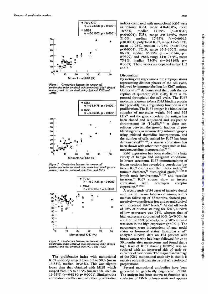
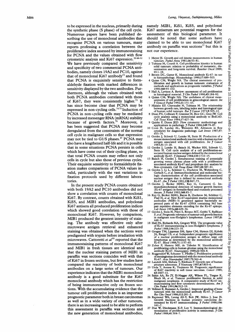
indices compared with monoclonal Ki67 were0001) as follows: KiSl, range 4-0-46-0%, mean
18-53%, median 14-25% (r=0-8348;0001) p<O0OOO1); KiS5, range 2-0-72-5%, mean
21-5%, median 15-75% (r=0-66945;p<0 0001); polyclonal Ki67, range 1 0-58 5%,mean 17-23%, median 17-25% (r=0-7339;p<0 0001); PC10, range 65-5-100%, mean86-9%, median 86-25% (r= -0-0144; p=0-9399); and 19A2, range 44-0-95-5%, mean75-1%, median 78-5% (r=0-18195; p=03359). These values are depicted in figs 1, 2and 3.
o 10 20 30 40 50 DiscussionMonoclonal Ki67 (%) By sorting cell suspensions into subpopulations
Figure 1 Comparison between the tumour cell representing distinct phases of the cell cycle,proliferative index obtained with monoclonal Ki67 frozen followed by immunolabelling for Ki67 antigen,sections) and that obtained with polyclonal Ki67 and Gerdes et al'2 demonstrated that, with the ex-MIB1. ception of quiescent cells (GO), Ki67 is ex-
pressed throughout the cell cycle. The Ki67* KiSl molecule is known to be a DNAbinding protein
(r = 0.83475, p < 0.0001) that probably has a regulatory function in cello KiS5 proliferation. The Ki67 antigen is a bimolecular
(r= 0.66945, p < 0.0001) complex of molecular weight 345 and 395kDa'4 and the gene encoding the antigen has
80 - been cloned and sequenced and assigned to70 - chromosome 10 (10q25).6061 A close cor-60 - o relation between the growth fraction of pro-50 - liferating cells, as measured by autoradiography40 using tritiated thymidine incorporation, and30 _ ° o o the number of cells stained by Ki67 has been20 a- * +++- °- *demonstrated'26264; a similar correlation has0 _ been shown with other techniques such as bro-10 moexyrdie65167.0 I! I modeoxyuridine incorporation.0 10 20 30 40 50 Ki67 expression has been studied in a large
Monoclonal Ki67 () variety of benign and malignant conditions.In breast carcinoma Ki67 immunostaining of
Figure 2 Comparison between the tumour cell frozen sections has revealed a correlation be-proliferative index obtained with monoclonal Ki67 (frozen tween Ki67 counts and the mitotic index,68-70sections) and that obtained with KiSl and KiS5. t 7' histologcal grade,323568-70tumour diameter, hsoola rd,-
lymph node involvement,32334' and vascular*Pcio invasion.33 Ki67 counts show an inverse
(r = -0-01438, p = 0.9399) correlation with oestrogen receptoro19A231672(r - 0.18195, p = 0.3359) expression.3569 72A recent study of 54 cases of invasive ductal
100 * o and nine of invasive lobular carcinoma, with a90 0 median follow up of 37 months, showed pro-80 gressivelyworse disease free and overall survival70 0 0 with increased Ki67 levels.40 At cut off levels60 - 0-o of 12% of nuclear staining for Ki67, survival50 0 0 of low expressers was 95%, whereas that of40 high expressers approached 60% (p<005). At
20 a cut off of 16% positivity, only 50% survival10 was seen in the high expressers (p<001). The0 0 1 O 0 0 parameters were independent of age, nodal
o1020 30 40 50 status or hormonal status. Bouzubar et a170Monoclonal Ki67 (%) analysed survival data on 124 patients with
breast cancer who had been followed for up toFigure 3 Comparison between the tumour cell 30 moth afe matcoyadfudtarproliferative index obtained with monoclonal Ki67 (frozen hgmontlsalfer mastectomy andg ourd that asections) and that obtained with PC1O and 19A2. high level of Ki67 staining (>20%) was as-
sociated with an increased risk of early re-currence ofcarcinoma. The major disadvantage
The proliferative index with monoclonal of the Ki67 monoclonal antibody is that it isKi67 antibody ranged from 0 5 to 50% (mean reactive only in frozen tissue or fresh cytological13-83%, median 10-25%). This was slightly preparations.lower than that obtained with MIB1 which Several monoclonal antibodies have beenranged from 2-5 to 52-5% (mean 16%, median generated to genetically engineered PCNA.13-75%) (r= 0-8180; p<00001)). Similarly, the The antigen has been shown to function as acorrelation coefficience of other proliferative co-factor of DNA polmyerase-6 and appears
40
30
20
10
M85
v -- ;;u
j
I
j
j
on 4 August 2018 by guest. P
rotected by copyright.http://m
p.bmj.com
/C
lin Mol P
athol: first published as 10.1136/mp.48.2.M
83 on 1 April 1995. D
ownloaded from
Leong, Vinyuvat, Suthipintawong, Milios
to be expressed in the nucleus, primarily duringthe synthetic phase (S phase) of the cell cycle.Numerous papers have been published de-scribing the use of monoclonal antibodies thatrecognise PCNA on various tumours, manyreports professing a correlation between theproliferative index assessed by immunostainingfor PCNA and the values obtained with flowcytometric analysis and Ki67 expression.3"5"We have previously compared the sensitivityand specificity of two commercial PCNA anti-bodies, namely clones 1 9A2 and PC10, againstthat of monoclonal Ki67 antibody73 and foundthat PCNA is exquisitely sensitive to form-aldehyde fixation with marked differences insensitivity displayed by the two antibodies. Fur-thermore, although the values obtained withboth PCNA antibodies correlated with thoseof Ki67, they were consistently higher.73 Ithas since become clear that PCNA may beexpressed in non-cycling cells.7475 Induction ofPCNA in non-cycling cells may be mediatedby increased messenger RNA (mRNA) stabilitybecause of growth factors.76 Moreover, ithas been suggested that PCNA may becomederegulated from the constraints of the normalcell cycle in malignant cells so that expressionmay not be tied to GI/S phases.47 PCNA mayalso have a lengthened half-life and it is possiblethat in some situations PCNA persists in cellswhich have come out of their cycling phase, sothat total PCNA counts may reflect not onlycells in cycle but also those of previous cycles.Their exquisite sensitivity to formaldehyde fix-ation makes comparisons of PCNA values in-valid, particularly with the vast variations infixation protocols used by different labora-tories.
In the present study PCNA counts obtainedwith both 19A2 and PC 10 antibodies did notshow a correlation with counts of monoclonalKi67. By contrast, counts obtained with KiS1,KiS5, and MIB 1 antibodies, and polyclonalKi67 antisera all produced proliferation indiceswhich showed good correlation with those ofmonoclonal Ki67. However, by comparison,MIB 1 produced the greatest intensity of stain-ing. The antibody was effective only aftermicrowave antigen retrieval and enhancedstaining was obtained when the sections werepredigested with trypsin before irradiation withmicrowaves. Cattoretti et al55 reported that theimmunostaining patterns of monoclonal Ki67and MIB 1 in fresh tissues are identical andthat the nuclear staining pattern of MIB 1 inparaffin wax sections coincides well with thatof Ki67 in frozen sections, but few studies havecompared the reactivity of both monoclonalantibodies on a large series of tumours. Ourexperience indicates that the MIB 1 monoclonalantibody is a good substitute for the Ki67monoclonal antibody which has the restrictionof being immunoreactive only on frozen sec-tions. With the accumulating evidence that thetumour cell proliferative index is an importantprognostic parameter both in breast carcinomasas well as in a wide variety of other tumours,there is an increasing need to be able to performthis assessment in paraffin wax sections andthe new generation of monoclonal antibodies,
namely MIB1, KiSl, KiS5, and polyclonalKi67 antiserum are potential reagents for theassessment of this biological parameter. Itshould be noted that some authors haveclaimed to be able to use monoclonal Ki67antibody on paraffin wax sections77 but this isnot our experience.
1 Meyer JS. Growth and cell kinetic measurements in humantumours. PatholAnnu 1981;16:53-81.
2 Tubiana M, Courdi A. Cell proliferation kinetics in humansolid tumours: relation to probability of metastatic, dis-semination and long-term survival. Raiother Oncol 1989;15:1-18.
3 Brown DC, Gatter K. Monoclonal antibody Ki-67: its usein histopathology. Histopathology 1990;17:489-503.
4 Quinn CM, Wright NA. The clinical assessment of pro-liferation and growth in human tumours: evaluation ofmethods and applications as prognostic variables. J Pathol1990;160:93-102.
5 Hall A, Levison A. Review: assessment of cell proliferationin histological material. J Clin Pathol 1990;43:184-92.
6 Quinn CM, Wright NA. The usefulness of clinical meas-urements of cell proliferation in gynecological cancer. IntJ Gynecol Pathol 1992;11:131-43.
7 Malaise EP, Chavaudra N, Tubiana M. The relationshipbetween growth rate, labelling index and histological typeof human solid tumours. EurJ7 Cancer 1973;9:305-12.
8 Dean PN, Colbeare F, Gratzner H, Rice GC, GrayJW. Cellcycle analysis using a monoclonal antibody to BrdUrD.Cell Tissue Kinet 1984;17:427-36.
9 Quirke P, Dyson JED. Flow cytometry: methodology andapplications in pathology. J Pathol 1986;149:79-87.
10 Coon JS, Landay AL, Weinstein RS. Advances in flowcytometry for diagnostic pathology. Lab Invest 1987;57:453-79.
11 Gerdes J, Schwab U, Lemke H, Stein H. Production of amouse monoclonal antibody reactive with a human nuclearantigen associated with cell proliferation. Int J Cancer1983;31: 13-20.
12 Gerdes J, Lemke H, Baisch H, Wacker HH, Schwab U,Stein H. Cell cycle analysis of a cell proliferation-as-sociated human nuclear antigen defined by the monoclonalantibody Ki-67.
_Immunol 1984;133:1710-15.
13 Baisch H, Gerdes J. Simultaneous staining of potentiallygrowing versus plateau phase cells with a proliferationassociated antibody Ki-67 and propidium dye: analysis byflow-cytometry. Cell 7ssue Kinet 1987;20:387-91.
14 Gerdes J, Li L, Schluter C, Duchrow M, Wohlenberg C,Gerlach C, et al. Immunobiochemical and molecular bio-logic characterisation of the cell proliferation-associatednuclear antigen that is defined by monoclonal antibodyKi-67. Am JPathol 1991;138:867-73.
15 Gerdes J, Becker MHG, Key G, Cattoretti G. Im-munohistochemical detection of tumour growth fraction(Ki-67 antigen) in formalin-fixed and routinely processedtissues. J Pathol 1992;168:85-7.
16 Key G, Becker MHG, Baron B, Duchrow M, Schluter C,Flad H, et al. New Ki-67-equivalent murine monoclonalantibodies (MIBI-3) generated against bacterially ex-pressed parts of the Ki-67 cDNA containing 362 basepair repetitive elements encoding for the Ki-67 epitope.Lab Invest 1993a;68:629-36.
17 Gerdes J, Stein H, Pileri S, Rivano MT, Gobbi T, RalfkiaerF, et al. Prognostic relevance oftumour-cell growth fractionin malignant non-Hodgkin's lymphomas. Lancet 1987;ii:448-9.
18 Hall PA, Richards MA, Gregory WM. The prognostic valueof Ki-67 immunostaining in non-Hodgkin's lymphoma. JPathol 1988;154:223-35.
19 Grogan TM, Lippman SM, Spier CM, Slymen DJ, KybskiJA, Rangel CS, et al. Independent prognostic significanceof a nuclear proliferation antigen in diffuse large celllymphomas as determined by the monoclonal antibodyKi-67. Blood 1988;71:1157-60.
20 Zuber P, Hamou MF, de Tribolet N. Identification ofproliferating cells in human gliomas using the monoclonalantibody Ki-67. Neurosurgery 1988;22:364-8.
21 RoggendorfW, Schuster T, Pfeiffer J. Proliferative potentialof meningiomas determined with the monoclonal antibodyKi-67. Acta Neuropathol 1987;73:361-4.
22 Landolt AM, Shibata T, Kleihues P. Growth rate of humanpituitary adenomas. J Neurosurg 1987;67:803-6.
23 Ueda T, Aozasa K, Tsujimoto M. Prognostic significanceof Ki67 reactivity in soft tissue sarcomas. Cancer 1989;63:1607-11.
24 Sahin AA, Ro JY, El-Naggar AK, Wilson PL, Teague K,Block M, et al. Tumour proliferative fraction in solidmalignant neoplasms. A comparative study of Ki-67 im-munostaining and flow cytometric determinations. Am J'Clin Pathol 1991;96:512-19.
25 Vollmer E, Roessner A, Gerdes J. Improved grading ofbonetumours with the monoclonal antibody Ki-67. Y CancerRes Clin Oncol 1986;112:281-2.
26 Raymond WA, Leong AS-Y, Bolt 1W, Milios J, Jose JS.Growth fractions in human prostatic carcinoma de-termined by Ki-67 immunostaining. J Pathol 1988;156:161-7.
27 Due W, Dieckmann K-P, Loy V. Immunohistological de-termination of proliferative activity in seminomas. J7 ClinPathol 1988;41:304-7.
M86
on 4 August 2018 by guest. P
rotected by copyright.http://m
p.bmj.com
/C
lin Mol P
athol: first published as 10.1136/mp.48.2.M
83 on 1 April 1995. D
ownloaded from
Tumour cell proliferation markers
28 Shepherd NA, Richman PI, England J. Ki-67 derived pro-liferative activity in colorectal adenocarcinoma with prog-nostic correlations. J Pathol 1988;155:213-19.
29 Grigoni WF, D'Errico A, Bacci F. Primary liver neoplasms:evaluation of proliferative index using MoAb Ki-67. JPathol 1989;158:23-9.
30 Kaudewitz P, Braun-Falco 0, Ernst M, Landthaler M,Stolz W, Gerdes J. Tumor cell growth fractions in humanmalignant melanomas and the correlation to histo-pathologic tumour grading. AmJPathol 1989;134: 1063-8.
31 Gatter KC, Dunhill MS, Gerdes J, Stein H, Mason DYNew approach to assessing lung tumours in man. Jf ClinPathol 1986;39:590-3.
32 Lelle RJ, Heidenreich W, Stauch G, Gerdes J. The cor-relation of growth fraction with histologic grading andlymph node status in human mammary carcinoma. Cancer1987;59:83-8.
33 Charpin C, Andrac L, Vacheret H. Multiparametric evalu-ation (SAMBA) of growth fraction (monoclonal Ki-67)in breast carcinoma tissue sections. Cancer Res 1988;48:4368-74.
34 Raymond WA, Leong AS-Y. Nucleolar organizer regionsrelate to growth fractions in human breast carcinoma.Hum Pathol 1989a;20:741-6.
35 Raymond WA, Leong AS-Y. The relationship betweengrowth fractions and oestrogen receptors in human breastcarcinoma, as determined by immunohistochemical stain-ing. Jf Pathol 1989b;158:203-11.
36 van Diest PJ, Martze-Cok E, Baak JPA. Prognostic value ofproliferative activity in lymph node metastases of patientswith breast cancer. J Clin Pathol 199 1;44:416-18.
37 Gasparini G, Pozza F, Bevilacqua P. Growth fraction (Ki-67 antibody) determination in early stage breast cancer.Histopathologic clinical and prognostic correlations. Breast1992;1:92-9.
38 Leonardi E, Girlando S, Serio G, Mauri FA, Perrone G,Scampini S, et al. PCNA and Ki-67 expression in breastcarcinoma: correlations with clinical and biological vari-ables. Jf Clin Pathol 1992;45:416-19.
39 Barbareschi M, Girlando S, Mauri FM, Forti S, Eccher C,Mauri FA, et al. Quantitative growth fraction evaluationwith MIB1 and Ki-67 antibodies in breast carcinomas.Am J Clin Pathol 1994;102:171-5.
40 WWmtzer HO, Zipfel I, Schulte-Monting J, Hellerich U, vonKleist S. Ki-67 immunostaining in human breast tumoursand its relationship to prognosis. Cancer 1991;67:421-8.
41 Sahin AA, Ro J, Ro JY, Block MB, El-Naggar AK, OrdonezNG, Fritsche HA, et al. Ki-67 immunostaining in node-negative stage I/II breast carcinoma. Significant correlationwith prognosis. Cancer 1991;68:549-57.
42 Miyachi K, Fitzler MJ, Tan EM. Autoantibody to a nuclearantigen in proliferating cells. Jf Immunol 1978;121:2228-34.
43 Mathews MB, Bernstein RM, Franza BR, Garrels JI. Iden-tity of the proliferating cell nuclear antigen and cyclin.Nature 1984;303:374-6.
44 Celis JE, Celis A. Cell cycle dependant variations in thedistribution of the nuclear protein cyclin proliferating cellnuclear antigen in cultured cells: subdivision of S-phase.Proc Nad Acad Sci USA 1985;82:3262-6.
45 Kurki P, Ogata K, Tan EM. Monoclonal antibodies toproliferating cell nuclear antigen (PCNA)/cyclin as probesfor proliferating cells by immunofluorescence microscopyand flow cytometry. Jf Immunol Methods 1988;109:49-59.
46 Bravo R, Frank R, Blundell PA, MacDonald-Bravo H.Cyclin/PCNA is the auxiliary protein of DNA polymerasealpha. Nature 1987;326:515-17.
47 Hall A, Levison A, Woods L, Yu CC-W, Kellock DB,WatkinsJA, etal. Proliferating cell nuclear antigen (PCNA)immunolocalization in paraffin sections: an index of cellproliferation with evidence of deregulated expression insome neoplasms. J Pathol 1990;162:285-94.
48 Garcia RL, Coltrera MD, Gown AM. Analysis of pro-liferative grade using anti-PCN/cyclin monoclonal anti-bodies in fixed, embedded tissues. Comparison with flowcytometric analysis. Am J Pathol 1989;134:733-9.
49 Swanson SA, Brooks JJ. Proliferation markers Ki-67 andp105 in soft tissue lesions. Correlation with DNA flowcytometric characteristics. Am J Pathol 1990;137:1491-500.
50 Woods AL, Hall PA, Shepherd NP, Hauby AM, WaseemNH, Lane DP, et al. The assessment of proliferatingcell nuclear antigen (PCNA) immunostaining in primarygastro-intestinal lymphomas and its relationship to histo-logic grade, S + G2 +M phase fraction (flow cytometricanalysis) and prognosis. Histopathology 1991;19:21-7.
51 van Dierendonck JH, Wijsman JH, Keijzer R, van de VeldeCJH, Cornelisse CJ. Cell cycle-related staining patternsof anti-proliferating cell nuclear antigen monoclonal anti-bodies. Comparison with BrdUrd labelling and Ki-67staining. Am JPathol 1991;138:1165-72.
52 Carey FA, Fabbroni G, Lamb D. Expression of proliferatingcell nuclear antigen in lung cancer: a systematic study
and correlation with DNA ploidy. Histopathology 1992;20:499-503.
53 Kreipe H, Alm P, Olsson H, Haubert M, Fischer L, Par-waresch R. Prognostic significance of a formalin-resistantnuclear proliferation antigen in mammary carcinomas asdetermined by the monoclonal antibody Ki-SI. Am JPathol 1993;142:651-7.
54 Kreipe H, Wacker HH, Heidebrecht HJ, Haas K, HaubergM, Tiemann M, et al. Determination ofthe growth fractionin non-Hodgkin's lymphomas by monoclonal antibodyKi-S5 directed against a formalin-resistant epitope of theKi-67 antigen. AmJ Pathol 1993;142:1689-94.
55 Cattoretti G, Becker MHG, Key G, Duchrow M, SchlUterC, Galle J, et al. Monoclonal antibodies against re-combinant parts of the Ki-67 antigen (MIB1 and MlB3)detect proliferating cells in microwave-processed formalin-fixed paraffin sections. J7 Pathol 1992;168:357-63.
56 Key G, Larsen Petersen J, Becker MHG, Duchrow M,Schluter C, Askaa J, et al. New antiserum against Ki-67antigen suitable for double immunostaining of paraffinwax sections. J Clin Pathol 1993;46:1080-4.
57 Raymond WA, Leong AS-Y. Oestrogen receptor stainingof paraffin-embedded breast carcinomas following shortfixation in formalin: a comparison with cytosolic andfrozen section receptor analyses. J Pathol 1990;160:295-303.
58 Milios J, Leong AS-Y. The detection of tissue antigens inpreviously stained histological sections. Stain Technol 1987;62:411-16.
59 Leong AS-Y, Milios J. An assessment of the efficacy of themicrowave antigen-retrieval procedure on a range of tissueantigens. Appl Immunohistochem 1993;1:267-74.
60 Schonk DM, Kuijpers HJ, van Drunen E. Assignment ofthe gene(s) involved in the expression of the proliferation-related Ki-67 antigen to human chromosome 10. HumGenet 1989;83:297-9.
61 Fonatsch C, Buchrow M, Rieder H, Schluter C, Gerdes J.Assignment of the human Ki-67 gene (MK1 67) to 10q25-qter. Genomics 1991;11:476-7.
62 Meyer JS, Friedman E, McCrate M, Bauer WC. Predictionof early course of breast carcinoma by thymidine labelling.Cancer 1983;51:1879-86.
63 Schwarting R, Gerdes J, Niehus J, Jaeschke L, Stein H.Determination of the growth fraction in cell suspensionsby flow cytometry using the monoclonal antibody Ki-67.Jf Immunol Methods 1986;90:65-70.
64 Kamel OW, Franklin WA, Ringus JC, Meyer JS. Thymidinelabeling index and Ki-67 growth fraction in lesions of thebreast. Am JT Pathol 1989;134:107-13.
65 Silvestrini R, Costa A, Veneroni S, Del Bino G, Persici P.Comparative analysis of different approaches to investigatecell kinetics. Cell 7issue Kinet 1988;21:123-31.
66 Sasaki K, Matsumura K, Tsuji T, Shinozaki F, TakahashiM. Relationship between labeling indices of Ki-67 andBrdUrd in human malignant tumours. Cancer 1988;62:989-93.
67 Magaud J-P, Sargent I, Clarke PJ, French M, Rimokh R,Mason DY. Double immunocytochemical labeling of celland tissue samples with monoclonal anti-bromo-deoxyuridine. J Histochem Cytochem 1989;37:1517-27.
68 Barnard NJ, Hall PA, Lemoine NR, Kadar N. Proliferativeindex in breast cancer in situ by Ki-67 immunostainingand its relationship to clinical and pathological variables.J Pathol 1987;151:287-95.
69 McGurrin JF, Doria Ml, Dawson PJ. Assessment of tumorcell kinetics by immunohistochemistry in carcinomas ofthe breast. Cancer 1987;59: 1744-50.
70 Bouzubar N, Walker KJ, Griffiths K, Ellis IO, Elston CW,Robertson JFR, et al. Ki67 immunostaining in primarybreast cancer: pathological and clinical association. Br JCancer 1989;59:943-7.
71 Wrba F, Reiner A, Markis-Ritzinger E, Holzner JH, ReinerG, Spona J. Prognostic significance of immuno-histochemical parameters in breast carcinomas. PatholRes Pract 1988;283:277-83.
72 Veronese SM, Gambacorta M. Detection of Ki-67 pro-liferation rate in breast cancer. Correlation with clinicaland pathologic features. Am J Clin Pathol 199 1;95:30-4.
73 Leong AS-Y, Milios J, Tang SK. Is immunolocalisationof proliferating cell nuclear antigen (PCNA) in paraffinsections a valid index of cell proliferation? Appl Im-munohistochem 1993;1:127-35.
74 Schwarting R. Little missed markers and Ki-67. Lab Invest1993;68:597-9.
75 Coltrera MD, Skelly M, Gown AM. Anti-PCNA-antibodyPC10 yields unreliable proliferation indexes in routinelyprocessed, deparaffinised, formalin fixed tissue. Appl Im-munohistochem 1993;1: 193-200.
76 McCormick D, Hall PA. The complexities of proliferatingcell nuclear antigen. Histopathology 1992;21:591-4.
77 Cuevas E, Jones DB, Wright DH. Immunochistochemicaldetection of tumour growth fraction (Ki-67 antigen) informalin-fixed and routinely processed tissues. Jf Pathol1993;169:477-8.
M87
on 4 August 2018 by guest. P
rotected by copyright.http://m
p.bmj.com
/C
lin Mol P
athol: first published as 10.1136/mp.48.2.M
83 on 1 April 1995. D
ownloaded from