Embed Size (px)
Citation preview

Size-Dependent Accumulation ofPEGylated Silane-Coated Magnetic IronOxide Nanoparticles in Murine TumorsEsben K. U. Larsen,† Thomas Nielsen,‡ Thomas Wittenborn,‡ Henrik Birkedal,† Thomas Vorup-Jensen,†
Mogens H. Jakobsen,§ Leif Østergaard,‡ Michael R. Horsman,‡ Flemming Besenbacher,†,*Kenneth A. Howard,† and Jørgen Kjems†,*†Interdisciplinary Nanoscience Center (iNANO), Departments of Molecular Biology, Physics and Astronomy, Chemistry, and Medical Microbiology & Immunology, AarhusUniversity, DK-8000 Aarhus C, Denmark, ‡iNANO, Department of Experimental Clinical Oncology and Department of Neuroradiology, Danish National ResearchFoundations Center of Functionally Integrative Neuroscience Aarhus University Hospital, DK-8000 Aarhus C, Denmark, and §DTU Nanotech, Department of Micro- andNanotechnology, DK-2800 Kongens Lyngby, Denmark
Magnetic nanoparticles (MNPs)have been synthesized andused as contrast-enhancing
agents in magnetic resonance imaging(MRI) for diagnostic application in a di-verse range of diseases including cardio-vascular, neurological disorders, andcancer.1�3 MNP surface coating with dex-tran or citrate facilitates monodispersedparticles, colloidal stability, and biocom-patibility that are crucial requirements forclinical application.4,5 Furthermore, coat-ing the particles with oleic acid can cre-ate a hydrophobic surface that reducesparticle oxidation.6 Subsequent replace-ment of oleic acid with alternative com-pounds, such as silane, allows greaterflexibility with regard to surface chemis-try modification and biocompatibility.7�9
Commercially available end-labeled si-lane derivatives allow for the introduc-tion of different chemical moieties suchas amine groups or polyethylene glycol(PEG) onto the surface of MNPs.10 The hy-drophilic PEG molecules have been usedto reduce phagocytic capture of nanopar-ticles by cellular components of the im-mune system, leading to extended circu-lation and subsequent accumulation intumors as a consequence of the en-hanced permeability and retention (EPR)effect due to leaky vasculature and poorlymphatic drainage in tumors.11�15 TheEPR effect occurs because the newlyformed vessels in the tumor are irregularin shape, leaky with large openings, andhave poor lymphatic drainage. The EPReffect can be utilized for passive target-ing of nanoparticles in areas with in-
creased angiogenesis, where there is en-hanced permeability of the nanoparticlesout of the blood vessels and a longer re-tention time in the tumor.
This approach has been utilized in thedevelopment of the commercial PEGylatedliposomal Doxil as an anticancer treat-ment.16 The application and characteriza-tion of PEGylated approaches for silatedPEG MNP-mediated imaging of tumors isunexplored.
In this work, we describe the prepara-tion and characterization of silane-PEG-coated MNPs using a simple synthesismethod based on the use of biocompati-ble silane-PEG as a coating agent. The ef-fect of size on phagocytic capture ofPEGylated MNPs in vitro and tumor accumu-lation in vivo using MRI is determined foroptimal design of diagnostic nanoparticles.
*Address correspondence [email protected], [email protected].
Received for review April 2, 2009and accepted June 22, 2009.
Published online July 2, 2009.10.1021/nn900330m CCC: $40.75
© 2009 American Chemical Society
ABSTRACT Magnetic nanoparticles (MNP) can be used as contrast-enhancing agents to visualize tumors by
magnetic resonance imaging (MRI). Here we describe an easy synthesis method of magnetic nanoparticles coated
with polyethylene glycol (PEG) and demonstrate size-dependent accumulation in murine tumors following
intravenous injection. Biocompatible iron oxide MNPs coated with PEG were prepared by replacing oleic acid
with a biocompatible and commercially available silane-PEG to provide an easy and effective method for chemical
coating. The colloidal stable PEGylated MNPs were magnetically separated into two distinct size subpopulations
of 20 and 40 nm mean diameters with increased phagocytic uptake observed for the 40 nm size range in vitro. MRI
detection revealed greater iron accumulation in murine tumors for 40 nm nanoparticles after intravenous
injection. The enhanced MRI contrast of the larger MNPs in the tumor may be a combined result of the size-
dependent extravasation and capture by macrophages in the tumor, providing important considerations for
improved bioimaging approaches.
KEYWORDS: magnetic resonance imaging · cancer · magnetite nanoparticles · drugdelivery · ultrasmall superparamagnetic iron oxide (USPIO) particles · polyethyleneglycol
ARTIC
LE
www.acsnano.org VOL. 3 ▪ NO. 7 ▪ 1947–1951 ▪ 2009 1947

RESULTS AND DISCUSSIONIron oxide MNPs were produced and coated with
2-methoxy polyethyleneoxy propyltrimethoxysilane(silane-PEG) by a two-step method; in the first step, ironoxide MNPs were prepared by co-precipitation of Feions with oleic acid under basic conditions followed bya second step in toluene, where oleic acid was replacedby a silane group (Figure 1).
Fe3O4 nanoparticles were synthesized according tothe method of Sun et al.7 Replacement of oleic acid
with silane-PEG was per-formed by a ligand exchangereaction in a toluene solventin the presence of the basetriethylamine and water. Ad-dition of water converts theSi(OMe)3 to the silanol form,enabling efficient reactionwith the oxygen on the ironoxide particles and oleic acidreplacement.17 The silanolforms polymer networks upto three binding sites per Siatom to the iron oxide, result-ing in a more stable coatcompared to oleic acid.8 Thesilane-PEG-coated nanoparti-cles were soluble in aqueousand organic conditions and
stable to aggregation in aqueous buffer for more than6 months at room temperature (data not shown).
After purification by a toluene/pentane wash, MNPswith oleic acid and silane/PEG coatings were analyzedby IR spectroscopy (Figure 2). From the IR spectra ofiron oxide particles coated with oleic acid, vibration fre-quencies within the crystal structure compatible withFe�O and FeO�H bonds were detected (at 577 and3400 cm�1, respectively). The peak at 1406 cm�1 was at-tributed to C�H bending vibrations. The broad peakfrom 1500 to 1700 cm�1 represented the stretching vi-bration of C�C, CAC, and CAO bonds in the oleic acidcarbon chain and the acid group. The oleic acid hydro-carbon chain had a distinct C�H stretching vibration at2850�2950 cm�1. Hence, all of the peaks are in accor-dance with previously reported spectra of oleic-acid-coated MNPs.7
In the IR spectra of silane-PEG-coated MNPs, theSi�O�Fe and Fe�OH peaks were detected in the fre-quency regimen between 500 and 1000 cm�1. The peakat 1105 cm�1 most probably reflects C�O�C stretch-ing vibration from the ether group. The C�H bendingvibrations from the carbon chain from 1200 to 1500cm�1 were difficult to assign. The peak, however, at1344 cm�1 most likely derives from the C�H bendingin the ether groups. Our results are in agreement withpreviously reported IR spectra for silane-PEG-coatedMNPs.17 The spectra are similar to the spectra of thepure silane-PEG spectra except that the pure silane-PEG does not have an absorption from iron between500 and 600 cm�1.
Transmission electron microscopy (TEM) analysis re-vealed a diameter of 8.0 � 1.4 nm for the oleic-acid-coated MNP and 8.2 � 1.7 nm for the silane/PEG-coatedparticles (Supporting Information Figure S1). X-ray dif-fraction of silane-PEG-coated MNPs showed a crystal-linic Fe3O4 core structure with diameters between 11and 15 nm (Table S2 and Figure S2 in Supporting Infor-
Figure 1. Preparation of PEGylated nanoparticles. Precipitation of iron ions in the presence of oleic acidyields oleic-acid-coated nanoparticles. Ligand exchange with silane-PEG replaces the oleic acid with si-lane, resulting in PEGylated MNPs.
Figure 2. Infrared spectra of (a) oleic-acid-coated MNPs, (b) silane-PEG-coated MNPs, and (c) pure silane-PEG.
ART
ICLE
VOL. 3 ▪ NO. 7 ▪ LARSEN ET AL. www.acsnano.org1948

mation). The hydrodynamic radius of the MNPs mea-sured by dynamic light scattering (DLS) showed a peakdiameter of 26 nm and a polydispersity index of 0.24 forthe PEGylated particles (Supporting Information FigureS3). This supports that the particles were discretePEGylated particles without interparticle cross-linkingpreviously observed with the silane coating process.18
The surface charge measured by the � potentialmeasurement of the PEG-coated particles revealed anet charge between �27 mV (pH � 2) and �18 mV (pH� 10) with the isoelectric point at pH � 5 (SupportingInformation Figure S4). These charges are probably de-rived from the ionizable hydroxyl groups on the iron ox-ide surface. Furthermore, the PEG layer inhibited par-ticle aggregation over a period of evaluation of 1month. The molecular density of the silane-PEG coaton the surface of MNPs was characterized by a thermo-gravimetric analysis performed over a temperaturerange of 20�900 °C (Supporting Information FigureS5). On the basis of the total weight loss of 30% (w/w),and assuming an average size of 8 nm, we estimated asilane-PEG surface density of 10.5 molecules per nm2
MNP surface. The cellular cytotoxicity in HeLa cells wasmeasured after 24 h incubation by a colorimetric MTTcell viability assay to determine the clinical feasibility ofthe nanoparticles (Supporting Information Figure S6).A 90�100% cell viability was observed in the cells incu-bated with silane-PEG-coated iron oxide at 1.6�200�g/mL concentration, suggesting the nontoxic natureof the MNPs.19
Two batches of differently sized silane-PEG-coatedparticles were synthesized by magnetic separation us-ing a neodymium magnet attached to a MACS columnin which larger nanoparticles were retained and smallerparticles contained within the effluent. DLS measure-ments revealed effluent-containing small MNPs with amean diameter of �20 nm, whereas the retained MNPswere significantly larger with a mean diameter of �40nm. TEM analysis revealed iron oxide cores with a meandiameter at 4.6 � 1.4 nm for the 20 nm particles com-pared to 9.4 � 1.9 nm for the larger 40 nm particles (Fig-ure 3).
The macrophage cell line RAW 264.7 was used as acellular model for phagocytic capture of the nanoparti-cles at a concentration range of 0.1�0.4 mg/mL over a24 h incubation. After repeated cell washes, the totaliron and protein contents in the cell culture were mea-sured using ferrozin and Bradford assay, respectively(Figure 4). Compared to the 20 nm particles, the up-take of 40 nm particles exhibited an �8-fold increase,suggesting that the phagocytic uptake is size-dependent. However, the 40 nm particles have 8 timeslarger volume compared to the 20 nm particles, whichindicates a similar number of particle uptake. The size-dependent nature of macrophage phagocytosis hasbeen described previously for various other types ofnanoparticles with larger particles showing greater up-
take.20 Studies of macrophage uptake of iron oxide
MNPs has revealed a dependence on the polyanion
scavenger receptor SR-A, which can recognize nega-
tive charged particles.21 Receptor-mediated uptake can
explain the increased uptake, in weight, of the larger
nanoparticles as each nanoparticle is taken up
specifically.
Interestingly, the PEG coat did not inhibit cellular up-
take, promoting the use of the 40 nm magnetic nano-
particles for macrophage imaging applications in vivo.
Mice bearing subcutaneously implanted foot tu-
mors were examined by MRI before and after intrave-
nous injection with either 20 or 40 nm MNPs or a non-
separated mixture of MNPs (Figure 5). As a measure of
accumulation, the relaxation rate R2 was used due to
the correlation with accumulation of nanoparticles.
Before injection, the R2 value in the tumor was mea-
sured to be 20 s�1. After 24 h, this increased to over 30
s�1 for mice injected with the nonseparated particles
and the larger 40 nm particles (Figure 5a,c). In contrast,
the small 20 nm particles did not result in any increased
contrast within the tumor (Figure 5b).
Figure 3. Size range of magnetically separated PEGylated iron oxidenanoparticle analyzed by TEM. (a) Small 20 nm silane-PEG-coatedMNPs have an iron oxide core with an average size of 4.6 � 1.4 nm.(b) Larger 40 nm silane-PEG-coated MNPs have an average iron oxidecore of 9.4 � 2.0 nm (scale bar � 20 nm).
Figure 4. Uptake of different sized iron oxide nanoparticles inmacrophage-like cells (RAW 264.7). Concentration-dependent up-take was observed, with approximately 8-fold lower uptake ofthe small (20 nm) particles compared to the larger (40 nm) par-ticles. The mean cellular uptake and standard deviation error barsare shown. All samples were performed in triplicates.
ARTIC
LE
www.acsnano.org VOL. 3 ▪ NO. 7 ▪ 1947–1951 ▪ 2009 1949
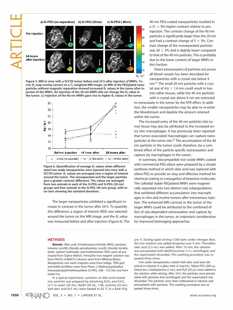
The larger nanoparticles exhibited a significant in-
crease in contrast in the tumor after 24 h. To quantify
this difference, a region of interest (ROI) was selected
around the tumor on the MRI image, and the R2 value
was measured before and after injection (Figure 6). The
40 nm PEG-coated nanoparticles resulted ina 31 � 9% higher contrast relative to pre-injection. The contrast change of the 40 nmparticles is significantly larger than the 20 nmand had a contrast change of 5 � 3%. Con-trast change of the nonseparated particleswas 20 � 3% and is slightly lower comparedto that of the 40 nm particles. This is probablydue to the lower content of larger MNPs inthis fraction.
Direct extravasation of particles out acrossall blood vessels has been described fornanoparticles with a crystal size below 9nm.22 The small 20 nm particles with a crys-tal size of 4.6 � 1.4 nm could result in lossinto other tissues, while the 40 nm particleswith a crystal size above 9 nm are restricted
to extravasate in the tumor by the EPR effect. In addi-tion, the smaller nanoparticles may be able to re-enterthe bloodstream and deplete the amount retainedwithin the tumor.
The increased entry of the 40 nm particles into tu-mor tissue may also be attributed to the increased en-try into macrophages. It has previously been reportedthat tumor-associated macrophages can capture nano-particles at the tumor site.23 The accumulation of the 40nm particles in the tumor could, therefore, be a com-bined effect of the particle-specific extravasation andcapture by macrophages in the tumor.
In summary, biocompatible iron oxide MNPs coatedwith commercial PEG-silane were prepared by a simplesynthesis method in which oleic acid was replaced withsilane-PEG to provide an easy and effective method forchemical coating or conjugation of bioactive molecules.The colloidal stable PEGylated MNPs were magneti-cally separated into two distinct size subpopulationsthat exhibited different accumulation into macroph-ages in vitro and murine tumors after intravenous injec-tion. The enhanced MRI contrast in the tumor of thelarger MNPs could be attributed to the combined ef-fect of size-dependent extravasation and capture bymacrophages in the tumor, an important considerationfor improved bioimaging approaches.
METHODSMaterials. Oleic acid, N-hydroxysuccinimide (NHS), pentane,
toluene, iron(III) chloride pentahydrate, iron(II) chloride tetrahy-drate, sodium hydroxide, and triethylamine (TEA) were all pur-chased from Sigma-Aldrich. FerroZine iron reagent solution wasfrom HACH; msMACH columns were from Miltenyi Biotec.Neodymium rare earth magnets were from Indigo. TEM gridand kohle lochfilme were from Plano. 2-Methoxy(polyethyl-eneoxy)propyltrimethoxysilane (Si-PEG, 596�725 Da) wsa fromGelest Inc.
In a typical experiment, synthesis of oleic-acid-coatediron particles was prepared by dissolving FeCl3 and FeCl2
(2:1) in water (20 mL). NaOH (50 mL, 1 M), acetone (25 mL),and oleic acid (0.5 mL) were heated to 83 °C in a flask (Fig-
ure 1). During rapid stirring (1200 rpm) under nitrogen flow,the iron solution was added dropwise over 5 min. Thereafter,oleic acid (2.5 mL) was added. After 10 min, the solutionwas precipitated with MeOH/acetone (1:1), centrifuged, andthe supernatant discarded. This washing procedure was re-peated three times.
Iron oxide nanoparticles coated with oleic acid were dis-solved in toluene in a glass tube (2 mg/mL). Silane-PEG (200 �L,Gelest Inc.), triethylamine (1 mL), and H2O (20 �L) were added tothe solution while stirring. After 24 h, the particles were precipi-tated with pentane and centrifuged, and the supernatant wasdiscarded. The particles were then redissolved in toluene andprecipitated with pentane. This washing procedure was re-peated three times.
Figure 5. MRI in mice with a SCCVII tumor before and 24 h after injection of MNPs. Tu-mor R2 map overlay (arrow) on a T2-weighted MRI image. (a) MRI of the PEGylated nano-particles without magnetic separation showed increased R2 values in the tumor after in-jection of the MNPs. (b) Injection of the 20 nm MNPs did not change the R2 value inthe tumor. (c) Injection of the 40 nm MNPs gave rise to higher R2 values in the tumor.
Figure 6. Quantification of average R2 values when differentsized iron oxide nanoparticles were injected into mice with aSCCVII tumor. R2 values are averaged over a region of interestaround the tumor. The nonseparated and the larger particlesgive a greater contrast difference. The values are averagedfrom two animals in each of the Si-PEG and Si-PEG (20 nm)groups and four animals in the Si-PEG (40 nm) group, with er-ror bars showing the standard deviation.
ART
ICLE
VOL. 3 ▪ NO. 7 ▪ LARSEN ET AL. www.acsnano.org1950

Different fractions of MNPs coated with silane-PEG were pre-pared by running the samples through a magnetic msMACS col-umn (Miltenyi Biotec) with a neodymium magnet attached.
To measure uptake in macrophages, the iron oxide nanopar-ticles were added to RAW cells and incubated for 24 h. Afterwashing and harvesting the cells by trypsin treatment, the cellswere lysed in passive lysis buffer from Promega, and the proteincontent in the cell lysate was measured with a Bradford assay.The iron concentration was determined with ferrozin assay afterdissolving in HCl.
Cytotoxicity was determined using a tetrazolium-based vi-ability assay.
SCCVII tumors were implanted into the right rear foot of fe-male C3H/Hentac mice and grown to 200 mm3. The MNPs werediluted in saline to the concentration of 1.25 mg Fe/mL and in-jected intravenously at the dose of 2.5 mg/kg animal bodyweight. MRI was performed using a 3 T MR scanner (Signa Ex-cite HD). R1, R2, and R2* spin echo inversion recovery, gradientecho, and spin echo sequence relaxometry was performed priorto and at different time points following intravenous contrastagent administration. All experiments were performed under na-tional and European Union approved guidelines for animalwelfare.
IR spectroscopy was performed using a Perkin-Elmer Para-gon 1000 FTIR spectrometer. The samples were air-dried andthen mixed with potassium bromide and pressed to a KBr disk.Thermal gravimetric analysis (TGA) was performed using aNetsch STA 409 thermal analyzer. The sample was heated fromroom temperature to 1100 °C at 10 K/min. Photon correlationscattering and � sizing was performed with a Zetasizer (MalvernInstruments, Malvern, UK).
Transmission electron microscopy was performed onsamples air-dried onto 300 mesh copper grids and visualized us-ing a 200 kV Philips CM20 microscope. For each MNP formula-tion, the mean size value was calculated � standard deviationbased on more than 50 particles. X-ray powder diffraction wasmeasured on a Bruker D8 powder diffractometer using Cu K�1 ra-diation ( � 1.54056 Å) with an Fe fluorescence suppressingdetector.
Acknowledgment. This work was supported by grants fromThe Danish Council for Strategic Research/Programme Commis-sion on Nanoscience, Biotechnology, and IT (NABIIT) and theDanish National Research Council, and from the CarlsbergFoundation.
Supporting Information Available: Additional experimental de-tails, figures, and table. This material is available free of chargevia the Internet at http://pubs.acs.org.
REFERENCES AND NOTES1. Wickline, S A.; Neubauer, A. M.; Winter, P. M.; Caruthers,
S. D.; Lanza, G. M. Molecular Imaging and Therapy ofAtherosclerosis with Targeted Nanoparticles. J. Magn.Reson. Imaging 2007, 25, 667–680.
2. Jain, T. K.; Reddy, M. K.; Morales, M. A.; Leslie-Pelecky, D. L.;Labhasetwar, V. Biodistribution, Clearance, andBiocompatibility of Iron Oxide Magnetic Nanoparticles inRats. Mol. Pharmaceutics 2008, 5, 316–327.
3. McCarthy, J. R.; Weissleder, R. Multifunctional MagneticNanoparticles for Targeted Imaging and Therapy. Adv.Drug Delivery Rev. 2008, 60, 1241–1251.
4. LaConte, L.; Nitin, N.; Bao, G. Magnetic NanoparticleProbes. Mater. Today 2005, 8, 32–38.
5. Wang, Y.-X. J.; Hussain, S. M.; Krestin, G. P.Superparamagnetic Iron Oxide Contrast Agents:Physicochemical Characteristics and Applications in MRImaging. Eur. Radiol. 2001, 11, 2319.
6. Maity, D.; Agrawal, D. C. Synthesis of Iron OxideNanoparticles under Oxidizing Environment and TheirStabilization in Aqueous and Non-aqueous Media. J. Magn.Magn. Mater. 2007, 308, 46.
7. Sun, Y.; Ding, X.; Zheng, Z.; Cheng, X.; Hua, X.; Peng, Y.Magnetic Separation of Polymer Hybrid Iron OxideNanoparticles Triggered by Temperature. Chem. Commun.2006, 26, 2765–2767.
8. De Palma, R.; Peeters, S.; Van Bael, M. J.; Van den Rul, H.;Bonroy, K.; Laureyn, W.; Mullens, J.; Borghs, G.; Maes, G.Silane Ligand Exchange to Make HydrophobicSuperparamagnetic Nanoparticles Water-Dispersible.Chem. Mater. 2007, 19, 1821–1831.
9. Fan, Q.-L.; Neoh, K.-G.; Kang, E.-T.; Shuter, B.; Wang, S.-C.Solvent-Free Atom Transfer Radical Polymerization for thePreparation of Poly(ethyleneglycol) monomethacrylate)-Grafted Fe3O4 Nanoparticles: Synthesis, Characterizationand Cellular Uptake. Biomaterials 2007, 28, 5426–5436.
10. Mornet, S.; Portier, J.; Duguet, E. A Method for Synthesisand Functionalization of Ultrasmall SuperparamagneticCovalent Carriers Based on Maghemite and Dextran. J.Magn. Magn. Mater. 2005, 293, 127–134.
11. Allen, T. M. The Use of Glycolipids and HydrophilicPolymers in Avoiding Rapid Uptake of Liposomes by theMononuclear Phagocyte System. Adv. Drug Delivery Rev.1994, 13, 285–309.
12. Folkman, J. What is the Evidence That Tumors areAngiogenesis Dependent? J. Natl. Cancer Inst. 1990, 82, 4–7.
13. Brigger, I.; Dubernet, C.; Couvreur, P. Nanoparticles inCancer Therapy and Diagnosis. Adv. Drug Delivery Rev.2002, 54, 631–651.
14. Miller, J. C.; Pien, H. H.; Sahani, D.; Sorensen, G. A.; Thrall,J. H. Imaging Angiogenesis: Applications and Potential forDrug Development. J. Natl. Cancer Inst. 2005, 97, 172–187.
15. Iyer, A. K.; Khaled, G.; Fang, J.; Maeda, H. Exploiting theEnhanced Permeability and Retention Effect for TumorTargeting. Drug Discovery Today 2006, 11, 812–818.
16. Papahadjopoulos, D.; Allen, T. M.; Gabizon, A.; Mayhew, E.;Matthay, K.; Huang, S. K. Sterically Stabilized Liposomes:Improvements in Pharmacokinetics and AntitumorTherapeutic Efficacy. Proc. Natl. Acad. Sci. U.S.A. 1991, 88,11460–11464.
17. Zhang, Y.; Kohler, N.; Zhang, M. Surface Modification ofSuperparamagnetic Magnetite Nanoparticles and TheirIntracellular Uptake. Biomaterials 2002, 23, 1553–1561.
18. Quinton, J.; Thomsen, L.; Dastoor, P. Adsorption ofOrganosilanes on Iron and Aluminium Oxide Surfaces.Surf. Interface Anal. 1997, 25, 931–936.
19. Mosmann, T. Rapid Colorimetric Assay for Cellular Growthand Survival: Application to Proliferation and CytotoxicityAssays. J. Immunol. Methods 1983, 65, 55–63.
20. Howard, K. A.; Dash, P. R.; Read, M. L.; Ward, K.; Tomkins,L. M.; Nazarova, O.; Ulbrich, K.; Seymour, L. W. Influence ofHydrophilicity of Cationic Polymers on the BiophysicalProperties of Polyelectrolyte Complexes Formed by Self-Assembly with DNA. Biochim. Biophys. Acta 2000, 1475,245–255.
21. Raynal, I.; Prigent, P.; Peyramaure, S.; Najid, A.; Rebuzzi, C.Macrophage Endocytosis of Superparamagnetic IronOxide Nanoparticles. Invest. Radiol. 2004, 39, 56–63.
22. Zimmer, J. P.; Kim, S.-W.; Ohnishi, S.; Tanaka, E.; Frangioni,J. V.; Bawendi, M. G. Size Series of Small Indium Arsenide-Zinc Selenide Core�Shell Nanocrystals and TheirApplication to In Vivo Imaging. J. Am. Chem. Soc. 2006,128, 2526–2527.
23. Corot, C.; Petry, K. G.; Trivedi, R.; Saleh, A.; Jonkmanns, C.;Le Bas, J. F.; Blezer, E.; Rausch, M.; Brochet, B.; Foster-Gareau, P.; Baleriaux, D. Macrophage Imaging in CentralNervous System and in Carotid Atherosclerotic PlaqueUsing Ultrasmall Superparamagnetic Iron Oxide inMagnetic Resonance Imaging. Invest. Radiol. 2004, 39,619–625.
ARTIC
LE
www.acsnano.org VOL. 3 ▪ NO. 7 ▪ 1947–1951 ▪ 2009 1951