Embed Size (px)
Citation preview
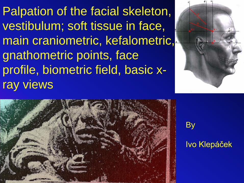
Palpation of the facial skeleton, vestibulum; soft tissue in face, main craniometric, kefalometric, gnathometric points, face profile, biometric field, basic x-ray views
By
Ivo Klepáček

SkullSkinSubdermisMuscular arrangementFat padInterdental relationsIntermaxillary relations
Morphologic structures having influence on profile formation and face relief

from Petrovický et al. 2001
Infratemporal region:a) pterygomandibular spaceb) interpterygoid space (superficial part)c) osseous part of infratemporal fossa (deep part)pterygopalatine fossalateral neck triangle (15, 16)a) omotrapezoid triangleb) omoclavicular triangle

Krajiny děleny
převážně podle
průmětu kostí svalů
Regiones are selected following bone and muscle
structures
To deep region

Points, lines and planes
used in dentistry
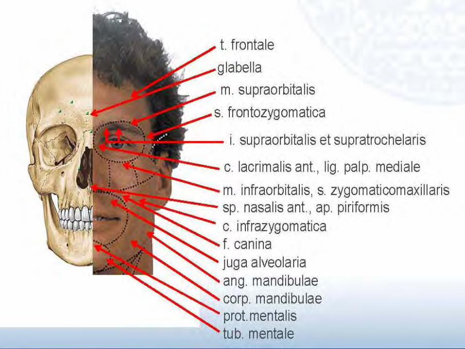
Overview of basic points, lines and planes in relation to face, dental arches and facial skeleton


Gnathometric points•Incisale inferius (Ii) – lower incisal point – crosspoint of both the lines parallel with incisal margines of first lower incisors•Incisale superius (Is) – upper incisal point - crosspoint of both the lines parallel with incisal margines of first upper•Infradentale (Id) – point more up on interalveolar septum between upper incisors.•Labrale inferius (Li) – on the most ventral part of lower lip.•Labrale superius (Ls) – on the most ventral part of upper lip•Mentale (Mn) – the deepest point inside mental canal•Nasion (Na) – on the nose root•Nasospinale (Ns) – in the midline on the base of anterior nasal spine•Orale (Ol) – between ventral incisors on the dorsal margine of their alveolar process (ventral margin of osseous palate)•Pogonion (Pg) – ventral margine of mental protuberence and on skin covering it.•Prosthion (Pr) – between both the first upper incisors on the alveolar margin ventrally•Punctum S (S) - middle of turcic sella•Staphylion (St) – on the top of posterior nasal spine (margine of the hard palate)•Stomion (Sto) – point where upper and lower lips are touching each other•Subnasale (Sn) – on the fusion between columella and philtrum•Subspinale (Sb, after Downs orthodontic upper point A) – labelling position of the upper apical basis); it is in the middle distance between akanthion and prosthion.. It lies on ventral surface of alveolar process. •Supramentale (Sm, after Downs orthodontic lower point B) – labelling position of the lower apical basis); it is on the ventral surface of osseous chin at level of tops of lower first incisors•Menton (Me) – the most dosrally and distally on the osseous chin

OBLIČEJ FACE
Inervační zóny, tukové těleso tváře, inervace v dutině ústní, cévy
a uzliny obličeje
Nerves in face, Head´s zones, fat pad, oral cavity innervation,
vessels and lymph nodes in face

Tloušťka kůže a podkožíLinie štěpnosti
Thickness of the skin and subcutis Cleavage lines
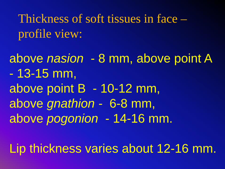
above nasion - 8 mm, above point A - 13-15 mm, above point B - 10-12 mm, above gnathion - 6-8 mm, above pogonion - 14-16 mm.
Lip thickness varies about 12-16 mm.
Thickness of soft tissues in face –profile view:

Kraissl´s (Kraissl 1951)
Langer´s Lines of skin fissionability (skin cleavage)
Kraissl: skin elongates parallel with direction of dermis bundles
On dead bodies
On living bodies

Skin cleavage (right, black lines) and location of incisions (left, pink furrows)
Corrugation – furrows are perpendicular to fibres inside mimic muscles. a – nasolabial groove, b –angular groove, c –mentolabial groove
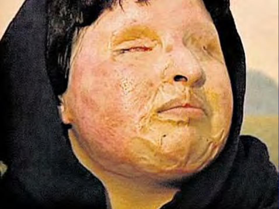

Fibromatosa ve tváři
Fibromatosis in face

Areae nervinae Head zones)

Headovy zónyHead´s zones

M.Chovanec - http://anat.lf1.cuni.cz/preinfo.html
n. V. examination

Muscles of facial expression
mm. faciales
Innervation from facial n. (nervus cranialis septimus; VII.)
Muscles: – of skull vault – of face (proper facial)
• Mm. surrounding orbit• Mm. surrounding
external nose• Mm. surrounding oral
cavity
VII – FACIALIS:follows structures of 2nd branchial arch

trigonum mortis
Mimické svaly cévy obličeje Mimic muscles face vessels

Depressor anguli orisAroganceaversion Zygomaticus
laugh
Levator labii superioriscry Levator alae nasi
Hopeless cryPetrovický a spol. 2001
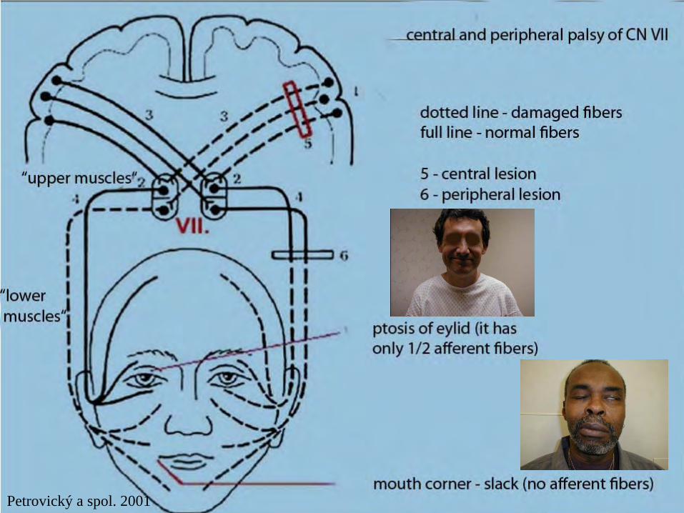
Petrovický a spol. 2001

Muscles around lips
• M. orbicularis oris– Circular arrangement, support lips, controls lip
parts and fuses with other mimic muscles, • M. buccinator
– From upper and lower jaw– to angulus oris

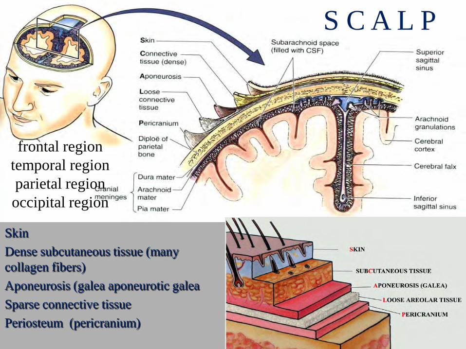
SkinConnective tissueAponeurosis (epicranial membrane)
Loose areolar tissuePericranium
S C A L P
SkinDense subcutaneous tissue (many collagen fibers)Aponeurosis (galea aponeurotic galeaSparse connective tissuePeriosteum (pericranium)
frontal region temporal regionparietal region occipital region

Epicranial (vault) muscle
• M. epicraniuscomposed from the two units– m. occipito-frontalis
• Frontal belly, occipital belly– Skin and above superior nuchal line– To aponeurotic galea
– M. temporoparietalis• Skin above auricle• to aponeurotic galea

Veins - from v. venae jugularis externa,
supraorbitalis, occipitalis
arteriae upraorbitales, superficialestemporales, posteriores
auriculares, occipitalesnervi cervicales and
trigeminal V1,V2branches
In subaponeurotic space


Brýlový hematomHematoma in eylids “spectacle haematoma“
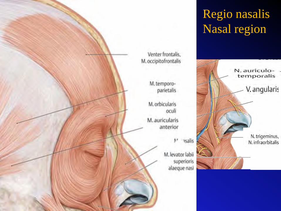
Regio nasalisNasal region

Regio buccalis
Buccal region


Regio buccalisBuccal region

Regio buccalis et infraorbitalis
Corpus adiposum buccaeBichatův polštář


Regio parotideo masseterica
Parotideo masseteric region!
Nodi lymphatici parotidei Parotid nodes


Septum styloideum
Mediální stěna spatium parotideumMedial wall of parotid space

Tumour of parotid gl. Compress CN VII. – ipsilateral peripheral palsy
(Bell´s palsy).
Ptosis of mouth corner and lower eylid.

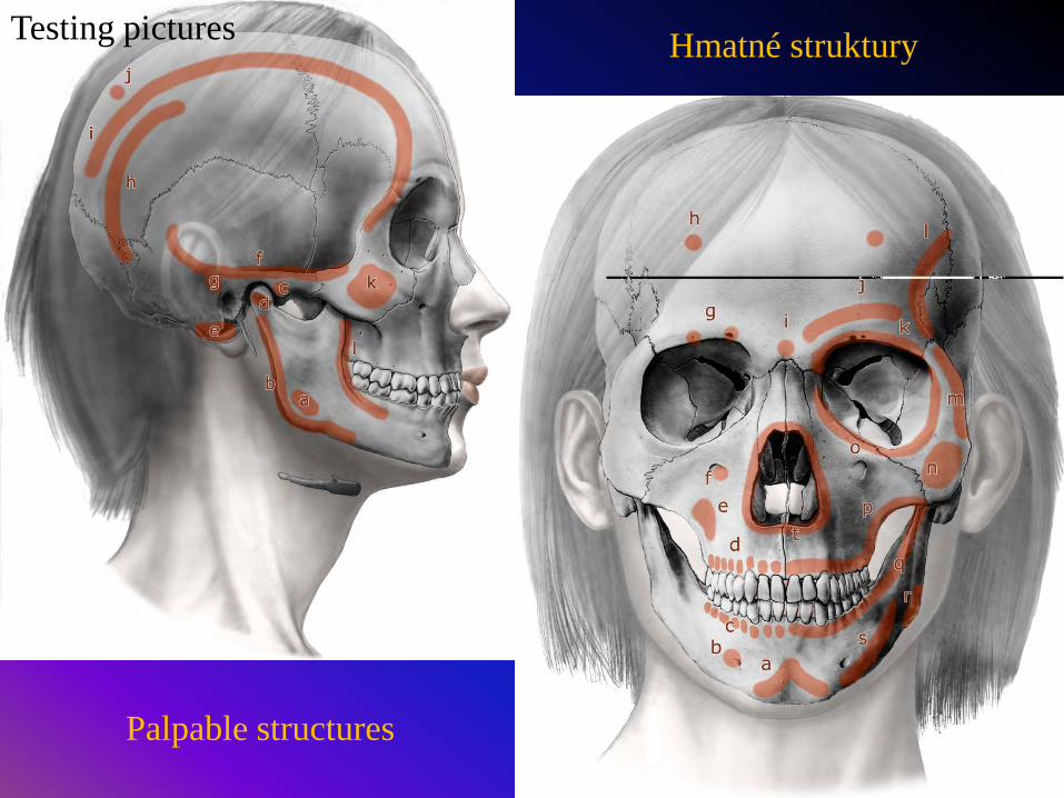
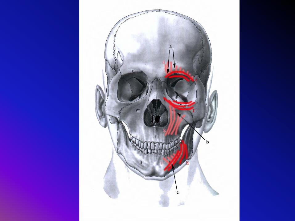
Hmatné struktury
Palpable structures
Testing pictures

a Plica sublingualisb Caruncula
sublingualisc Frenulum labii
inferiusd Plica buccogingivalise Frennulum linguaef Plica fimbriata
Plicae gingivolabiales* Area sublingualis** Area submandibularis
Canalis paralingualis Paralingual canal =between hyoglossus and genioglossus
Spodina dutiny ústní cavum oris bottom

Mucous relief in area where punction occurs. (field blocking, conduction anaesthesia). Inferior alveolar nerve can be blocked. Direction –between anterior margine and pterygomandibular ligament (to the mucous triangular fossa). a – margo anterior, b- ligamentum pterygomandibulare, c – arcus palatoglossus1 – projection of buccal nerve, 2 – projection of lingual nerve, 3 – inferior alveolar nerve

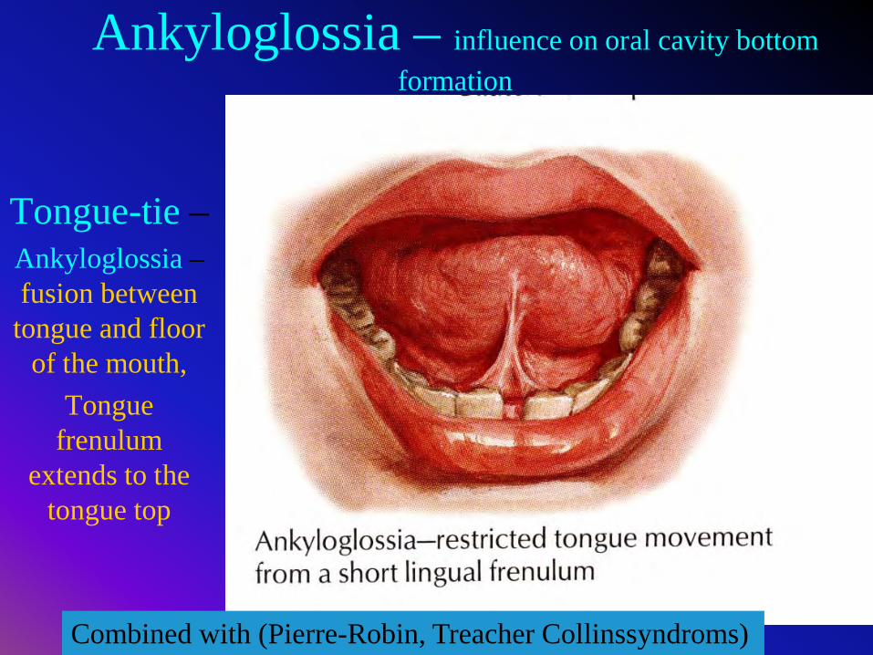
Ankyloglossia – influence on oral cavity bottom formation
Tongue-tie –Ankyloglossia –fusion between
tongue and floor of the mouth,
Tongue frenulum
extends to the tongue top
Combined with (Pierre-Robin, Treacher Collinssyndroms)

AnkyloglossiaTongue-tie

Tongue presses on alveolar
crest, which is deviated ventrally

Palate is compressed and deformed; middle palatal suture is wide
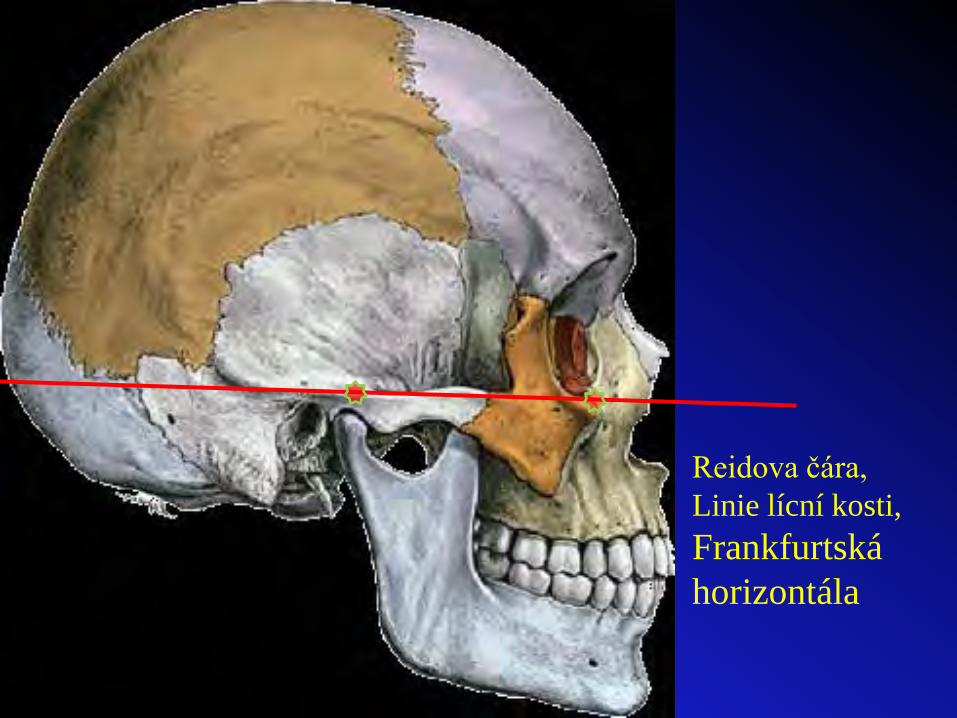
Reidova čára,Linie lícní kosti,Frankfurtská horizontála

•Frankfurter horizontale, eye ear line (linea horizontalis auriculoorbitalis) German line, Reid´s line (R. W. Reid, Scottish anatomist, 1851-1939) -Line crossing the lowest point of osseous orbital margine – punctum medioorbitale and upper margine of external acoustic opening - porus acusticus externus. It connects orbitale and porion points (or lower orbital margine and tragion).It is parallel with zygomatic arch. It serves for basic orientation of head or skull in space through e.g. X-ray examination. Between Reid´s line and Camper prosthetic line there is 10o až 15o angle.

Determination of occlusal plane using Camper´s plane; Its relation to the maxillary (b) and mandibular (d) planes

Occlusal plane (bite plane, planum occlusale) It is a plane crossing upper incisal point and tops of both the mesiobuccal tubercles of upper first molars. It is a plane crossing lower incisal point and tops of distobuccal tubercles of second lowr molars. It is at level wherer upper and lower lips are touching each other.
Other definitions -it is a plane crossing upper incisal pint and tops od distobuccal tubercles of second lower molars.
It is a plane crossing upper surface of lower lip, tops of canines, and retromolar triangle or tubercle on both the sides.It is a remnant of parodontium of wisdom tooth; alveolar bone below is not absorbed.
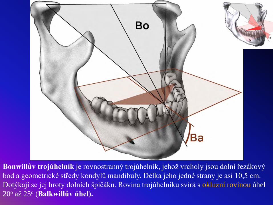
Bonwillův trojúhelník je rovnostranný trojúhelník, jehož vrcholy jsou dolní řezákový bod a geometrické středy kondylů mandibuly. Délka jeho jedné strany je asi 10,5 cm. Dotýkají se jej hroty dolních špičáků. Rovina trojúhelníku svírá s okluzní rovinou úhel 20o až 25o (Balkwillův úhel).
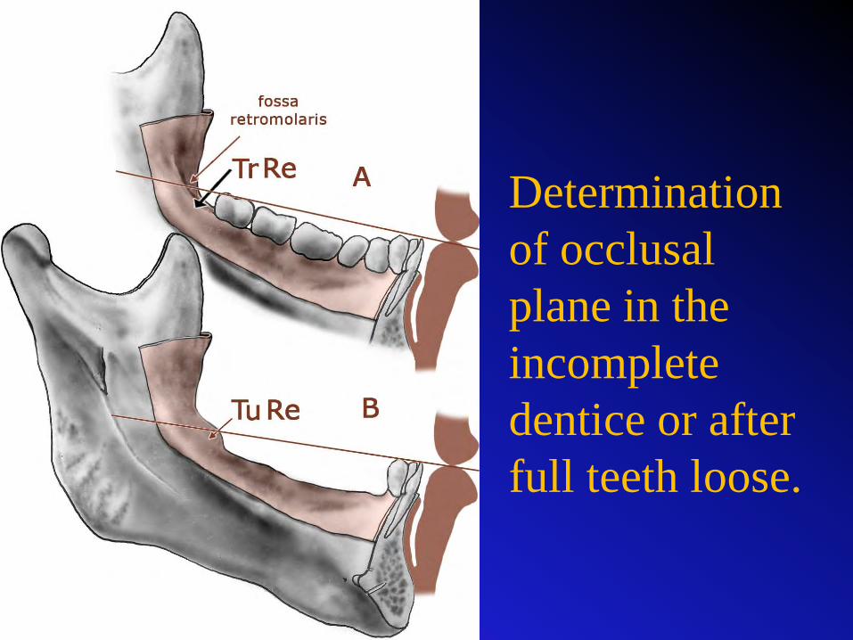
Determination of occlusal plane in the incomplete dentice or after full teeth loose.

18 / 8
Biorbitalis lineBiangularis line

Monson (Wilson) curve
Spee curve
Ferdinand Graf von Spee(1855-1937), German embryologist
George S. Monson (1869-1933), am. dentist

Skeletodental ní analýza, telerengenografiePravidelný a dobře utvářený chrup je výsledkem harmonického růstu a diferenciace všech obličejových komponent.
Skeletodental analysis, telerentgenography
Harmonic and regular dentice – it is a result of harmonic growth and diferentiation of all face components.

Connie Culp
Defekt po střelném zranění a jeho oprava.
Rok 2004

Biometric field
Biometric field is an imaginary area (seen from the lateral view) Margines: - line crossing lower orbital margine in midline (perpendiculare orbitale, Simonov´s orbital line), -line crossing nasion (perpendiculare nasion) –frankfurter line FH and- line crossing lower chin surface (parallel with FH).

•Simonov ´s line (orbital line, linea orbitalis, POr line) crosses vertically midorbitale point on lower orbitale margin. •Ricketts´s R line (aesthetic line) connects nose tip and skin pogonion. Distances from R line: labiale (labrale) superius: -1 to 2 mm; labiale (labrale) inferius: 0 to 2 mm.•Aesthetic plane (planum esteticum) crosses nose tip and skin pogonion.•Holdaway ´s H line connects skin pogonion, labiale (labrale) superius and ventral margine of nostril. Nose profile and lip are giving symetric “S“ curve line.The most dorsal point of this line is optimaly found 5 ± 2 mm dorsally of H line.
Aesthetic line R
Line H

Nose profile and lip are giving symetric “S“ curve line.The most dorsal point of this line is optimaly found 5 ± 2 mm dorsally of H line.
30o
90-110o

Aesthetic requests
Arrangement of mm. of facial expression relates to forms
of skull bones.
What can be accepted: face with small protrusion of lower
jaw (dental protrusion). Lower one third of face can be longer about 5-10 mm than
middle one third of face.WRONG: short lower one third of face in comparison with
middle one third of face (deep bite).

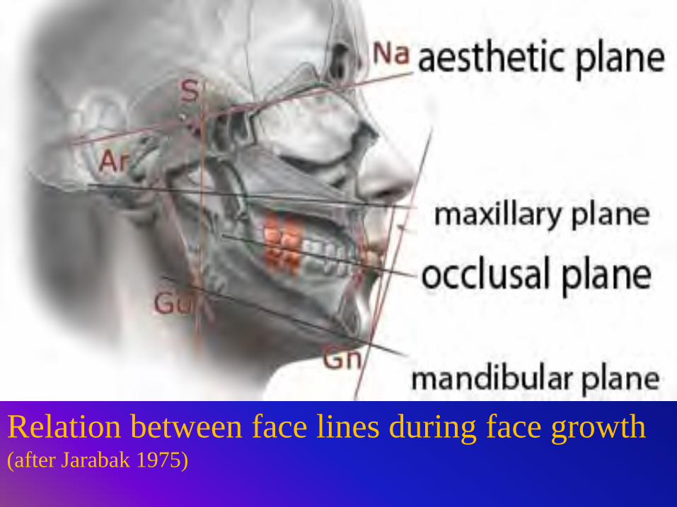
Relation between face lines during face growth (after Jarabak 1975)

Profile face line It helps to determine sagittal relations between jaws and chin position due to amount of soft tissues in face.
Relation between face lines during face growth (after Jarabak 1975)

Kroenlein lines for detection of
arteriesPoint K (for compression of facial artery). Two lines are crossed – line crossing middle of zygoma (linea verticalis zygomatica) and frankfurter line. Point K1 serves for compression of frontal branch of temporal arteryPoint K2 serves for compression of parietal brach from the same artery.
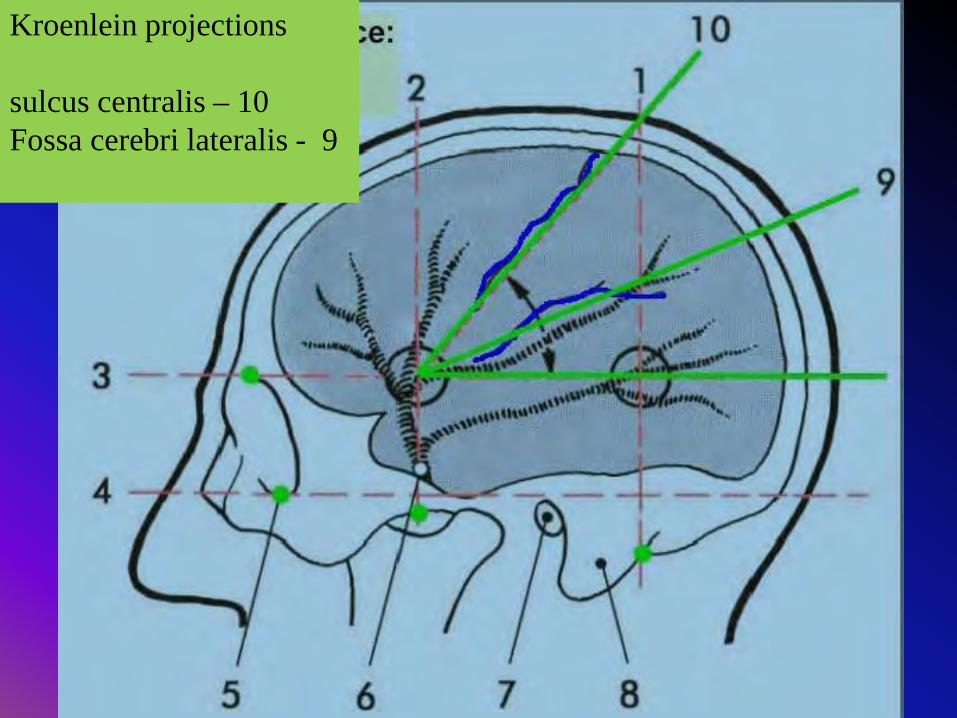
Kroenlein projections
sulcus centralis – 10Fossa cerebri lateralis - 9

Palpable skull structures and internal bone
structures seen on X – ray photos
Main X – ray projections and views

Sinus frontalis
Sella turcica
Sinus sphenoidalis
Meatus ac. ext.
Vomer
Processus mastoideus
Proc. styloideus
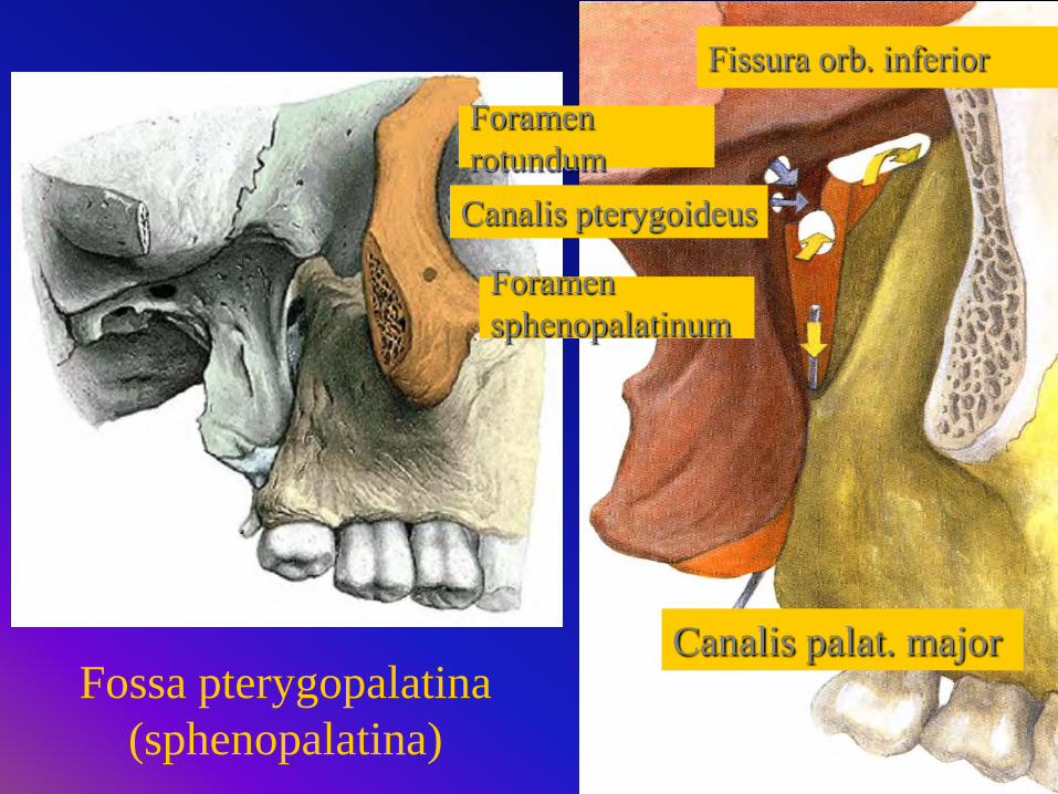
Fossa pterygopalatina (sphenopalatina)
Canalis pterygoideus
Canalis palat. major
Fissura orb. inferior
Foramen rotundum
Foramen sphenopalatinum

Lateral view

Testing picture

Posteroanterior projection– for details of facial skeleton
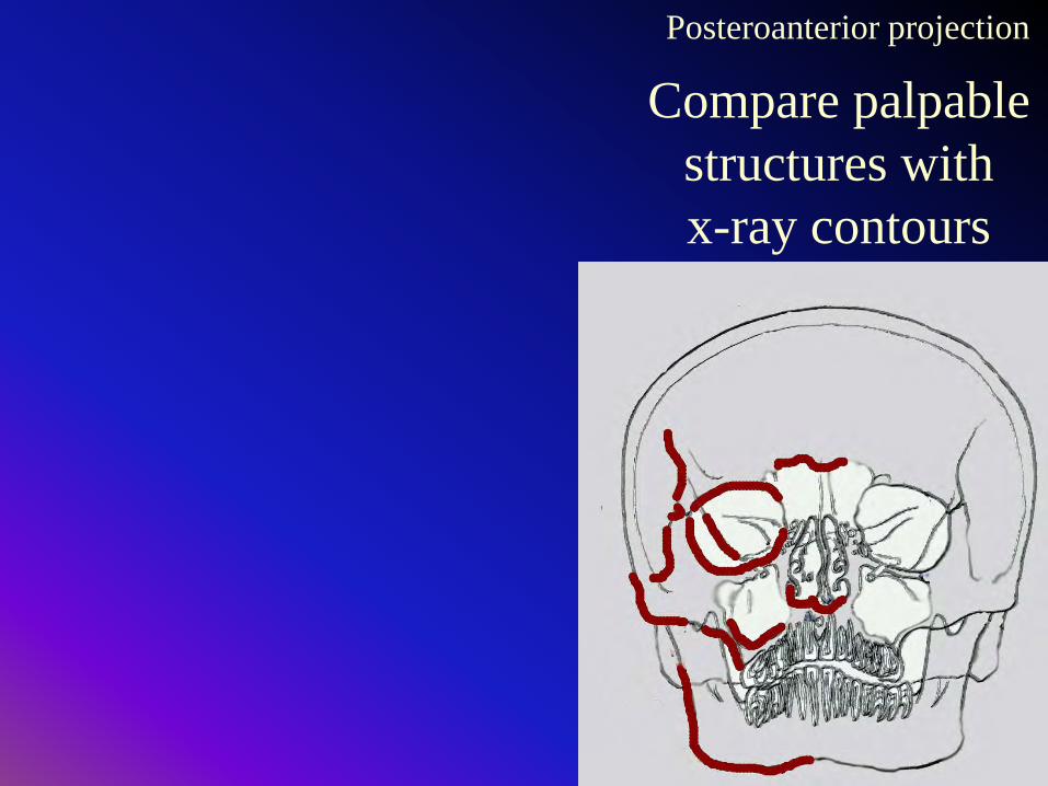
Posteroanterior projection
Compare palpable structures with x-ray contours


Testing picture

Lower oblique posteroanterior projection lower view

Clementschitsch view
Modified posteroanterior lower oblique projection


Testing picture

Waters view
Upper oblique posteroanterior projection


Testing picture


Pillar teeth of permanent dentice


Testing picture

Clinical remarks
Mucoele – labial small gland is enlarged Ranula (frog) – relates to sublinguali gland
Sialolite (calculus) inside submandibular duct

Salivary glands

Gland parts: Acini, grouped to lobes, septae, capsule

X- ray pictures:Paroticsublingualandsubmadibular Glands with ducts

Panoramatický snímek
panoramic X – ray photo


Berkowitz et al.: Oral Anatomy, Histology and Embryology. 3rd ed.. Mosby 2002Woelfel, Scheid: Dental Anatomy, 6th ed. Williams & Wilkins, 2002Feneis, Dauber: Pocket Atlas of Human Anatomy. Georg Thieme, 2007Weber: Memorix Zahnmedizin. 2nd. ed., Georg Thieme Verlag 2003Schuenke,Schulte,Schumacher: Head and Neuroanatomy. Thieme, 2006Fehrenbach,Herring: Anatomy of the Head and Neck. 3rd ed., Saunders Elsevier, 2007Snell: Clinical Anatomy for Medical Students. Williams and Wilkins, 2004 Moore, Agur: Essential Clinical Anatomy, Williams and Wilkins 2002Lang: Clinical Anatomy of the Masticatory Apparatus and Peripharyngeal Spaces. Stuttgart, Thieme, 1995White, Pharoah: Oral Radiology: Principles and Interpretation 5th ed., Mosby, 2003Bath-Balogh: Workbook for Illustrated Dental Embryology, Histology and Anatomy. 2nd ed. 2005, SaundersWhaites: Essentials of Dental Radiography and Radiology. 4th ed., 2006Churchill Livingstone Ivo Klepáček, J. Mazánek et al.: Klinická anatomie ve stomatologii. Grada 2002Own archive
see: www.lf1.cuni.czor: http://anat.lf1.cuni.cz/aindex.html
Sources


![Craniometric Analysis of the Hindbrain and Craniocervical ...epubs.surrey.ac.uk/813560/1/PLOS ONE Craniometric... · [25–27]. The Chihuahua is the smallest known dog breed for which](https://img.pdfslide.us/doc/110x75/5f6bfefecd418f439b108db3/craniometric-analysis-of-the-hindbrain-and-craniocervical-epubs-one-craniometric.jpg)