Embed Size (px)
Citation preview
AMERICAN JOURNAL OF PHYSICAL ANTHROPOLOGY 100:531-544 (1996)
Paleoepidemiology, Healing, and Possible Treatment of Trauma in the Medieval Cemetery Population of St. Helen-on-the-Walls, York, England
A.L. GRAUER AND C.A. ROBERTS Department of Sociology and Anthropology, Loyola University of Chicago, Chicago, Illinois 60626 (A.L. GJ; Department of Archaeological Sciences, University of Bradford, Bradford, West Yorkshire, England BD7 IDP (C.A.R.)
KEY WORDS Paleopathology, Long bone fractures, Trauma
ABSTRACT Traumatic lesions are commonly found in the archeological record and have potential to provide insight into the lives of past populations. This paper examines patterns of long bone fractures in the British medieval population of St. Helen-on-the-Walls from York (approximately 1100-1550) in an effort to determine patterns of healing and evidence for treatment. Long bones were macroscopically and radiologically examined. Clinical data were used to assess whether a fracture had successfully or unsuccessfully healed. The results indicate that fractures of the radius and ulna were most common. Males displayed more fractures than women. Most fractures were healed, well aligned, and without substantial deformity. Lack of evidence for deformity in bones likely to be severely affected by fracture implied that immobilization and possibly reduction was practiced on even the poorest residents of the medieval city. o 1996 Wiley-Liss, Inc
Traumatic lesions are common abnormali- ties observed in skeletal populations. Re- ports of trauma, however, are often limited to case studies and tend to highlight cranial trauma or postcranial fractures clearly asso- ciated with violence (e.g., Wells, 1963; Cour- ville, 1965a,b; Manchester and Elmhirst, 1980). The overall frequency rates within a given population (e.g., Manchester, 1978; Wells, 1982; Zivanovic, 1984) or changes in frequency rates over time and between popu- lations (e.g., Wood-Jones, 1910; Brothwell, 1961; Angel, 1974; Steinbock, 1976; Grimm, 1980, 1983; Jurmain, 1983, 1991) are also commonly explored. There is, however, a cur- rent trend toward placing trauma into a broader biocultural perspective (e.g., Merbs, 1983; Roberts, 1991; Lovejoy and Heiple, 1981). These studies have attempted to ex- plore the etiology of fractures and the biolog- ical and sociocultural environment within which the population operated.
Consideration of fracture types, healing
patterns, and complications help build infer- ences concerning the etiology of trauma and the possibility of treatment. Lovejoy and Heiple (19811, for instance, in their analysis of the Libben site, explore the rate of frac- tures for particular bones and the presence of healing and distortion. They infer from their data that the Late Woodland popula- tion successfully treated fractures. Simi- larly, Merbs (1983) in his examination of the Sadlermiut weaves information concerning cultural behavior with the frequency and patterns of trauma detected within the popu- lation.
The assessment, however, of archeological material can be difficult to interpret. Obser- vations of dry bone specimens pose several
Received July 31, 1995; accepted March 22, 1996. Address reprint requests to A.L. Grauer, Department of Sociol-
ogy and Anthropology, Loyola University of Chicago, 6525 N. Sheridan Rd., Chicago, IL 60626.
0 1996 WILEY-LISS, INC
532 A.L. GRAUER AND C.A. ROBERTS
limitations. First, determining fracture fre- quency rates can be hindered by fragmen- tary skeletal remains or poor preservation. Even with good preservation, cases of perimortem fracture may pass undetected since their appearance can mimic postmor- tem damage. Conversely, well-remodeled fractures of long bones (including stress fractures) can pass undetected during macroscopic and x-ray investigation. Well- remodeled lesions can make the determina- tion of fracture type difficult. Determining the age of the individual when the trauma occurred is also problematic, if not impossi- ble in most instances. Lastly, inferring that a fracture was treated is particularly difficult since splinted bones are rarely found and materials used for treatment were proba- bly biodegradable.
Notwithstanding the limitations, the po- tential for the study of traumatic lesions in human skeletal remains is considerable (Roberts, 1988). This paper examines the patterns of traumatic lesions in adults, spe- cifically long bone fractures, within a late medieval British cemetery population from St. Helen-on-the-Walls, York. By combining paleopathological information with avail- able archeological and historical data, our goal is to understand the etiology of the le- sions and to assess the possibility of treat- ment of fractures in medieval Britain.
MATERIALS AND METHODS The sample
The late medieval skeletal material from the cemetery of St. Helen-on-the-Walls (ap- proximately AD 1100-1550) in York, En- gland, was chosen for this study for several reasons. First, the total population (n = 1,014) provided reasonable sample sizes. Second, the quality of preservation of the bones allowed for macroscopic and radio- graphic assessment (Dawes and Magilton, 1980). Lastly, extensive archeological and historic research, pertaining to this and other medieval British urban centers, pro- vided insight into the biological and sociocul- tural environment.
The St. Helen-on-the-Walls cemetery yielded total of 1,014 discrete individuals (Grauer, 19891, representing two-thirds of
the original graveyard (Dawes and Magilton, 1980). Of the 685 adults, 334 were assigned to specific age-at-death intervals; 351 could be classified only as adult (20+ years old). The determination of age at death was made using as many independent techniques as possible, including dental eruption, forma- tion and attrition rates (Moorees et al., 1963a,b; Miles, 1963; Brothwell, 1981, 1989; Lovejoy, 1985), epiphyseal and sutural fu- sion (McKern and Stewart, 1957; Krogman and Iscan, 1986; Ubelaker 1989a,b), and pu- bic symphyseal morphology (Todd, 1921a,b). A total of 533 adult skeletons were assigned a sex (with or without the further assign- ment of an age-at-death interval) using inde- pendent assessments of dimorphic features of the 0s coxae and cranium (Meindl et al., 1985; Buikstra and Meilke, 1985; Bass, 1987; Ubelaker, 1989a).
As seen in Table 1, the highest proportion of skeletons were placed within the 25-34.9 age-at-death interval. This mortality pat- tern, along with documentary evidence, sug- gested from past research that adult migra- tion was an important demographic feature (Grauer, 1991a). The adult mortality pattern by sex (Table 1) indicated that the peak age- at-death for females occurred at 25-34.9 years old and at 35-45 years old for males, a statistically significant difference (Grauer, 1991a). This difference was attributed by Grauer (1991b) to the effects of delayed age at marriage (and thus maternity) and the high rate of female immigration.
Recording of fractures The term fracture refers to traumatic
events which lead to a complete or partial break in the continuity of a bone. There are many types of fractures discussed in clinical literature (Watson-Jones and Coltart, 1976; Ostrum et al., 1994). Causes of fracture, as well as the healing process, are also well documented (Rockwood and Green, 1975; Crawford-Adams, 1983). The type of fracture an individual sustains will give an indication of the type of force that acted upon the bone (Merbs, 1989). Transverse fractures, for in- stance, are caused by direct impact to a par- ticular area, while oblique fractures are caused by direct force applied at a dis- tance from the site o f the fracture. In both
TRAUMA IN MEDIEVAL YORK 533
TABLE 1. Mortality patterns of skeletons from the St. Helen-on-the- Walls cemetery population
Female Males Age at death n % total n % n % Total %
0 4 . 9 90 14.2 5-9.9 100 15.8 10-14.9 65 10.3 15-19.9 51 8.0 20-24.9 24 3.8 13 7.6 9 6.0 22 6.9 25-34.9 108 17.1 65 38.2 39 26.0 104 32.5 3544.9 98 15.5 44 25.9 54 36.0 98 30.6 45-54.9 70 11.0 36 21.2 34 22.7 70 21.9 55-64.9 27 4.3 12 7.1 14 9.3 26 8.1 Subtotal 633 Undeterminable 381 Total 1,014 170 150 320
archeological and clinical material it is possi- ble to record most fracture types (Roberts, 1988).
Macroscopic recording. The long bones of all skeletons from St. Helen-on-the-Walls were macroscopically evaluated (n = 4,938). Detailed examinations were made of the shafts of the humerus, radius, ulna, femur, tibia, and fibula, since the debilitating na- ture of these fractures would more likely compel the sufferer to seek treatment, if available, than would fractures of the smaller bones of the hands or feet.
Fractures, when detected, were recorded by position along the shaft (i.e., proximal third, mid shaft, distal third). Location of a fracture can occasionally be associated with soft tissue complications (Bodine and Lieber, 1994) and influence the healing process. The type of fracture was also recorded. The asso- ciation of specific types of fractures with par- ticular actions, occupations, and accidents is well noted in the clinical literature (Merbs, 1989) and holds promise for archeo- logical evaluation. Identifying the type of fracture can lead to the identification of the cause, an assessment of the ability to heal, and the potential for complications (Rob- erts, 1988).
The presence and results of healing were also recorded. Comparisons between frac- tured and unaffected bone (when available) from the same individual provided a means to recognize and quantify loss of length (or shortening). Bone shortening indicated that an overlap of the fracture fragments had oc- curred and subsequently suggested that
fracture reduction was not attempted or had failed. Rotational or linear deformity was also recorded by comparing the fractured bone with the contralateral side (or, if un- available, with a normal reference bone). The presence of this deformity was assumed to indicate that the fracture had not been treated or had been treated ineffectively. Since linear deformity is, and would have been in antiquity, easier to reduce than a rotational deviation, associating type of frac- ture with healing was important.
Alignment of the healed fracture bone was recorded as a means to recognize the practice of reduction and splinting. The presence of good alignment or angulation, good apposi- tion or partial apposition, overlap, or distrac- tion was recorded when possible. Since malalignment and other deformities of healed bone can place stress on adjacent joints, the presence of degenerative joint dis- ease was recorded, even though the develop- ment of joint disease could have occurred prior to the fracture.
Evidence of nonspecific infection, ap- pearing as periosteal reaction, was also re- corded when associated with a fracture. Periostitis and osteomyelitis occur as com- plications of compound fractures and can de- lay or prevent healing. In this study the pres- ence of localized periosteal reaction was assumed to indicate that the fracture was compound and resulted from the direct spread of environmental or commensal or- ganisms from the skin surface to the exposed fractured bone. Nonlocalized periosteal reac-
534 A.L. GRAUER AND C.A. ROBERTS
tion was regarded as unrelated to the fracture.
Radiographic recording. The evaluation of trauma in ancient populations, by neces- sity, relies heavily on the use of radiography to assess the injury and healing process. Fol- lowing gross examination, each fracture was radiographed in standard views and in two projections at right angles to each other, anteroposteriorly (AP) and mediolaterally (ML). From each radiograph a determina- tion of fracture type, aspects of healing, and the presence and degree of displacement of fracture fragments was made. The presence of other pathological lesions on the same bone was also noted. Radiographic determi- nation of fracture type is of considerable im- portance in paleopathology. A well-remod- eled bone often masks the type of fracture originally present or, worse, can be mis- leading.
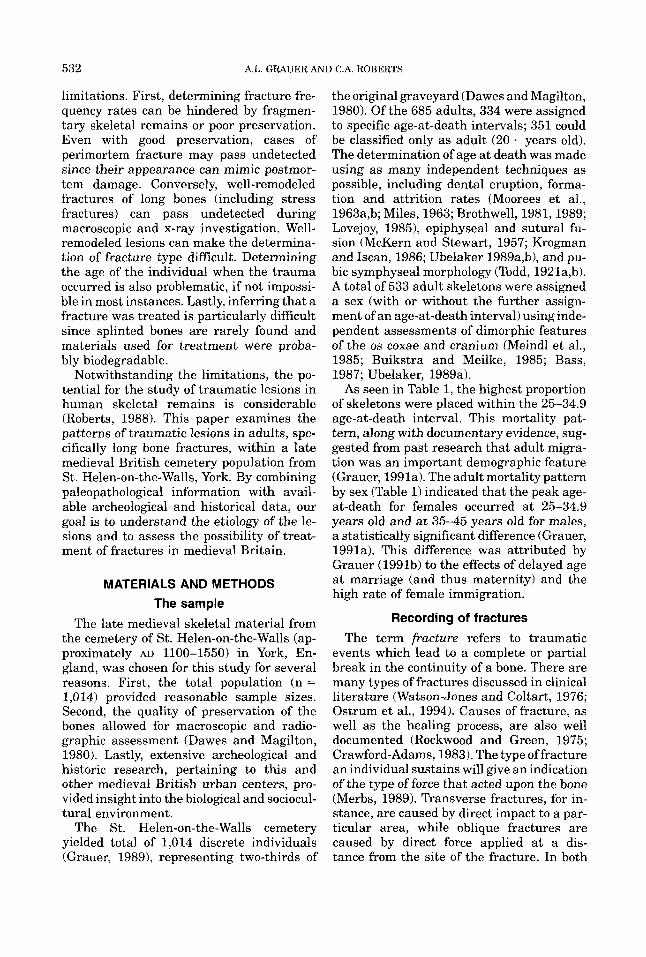
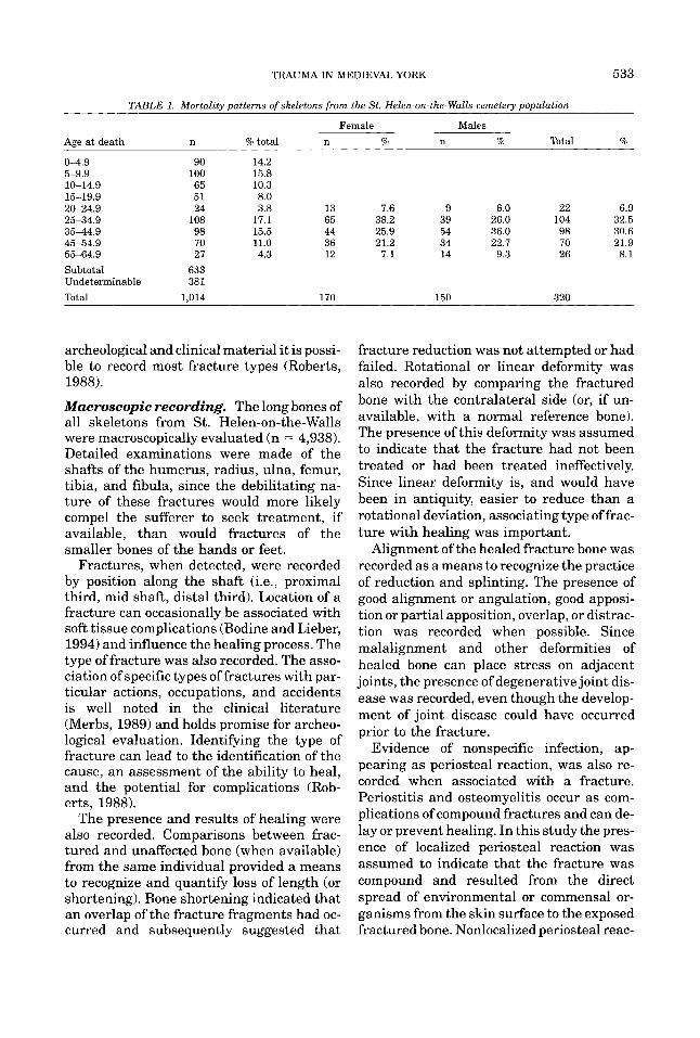
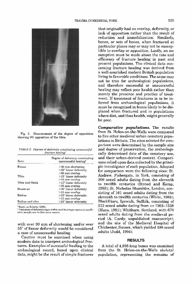
The presence andlor degree of displace- ment of each fracture was also recorded. Based on the work of Roberts (19881, the degree of angulation was measured by re- cording the direction and degree of displace- ment of the distal fragment using a standard ruler and protractor (Fig. 1). Angulation was recorded as anterior, posterior, medial, or lateral. Bone fracture fragment overlap and apposition (Figs. 2 , 3 ) were recorded by milli- meters offset and percent apposition, respec- tively.
Radiographs and medical records of mod- ern fractures were examined from the Na- tional Orthopaedic Hospital, Stanmore, to create a model for determining successful and unsuccessful healing (Roberts, 1988). Modern cases of fracture were included in the study based on three criteria: 1) the pa- tient’s fracture was not the result of an acci- dent involving modern technology; 2 ) the fracture was treated conservatively (i.e., with simple reduction and splinting); and 3 ) the orthopaedic surgeon concluded that healing was successful. From this data crite- ria were established for assessing archaeo- logical cases of successful and unsuccessful fracture healing according to the degrees of deformity, overlap of fragments, and angula- tion (Table 2) . A healed fracture Of a tibia, for instance, found in the archeological record
Fig. 1. Measurement of the degree of angulation de- formity of a Colles fracture of the radius.
Fig. 2. Measurement of the degree of bone overlap of the fibula.
TRAUMA IN MEDIEVAL YORK 535
Fig. 3. Measurement of the degree of apposition showing 0% apposition of the tibia.
TABLE 2. Degrees of deformity constituting unsuccessful fracture healing'
Degree of deformity constituting Bone unsuccessful healing'
Femur >30 mm shortening >35" linear deformity >50 mm overlap
Tibia >15" linear deformity >10 mm overlap
Tibia and fibula >15" linear deformity >35 mm overlap
Humerus >Zoo linear deformity >15 mm overlap
Radius >25" linear deformity > 15 mm overlap
Radius and ulna >25" linear deformitv
'Based on Roberts (1988).
cient sample size to determine results. Omissions ofdeformity types within each bone type represent insuffi-
with over 30 mm of shortening andlor over 35" of linear deformity would be considered a case of unsuccessful healing.
Caution must be exercised when using modern data to interpret archeological frac- tures. Examples of successful healing in the archeological record, based upon clinical data, might be the result of simple fractures
that originally had no overlap, deformity, or lack of apposition rather than the result of reduction and immobilization. Similarly, bones, or sets of bones, when fractured at particular places may or may not be suscep- tible to overlap or apposition. Lastly, an as- sumption must be made about the rate and efficiency of fracture healing in past and present populations. The clinical data con- cerning fracture healing was derived from a well-nourished modern British population living in favorable conditions. The same may not be true for archeological populations, and therefore successful or unsuccessful healing may reflect poor health rather than merely the presence and practice of treat- ment. If treatment of fractures is to be in- ferred from archaeological populations, it must be recognized in bones likely to be dis- placed when fractured and in populations where diet, and thus health, might generally be poor.
Comparative populations. The results from St. Helen-on-the-Walls were compared to five other medieval urban cemetery popu- lations in Britain. The sites selected for com- parison were determined by the sample size and degree of preservation, the archeologi- cally determined date of the cemetery use, and their urban-derived context. Compari- sons relied upon data collected by the princi- ple investigator of each population. Adopted for comparison were the following sites: St. Andrew, Fishergate, in York, consisting of 309 sexed adults dating from the eleventh to twelfth centuries (Stroud and Kemp, 1993); St. Nicholas Shambles, London, con- sisting of 161 sexed adults dating from the eleventh to twelfth centuries (White, 1988); Blackfriars, Ipswich, Suffolk, consisting of 212 sexed adults dating from AD 1263-1538 (Mays, 1991); Whithorn, Scotland, with 670 sexed adults dating from the medieval pe- riod (A. Cardy, unpublished manuscript); and the site of the Medieval Hospital of Chichester, Sussex, which yielded 198 sexed adults (Judd, 1994).
R ES U LTS A total of 4,938 long bones was examined
from the St. Helen-on-the-Walls skeletal population, representing the remains of
536 A.L. GRAUER AND C.A. ROBERTS
TABLE 3. Frequency of fracture by bone in St. Helen-on- the- Walls
TABLE 4. Age and sex distribution of individuals displaying long bone fracture in the St. Helen-on-the- Walls
cemeterv uouulation “ 1 I
Fractures Fractures % Bone (%) Age a t death Females (n) Males (n) Total (n) (70) Bone ._
20-24.9 0 0 0 0 L humerus 424 3 .7 Rhumerus 427 4 0.9 Humerus 0.8 25-34.9
35-44.9 L radius 383 4 1.0 R radius 387 6 1.5 Radius
55 + L ulna 372 5 1.3 R ulna 380 6 1.6 Ulna L femur 474 0 0
1 1 2 6.7 3 6 9 30.0 3 0 3 10.0 0 1 1 3.3
15 50.0
1.3 45-54.9
1.5 Adult (age 5 10 indeterminable)
R L R
femur 463 1 .2 Femur 0.1 tibia 431 4 .9 tibia 433 2 .5 Tibia 0.7
L fibula 363 5 1.4 R fibula 362 1 .5 Fibula 0.8 Total 4.938 41 .8
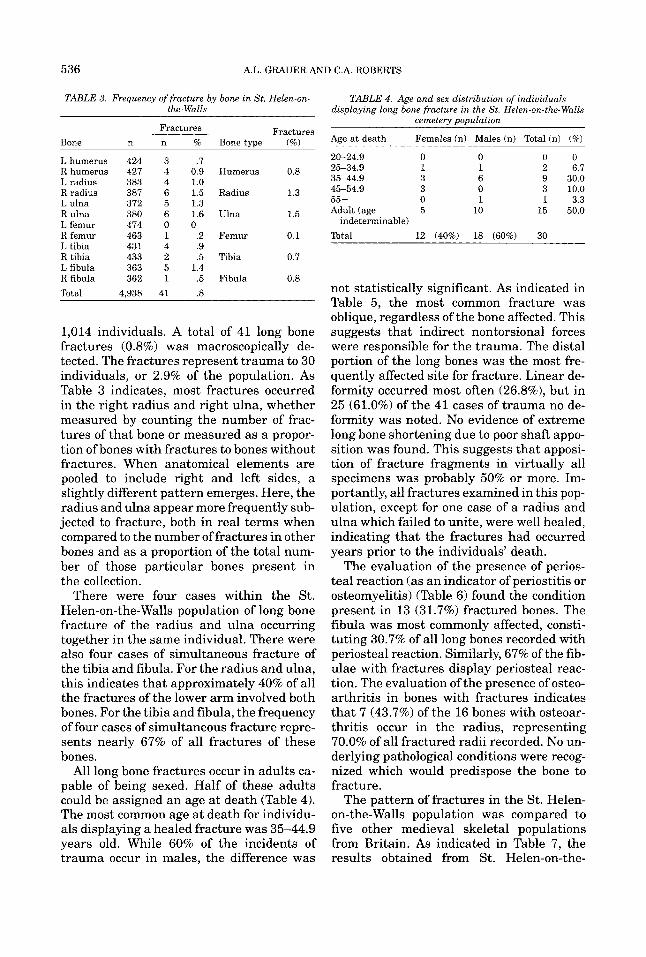
1,014 individuals. A total of 41 long bone fractures (0.8%) was macroscopically de- tected. The fractures represent trauma to 30 individuals, or 2.9% of the population. As Table 3 indicates, most fractures occurred in the right radius and right ulna, whether measured by counting the number of frac- tures of that bone or measured as a propor- tion of bones with fractures to bones without fractures. When anatomical elements are pooled to include right and left sides, a slightly different pattern emerges. Here, the radius and ulna appear more frequently sub- jected to fracture, both in real terms when compared to the number of fractures in other bones and as a proportion of the total num- ber of those particular bones present in the collection.
There were four cases within the St. Helen-on-the-Walls population of long bone fracture of the radius and ulna occurring together in the same individual. There were also four cases of simultaneous fracture of the tibia and fibula. For the radius and ulna, this indicates that approximately 40% of all the fractures of the lower arm involved both bones. For the tibia and fibula, the frequency of four cases of simultaneous fracture repre- sents nearly 67% of all fractures of these bones.
All long bone fractures occur in adults ca- pable of being sexed. Half of these adults could be assigned an age at death (Table 4). The most common age at death for individu- als displaying a healed fracture was 35-44.9 years old. While 60% of the incidents of trauma occur in males, the difference was
Total 12 (40%) 18 (60%) 30
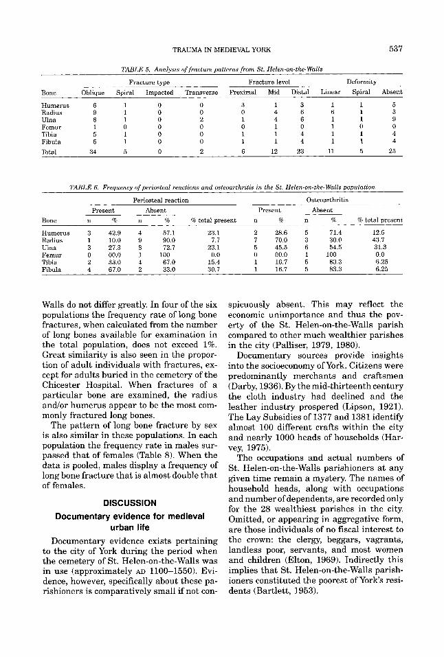
not statistically significant. As indicated in Table 5, the most common fracture was oblique, regardless of the bone affected. This suggests that indirect nontorsional forces were responsible for the trauma. The distal portion of the long bones was the most fre- quently affected site for fracture. Linear de- formity occurred most often (26.8%), but in 25 (61.0%) of the 41 cases of trauma no de- formity was noted. No evidence of extreme long bone shortening due to poor shaft appo- sition was found. This suggests that apposi- tion of fracture fragments in virtually all specimens was probably 50% or more. Im- portantly, all fractures examined in this pop- ulation, except for one case of a radius and ulna which failed to unite, were well healed, indicating that the fractures had occurred years prior to the individuals’ death.
The evaluation of the presence of perios- teal reaction (as an indicator of periostitis or osteomyelitis) (Table 6) found the condition present in 13 (31.7%) fractured bones. The fibula was most commonly affected, consti- tuting 30.7% of all long bones recorded with periosteal reaction. Similarly, 67% of the fib- ulae with fractures display periosteal reac- tion. The evaluation of the presence of osteo- arthritis in bones with fractures indicates that 7 (43.7%) of the 16 bones with osteoar- thritis occur in the radius, representing 70.0% of all fractured radii recorded. No un- derlying pathological conditions were recog- nized which would predispose the bone to fracture.
The pattern of fractures in the St. Helen- on-the-Walls population was compared to five other medieval skeletal populations from Britain. As indicated in Table 7, the results obtained from St. Helen-on-the-
TRAUMA IN MEDIEVAL YORK 537
TABLE 5. Analysis of fracture patterns from St. Helen-on-the- Walls
Fracture type Fracture level Deformity
Bone Obliaue Suiral Imuacted Transverse Proximal Mid Distal Linear Spiral Absent
Humerus 6 1 0 0 3 1 3 1 1 5 Radius 9 1 0 0 0 4 6 6 1 3 Ulna 8 1 0 2 1 4 6 1 1 9 Femur 1 0 0 0 0 1 0 1 0 0 Tibia 5 1 0 0 1 1 4 1 1 4 Fibula 5 1 0 0 1 1 4 1 1 4 Total 34 5 0 2 6 12 23 11 5 25
TABLE 6. Frequency of periosteal reactions and osteoarthritis in the St. Helen-on-the- Walls population
Periosteal reaction Osteoarthritis Present Absent Present Absent
Bone n % n % % total present n % n % % total present
Humerus 3 42.9 4 57.1 23.1 2 28.6 5 71.4 12.5 Radius 1 10.0 9 90.0 7.7 7 70.0 3 30.0 43.7 Ulna 3 27.3 8 72.1 23.1 5 45.5 6 54.5 31.3 Femur 0 00.0 1 100 0.0 0 00.0 1 100 0.0 Tibia 2 33.0 4 67.0 15.4 1 16.7 5 83.3 6.25 Fibula 4 67.0 2 33.0 30.7 1 16.7 5 83.3 6.25
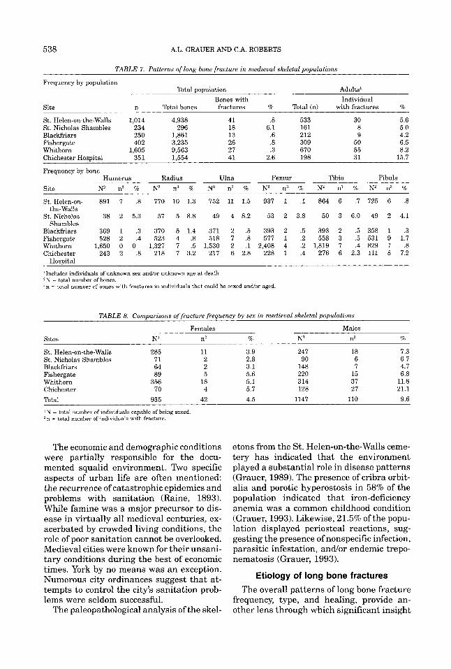
Walls do not differ greatly. In four of the six populations the frequency rate of long bone fractures, when calculated from the number of long bones available for examination in the total population, does not exceed 1%. Great similarity is also seen in the propor- tion of adult individuals with fractures, ex- cept for adults buried in the cemetery of the Chicester Hospital. When fractures of a particular bone are examined, the radius andor humerus appear to be the most com- monly fractured long bones.
The pattern of long bone fracture by sex is also similar in these populations. In each population the frequency rate in males sur- passed that of females (Table 8). When the data is pooled, males display a frequency of long bone fracture that is almost double that of females.
DISCUSSION Documentary evidence for medieval
urban life Documentary evidence exists pertaining
to the city of York during the period when the cemetery of St. Helen-on-the-Walls was in use (approximately AD 1100-1550). Evi- dence, however, specifically about these pa- rishioners is comparatively small if not con-
spicuously absent. This may reflect the economic unimportance and thus the pov- erty of the St. Helen-on-the-Walls parish compared to other much wealthier parishes in the city (Palliser, 1979, 1980).
Documentary sources provide insights into the socioeconomy of York. Citizens were predominantly merchants and craftsmen (Darby, 1936). By the mid-thirteenth century the cloth industry had declined and the leather industry prospered (Lipson, 1921). The Lay Subsidies of 1377 and 1381 identify almost 100 different crafts within the city and nearly 1000 heads of households (Har- vey, 1975).
The occupations and actual numbers of St. Helen-on-the-Walls parishioners a t any given time remain a mystery. The names of household heads, along with occupations and number of dependents, are recorded only for the 28 wealthiest parishes in the city. Omitted, or appearing in aggregative form, are those individuals of no fiscal interest to the crown: the clergy, beggars, vagrants, landless poor, servants, and most women and children (Elton, 1969). Indirectly this implies that St. Helen-on-the-Walls parish- ioners constituted the poorest of York’s resi- dents (Bartlett, 1953).
538 A.L. GRAUER AND C.A. ROBERTS
TABLE 7. Patterns of long bone fracture in medieval skeletal populations
Frequency by population Total population Adults'
Bones with Individual Site n Total bones fractures % Total (n) with fractures %
St. Helen-on-the-Walls 1,014 4,938 41 .8 533 30 5.6 St. Nicholas Shambles 234 296 18 6.1 161 8 5.0 Blackfriars 250 1,861 13 .6 212 9 4.2 Fishergate 402 3,235 26 .8 309 50 6.5 Whithorn 1,605 9,563 27 .3 670 55 8.2 Chichester Hospital 351 1,554 41 2.6 198 31 15.7
Frequency by bone
Site N2 n3 % N2 n3 % N2 n3 % N2 n3 % N2 n? % N2 n3 %
St. Helen-on- 891 7 .8 770 10 1.3 752 11 1.5 937 1 .1 864 6 .7 725 6 .8
St. Nicholas 38 2 5.3 57 5 8.8 49 4 8.2 53 2 3.8 50 3 6.0 49 2 4.1
Blackfriars 369 1 .3 370 5 1.4 371 2 .5 393 2 .5 393 2 .5 358 1 .3 Fishergate 528 2 .4 523 4 .8 518 7 .8 577 1 .2 558 3 .5 531 9 1.7 Whithorn 1,650 0 0 1,327 7 .5 1,530 2 .1 2,408 4 .2 1,819 7 .4 829 7 .8 Chichester 243 2 .8 218 7 3.2 217 6 2.8 228 1 .4 276 6 2.3 111 8 7.2
Humerus Radius Ulna Femur Tibia Fibula
the-Walls
Shambles
HosDital
'Includes individuals of unknown sex and/or unknown age a t death. 2 N = total number of bones. , n = total number of bones with fractures in individuals that could be sexed andor aged
TABLE 8. Comuarisons of fracture freauencv b y sex in medieval skeletal populations
Males - Females Sites N' n2 % N' n2 %
St. Helen-on-the-Walls 285 11 3.9 247 18 7.3 St. Nicholas Shambles 71 2 2.8 90 6 6.7 Blackfriars 64 2 3.1 148 7 4.7 Fishergate 89 5 5.6 220 15 6.8 Whithorn 356 18 5.1 314 37 11.8 Chichester 70 4 5.7 128 27 21.1 Total 935 42 4.5 1147 110 9.6
' N = total number of individuals capable of being sexed. z n = total number of individuals with fracture.
The economic and demographic conditions were partially responsible for the docu- mented squalid environment. Two specific aspects of urban life are often mentioned: the recurrence of catastrophic epidemics and problems with sanitation (Raine, 1893). While famine was a major precursor to dis- ease in virtually all medieval centuries, ex- acerbated by crowded living conditions, the role of poor sanitation cannot be overlooked. Medieval cities were known for their unsani- tary conditions during the best of economic times. York by no means was an exception. Numerous city ordinances suggest that at- tempts to control the city's sanitation prob- lems were seldom successful.
The paleopathological analysis of the skel-
etons from the St. Helen-on-the-Walls ceme- tery has indicated that the environment played a substantial role in disease patterns (Grauer, 1989). The presence of cribra orbit- alia and porotic hyperostosis in 58% of the population indicated that iron-deficiency anemia was a common childhood condition (Grauer, 1993). Likewise, 21.5% ofthe popu- lation displayed periosteal reactions, sug- gesting the presence of nonspecific infection, parasitic infestation, andlor endemic trepo- nematosis (Grauer, 1993).
Etiology of long bone fractures
The overall patterns of long bone fracture frequency, type, and healing, provide an- other lens through which significant insight
TRAUMA IN MEDIEVAL YORK 539
into the environment and conditions facing the poor inhabitants of medieval York can be made. First, it appears that long bone fractures were uncommon. This is not an artefact of poor skeletal preservation since the number of individuals displaying frac- tures is low among the best preserved group: aged and sexed adults. The lower arm, espe- cially the right side, displayed the greatest number of long bone fractures. Evidence of fracture was found in males more often than in females. These data appear to corroborate the documentary evidence. For instance, the nature of medieval cities as sites of craft production and commerce suggests that the majority of citizens would be employed or engaged in light labor-that is, labor requir- ing proportionately greater use of the arms than other body parts. Since Calvin (1982) has reported that 90% of humans can be categorized as right-handed, it is not sur- prising to find that the majority of injuries occurred to this side of the body. It also is no surprise that males more frequently display long bone fractures. Medieval Britain was notoriously patriarchal, denying member- ship to craft guilds and the participation in many occupations to women under normal circumstances. For women, employment was usually gained in household service or as independent traders (Goldberg, 19861, per- haps minimizing their susceptibility to trauma.
It is difficult to determine the etiology of the fractures in the St. Helen-on-the-Walls population. While the majority of forearm fractures occurred to the middle or distal shafts, the occurrence of transverse frac- tures is low. Transverse fractures to the ulna in particular can be interpreted as a sign of violence caused by direct blows with angu- lated force (Crawford-Adams, 1983). These fractures, known as parry fractures, occur as the victim attempts to ward off an of- fender by shielding hisherself with an arm. Subsequently, the two cases of transverse fracture to the ulna might have been caused by interpersonal violence.
The majority of ulna fractures in this pop- ulation are oblique. This indicates the pres- ence of an indirect force acting at a distance from the site of fracture, resulting in an un- even bending of the bone. Hence, most of the ulna fractures were likely the result of acute
injury provoked by a fall or similar unfortu- nate occurrence.
The patterns of fractures to the radius present similar results. Most of the fractures occur to the mid or distal shaft, and all except one is oblique. Most fractures occurring to the distal radial shaft appear to be Colles’s fractures, a condition caused by static and dynamic forces working against each other, commonly the result of falling on an out- stretched arm. The result of this shearing trauma to the radius is the posterior dis- placement of the distal portion of the bone (Ortner and Putschar, 1981). The presence of this condition suggests again an accidental rather than violent etiology.
The location and type of fracture found in other long bones (humerus, femur, tibia, and fibula) repeat the patterns discussed above. The majority of the fractures are oblique and at the distal ends. While assigning specific causes of trauma is impossible, as is relating the trauma to specific occupations or actions, it appears from the data that long bone frac- tures suffered in medieval York are remark- ably similar to those associated with twenti- eth-century life. That is, the most commonly fractured long bones are the radius and ulna and the tibia and fibula, and the most com- mon causes are accidents (Buhr and Cooke, 1959; Garraway et al., 1979; Fife and Baran- cik, 1985).
The possibility of medieval treatment Evidence for the availability of fracture
treatment in late medieval Europe is plenti- ful (Clark, 1937). There were orthopaedic surgeons, specific individuals known as bonesetters, and barber surgeons who occa- sionally reduced, manipulated, and splinted bones. While many of the methods of bone repair appear to be based upon Greco-Roman concepts of disease and injury, illustrations exist showing sophisticated methods of frac- ture treatment. No references to fracture treatment in Britain remain.
Synthesizing modern clinical data on heal- ing with data collected from the St. Helen- on-the-Walls population may provide insight into treatment in Britain. Causes and effects of fractures, along with complications and their effect on healing, are provided by clini- cal data. Comparing these data with the St. Helen-on-the-Walls population allows for
540 A.L. GRAUER AND C.A. ROBERTS
speculation concerning the treatment of fractures.
Clinical data suggests that fractures of the surgical or anatomical neck of the humerus are usually impacted and, therefore, are sta- ble (Crawford-Adams, 1983). Treatment commonly involves support in a sling with encouragement to exercise the shoulder and elbow joints. Unimpacted fractures are given the same treatment but restricted from movement at the shoulder. Three cases of fracture of the proximal humerus, one at the surgical neck and two at the anatomical neck, were found in the St. Helen-on-the- Walls population. Of these only one appeared to be impacted and had healed well from an oblique fracture. This stable fracture, how- ever, may have been accompanied by subse- quent infection, as severe localized perios- teal reaction with cortical bone involvement was noted. The two fractures of the anatomi- cal neck were well healed with no sign of infection. One case, however, displayed se- vere osteoarthritis of the head of the hu- merus and the glenoid fossa. The bone was also substantially shorter than the other side, indicating that the fracture had oc- curred at a young age and had disrupted normal length development.
Mid-shaft and supracondylar fractures (located at the distal third of the humerus shaft) usually unite after reduction and im- mobilization of the bone in a plaster cast. Delayed or non-union, however, occurs more often in the humerus than in any other bone (Campbell, 1937), possibly due to the impos- sibility of securing complete immobilization of the fracture andlor the difficulty in main- taining coaption of the fragments and pre- venting a gap between them. Although the weight of the arm can assist with reduction of the fracture, it can also produce a gap between the fragments. Supracondylar frac- tures are complicated by the risk of damage to the brachial artery, median nerve, and radial nerve. One case of mid-shaft fracture and three cases of supracondylar fracture were recorded in the St. Helen-on-the-Walls population. All cases were well healed. The mid-shaft fracture was spiral and displayed a linear deformity with slight rotation and poor alignment and signs of localized perios- teal reaction. Of the three supracondylar
fractures, none displayed deformity, only one displayed localized periosteal reaction, and another displayed osteoarthritic changes to the distal articular surface.
The overall assessment of healing pat- terns of fractures in the humerus leads to the conclusion that in most instances the repercussions of fracture were mild. This finding seems unlikely when the clinical in- formation is considered, unless the sufferers of humerus fractures were particularly lucky in their patterns of healing or if treatment, most likely in the form of immobilization, was being administered to some degree.
Clinical data suggests that fractures of the radius and ulna are extremely common in modern human populations. Accurate reduc- tion of these fractures is functionally vital; even slight displacement on healing may dis- turb the relationship between the radius and ulna and impair rotation of the forearm. De- layed or non-union of these long bones is also common, especially at the junction of the mid and distal thirds of the ulna shaft. In cases of marked malunion, subluxation of the infe- rior radio-ulna joint may occur. Reduction and immobilization in a plaster cast is the standard treatment in clinical contexts, ex- cept in cases such as Smith’s fracture (a frac- ture of the distal end of the radius with ante- rior displacement), which must be corrected operatively due to its unstable nature.
The majority of fractures to the ulna (10/11) and radius (10/10) in the St. Helen- on-the-Walls population occur to the mid and distal shafts. In four instances the ulna and radius appear to have been fractured to- gether. In three of these cases the fractures to both bones are well healed with no defor- mity or osteoarthritis. In one case of radius and ulna fracture, healing did not take place. Localized periosteal reaction is present in one individual with healed radius and ulna fractures. The successful healing of these bones is particularly surprising in light of the instability that fractures of these two bones cause. It appears highly likely that treatment was sought and successfully ad- ministered in these cases.
This is not necessarily true for instances when only one bone was fractured. In cases of radial fracture, seven out of ten instances displayed deformity of the bone, while only
TRAUMA IN MEDIEVAL YORK 54 1
one out of eleven cases of fracture to the ulna resulted in deformity. Connolly (1988) suggests that modern mid-shaft fractures of the ulna are easy t o reduce. This may sug- gest that in instances when the ulna was fractured in medieval populations the radius successfully served as a natural splint or that the nature of the fracture insured that treatment would be particularly successful. The same, however, does not appear to be true in instances when only the radius was fractured.
The pattern of osteoarthritis substanti- ates this contention Arthritic lipping was noted on the articular facets of 70% of the radii displaying fracture. It appears, then, that rarely did a fracture of a radius success- fully heal without deformity and subsequent development of arthritis in the adjacent joints as a sequel to the trauma. The instabil- ity of this bone when fractured, due to its rotation around the ulna when not immobi- lized, resulted in deformity and osteoarthri- tis. This finding suggests that either treat- ment of this fracture was usually unsuccessful or that when the radius was fractured immobilization was not practiced.
Fractures to the tibia and fibula are also common in modern populations. Reduction and immobilization is standard treatment for these fractures. Fibula fractures without associated tibia1 fractures are rare and often do not require intervention because of the natural splint that the tibia provides. De- layed or non-union of the tibia and fibula is a common complication. The distal shafts are particularly vulnerable due to the lack of surrounding soft tissues in the lower leg, reducing the amount of blood coming from the soft tissue (Rhinelander, 1974). Mal- union may lead to osteoarthritis of the knee andor ankle joints because of changed me- chanical stress.
Six instances of fracture of the tibia and fibula were noted in the St. Helen-on-the- Walls population. The distal ends are the most prevalent location for fracture. There are four cases within this population of the tibia and fibula being fractured together; two of these instances appear within the same individual. Perplexingly, while the fractures in this individual were well healed, the trauma to the right side resulted in substan-
tial deformity and osteoarthritis, while the fractures of the tibia and fibula of the left side healed with virtually no deformity. In total, half of the cases of fracture of the tibia (two out of four) and fibula (two out of four) in the St. Helen’s population resulted in de- formity. The relatively small number of frac- tures to these bones, along with the varying results, precludes a determination of the presence of treatment for these fractures. Similarly, the varying rate of periosteal reac- tion in the bones (tibia 33% and fibula 67%) may be an artifact of small sample sizes rather than a clear indication that the fibula was more susceptible to compound fracture (and therefore infection) than the tibia.
Only one case of fracture of the femur was noted in the St. Helen-on-the-Walls popula- tion. This oblique, mid-shaft fracture dis- played linear deformity and no signs of 0s- teoarthritis or periosteal reaction. The particularly low prevalence of fracture to this bone does not appear to be uncommon within medieval archeological populations from Britain (Table 6). Further, it implies that the femur was as unlikely a candidate for fracture in medieval urban populations as it is, reported by Buhr and Cooke (1959) and Fife and Barancik (1985), in modern populations.
The analysis of periosteal reaction and 0s- teoarthritis associated with fractures may also be used to indirectly detect the availabil- ity and/or efficacy of medical treatment. The data from the St. Helen-on-the-Walls popu- lation suggests that periosteal reaction was present in 31.7% of the bones displaying fractures. This proportion appears to be low in light of the historical data indicating the unsanitary conditions in York. Similarly, Grauer (1993) has suggested that the high proportion of adults in the skeletal popula- tion displaying remodeled periosteal reac- tion (77%) may indicate the presence of chronic infectious agents affecting the popu- lation. It is surprising, therefore, that only 31.7% of the fractured bones of adults dis- play pathological change associated with the presence of infection. Two reasons for this pattern are offered: the prevalence of com- pound fracture in the St. Helen-on-the-Walls population was low (thereby not exposing the bone or internal tissue to the environ-
542 A.L. GRAUER AND C.A. ROBERTS
ment), or attempts a t intervention were made and often were successful.
Population comparisons Comparisons of fracture patterns high-
lighted several trends and suggest that St. Helen-on-the-Walls was similar to other me- dieval urban skeletal populations. First, long bone fractures appear to be uncommon in all populations examined. The higher fre- quency found in St. Nicholas Shambles is likely due to poor long bone preservation, since the percentage of long bones available for examination per individual was low. The higher percentage of adults with fractures a t Chichester Hospital is likely due to the unusual nature of hospital cemetery sam- ples. Second, the radius andlor humerus are the most commonly fractured bones. Lastly, the higher number of males displaying long bone fractures a t St. Helen’s is corroborated by the same trend in every other population examined. Although fracture healing pat- terns were not examined in the comparative groups, the overall trends in fracture fre- quency and type suggest that hazards in me- dieval urban centers were similar. Future research into healing patterns will provide greater insight into the availability of treat- ment throughout Britain and the effects of varying socioeconomic environments.
CONCLUSIONS The analysis of patterns of long bone frac-
tures in the St. Helen-on-the-Walls popula- tion has shown that virtually all detectable cases of fractures in adult skeletons were well healed. The majority of the fractures were well aligned and without substantial deformity. Of particular interest were the low numbers of deformity in bones likely to be severely affected by fracture, such as in the humerus and instances in which the ra- dius and ulna were both fractured. The suc- cessful healing of these fractured bones im- plies that immobilization and possibly reduction was practiced. The analysis of the type of fracture suggests that trauma to the long bones was not attributable to heavy la- bor or violent activities. Males, however, in the St. Helen-on-the-Walls population, as well as in the comparative medieval popula- tions, display twice the number of long bone
fractures as females. In all, the patterns of fracture type, location, and healing suggest that some form of treatment was available for the poorest residents of York and, like- wise, was often successful.
ACKNOWLEDGMENTS We thank Peter Addyman and the York
Archaeological Trust for providing access to the St. Helen-on-the-Walls skeletal collec- tion, Elizabeth Hartley and the Yorkshire Museum for providing a work space and sup- plies, and Dr. Denis Stoker and staff of the National Orthopaedic Hospital for allowing access to modern x-rays and patient records. Thanks also to Amanda Cardy, Simon Mays, Bill White, and Margaret Judd for access to unpublished data, Jean Brown of the De- partment of Archaeological Sciences, Uni- versity of Bradford, for the photographs, and the AJPA reviewers for their comments. This research was supported in part by NSF grant SBR-9350256.
LITERATURE CITED Angel J L (1974) Pattern of fractures from the Neolithic
to modern times. Anthropologica Kozlemenyek 18:9-18.
Bartlett N (1953) The Lay Poll Tax Returns for the City of York in 1381. East Riding Historical Transactions (offprint).
Bass WM (1987) Human Osteology: A Laboratory and Field Manual. 3rd ed. Columbus, Missouri: Missouri Archaeological Society.
Bodine SC, and Lieber RL (1994) Peripheral nerve physi- ology, anatomy, and pathology. In SR Simon (ed.): Or- thopaedic Basic Science. Rosemont, IL: American Academy of Orthopaedic Surgeons, pp. 325-396.
Brothwell D (1961) Early man in the British Isles. Jour- nal of the Royal Anthropological Institute 91:318-345.
Brothwell D (1981) Digging Up Bones, 3rd ed. Ithaca, Ny: Cornell University Press.
Brothwell D (1989) The relationship of tooth wear to aging. In M y Iscan (ed.): Age Markers in the Human Skeleton. Springfield: C.C. Thomas.
Buhr AJ, and Cook AM (1959) Fracture patterns. Lan- cet 1:531-536.
Buikstra JE , and Swegle M (1989) Demography, diet, and health. In RI Gilbert, Jr. and J H Mielke (eds): The Analysis of Prehistoric Diets. New York, Academic Press. pp 359422.
Calvin WA (1982) Did throwing stones shape hominid brain evolution? Ethnol. and Sociobiol. 3:115-124.
Campbell WC (1937) Ununited fractures of the shaft of the humerus. Ann. Surg. 105:135-149.
Clark WA (1937) History of Fracture Treatment up to the 16th Century. J. Bone Joint Surg. 19:47-63.
Connolly JF (1988) Fracture Complications: Recogni-
TRAUMA IN MEDIEVAL YORK 543
tion, Prevention, and Management. Chicago: Year Book Medical Publishers Inc.
Courville CB (1965a) War wounds of the cranium in the Middle Ages. 1. As disclosed in the skeletal material from the Battle of Visby (1361). Bull. Los Angeles Neurol. SOC. 30t27-33.
Courville CB (1965b) War wounds of the cranium in the Middle Ages. 2. As noted in the skulls of the Sedlec Ossuary near Kuttenberg, Czechoslovakia. Bull. Los Angeles Neurol. SOC. 30~34-44.
Crawford-Adams J (1983) Outline of Fractures. London: Churchill Livingstone.
Darby HC (1936) Economic geography of England, A.S. 100-1250. In HC Darby (ed.): An Historical Geogra- phy of Emgland Before AD. 1800. Cambridge: Cam- bridge University Press, pp. 165-229.
Dawes JD, and Magiltan JR (1980) The cemetery of St. Helen-on-the-Wdls, Aldwark. In PV Addyman (ed.): The Archaeology of York, Vol. 12, Fascicule 1: The Medieval Cemeteries. London: CounciT for British Ar- chaeology for York Archaedogicd Trust.
Elton GR (1969) England 1200-1640. London: Sources of History Ltd., Hodder and Stowghton, Ltd.
Fife D, and Barancik JI (1985) Nartheastern Ohio traum,a study 3: Incidence of fractures. Ann. Emerg. Med. 14244-248.
Garraway WM, Stauffer RN, Kurland LT, and OFallon WM (1979) Limb fractures in a defined population. Mayo Clin. Proc. 54t701-707.
Goldberg PJP (1986) Female labmw: Service and mar- riage in the late mediaeval urban north. Northern History 12r18-38.
Grauer AL (1989) Health, Disease and Status in Medi- eval York. Ph.D. dissertation. Amlherst: University of Massachusetts.
Grauer AL (1991a) Patterns of life and death: The palaeodemography of medieval Yark. In H Bush and M Zvelebil (eds.): Health in Past Societies: Biocultural Interpretations of Human Skeletal Remains in Ar- chaeological Contexts. British Archaeological Reports International Series 567. O d d T m p u s Reparatum, pp. 67-80.
Grauer AL (1991b) Life patterns of m e n from medi- eval York. In D Walde and ND Willows (eds.): The Archaeology of Gender. P r o c d m g s of the Twenty- Second Annual Conference of the Archaeological Asso- ciation of the University of Calgary. Calgary: The Chacmool Archaeological Association of the Univer- sity of Calgary, pp. 407418.
Grauer AL (1993) Patterns of anemia and infection from medieval York, England. Am. J . Phys. Anthropol. 91 r203-2 13.
Grimm H (1980) Sex differences in the frequency of bone fracture in Prehistoric and Historic t i m a In I Schwi- detzky, B Chiarelli, and 0 Necrasov (eds.): Physical Anthropology of European Populations. New York: Mouton Publishers, pp. 347-349.
Grimm H (1983) Sex differences in the incidence of trauma in human skeletons. Nova Acta Leopold NF55t73--77.
Harvey J (1975) York. York: B.T. B a t s f d . Judd M (1994) Fracture Patterns in Two Populations
from Medieval Britain. Masters thesis. Bradford: Uni- versity of Bradford.
Jurmain RD (1991) Paleoepidemiology of trauma in a prehistoric central California population. In D Ortner and A Aufderheide (eds.): Human Paleopathology: Current Syntheses and Future Options. Washington, DC: Smithsonian Institution Press, pp. 241-248.
Jurmain RD (1983) Paleopathology of a native Califor- nian skeletal population. Am. J . Phys. Anthropol. 6 0 2 11-2 12.
Krogman WM, and Iscan MY (1986) The Human Skele- ton in Forensic Medicine. Springfield: C.C. Thomas.
Lipson E (1921) The History of the Woolen and Worsted Industries. London: A. & C. Black Ltd.
Lovejoy CO (1985) Dental wear in the Libben population: Its functional pattern md role in the determination of aduEt skeketal age at death. Am. 6. Phys. Anthro-
Lovejoy CO, and Heiple KG (1981) The analysis of frac- tures in skeletal populations with an example from the Libben site, Ottawa County, Ohio. Am. J . Phys. Anthropol.. 55:529-541.
M a n c h t e r K (1978,) Pal’q&hdogy of a Royalist garri- son. Ossa 5%-33.
ManChester K, and Bmhksk OEC (1980) Forensic as- pects of an AngtaSaxan injury. Ossa 7t179-188.
Mays S (1991) TheMedbmalBuriab From the Blackfri- a r s Friaryn S b l : Skreet, Ipswich, Suffolk. Ancient Monuments Laboratory Report 16/91.
McKern T, and Stewart TT) (1957) Skeletal Age Changes in Young American Males, Analyzed from the Stand- point of Identification. Technical Report EP-45. Na- tick, MA: Headquarters, Quartermaster Research and Development Command.
Meindl Ets; Lovejoy CO, Mensforth RM, and Carlos LD (198EdAccuracy and direction of error in sexing of the skele$on: impIications for paleodemography. Am. Jour. Phys. Anthropol. 68.2945.
Merbs CF (1983) Patterns of Activity-Induced Pathology in aCanadian Inuit Population. Ottawa: National Mu- seum of Man Mercury Series, Archaeological Survey of Canada Paper No. 119.
M h CF (19891, ‘hamma. In MY Iscan and KAR Ken- nedy (ed’s.): Reconstruction of Life From the Skeleton. New Yak: Wiley-ks, pp. 161-189.
Miles A (1963) Derrtitim in the estimation of age. J . Dent. Res. 42555-263.
Moorees CFA, Fanning EA, and Hunt EE (1963a) For- mation and resorption of three deciduous teeth. Am.
Moorees CFA, Fanning EA, and Hunt EE (196313) Age formation by stages for the permanent teeth. J. Dent. Res. 42t1490-1502.
Ortner DJ, and Putschar WGJ (1981) Identification of Pathological Conditions of Human Skeletal Re- mains. Smithsonian Contributions to Anthropology No. 28- Washington DC: Smithsonian Institution Press.
Ostrum RF, Chao F E Y , Bassett CAL, Brighton CT, Ein- horn TA, Lucas TS, Aro HT, and Spector M (1994) Bone injuT, regeneration, and repair. In SR Simon (ed.): Orthopaedic Basic Science. Rosemont, IL: Amer- ican Academy of Orthapaedic Surgeons, pp. 277-324.
Palliser DM (1979) Tudor York. Oxford: Oxford Univer- sity Press.
Palliser DM (1980) Location and history. In JRMagilton:
pol. 68~47-56.
J. Phys. Anth*~ogol. 21~205-213.
544 A.L. GRAUER AND C.A. ROBERTS
The Church of St. Helen-on-the-Walls, Aldwark. In PV Addyman (ed. j: The Archaeology of York, Vol. 10, Fascicule 1: The Medieval Walled City Northeast of the Ouse. British Archaeological Reports. London: Council for British Archaeology for the York Archaeo- logical Trust, pp. 2-14.
Raine, J (1893) York. London: Longmans, Green and Company.
Rhinelander FW (1974) Tibia1 blood supply in relation to fracture healing. Clin. Orthop. 105:34-81.
Roberts CA (1988) Trauma and Its Treatment in British Antiquity. Ph.D. dissertation. Bradford, U K Univer- sity of Bradford.
Roberts CA (1991) Trauma and treatment in the British Isles in the Historic Period: A design for multidiscipli- nary research. In D Ortner and A Aufderheide (eds.) Human Paleopathology: Current Syntheses and Fu- ture Options. Washington DC: Smithsonian Institu- tion Press, pp. 225-240.
Rockwood CA, and Green DP (1975) Fractures. Philadel- phia, PA Lippincott.
Steinbock RT (1976) Paleopathological Diagnosis and Interpretation. Springfield, IL: C.C. Thomas.
Stroud G, and Kemp RL (1993) Cemeteries ofthe church and priory of St. Andrew, Fishergate. In PV Addyman (ed.): The Archaeology of York, Vol. 12, Fascicule 2: The Medieval Cemeteries. York: Council for British Archaeology for the York Archaeological Trust.
Todd TW (1921a) Age changes in the pubic bone: I: The male white pubis. Am. J. Phys. Anthropol. 3:285-334.
Todd TW (1921b) Age changes in the pubic bone 111: The pubis of the white female. IV The pubis of the female
white-negro hybrid. Am. Jour. Phys. Anthropol. 4:l-70.
Ubelaker DH (1989a) Human Skeletal Remains, 2nd ed. Washington DC: Taraxacum Press.
Ubelaker DH (198913) The estimation of age a t death from immature human bone. In MY Iscan (ed.): Age Markers in the Human Skeleton. Springfield: C.C. Thomas.
Watson-Jones R, and Coltart WD (1976) Fractures and Joint Injuries, Vols. 1-2. London: Churchill Liv- ingstone.
Wells C (1963) The human skeleton from Cox Lane, Ipswich. Proc. Suffolk Inst. Arch. 29:329-333.
Wells C (1982) The human burials. In A McWhirr, L Viner, and C Wells: Romano-British Cemeteries at Cir- encester. Cirencester, U K Cirencester Excavation Committee, pp. 135-202.
White W (1988) Skeletal Remains From the Cemetery of St. Nicholas Shambles, City of London. London: The London and Middlesex Archaeological Society.
Wood-Jones F (1910) Fractured bones and dislocations. In G Elliot-Smith and F Wood-Jones (eds. j: Archaeo- logical Survey of Nubia. Report for 1907-8, Vol. 2: Report of Human Remains. Cairo: National Printing Department, pp. 293-342.
Zivanovic S (1984) The changing pattern of injuries and fractures of bones in medieval Serbian population. In V. Capecchi and ER Massa (eds): Proceedings of the 5th European Paleopathology Association Meeting, Siena, Italy. Siena, Italy: University of Siena, pp. 379-385.