Embed Size (px)
Citation preview
Construction and Characteristic Analysis of Omp10 Deletion Mutant of Brucella abortusTiansen Li1, Meiling Huang2, Zhen Wang1, Fei Guo3, Hui Zhang1,* and Chuangfu Chen1,*1College of Animal Science and Technology, Shihezi University, 832000, Shihezi, Xinjiang, China2College of Life Science, Shihezi University, 832000, Shihezi, Xinjiang, China3College of Medicine, Shihezi University, 832000, Shihezi, Xinjiang, China
Article InformationReceived 23 July 2016Revised 15 December 2016Accepted 24 February 2017Available online 14 September 2017
Authors’ ContributionTL, MH and FG performed experiments. ZW designed and conducted analysis and HZ performed research. CC did supervision of the work and provision of funds.
Key wordsBrucella, Outer membrane protein, Brucella-containing vacuole, Lysosome, Apoptosis.
Brucella is pathogenic bacteria that cause animal and human brucellosis. Currently, the mechanism behind the pathogenesis of Brucella remains unclear. For this reason omp10 mutant was constructed to examine the impacts of the outer membrane protein 10 (Omp10) on bacterial survival, virulence, phagosome-lysosome fusion, and apoptosis induction, as assessed in appropriate in vitro (cell culture) and animal models. Results showed the omp10 mutant was dramatically attenuated for survival in macrophages and mice than the parent strain 2308. With decrease in the spleen/body weight ratios of mice infected with omp10 mutants were noted, inhibition of phagosome-Lysosome fusion was dramaticly impaired, and profound increase of apoptosis rate after exposure of RAW264.7 to omp10 mutants at 12 h, 24 h and 48 h compared to RAW264.7 exposed to 2308. Results indicate that Δomp10 mutant would affect its survival and pathogenicity.
INTRODUCTION
Brucella is gram-negative bacteria and host specific intracellular pathogens that cause human disease and
infection of livestock, leading to significant worldwide economic losses. Brucella abortus is the causative agent of brucellosis in cattle which results in abortions or birth of weak calves. In humans, chronic Brucella infection can lead to reproductive defects as well as undulant fever. It is also wide-spread zoonotic infection in the developing as well as many developed countries (Boschiroli et al., 2001; Seleem et al., 2010). The mechanism of entry into macrophage and intracellular trafficking varies based on the opsonization status of the Brucella (Gorvel and Moreno, 2002). Brucella is able to multiply within professional and nonprofessional phagocytes, but the exact mechanisms whereby B. abortus intracellularly parasitize the hosts are still unclear (Kim et al., 2012).
Outer membrane proteins (Omps) are important immunogens in most of the gram negative bacteria (Moriyón and López-Goñi, 2010). The molecular characterization of several Omps has been reported over the past years (Caro-Hernández et al., 2007). The gene
* Corresponding author: [email protected]; [email protected]/2017/0005-1809 $ 9.00/0Copyright 2017 Zoological Society of Pakistan
omp25, omp31 and omp2b encoding the major 25-, 31-, and 36-kDa Brucella Omps, respectively, have been cloned and sequenced. Omp2b functions as a porin. The cloning and sequencing of the gene encode three less abundant (minor) Omps has been previously reported. These three minor Omps are expressed in all six species and all of their biovars (Caro-Hernández et al., 2007; Estein et al., 2003; Jubier-Maurin et al., 2001; Mobasheri et al., 1997; Tibor et al., 1999). Omp10 and Omp19 are among the most abundant proteins in the outer membrane of Brucella, having similar antigenic determinant with rhizobia bacteria. Decreased virulence was shown by Δomp19 mutant strains (Pasquevich et al., 2009; Tibor et al., 2002; Verstreate et al., 1982).
Brucella has the ability to enter phagocytic and non-phagocytic cells by endocytosis, among which macrophages are major targets in the process of infection, and survive in Brucella-containing vacuole (BCV) (Pizarro-Cerdá et al., 1998a). Typically, survival of Brucella relies upon avoiding fusion of the intermediate BCVs with lysosomes. Cyclic glucans have been proven to modulate maturation of BCVs to avoid fusion with lysosomes (Arellano-Reynoso et al., 2005). It also has been proven that the rough LPS mutants of B. suis lacking the LPS O-chain entered cells by lipid raft-independent pathway and BCV fuse rapidly with lysosomes (Porte et al., 2003).
In recent years, chronic Brucella infection have trended upward, which is seriously harmful to human
A B S T R A C T
Pakistan J. Zool., vol. 49(5), pp 1809-1816, 2017. DOI: http://dx.doi.org/10.17582/journal.pjz/2017.49.5.1809.1816
1810
health and economic development (Olsen and Stoffregen, 2005). However, the mechanisms underlying the pathogenesis of Brucella remains unclear. Omp10 is the major outer membrane protein of Brucella and also key virulence factor closely related to intracellular survival of Brucella (Tibor et al., 2002). Here we focused on the role of the Omp10 in intracellular survival and pathogenicity in infected mice. This study might reveal further pathogenic mechanisms of Brucella.
MATERIALS AND METHODS
Bacterial strains, cell lines, miceB. abortus strain 2308 was obtained from the Center
of Chinese Disease Prevention and Control (Beijing, China) and B. abortus 2308 Δomp10 deletion mutant was engineered for this study. The B. abortus strains were cultured in tryptone soya agar (TSA) or tryptone soya broth (TSB) (OXOID, England). Following two days of incubation, pure culture of each strain was harvested in normal saline and pelleted by centrifugation (1700 xg). The pellet of each strain was washed twice with phosphate-buffered saline (PBS) (1M, PH=7.4). Escherichia coli strain DH5α was grown on Luria-Bertani (Difco, Becton Dickinson) plates or in broth overnight at 37°C with or without ampicillin (50 mg/liter). The RAW264.7 murine macrophage was purchased from Cell Resource Center, IBMS, CAMS/PUMC (Beijing, China), which was maintained at 37°C in 5% CO2 atmosphere in DMEM (Gibco, USA) containing 10% fetal bovine serum (FBS) (Gibco, USA). Cells were plated in 6-well tissue culture plates (Nalge Nunc International, Naperville, and III). Six-week-old BALB/c female mice were obtained from Experimental Animal Center of Academy Military Medical Science (Beijing, China). All experimental procedures and animal care were performed in compliance with institutional animal care regulations
Construction of the 2308 Δomp10 deletion mutantThe 2308 Δomp10 deletion mutant was constructed
in 2308 as previously described with some modifications. The sequence upstream of the omp10 gene was amplified from B. abortus 2308 with the primer pair 5’-GGA TCC TAT AGG GCT GGA GCC ATT CT-3’and 5’-CTA ATG GAG AGC ATG GAC TCC CTT TTT GGA AAA CAG AA -3’ the sequence downstream of omp10 was amplified with the primer pair 5’-GTC CAT GCT CTC CAT TAG AAA-3’ and 5’-GAG CTC GCG TGA AGG GTA TCC ATA TC-3’. These two products were ligated to one another via overlapping PCR with BamH I and Sac I sites (underlined) engineered between the two sequences. And a SacB gene (1477bp), which is a selectable marker gene
from B. subtilis. Primer sequences are the following: SacB-F, 5’-GAG CTC GGG CTG GAA GAA GCA GAC CGC TA-3’, SacB-R, 5’-GAG CTC GCT TAT TGT TAA CTG TTA ATT GTC C-3’, and inserted within the vector at the unique Sac I site (underlined). Those products were then ligated to pGEM-7zf+ to generate suicide plasmid pGEM-omp10-SacB (pOS). Competent B. abortus 2308 was electroporated with pOS and potential 2308 Δomp10 deletion mutant was isolated by its Ampr and sucrose phenotype. The mutant was further confirmed by PCR amplification with primers omp10-I-F (5’-GTG CCG AGA TTG ACC AGC-3’) and omp10-I-R (5’-GCC ACT ATG CCT ACC ACC C-3’), which are located in upstream and downstream of homologous arm of omp10, respectively. PCR products were sequenced to confirm the product.
Determination of intracellular viabilityMacrophages were seeded in six-well plates and filled
with DMEM with 10% FBS, infected at a multiplicity of infection (MOI) of 1:100 at 37 °C with B. abortus 2308 and B. abortus 2308 Δomp10. 45 min incubation, the cells were washed 3 times with medium without antibiotics and then incubated with 50 μg/ml of gentamicin for 1 h to kill extracellular bacteria. After 1, 4, 12, 24, 48 h post-infection and bacteria were collected. And the live bacteria were enumerated by plating on TSA plates. All assays were performed in triplicate and repeated at least three times.
Virulence determination in miceTo test the virulence of B. abortus 2308 Δomp10
deletion mutant, groups of six-week-old female BALB/c mice were inoculated intraperitoneally (i.p.) with 1×10⁵
CFU of B. abortus 2308 Δomp10 deletion mutant and B. abortus 2308. At 3, 5, 7, 14 and 28 days post-inoculation, five mice from each treatment group were euthanized by CO2, spleens were weighed, and bacterial survival was determined following homogenization of the mouse spleens in 1 ml of saline. Serial dilutions of the spleen homogenates were placed on TSA plates for bacterial enumeration and to assess the virulence of each strain.
For pathological studies, spleens and livers from infected and PBS-treated mice were fixed in 10% neutral buffered formalin, processed and stained with hematoxylin and eosin or Giemsa stain.
Phagosome-lysosome fusion measurementCells were seeded on glass coverslips and infected
by all of the strains as described above. After 4, 12, 24, 48 h post-infection, cells were washed three times with PBS (1M, PH=7.4), 5 min each time. Green fluorescence from Cell Navigator Lysosome Staining Kit was added to cells in 6-well plate and incubated for 2 h. Then cells
T. Li et al.
1811
were washed three times before and fixed for 30 min in 4% paraformaldehyde. Then the cells were washed three times again and permeabilized for 10 min in 0.3% Triton at room temperature. Then the cells were incubated for 20 min by 1% Bovine Serum Albumin (BSA). Coverslips were washed three times with PBS (1M, PH=7.4). Then coverslips were incubated with the primary antibodies in PBS sheep anti-B.abortus IgG antibodies for 2 h at 37°C or overnight at 4°C, washed in PBS, and then incubated with the secondary antibodies Rhodamine (TRITC) conjugated AffiniPure donkey anti-sheep IgG antibodies for 1 h at 37°C. The coverslips were mounted onto glass slide using 50% glycerinum. Cells were observed by confocal laser scanning microscope (Zeiss LSM710) used 64×oil immersion objectives. To determine the percentage of bacteria that colocalized with the lysosome marker 100 intracellular bacteria were counted. The assays were performed three times.
Flow cytometryApoptosis of RAW264.7 was studied by flow
cytometric analysis. After the infection of RAW264.7 by Δomp10 and 2308, about 1-5×10⁵ cells of each sample were collected, suspended with Binding Buffer and treated by Annexin V-FITC and PI at room temperature. After 5-15 min reaction in the dark we detected the apoptosis of RAW264.7 using flow cytometer. The assays were performed in three times.
Statistical analysisTo determine the significance of differences observed
in our experiments, pairwise comparisons were performed by Scheffe tests, after a two-way analysis of variance providing the residual mean square estimate with the highest available degree of freedom number.
RESULTS
B. abortus 2308 ∆omp10 mutant is attenuated compared with B. abortus 2308 for survival in RAW264.7 murine macrophages
RAW264.7 macrophages were infected with the B. abortus 2308 and Δomp10 mutant at a MOI of 1:100 and the bacteria the survived were enumerated. At 0 h, there were no significant difference in the number of survived bacteria between the Δomp10 mutants and B. abortus 2308 strains. 4 h post-infection, the number of bacteria had an initial decrease in both group. By 12 h post-infection, there was a 1.12-log (P <0.05) decrease in the bacteria number of B. abortus 2308 Δomp10 mutant compared with that of B. abortus 2308. At 24 h post-infection, there was a 2.89-log (P <0.01) decrease in the bacteria number of B.
abortus 2308 Δomp10 mutant compared with that of B. abortus 2308. And at 48 h post-infection, the decrease in the number of B. abortus 2308 Δomp10 mutant was more significant (3.86-log fold; P <0.01) compared with B. abortus 2308 (Fig. 1).
Fig. 1. Intracellular survival of B. abortus 2308(♦), Δomp10 (■) mutants in murine RAW264.7 cells. Macrophages were infected with the strains, and incubation at 37 oC for the indicated time. Bacteria intracellular growth in RAW264.7 cells was determined at different incubation time points by counting the viable intracellular bacteria. Each point represents the mean±standard deviation of three experiments. Statistically significant differences between the bacterial growth of the parent strain and the mutants are indicated by asterisks (***, P<0.001).
Fig. 2. Survival of B. abortus 2308 Δomp10 in the mouse model. Each group of mice was infected intraperitoneally with B. abortus 2308, Δomp10 mutant strains. Mice were infected by intraperitoneal injection with 1×10⁵ Brucella. The values are shown as means ± SEM of samples from 10 mice. Each point represents the mean±standard deviation of three experiments. Statistically significant differences between the bacterial growth of the parent strain and the mutants are indicated by asterisks (***, P<0.001).
Omp10 Deletion Mutant of Brucella abortus 1811
1812
Survival of B. abortus 2308 Δomp10 in the mouse modelTo evaluate the survival capability of mutants, the
mice were inoculated i.p 1×10⁵ CFU of B. abortus 2308 and B. abortus 2308 Δomp10. Compared to B. abortus 2308, splenic CFU in B. abortus 2308 Δomp10 infected mice were significantly reduced (P <0.01) at 7, 14, 21, 28, 35, 42 days. The counts of bacteria in spleens from mice infected with B. abortus 2308 Δomp10 mutants were always significantly lower than B. abortus 2308-infected mice during the 2-8 weeks, which indicates that survival capability of B. abortus 2308 Δomp10 mutant was attenuated (Fig. 2).
Fig. 3. The changes of spleen weight/body weight at different time points post-infection. At different time points (3d, 5d, 7d, 14d, 28d) after infection, five mice were sacrificed, quickly removed the spleen and weighed. Statistically significant differences between the bacterial growth of the parent strain and the mutants are indicated by asterisks (***, P<0.001).
Virulence of B. abortus 2308 Δomp10 in the mouse modelDecreases in the spleen/body weight ratios of
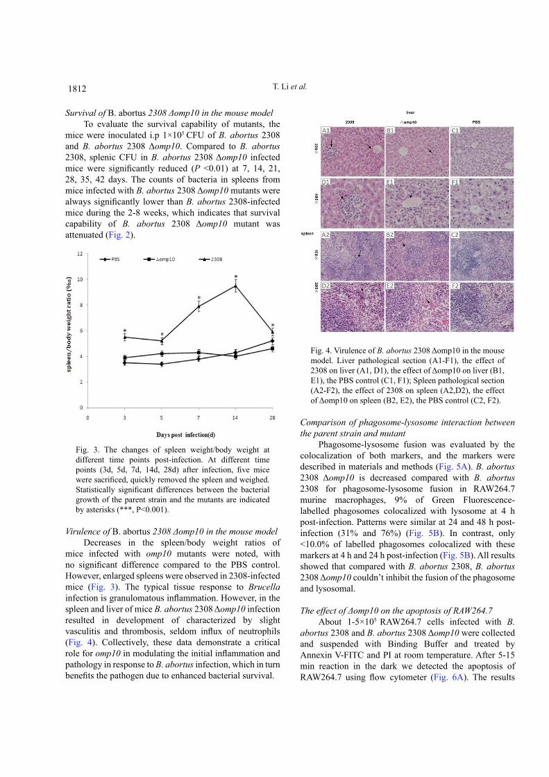
mice infected with omp10 mutants were noted, with no significant difference compared to the PBS control. However, enlarged spleens were observed in 2308-infected mice (Fig. 3). The typical tissue response to Brucella infection is granulomatous inflammation. However, in the spleen and liver of mice B. abortus 2308 Δomp10 infection resulted in development of characterized by slight vasculitis and thrombosis, seldom influx of neutrophils (Fig. 4). Collectively, these data demonstrate a critical role for omp10 in modulating the initial inflammation and pathology in response to B. abortus infection, which in turn benefits the pathogen due to enhanced bacterial survival.
Fig. 4. Virulence of B. abortus 2308 Δomp10 in the mouse model. Liver pathological section (A1-F1), the effect of 2308 on liver (A1, D1), the effect of Δomp10 on liver (B1, E1), the PBS control (C1, F1); Spleen pathological section (A2-F2), the effect of 2308 on spleen (A2,D2), the effect of Δomp10 on spleen (B2, E2), the PBS control (C2, F2).
Comparison of phagosome-lysosome interaction between the parent strain and mutant
Phagosome-lysosome fusion was evaluated by the colocalization of both markers, and the markers were described in materials and methods (Fig. 5A). B. abortus 2308 Δomp10 is decreased compared with B. abortus 2308 for phagosome-lysosome fusion in RAW264.7 murine macrophages, 9% of Green Fluorescence-labelled phagosomes colocalized with lysosome at 4 h post-infection. Patterns were similar at 24 and 48 h post-infection (31% and 76%) (Fig. 5B). In contrast, only <10.0% of labelled phagosomes colocalized with these markers at 4 h and 24 h post-infection (Fig. 5B). All results showed that compared with B. abortus 2308, B. abortus 2308 Δomp10 couldn’t inhibit the fusion of the phagosome and lysosomal.
The effect of Δomp10 on the apoptosis of RAW264.7About 1-5×10⁵ RAW264.7 cells infected with B.
abortus 2308 and B. abortus 2308 Δomp10 were collected and suspended with Binding Buffer and treated by Annexin V-FITC and PI at room temperature. After 5-15 min reaction in the dark we detected the apoptosis of RAW264.7 using flow cytometer (Fig. 6A). The results
T. Li et al.
1813 Omp10 Deletion Mutant of Brucella abortus 1813
showed that B. abortus 2308 Δomp10 compared with B. abortus 2308 couldn’t inhibit cell apoptosis (Fig. 6B),
indicating that the cells apoptosis was increased in B. abortus 2308 Δomp10 groups.
Fig. 5. Interaction of B. abortus 2308- and mutants-containing phagosomes with lysosomes in RAW264.7 macrophages. B. abortus were labeled with sheep anti-B. abortus IgG antibodies and Rhodamine (TRITC) conjugated AffiniPure donkey anti-sheep IgG antibody, lysosomes were labeled with Cell NavigatorTM Lysosomes Staining Kit Green Fluorescence. A, cells were fixed at different time points after infection. Confocal images of cells containing B. abortus were obtained at 4 h post-infection; B percentage of phagosome-lysosome fusion at different time points after infection. Fusion was evaluated by the colocalization of markers, Green fluorescein and TRD. To determine the percentage of fusion, bacteria were analyzed at each time point.
Fig. 6. Flow cytometric analysis of RAW264.7 macrophages exposed to B. abortus 2308 and omp10 mutants. RAW264.7 macrophages were suspended with Binding Buffer and treated by Annexin V-FITC and PI at room temperature. A, cells were collected at different time points after infection, cell apoptosis images containing B. abortus. B, percentage of apoptosis at different time points after infection.
1814 T. Li et al.
DISCUSSION
Omps of Gram-negative bacteria, play an important role in bacterial pathogenesis (Krinos et al. 1999). Brucella Omps are essential for antibiotic resistance, virulence regulation, and other unknown functions in bacteria (Verstreate et al., 1982). Brucella Omp10 belongs to lipoprotein (Tibor et al., 2002), which is one of the most abundant proteins in the outer membrane of Brucella, has been shown to play key roles in nutrient uptake, signal transduction, adhesion and antibiotic resistance (Kim et al., 2012).
We constructed the deletion mutant of omp10, which showed that the intracellular viability of the omp10 mutants in RAW264.7 cells were decreased compared to that of the parent strains. Surface properties changed by Omp10 may influence the intracellular viability of bacteria.
B. abortus inhibited phagosome-lysosome fusion after uptake by macrophages, allowing survival and replication (Pizarro-Cerdá et al., 1998b; Weber et al., 2009). However, inhibition of phagosome-Lysosome fusion was strikingly impaired by omp10 mutants, suggesting that the lipoprotein contributes to intracellular trafficking of B. abortus within phagocytes. B. abortus could inhibit apoptosis of macrophages through the dependence of O side chain of polysaccharides after infection (Maria-Pilar et al., 2005). However, suppression of apoptosis was attenuated by omp10 mutants as shown in our study, since deletion of the omp10 may affect the Brucella outer membrane lipid, and influence the surface properties of the Brucella lipopolysaccharide (Tibor et al., 1999). We also found that survival of omp10 mutants in mouse model was attenuated, correlated with defective intracellular survival as assessed in RAW264.7 cell model. Indeed, lipoproteins are crucial for the survival of B. abortus in mice. Splenomegaly induced by B. abortus was also ameliorated by omp10 mutants at 2 weeks post-inoculation. These results, together with previous reports suggest that lipoprotein is the key mediator of the pro-inflammatory response (Giambartolomei et al., 2004), as well as vital factor needed to evade lysosome fusion to multiply inside the phagocytes (Kim et al., 2002; Naroeni et al., 2001; Naroeni and Porte, 2002; Porte et al., 2003; Starr et al., 2008). Other pathogenic bacteria are known to avoid fusion with lysosome as has been demonstrated with Mycobacterium avium and Mycobacterium tuberculosis (Armstrong and Hart, 1971). Legionella is another intracellular pathogen that is well adapted to life inside phagosome (Amer et al., 2006). Virulent strain 2308 possesses mechanisms to escape lysosome degradation (Pizarro-Cerdá et al., 1998b). In contrast, it is clear that the Δomp10 mutant can invade cells but multiplies poorly
because the bacteria degrad after the Brucella-containing phagosomes fuse with lysosome. The results obtained here suggest that Δomp10 would be a good vaccine against B. abortus (Moreno et al., 1981).
CONCLUSION
Collectively, the results from our study showed that Δomp10 mutant could be attributed to altered outer membrane properties which would influence its intracellular survival and pathogenicity. Further studies are necessary to establish Omp10 function in exploiting a good vaccine against B. abortus infection.
ACKNOWLEDGEMENTS
This work was supported by the National Science and Technology Major Project (2013DFR31110), and the National Natural Science Foundation of China (31572494, 31260596, 31460650, 31360610, 31402166).
Conflict of interest statementAuthors declare no conflict of interest.
REFERENCES
Amer, A., Franchi, L., Kanneganti, T.D., Body-Malapel, M., Özören, N., Brady, G., Meshinchi, S., Jagirdar, R., Gewirtz, A. and Akira, S., 2006. Regulation of legionella phagosome maturation and infection through flagellin and host Ipaf. J. biol. Chem., 281: 35217-35223. https://doi.org/10.1074/jbc.M604933200
Arellano-Reynoso, B., Lapaque, N., Salcedo, S., Briones, G., Ciocchini, A.E., Ugalde, R., Moreno, E., Moriyón, I. and Gorvel, J.P., 2005. Cyclic β-1, 2-glucan is a Brucella virulence factor required for intracellular survival. Nature Immunol., 6: 618-625. https://doi.org/10.1038/ni1202
Armstrong, J. and Hart, P.A., 1971. Response of cultured macrophages to Mycobacterium tuberculosis, with observations on fusion of lysosomes with phagosomes. J. exp. Med., 134: 713-740. https://doi.org/10.1084/jem.134.3.713
Boschiroli, M.L., Foulongne, V. and O’Callaghan, D., 2001. Brucellosis: a worldwide zoonosis. Curr. Opin. Microbiol., 4: 58-64. https://doi.org/10.1016/S1369-5274(00)00165-X
Caro-Hernández, P., Fernández-Lago, L., de Miguel, M.J., Martín-Martín, A.I., Cloeckaert, A., Grilló, M.J. and Vizcaíno, N., 2007. Role of the Omp25/Omp31 family in outer membrane properties and
1815 Omp10 Deletion Mutant of Brucella abortus 1815
virulence of Brucella ovis. Infect. Immun., 75: 4050-4061. https://doi.org/10.1128/IAI.00486-07
Estein, S.M., Cassataro, J., Vizcaíno, N., Zygmunt, M.S., Cloeckaert, A. and Bowden, R.A., 2003. The recombinant Omp31 from Brucella melitensis alone or associated with rough lipopolysaccharide induces protection against Brucella ovis infection in BALB/c mice. Microb. Infect., 5: 85-93. https://doi.org/10.1016/S1286-4579(02)00075-8
Giambartolomei, G.H., Zwerdling, A., Cassataro, J., Bruno, L., Fossati, C.A. and Philipp, M.T., 2004. Lipoproteins, not lipopolysaccharide, are the key mediators of the proinflammatory response elicited by heat-killed Brucella abortus. J. Immunol., 173: 4635-4642. https://doi.org/10.4049/jimmunol.173.7.4635
Gorvel, J.P. and Moreno, E., 2002. Brucella intracellular life: from invasion to intracellular replication. Vet. Microbiol., 90: 281-297. https://doi.org/10.1016/S0378-1135(02)00214-6
Jubier-Maurin, V., Boigegrain, R.A., Cloeckaert, A., Gross, A., Alvarez-Martinez, M.T., Terraza, A., Liautard, J., Köhler, S., Rouot, B. and Dornand, J., 2001. Major outer membrane protein Omp25 of Brucella suis is involved in inhibition of tumor necrosis factor alpha production during infection of human macrophages. Infect. Immun., 69: 4823-4830. https://doi.org/10.1128/IAI.69.8.4823-4830.2001
Kim, D.H., Lim, J.J., Lee, J.J., Kim, D.G., Lee, H.J., Min, W., Kim, K.D., Chang, H.H., Rhee, M.H. and Watarai, M., 2012. Identification of genes contributing to the intracellular replication of Brucella abortus within HeLa and RAW 264.7 cells. Vet. Microbiol., 158: 322-328. https://doi.org/10.1016/j.vetmic.2012.02.019
Kim, S., Watarai, M., Makino, S.I. and Shirahata, T., 2002. Membrane sorting during swimming internalization of Brucella is required for phagosome trafficking decisions. Microb. Pathog., 33: 225-237. https://doi.org/10.1006/mpat.2002.0531
Krinos, C., High, A. and Rodgers, F., 1999. Role of the 25 kDa major outer membrane protein of Legionella pneumophila in attachment to U-937 cells and its potential as a virulence factor for chick embryos. J. appl. Microbiol., 86: 237-244. https://doi.org/10.1046/j.1365-2672.1999.00667.x
Maria-Pilar, J.D.B., Dudal, S., Dornand, J. and Gross, A., 2005. Cellular bioterrorism: how Brucella corrupts macrophage physiology to promote invasion and proliferation. Clin. Immunol., 114: 227-238. https://doi.org/10.1016/j.clim.2004.07.010
Mobasheri, H., Ficht, T., Marquis, H., Lea, E. and Lakey, J., 1997. Brucella Omp2a and Omp2b porins: single channel measurements and topology prediction. FEMS Microbiol. Lett., 155: 23-30. https://doi.org/10.1111/j.1574-6968.1997.tb12681.x
Moreno, E., Speth, S., Jones, L.M. and Berman, D.T., 1981. Immunochemical characterization of Brucella lipopolysaccharides and polysaccharides. Infect. Immun., 31: 214-222.
Moriyón, I. and López-Goñi, I., 2010. Structure and properties of the outer membranes of Brucella abortus and Brucella melitensis. Int. Microbiol., 1: 19-26.
Naroeni, A., Jouy, N., Ouahrani-Bettache, S., Liautard, J.P. and Porte, F., 2001. Brucella suis-impaired specific recognition of phagosomes by lysosomes due to phagosomal membrane modifications. Infect. Immun., 69: 486-493. https://doi.org/10.1128/IAI.69.1.486-493.2001
Naroeni, A. and Porte, F., 2002. Role of cholesterol and the ganglioside GM1 in entry and short-term survival of Brucella suis in murine macrophages. Infect. Immun., 70: 1640-1644. https://doi.org/10.1128/IAI.70.3.1640-1644.2002
Olsen, S.C. and Stoffregen, W., 2005. Essential role of vaccines in brucellosis control and eradication programs for livestock. Expert Rev. Vaccines, 4: 915-928. https://doi.org/10.1586/14760584.4.6.915
Pasquevich, K.A., Estein, S.M., Samartino, C.G., Zwerdling, A., Coria, L.M., Barrionuevo, P., Fossati, C.A., Giambartolomei, G.H. and Cassataro, J., 2009. Immunization with recombinant Brucella species outer membrane protein Omp16 or Omp19 in adjuvant induces specific CD4+ and CD8+ T cells as well as systemic and oral protection against Brucella abortus infection. Infect. Immun., 77: 436-445. https://doi.org/10.1128/IAI.00123-09
Pizarro-Cerdá, J., Méresse, S., Parton, R.G., van der Goot, G., Sola-Landa, A., Lopez-Goñi, I., Moreno, E. and Gorvel, J.P., 1998a. Brucella abortus transits through the autophagic pathway and replicates in the endoplasmic reticulum of nonprofessional phagocytes. Infect. Immun., 66: 5711-5724.
Pizarro-Cerdá, J., Moreno, E., Sanguedolce, V., Mege, J.L. and Gorvel, J.P., 1998b. Virulent Brucella abortus prevents lysosome fusion and is distributed within autophagosome-like compartments. Infect. Immun., 66: 2387-2392.
Porte, F., Naroeni, A., Ouahrani-Bettache, S. and Liautard, J.P., 2003. Role of the Brucella suis lipopolysaccharide O antigen in phagosomal genesis and in inhibition of phagosome-lysosome
1816 T. Li et al.
fusion in murine macrophages. Infect. Immun., 71: 1481-1490. https://doi.org/10.1128/IAI.71.3.1481-1490.2003
Seleem, M.N., Boyle, S.M. and Sriranganathan, N., 2010. Brucellosis: a re-emerging zoonosis. Vet. Microbiol., 140: 392-398. https://doi.org/10.1016/j.vetmic.2009.06.021
Starr, T., Ng, T.W., Wehrly, T.D., Knodler, L.A. and Celli, J., 2008. Brucella intracellular replication requires trafficking through the late endosomal/lysosomal compartment. Traffic, 9: 678-694. https://doi.org/10.1111/j.1600-0854.2008.00718.x
Tibor, A., Decelle, B. and Letesson, J.J., 1999. Outer membrane proteins Omp10, Omp16, and Omp19 of Brucella spp. are lipoproteins. Infect. Immun., 67: 4960-4962.
Tibor, A., Wansard, V., Bielartz, V., Delrue, R.M., Danese, I., Michel, P., Walravens, K., Godfroid, J. and Letesson, J.J., 2002. Effect of omp10 or omp19 deletion on Brucella abortus outer membrane properties and virulence in mice. Infect. Immun., 70: 5540-5546. https://doi.org/10.1128/IAI.70.10.5540-5546.2002
Verstreate, D., Creasy, M., Caveney, N., Baldwin, C., Blab, M. and Winter, A., 1982. Outer membrane proteins of Brucella abortus: isolation and characterization. Infect. Immun., 35: 979-989.
Weber, S.S., Ragaz, C. and Hilbi, H., 2009. Pathogen trafficking pathways and host phosphoinositide metabolism. Mol. Microbiol., 71: 1341-1352. https://doi.org/10.1111/j.1365-2958.2009.06608.x


![INGLES- RICARDO DAVID Vol. 3 Pamphlets and Papers 1809-1811 [1809].pdf](https://img.pdfslide.us/doc/110x75/577cdadc1a28ab9e78a6bace/ingles-ricardo-david-vol-3-pamphlets-and-papers-1809-1811-1809pdf.jpg)