Embed Size (px)
Citation preview
www.caymanchem.comCustomer Service 800.364.9897Technical Support 888.526.53511180 E. Ellsworth Rd · Ann Arbor, MI · USA
PAD4 (human) ELISA Kit
Item No. 501460
3GENERAL INFORMATION
TABLE OF CONTENTS GENERAL INFORMATION 3 Materials Supplied
3 Safety Data
4 Precautions
4 If You Have Problems
4 Storage and Stability
4 Materials Needed but Not Supplied
INTRODUCTION 5 Background
5 About This Assay
6 Principle of the Assay
7 Definition of Key Terms
PRE-ASSAY PREPARATION 8 Buffer Preparation
8 Sample Preparation
9 Sample Matrix Properties
ASSAY PROTOCOL 14 Preparation of Assay-Specific Reagents
16 Plate Set Up
17 Performing the Assay
ANALYSIS 19 Calculations
20 Performance Characteristics
RESOURCES 23 Troubleshooting
25 Plate Template
26 References
27 Notes
27 Warranty and Limitation of Remedy
GENERAL INFORMATION
Materials Supplied
Item Number Item 96 wells Quantity/Size
401462 Anti-PAD4 (human) ELISA Strip Plate 1 plate
401461 Anti-PAD4 (human) HRP Conjugate 1 vial/1.5 ml
401464 PAD4 (human) ELISA Standard 2 vials
400054 Immunoassay Buffer B Concentrate (10X) 1 vial/10 ml
400062 Wash Buffer Concentrate (400X) 1 vial/5 ml
400035 Polysorbate 20 1 vial/3 ml
400074 TMB Substrate Solution 1 vial/12 ml
10011355 HRP Stop Solution 1 vial/12 ml
400012 96-Well Cover Sheet 1 cover
If any of the items listed above are damaged or missing, please contact our Customer Service department at (800) 364-9897 or (734) 971-3335. We cannot accept any returns without prior authorization.
! WARNING: THIS PRODUCT IS FOR RESEARCH ONLY - NOT FORHUMAN OR VETERINARY DIAGNOSTIC OR THERAPEUTIC USE.
Safety DataThis material should be considered hazardous until further information becomes available. Do not ingest, inhale, get in eyes, on skin, or on clothing. Wash thoroughly after handling. Before use, the user must review the complete Safety Data Sheet, which has been sent via email to your institution.
5INTRODUCTION4 GENERAL INFORMATION
PrecautionsPlease read these instructions carefully before beginning this assay.The reagents in this kit have been tested and formulated to work exclusively with Cayman’s PAD4 (human) ELISA Kit. This kit may not perform as described if any reagent or procedure is replaced or modified. The Stop Solution provided with this kit is an acid solution. Please wear appropriate personal protection equipment (e.g., safety glasses, gloves, and lab-coat) when using this material.
If You Have ProblemsTechnical Service Contact Information
Phone: 888-526-5351 (USA and Canada only) or 734-975-3888Fax: 734-971-3640Email: [email protected]
In order for our staff to assist you quickly and efficiently, please be ready to supply the batch number of the kit (found on the outside of the box).
Storage and StabilityThis kit will perform as specified if stored at -20°C and used before the expiration date indicated on the outside of the box.
Materials Needed But Not Supplied1. A plate reader capable of measuring absorbance at 450 nm.2. Adjustable pipettes and a repeating pipettor.3. An orbital microplate shaker.4. A source of pure water; glass distilled or HPLC-grade water is
acceptable. NOTE: Ultra Pure water is available for purchase from Cayman (Item No. 400000)
INTRODUCTIONBackground Protein arginine deiminase 4 (PAD4) catalyzes the conversion of arginine residues to citrulline within cellular protein substrates, resulting in the loss of a positive charge, which can alter protein structure and/or function.1 It is expressed in neutrophils, as well as a variety of tissues, including the brain, liver, lung, and kidney.1-3 PAD4 has a key role in NETosis, a lytic form of cell death characterized by the release of neutrophil extracellular traps (NETs).1 Upon neutrophil activation, PAD4 translocates to the nucleus where it citrullinates histones, initiating chromatin decondensation and the release of NETs.2,4,5 Neutrophils isolated from Pad4-/- mice exhibit decreased citrullination of histone H3 under both basal and LPS-stimulated conditions and are defective for NET formation in response to stimulation with LPS, phorbol 12-myristate 13-acetate (PMA), or hydrogen peroxide.4 Citrullination of PAD4 targets contributes to disease progression and the generation of autoantibodies in patients with autoimmune diseases, including rheumatoid arthritis, Hashimoto’s encephalopathy, and multiple sclerosis.6-8 Additionally, PAD4 autoantibodies isolated from the serum of patients with rheumatoid arthritis are associated with disease severity.9
About This AssayCayman’s PAD4 (human) ELISA Kit is a sandwich assay that can be used to measure PAD4 in tissue culture medium, cell lysates, plasma, and serum. The standard curve spans the range of 0.16-10 ng/ml. NOTE: Rheumatoid factor in samples can give false positives and therefore should be removed from all samples before testing in the assay.8
6 INTRODUCTION 7INTRODUCTION
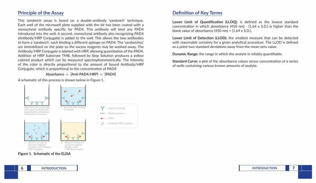
Principle of the AssayThis sandwich assay is based on a double-antibody ‘sandwich’ technique. Each well of the microwell plate supplied with the kit has been coated with a monoclonal antibody specific for PAD4. This antibody will bind any PAD4 introduced into the well. A second, monoclonal antibody also recognizing PAD4 (Antibody/HRP Conjugate) is added to the well. This allows the two antibodies to form a ‘sandwich’, each binding a different epitope on PAD4. The ‘sandwiches’ are immobilized on the plate so the excess reagents may be washed away. The Antibody/HRP Conjugate is labeled with HRP, allowing quantitation of the PAD4. Addition of HRP Substrate TMB, followed by Stop Solution produces a yellow colored product which can be measured spectrophotometrically. The intensity of the color is directly proportional to the amount of bound Antibody/HRP Conjugate, which is proportional to the concentration of PAD4.
Absorbance ∝ [Anti-PAD4/HRP] ∝ [PAD4]A schematic of this process is shown below in Figure 1.
Plates are pre-coated withthe capture an body andblocked with a proprietaryformula on of proteins.
2. Wash to remove all unbound reagents. Incubate with An body/ HRP Conjugate.
3. Wash to remove all unbound reagents. Develop with TMB/Stop Solu on.
1. Incubate with standard or sample. = Capture An�body
= Blocking proteins
= PAD4
= An�body/HRP Conjugate
Figure 1. Schematic of the ELISA
Definition of Key Terms
Lower Limit of Quantification (LLOQ): is defined as the lowest standard concentration in which absorbance (450 nm) - (1.64 x S.D.) is higher than the blank value of absorbance (450 nm) + (1.64 x S.D.).
Lower Limit of Detection (LLOD): the smallest measure that can be detected with reasonable certainty for a given analytical procedure. The LLOD is defined as a point two standard deviations away from the mean zero value.
Dynamic Range: the range in which the analyte is reliably quantifiable.
Standard Curve: a plot of the absorbance values versus concentration of a series of wells containing various known amounts of analyte.
8 PRE-ASSAY PREPARATION 9PRE-ASSAY PREPARATION
Sample Matrix Properties
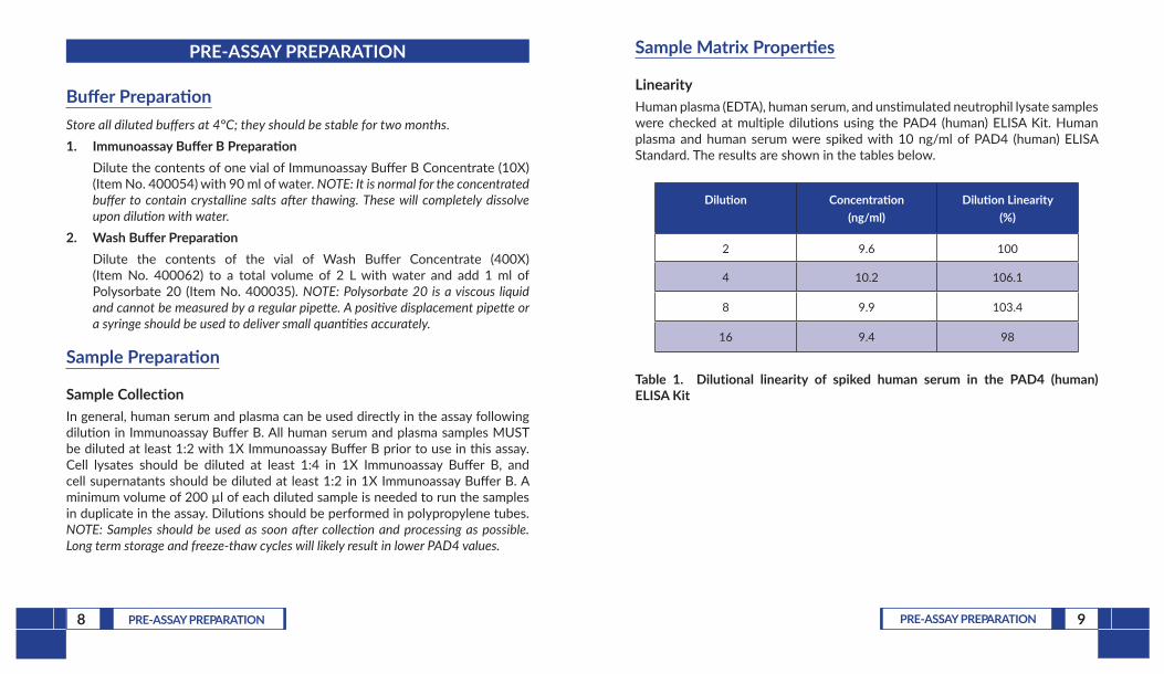
LinearityHuman plasma (EDTA), human serum, and unstimulated neutrophil lysate samples were checked at multiple dilutions using the PAD4 (human) ELISA Kit. Human plasma and human serum were spiked with 10 ng/ml of PAD4 (human) ELISA Standard. The results are shown in the tables below.
Dilution Concentration (ng/ml)
Dilution Linearity (%)
2 9.6 100
4 10.2 106.1
8 9.9 103.4
16 9.4 98
Table 1. Dilutional linearity of spiked human serum in the PAD4 (human) ELISA Kit
PRE-ASSAY PREPARATION
Buffer PreparationStore all diluted buffers at 4°C; they should be stable for two months.1. Immunoassay Buffer B Preparation
Dilute the contents of one vial of Immunoassay Buffer B Concentrate (10X) (Item No. 400054) with 90 ml of water. NOTE: It is normal for the concentrated buffer to contain crystalline salts after thawing. These will completely dissolve upon dilution with water.
2. Wash Buffer PreparationDilute the contents of the vial of Wash Buffer Concentrate (400X) (Item No. 400062) to a total volume of 2 L with water and add 1 ml of Polysorbate 20 (Item No. 400035). NOTE: Polysorbate 20 is a viscous liquid and cannot be measured by a regular pipette. A positive displacement pipette or a syringe should be used to deliver small quantities accurately.
Sample Preparation
Sample CollectionIn general, human serum and plasma can be used directly in the assay following dilution in Immunoassay Buffer B. All human serum and plasma samples MUST be diluted at least 1:2 with 1X Immunoassay Buffer B prior to use in this assay. Cell lysates should be diluted at least 1:4 in 1X Immunoassay Buffer B, and cell supernatants should be diluted at least 1:2 in 1X Immunoassay Buffer B. A minimum volume of 200 µl of each diluted sample is needed to run the samples in duplicate in the assay. Dilutions should be performed in polypropylene tubes. NOTE: Samples should be used as soon after collection and processing as possible. Long term storage and freeze-thaw cycles will likely result in lower PAD4 values.
10 PRE-ASSAY PREPARATION 11PRE-ASSAY PREPARATION
Dilution Concentration (ng/ml)
Dilution Linearity (%)
2 10.3 100
4 10.9 105.4
8 11.2 108.5
16 9.9 95.9
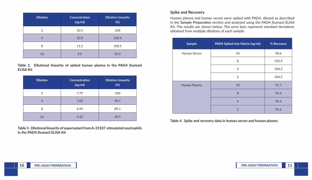
Table 2. Dilutional linearity of spiked human plasma in the PAD4 (human) ELISA Kit
Dilution Concentration (ng/ml)
Dilution Linearity (%)
2 7.79 100
4 7.02 90.1
8 6.94 89.1
16 5.42 69.5
Table 3. Dilutional linearity of supernatant from A-23187-stimulated neutrophils in the PAD4 (human) ELISA Kit
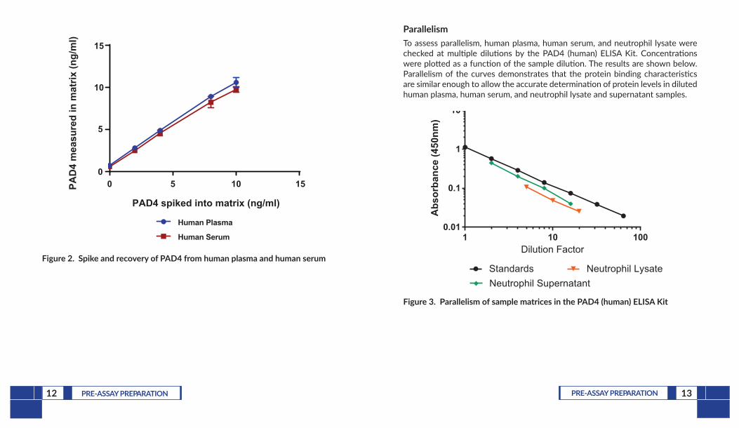
Spike and RecoveryHuman plasma and human serum were spiked with PAD4, diluted as described in the Sample Preparation section and analyzed using the PAD4 (human) ELISA Kit. The results are shown below. The error bars represent standard deviations obtained from multiple dilutions of each sample.
Sample PAD4 Spiked into Matrix (ng/ml) % Recovery
Human Serum 10 98.6
8 102.4
4 104.2
2 104.2
Human Plasma 10 91.7
8 95.4
4 99.4
2 95.6
Table 4. Spike and recovery data in human serum and human plasma
12 PRE-ASSAY PREPARATION 13PRE-ASSAY PREPARATION
Human Plasma
Human Serum
PAD
4 m
easu
red
in m
atrix
(ng/
ml)
15
10
5
00 5 10 15
PAD4 spiked into matrix (ng/ml)
Figure 2. Spike and recovery of PAD4 from human plasma and human serum
ParallelismTo assess parallelism, human plasma, human serum, and neutrophil lysate were checked at multiple dilutions by the PAD4 (human) ELISA Kit. Concentrations were plotted as a function of the sample dilution. The results are shown below. Parallelism of the curves demonstrates that the protein binding characteristics are similar enough to allow the accurate determination of protein levels in diluted human plasma, human serum, and neutrophil lysate and supernatant samples.
1 10 1000.01
0.1
1
10
Dilution Factor
Abs
orba
nce
(450
nm)
Standards Neutrophil LysateNeutrophil Supernatant
Figure 3. Parallelism of sample matrices in the PAD4 (human) ELISA Kit
14 ASSAY PROTOCOL 15ASSAY PROTOCOL
ASSAY PROTOCOL
Preparation of Assay-Specific Reagents
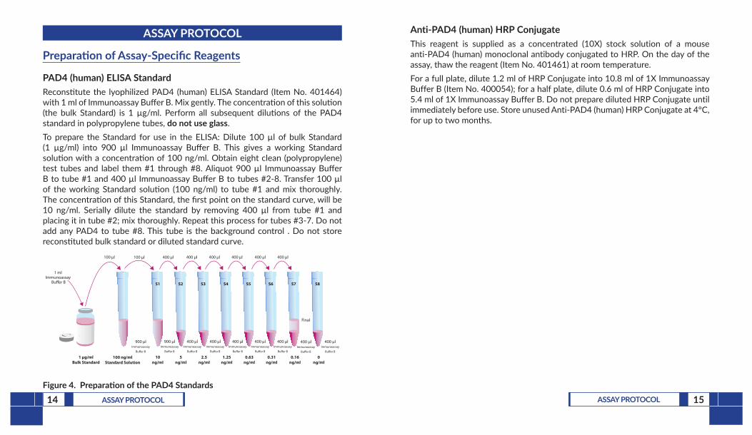
PAD4 (human) ELISA StandardReconstitute the lyophilized PAD4 (human) ELISA Standard (Item No. 401464) with 1 ml of Immunoassay Buffer B. Mix gently. The concentration of this solution (the bulk Standard) is 1 µg/ml. Perform all subsequent dilutions of the PAD4 standard in polypropylene tubes, do not use glass. To prepare the Standard for use in the ELISA: Dilute 100 µl of bulk Standard (1 µg/ml) into 900 µl Immunoassay Buffer B. This gives a working Standard solution with a concentration of 100 ng/ml. Obtain eight clean (polypropylene) test tubes and label them #1 through #8. Aliquot 900 µl Immunoassay Buffer B to tube #1 and 400 µl Immunoassay Buffer B to tubes #2-8. Transfer 100 µl of the working Standard solution (100 ng/ml) to tube #1 and mix thoroughly. The concentration of this Standard, the first point on the standard curve, will be 10 ng/ml. Serially dilute the standard by removing 400 µl from tube #1 and placing it in tube #2; mix thoroughly. Repeat this process for tubes #3-7. Do not add any PAD4 to tube #8. This tube is the background control . Do not store reconstituted bulk standard or diluted standard curve.
1 µg/mlBulk Standard
100 µl 400 µl 400 µl 400 µl 400 µl 400 µl
900 µlImmunoassay
Bu�er B
400 µlImmunoassay
Bu�er B
10ng/ml
S1 S2 S3 S4 S5 S6 S7 S8
5ng/ml
2.5ng/ml
1.25ng/ml
0.63ng/ml
0.31ng/ml
0.16ng/ml
0ng/ml
400 µlImmunoassay
Bu�er B
400 µlImmunoassay
Bu�er B
400 µlImmunoassay
Bu�er B
400 µlImmunoassay
Bu�er B
400 µlImmunoassay
Bu�er B
400 µlImmunoassay
Bu�er B
100 µl
900 µlImmunoassay
Bu�er B
100 ng/mlStandard Solution
400 µl
1 ml Immunoassay
Bu�er B
Final
Figure 4. Preparation of the PAD4 Standards
Anti-PAD4 (human) HRP ConjugateThis reagent is supplied as a concentrated (10X) stock solution of a mouse anti-PAD4 (human) monoclonal antibody conjugated to HRP. On the day of the assay, thaw the reagent (Item No. 401461) at room temperature.For a full plate, dilute 1.2 ml of HRP Conjugate into 10.8 ml of 1X Immunoassay Buffer B (Item No. 400054); for a half plate, dilute 0.6 ml of HRP Conjugate into 5.4 ml of 1X Immunoassay Buffer B. Do not prepare diluted HRP Conjugate until immediately before use. Store unused Anti-PAD4 (human) HRP Conjugate at 4°C, for up to two months.
16 ASSAY PROTOCOL 17ASSAY PROTOCOL
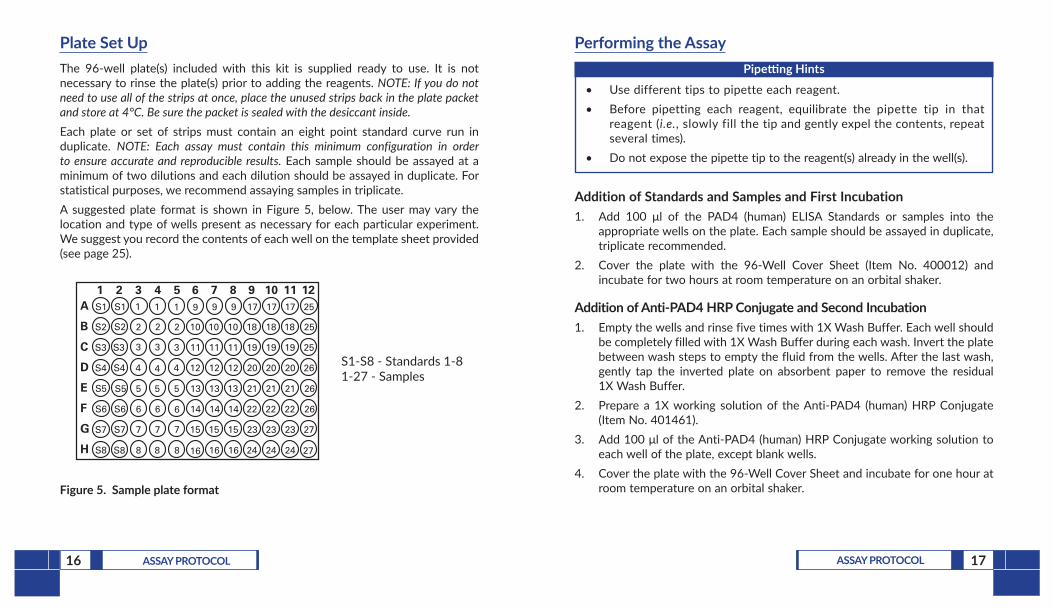
Plate Set UpThe 96-well plate(s) included with this kit is supplied ready to use. It is not necessary to rinse the plate(s) prior to adding the reagents. NOTE: If you do not need to use all of the strips at once, place the unused strips back in the plate packet and store at 4°C. Be sure the packet is sealed with the desiccant inside. Each plate or set of strips must contain an eight point standard curve run in duplicate. NOTE: Each assay must contain this minimum configuration in order to ensure accurate and reproducible results. Each sample should be assayed at a minimum of two dilutions and each dilution should be assayed in duplicate. For statistical purposes, we recommend assaying samples in triplicate.A suggested plate format is shown in Figure 5, below. The user may vary the location and type of wells present as necessary for each particular experiment. We suggest you record the contents of each well on the template sheet provided (see page 25).
S1-S8 - Standards 1-81-27 - Samples
A
B
C
D
E
F
G
H
1 2 3 4 5 6 7 8 9 10 11 12S1
S2
S3
S4
S5
S6
S7
S8 8
7
6
5
4
3
2
1
8
7
6
5
4
3
2
1
8
7
6
5
4
3
2
1
16
15
14
13
12
11
10
9
16
15
14
13
12
11
10
9
16
15
14
13
12
11
10
9
24
23
22
21
20
19
18
17
24
23
22
21
20
19
18
17
24
23
22
21
20
19
18
17 25
27
27
26
26
26
25
25
S1
S2
S3
S4
S5
S6
S7
S8
Figure 5. Sample plate format
Performing the Assay Pipetting Hints
• Use different tips to pipette each reagent.• Before pipetting each reagent, equilibrate the pipette tip in that
reagent (i.e., slowly fill the tip and gently expel the contents, repeat several times).
• Do not expose the pipette tip to the reagent(s) already in the well(s).
Addition of Standards and Samples and First Incubation1. Add 100 μl of the PAD4 (human) ELISA Standards or samples into the
appropriate wells on the plate. Each sample should be assayed in duplicate, triplicate recommended.
2. Cover the plate with the 96-Well Cover Sheet (Item No. 400012) and incubate for two hours at room temperature on an orbital shaker.
Addition of Anti-PAD4 HRP Conjugate and Second Incubation1. Empty the wells and rinse five times with 1X Wash Buffer. Each well should
be completely filled with 1X Wash Buffer during each wash. Invert the plate between wash steps to empty the fluid from the wells. After the last wash, gently tap the inverted plate on absorbent paper to remove the residual 1X Wash Buffer.
2. Prepare a 1X working solution of the Anti-PAD4 (human) HRP Conjugate (Item No. 401461).
3. Add 100 μl of the Anti-PAD4 (human) HRP Conjugate working solution to each well of the plate, except blank wells.
4. Cover the plate with the 96-Well Cover Sheet and incubate for one hour at room temperature on an orbital shaker.
19ANALYSIS18 ASSAY PROTOCOL
Development of the Plate1. Empty the wells and rinse five times with 1X Wash Buffer. Each well should
be completely filled with 1X Wash Buffer during each wash. Invert the plate between wash steps to empty the fluid from the wells. After the last wash, gently tap the inverted plate on absorbent paper to remove the residual 1X Wash Buffer.
2. Add 100 μl of TMB Substrate Solution (Item No. 400074) to each well of the plate.
3. Cover the plate with the 96-Well Cover Sheet and incubate for 30 minutes at room temperature on an orbital shaker.
4. DO NOT WASH THE PLATE. Add 100 μl of HRP Stop Solution (Item No. 10011355) to each well of the plate. Blue wells should turn yellow. NOTE: The stop solution in this kit contains an acid. Wear appropriate protection and use caution when handling this solution.
Reading the Plate1. Wipe the bottom of the plate with a clean tissue to remove fingerprints,
dirt, etc.2. Read the plate at a wavelength of 450 nm.
ANALYSISMany plate readers come with data reduction software that plots data automatically. If your plate reader is capable of analyzing the data, we recommend using a linear fit. Alternatively, a spreadsheet program can be used.
Calculations
Plotting the Standard Curve and Determining the Sample ConcentrationUsing computer reduction software, plot absorbance (linear y-axis) versus concentration (linear x-axis) for standards (S1-S8) and fit the data with a linear fit, or alternatively a four-parameter logistic equation. Using the equation of the line, calculate the concentration of PAD4 in each sample, making sure to correct for any sample dilution.
20 ANALYSIS 21ANALYSIS
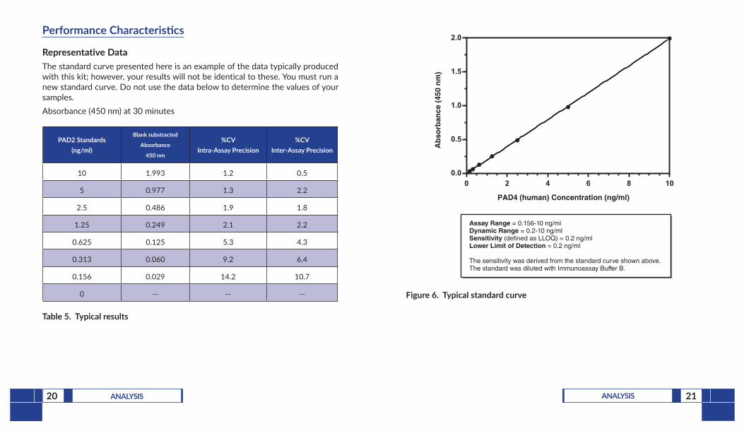
Performance Characteristics
Representative DataThe standard curve presented here is an example of the data typically produced with this kit; however, your results will not be identical to these. You must run a new standard curve. Do not use the data below to determine the values of your samples. Absorbance (450 nm) at 30 minutes
PAD2 Standards (ng/ml)
Blank substracted
Absorbance
450 nm
%CV Intra-Assay Precision
%CV Inter-Assay Precision
10 1.993 1.2 0.5
5 0.977 1.3 2.2
2.5 0.486 1.9 1.8
1.25 0.249 2.1 2.2
0.625 0.125 5.3 4.3
0.313 0.060 9.2 6.4
0.156 0.029 14.2 10.7
0 -- -- --
Table 5. Typical results
0 2 4 6 8 100.0
0.5
1.0
1.5
2.0
PAD4 (human) Concentration (ng/ml)
Assay Range = 0.156-10 ng/mlDynamic Range = 0.2-10 ng/mlSensitivity (defined as LLOQ) = 0.2 ng/mlLower Limit of Detection = 0.2 ng/ml
The sensitivity was derived from the standard curve shown above. The standard was diluted with Immunoassay Buffer B.
Ab
sorb
ance
(45
0 n
m)
Figure 6. Typical standard curve
23RESOURCES22 ANALYSIS
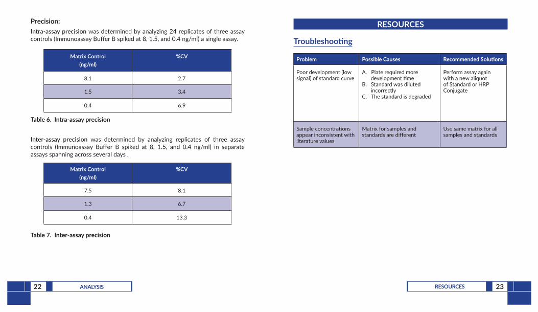
Precision:Intra-assay precision was determined by analyzing 24 replicates of three assay controls (Immunoassay Buffer B spiked at 8, 1.5, and 0.4 ng/ml) a single assay.
Matrix Control (ng/ml)
%CV
8.1 2.7
1.5 3.4
0.4 6.9
Table 6. Intra-assay precision
Inter-assay precision was determined by analyzing replicates of three assay controls (Immunoassay Buffer B spiked at 8, 1.5, and 0.4 ng/ml) in separate assays spanning across several days .
Matrix Control (ng/ml)
%CV
7.5 8.1
1.3 6.7
0.4 13.3
Table 7. Inter-assay precision
RESOURCES
Troubleshooting
Problem Possible Causes Recommended Solutions
Poor development (low signal) of standard curve
A. Plate required more development time
B. Standard was diluted incorrectly
C. The standard is degraded
Perform assay again with a new aliquot of Standard or HRP Conjugate
Sample concentrations appear inconsistent with literature values
Matrix for samples and standards are different
Use same matrix for all samples and standards
24 RESOURCES 25RESOURCES
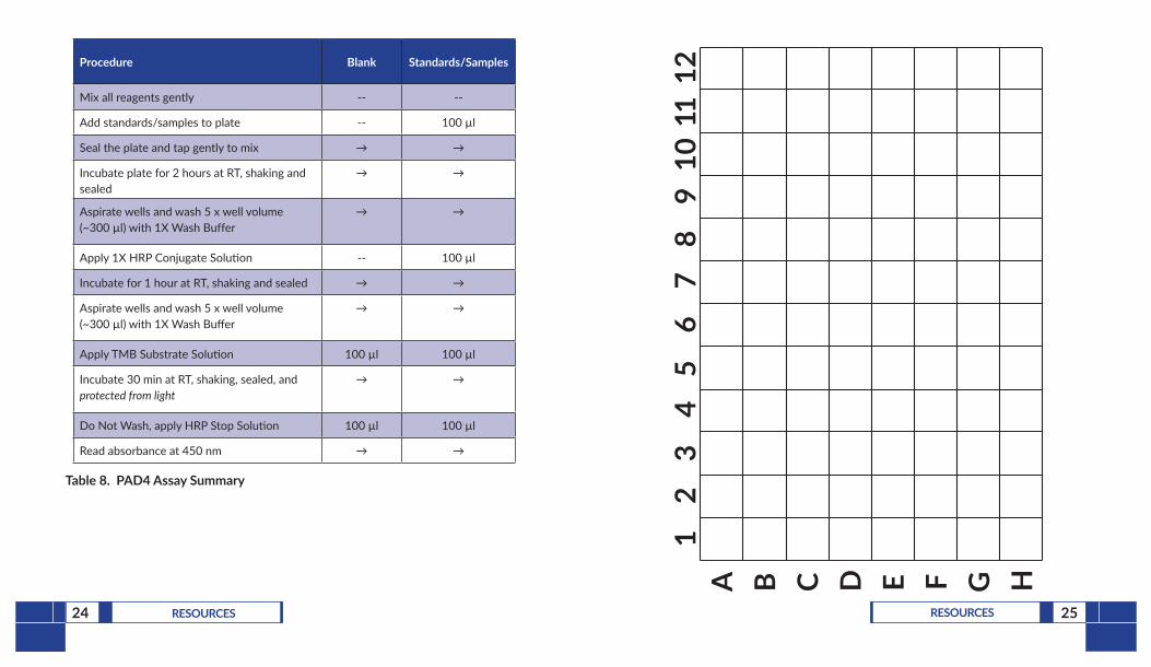
Procedure Blank Standards/Samples
Mix all reagents gently -- --
Add standards/samples to plate -- 100 µl
Seal the plate and tap gently to mix → →
Incubate plate for 2 hours at RT, shaking and sealed
→ →
Aspirate wells and wash 5 x well volume (~300 µl) with 1X Wash Buffer
→ →
Apply 1X HRP Conjugate Solution -- 100 µl
Incubate for 1 hour at RT, shaking and sealed → →
Aspirate wells and wash 5 x well volume (~300 µl) with 1X Wash Buffer
→ →
Apply TMB Substrate Solution 100 µl 100 µl
Incubate 30 min at RT, shaking, sealed, and protected from light
→ →
Do Not Wash, apply HRP Stop Solution 100 µl 100 µl
Read absorbance at 450 nm → →
Table 8. PAD4 Assay Summary
A B C D E F G H
12
34
56
78
910
1112
26 RESOURCES 27RESOURCES
References1. van Beers, J.J.B.C., Zendman, A.J.W., Raijmakers, R., et al. Peptidylarginine
deiminase expression and activity in PAD2 knock-out and PAD4-low mice. Biochimie 95(2), 299-308 (2013).
2. Demers, M., Wong, S.L., Martinod, K., et al. Priming of neutrophils toward NETosis promotes tumor growth. Oncoimmunology 5(5), e1134073 (2016).
3. Jones, J., Causey, C., Knuckley, B., et al. Protein arginine deiminase 4 (PAD4): Current understanding and future therapeutic potential. Curr. Opin. Drug Discov. Devel. 12(5), 616-627 (2009).
4. Li, P., Li, M., Lindberg, M.R., et al. PAD4 is essential for antibacterial innate immunity mediated by neutrophil extracellular traps. J. Exp. Med. 207(9), 1853-1862 (2010).
5. Thiam, H.R., Wong, S.L., Qiu, R., et al. NETosis proceeds by cytoskeleton and endomembrane disassembly and PAD4-mediated chromatin decondensation and nuclear envelope rupture. Proc. Natl. Acad. Sci. USA 117(13), 7326-7337 (2020).
6. Mastronardi, F.G. and Moscarello, M.A. Molecules affecting myelin stability: A novel hypothesis regarding the pathogenesis of multiple sclerosis. J. Neurosci. Res. 80(3), 301-308 (2005).
7. Arriza, J.L., Fairman, W.A., Wadiche, J.I., et al. Functional comparisons of three glutamate transporter subtypes cloned from human motor cortex. J. Neurosci. 14(9), 5559-5569 (1994).
8. Lundberg, K., Kinloch, A., Fisher, B.A., et al. Antibodies to citrullinated α-enolase peptide 1 are specific for rheumatoid arthritis and cross-react with bacterial enolase. Arthritis Rheum. 58(10), 3009-3019 (2008).
9. Halvorsen, E. H., Pollmann, S., Gilboe, I. M., et al. Serum IgG antibodies to peptidylarginine deiminase 4 in rheumatoid arthritis and associations with disease severity. Ann. Rheum. Dis. 67(3), 414-417 (2008).
NOTES
Warranty and Limitation of RemedyBuyer agrees to purchase the material subject to Cayman’s Terms and Conditions.Complete Terms and Conditions including Warranty and Limitation of Liability information can be found on our website.This document is copyrighted. All rights are reserved. This document may not, in whole or part, be copied, photocopied, reproduced, translated, or reduced to any electronic medium or machine-readable form without prior consent, in writing, from Cayman Chemical Company.©02/03/2021, Cayman Chemical Company, Ann Arbor, MI, All rights reserved. Printed in U.S.A.