Embed Size (px)
Citation preview

Cell Metabolism, Volume 15
Supplemental Information
p53-Induced Adipose Tissue Inflammation
Is Critically Involved in the Development
of Insulin Resistance in Heart Failure Ippei Shimizu, Yohko Yoshida, Taro Katsuno, Kaoru Tateno, Sho Okada, Junji
Moriya, Masataka Yokoyama, Aika Nojima, Takashi Ito, Rudolf Zechner, Issei
Komuro, Yoshio Kobayashi, and Tohru Minamino

Supplemental Experimental Procedures
CT Scan Analysis
The adiposity of mice was examined radiographically using CT (LaTheta, ALOCA)
according to the manufacturer’s protocol. We performed CT scanning at 2-mm intervals
from diaphragm to the bottom of abdominal cavity. Fat mass was evaluated as visceral fat /
body weight.
Western Blot Analysis
Whole tissue lysates were prepared in lysis buffer (10 mM Tris-HCl, pH 8, 140 mM NaCl,
5 mM EDTA, 0.025% NaN3, 1% Triton X-100, 1% deoxycholate, 0.1% SDS, 1 mM PMSF,
5 μg/ml leupeptin, 2 μg/ml aprotinin, 50 mM NaF, and 1 mM Na2VO3). The lysates
(20–30 μg) were resolved by SDS-polyacrylamide gel electrophoresis (PAGE). Proteins
were transferred to a polyvinylidene difluoride (PVDF) membrane (Millipore, Bedford,
MA), which was incubated with the primary antibody followed by anti-rabbit or anti-mouse
immunoglobulin-G conjugated with horseradish peroxidase (Jackson, West Grove, PA).
The primary antibodies used for Western blotting were as follows: anti-p53 antibody
(1C12) (Cell signaling) for mice, anti-p53 antibody (DO-1) (Santa Cruz) and
anti-phospho-p53 antibody (Ser15, Cell signaling) for human, and anti-actin antibody (Cell
signaling). For detection of mouse p53, we performed systemic perfusion with saline to
avoid contamination by immunoglobulin when harvesting the samples, because this could
lead to detection of nonspecific 50–55 kD bands. We also utilized horseradish
peroxidase-conjugated anti-mouse immunoglobulin G (Jackson, West Grove, PA) that only
bound to the light chains as the secondary antibody. This allowed us to detect the specific
band for p53 without any nonspecific bands, as confirmed by blots of samples from
systemically p53-deficient mice (Figure S2A).

RNA Analysis
Total RNA (10μg) was isolated from adipose tissue with RNAZol-B (Molecular Research
Center). Real-time PCR was performed by using the Light Cycler (Roche) with the Taqman
Universal Probe Library and the Light Cycler Master (Roche) according to the
manufacturer’s instruction.
Cell Culture
Human preadipocytes were purchased from Sanko (Tokyo, Japan), and were cultured
according to the manufacturer’s instructions. NIH3T3-L1 cells were cultured in high
glucose DMEM plus 10% fetal bovine serum. Small-interfering RNA targeting p53 or p50
was purchased from Invitrogen (Carlsbad, CA) and introduced into preadipocytes by using
Lipofectamine RNAiMax (Invitrogen) according to the manufacturer’s instructions.
Preadipocytes were treated with palmitic acid (Sigma, 500 μM).
Luciferase Assay
The reporter gene plasmid (1 μg) was transfected into NIH3T3-L1 cells at 24 hours before
the luciferase assay. The control vector encoding Renilla luciferase (0.1 μg) was
co-transfected as an internal control. Then the luciferase assay was carried out using a dual
luciferase reporter assay system (Promega, Madison, WI) according to the manufacturer’s
instructions. p55-A2-Luc, the luciferase reporter gene containing the κB binding sites, was
a kind gift from Dr. T Fujita (The Tokyo Metropolitan Institute of Medical Science, Tokyo,
Japan) (Fujita et al., 1993).
Statistical Analysis
Data are shown as the mean ± SEM. Differences between groups were examined by

Student’s t-test or ANOVA followed by Bonferroni’s correction for comparison of
means. For survival analysis, the Kaplan-Meier method and log-rank test were used. For all
analyses, P<0.05 was considered statistically significant.
Supplemental References
Fujita, T., Nolan, G.P., Liou, H.C., Scott, M.L., and Baltimore, D. (1993). The candidate
proto-oncogene bcl-3 encodes a transcriptional coactivator that activates through NF-kappa
B p50 homodimers. Genes Dev. 7, 1354-1363.

Figure S1. Pressure Overload Induces Systemic Insulin Resistance and Adipose Tissue
Inflammation
(A) Echocardiographic analysis of mice at indicated time points after TAC operation (n = 8).
FS, fractional shortening; LVDs, left ventricular systolic dimension.
(B) Food intake of mice at 4 weeks after sham oeration (Sham) or TAC (n = 6).
(C) Effect of treatment with a neutralizing antibody for TNF-α (TNF Nab) (1 μg/kg/week)
or control antibody (Cont IgG) on insulin sensitivity and glucose tolerance of mice at 6
weeks after TAC (n = 6). Data are shown as the means ± s.e.m. *P< 0.05, **P<001.

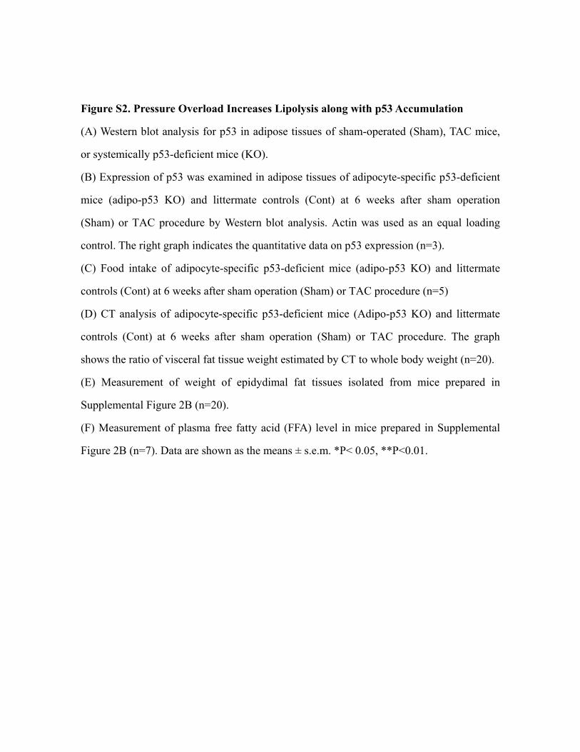
Figure S2. Pressure Overload Increases Lipolysis along with p53 Accumulation
(A) Western blot analysis for p53 in adipose tissues of sham-operated (Sham), TAC mice,
or systemically p53-deficient mice (KO).
(B) Expression of p53 was examined in adipose tissues of adipocyte-specific p53-deficient
mice (adipo-p53 KO) and littermate controls (Cont) at 6 weeks after sham operation
(Sham) or TAC procedure by Western blot analysis. Actin was used as an equal loading
control. The right graph indicates the quantitative data on p53 expression (n=3).
(C) Food intake of adipocyte-specific p53-deficient mice (adipo-p53 KO) and littermate
controls (Cont) at 6 weeks after sham operation (Sham) or TAC procedure (n=5)
(D) CT analysis of adipocyte-specific p53-deficient mice (Adipo-p53 KO) and littermate
controls (Cont) at 6 weeks after sham operation (Sham) or TAC procedure. The graph
shows the ratio of visceral fat tissue weight estimated by CT to whole body weight (n=20).
(E) Measurement of weight of epidydimal fat tissues isolated from mice prepared in
Supplemental Figure 2B (n=20).
(F) Measurement of plasma free fatty acid (FFA) level in mice prepared in Supplemental
Figure 2B (n=7). Data are shown as the means ± s.e.m. *P< 0.05, **P<0.01.


Figure S3. Inhibition of Lipolysis Ameliorates Adipose Tissue Inflammation during
Heart Failure
(A) CT analysis of mice at 6 weeks after sham operation (Sham) or TAC with or without
surgical denervation of the sympathetic nerves, guanethidine treatment, and acipimox
treatment (n=15).
(B) Measurement of plasma free fatty acid (FFA) level in mice prepared in Supplemental
Figure 3A (n=7).
(C and D) Hematoxylin and eosin staining of adipose tissues of mice prepared in
Supplemental Figure 3A. The graph indicates the quantitative data on the infiltration of
macrophages (n=4). Data are shown as the means ± s.e.m. *P< 0.05, **P<0.01.


Figure S4. Guanethidine Treatment Attenuates Adipose Tissue Inflammation and
Systemic Insulin Resistance
(A) Real-time PCR assessing the expression of Emr1, Tnf (Tnfα), Ccl2 (MCP1), and
Cdkn1a (p21) levels in adipose tissues of mice at 6 weeks after sham operation (Sham) or
TAC with or without guanethidine treatment (n=10).
(B) Expression of p53 was examined in adipose tissues of mice prepared in Supplemental
Figure 4A by Western blot analysis. The right graph indicates the quantitative data on p53
expression (n=3).
(C) Insulin tolerance test (ITT) and glucose tolerance test (GTT) of mice prepared in
Supplemental Figure 4A (n=16). Data are shown as the means ± s.e.m. *P< 0.05, **P<0.01.


Figure S5. Effects of Isoproterenol Treatment and Myocardial Ischemia on Adipose
Tissue Inflammation
(A) CT analysis of wild-type mice treated with or without isoproterenol (ISO) (n=5).
(B) Measurement of weight of epidydimal fat tissues isolated from mice prepared in
Supplemental Figure 5A (n=5).
(C) Measurement of plasma free fatty acid (FFA) level in mice prepared in Supplemental
Figure 5A (n=5).
(D) Echocardiography estimating systolic function (FS) and ventricular size (LVDs) of Atgl
heterozygous knockout mice (Atgl KO) and littermate controls (Cont) at 2 weeks after
sham-operation or TAC procedure (n=4). FS, fractional shortening; LVDs, left ventricular
end-systolic diameter.
(E) Insulin tolerance test (ITT) and glucose tolerance test (GTT) of wild-type mice at 6
weeks after sham operation (Sham) or MI operation (n=12).
(F) CT analysis of mice at 6 weeks after sham operation (Sham) or MI operation. The graph
shows the ratio of visceral fat tissue weight estimated by CT to whole body weight (n=9).
(G) Measurement of weight of epidydimal fat tissues isolated from mice prepared in
Supplemental Figure 5E (n=7).
(H) Expression of p53 was examined in adipose tissues of mice prepared in Supplemental
Figure 5E by Western blot analysis. Actin was used as an equal loading control. The right
graph indicates the quantitative data on p53 expression (n=3).
(I) Real-time PCR assessing the expression of Emr1, Tnf (Tnfα), and Ccl2 (MCP1) levels
in adipose tissue of mice prepared in Supplemental Figure 5E (n=6).
(J) Hematoxylin and eosin staining of adipose tissues in mice prepared in Supplemental
Figure 5E. The graph indicates the quantitative data on the infiltration of macrophages
(n=4). Scale bar, 50 μm.

(K) Real-time PCR assessing the expression of Emr1, Tnf (Tnfα), and Ccl2 (MCP1) levels
in adipose tissue of adipocyte-specific p53-deficient mice (adipo-p53 KO) and littermate
controls (Cont) at 6 weeks after sham operation or MI operation (n=5).
(L) Insulin tolerance test (ITT) and glucose tolerance test (GTT) of adipocyte-specific
p53-deficient mice (adipo-p53 KO) and littermate controls (Cont) at 6 weeks after sham
operation or MI operation (n=8). Data are shown as the means ± s.e.m. *P< 0.05, **P<0.01.


Figure S6. Influence of Inhibiting p53-induced Adipose Tissue Inflammation on
Cardiac Function
(A) Echocardiography estimating systolic function (FS) and ventricular size (LVDs) of
adipocyte-specific p53-deficient mice (adipo-p53 KO) and littermate controls (Cont) at 6
weeks after sham operation or MI operation (n=5).
(B) Pifithrin-α (PFT) was administered to adipose tissues of the MI mice at 2–4weeks after
operation, and systolic function (FS) and ventricular size (LVDs) were estimated before
(2w) and after (4w) treatment with PFT by echocardiography (n=5).
(C) Real-time PCR assessing the expression of Emr1, Tnf (Tnfα), and Ccl2 (MCP1) levels
in adipose tissue of mice at 6 weeks after sham operation (Sham) or MI operation with or
without pifithrin-α (PFT) treatment (n=6).
(D) Insulin tolerance test (ITT) and glucose tolerance test (GTT) of mice at 6 weeks after
sham operation (Sham) or MI operation with pifithrin-α (PFT) treatment (n=11).
(E–G) Echocardiography estimating systolic function (FS) and ventricular size (LVDs) of
mice at 6 weeks after sham-operation (Sham) or TAC with or without surgical denervation
of the sympathetic nerves (E) (n=10), guanethidine treatment (F) (n=6), or acipimox
treatment (G) (n=9). Data are shown as the means ± s.e.m. *P< 0.05, **P<0.01.


Figure S7. Mechanisms of p53-induced Adipose Tissue Inflammation during Heart
Failure
(A) The number of γ-H2AX-positive nuclei in preadipocytes treated with or without
palmitic acid (500 μM) in the presence of N-acetyl cysteine (NAC, 1 mM) or vehicle
(Cont) (n=5).
(B) Western blot analysis of p53 expression in preadipocytes treated with or without
palmitic acid (500 μM) in the presence of NAC (1 mM) or vehicle (Cont) (n=3).
(C) The number of p50-positive nuclei (white arrows) in adipose tissue of
adipocyte-specific p53 deficient mice (adipo-p53 KO) and littermate controls (Cont) at 6
weeks after sham-operation or TAC procedure was estimated by double-immunostaining
for p50 (red) and Wheat Germ agglutinin lectin (green). Nuclei were stained with Hoechst
dye (blue). Scale bar indicates 50 μm.
(D) Dihydroethidium (DHE) staining in adipose tissue from mice at 6 weeks after
sham-operation (Sham) or TAC with or without acipimox treatment. The graph indicates
DHE-positive area (n=4).
(E) The number of p50-positive nuclei in adipose tissue of mice at 6 weeks after
sham-operation (Sham) or TAC with or without acipimox treatment was estimated by
immunofluorescent staining for p50 (n=6).
(F) BAY 11-7082 or vehicle was administered into the adipose tissue of mice at 2–4 weeks
after TAC. Real-time PCR was performed to assess the expression of Tnf (Tnfα) and Ccl2
(MCP1) levels in adipose tissue of mice at 4 weeks after sham-operation (Sham) or TAC
with or without BAY 11-7082 treatment (n=4).
(G) Echocardiography to assess systolic function (FS) and ventricular size (LVDs) of mice
prepared in Supplemental Figure 7F (n=5).
(H) Insulin tolerance test (ITT) and glucose tolerance test (GTT) of mice at 6 weeks after

sham operation (Sham) or TAC with or without BAY 11-7082 treatment (n=8). Data are
shown as the means ± s.e.m. *P< 0.05, **P<0.01.