Embed Size (px)
Citation preview
p21-activated Kinase 1 Is Activated through the MammalianTarget of Rapamycin/p70 S6 Kinase Pathway and Regulatesthe Replication of Hepatitis C Virus in HumanHepatoma Cells*
Received for publication, October 30, 2006, and in revised form, January 22, 2007 Published, JBC Papers in Press, January 25, 2007, DOI 10.1074/jbc.M610106200
Hisashi Ishida, Kui Li, MinKyung Yi, and Stanley M. Lemon1
From the Center for Hepatitis Research, Institute for Human Infections and Immunity, and the Department of Microbiologyand Immunology, University of Texas Medical Branch, Galveston, Texas 77555-1018
Cellularmechanisms that regulate the replication of hepatitisC virus (HCV)RNAare poorly understood. p21-activated kinase1 (PAK1) is a serine/threonine kinase that has been suggested toparticipate in antiviral signaling. We studied its role in the cel-lular control of HCV replication. Transfection of PAK1-specificsmall interfering RNA enhanced viral RNA and protein abun-dance in established replicon cell lines as well as cells infectedwith chimeric genotype 1a/2a HCV, despite reducing cellularproliferation, suggesting specific regulation ofHCV replication.PAK1 knockdown did not reduce interferon regulatory factor3-dependent gene expression, indicating that this regulation isindependent of the retinoic acid-inducible gene I/interferonregulatory factor 3 pathway. On the other hand, LY294002 andrapamycin abolished PAK1 phosphorylation and enhancedHCV abundance, suggesting that themammalian target of rapa-mycin (mTOR) is involved in PAK1 regulation of HCV. Smallinterfering RNA knockdown of the mTOR substrate p70 S6kinase abrogated PAK1 phosphorylation and enhanced HCVRNA abundance, whereas overexpression of a constitutivelyactive alternate substrate, eukaryotic translation initiation fac-tor 4E-binding protein 1, increased cap-independent viraltranslation and viral RNAabundancewithout influencingPAK1phosphorylation. Similar data indicated thatmTOR is regulatedby both phosphatidylinositol 3-kinase/Akt and ERK. Takentogether, the data indicate that p70 S6 kinase activates PAK1and contributes to phosphatidylinositol 3-kinase- and ERK-me-diated regulation of HCV RNA replication.
Hepatitis C virus (HCV)2 is a globally important cause ofchronic hepatitis, cirrhosis, and hepatocellular carcinoma (1).
Approximately 170 million people are estimated to be infectedwith the virus worldwide (2). Classified within the family Flavi-viridae, HCVhas a positive sense single-stranded RNA genomeabout 9.6 kb in length (3). The precursor polyprotein encodedby the large open reading frame that spansmuch of the genomeis processed into at least 10 proteins (core, E1, E2, p7, NS2,NS3,NS4A, NS4B, NS5A, and NS5B) by cellular and viral proteases(4). Up to 50–80% of patients infected with HCV fail to elimi-nate the virus (2), leading to long term viral persistence and anincreased risk of liver-related morbidity and mortality.The viral mechanisms that promote the establishment of
persistent HCV infection remain poorly defined. In general,infected hosts mount a variety of innate and adaptive immuneresponses to ongoing replication of RNA viruses. Many viruseshave evolved mechanisms to evade or defeat these cellularresponses (reviewed in Ref. 5), and HCV is no different. Recentstudies have shown that HCV infection blocks viral activationof interferon regulatory factor 3 (IRF-3), a transcription factorthat plays a pivotal role in orchestrating innate intracellularantiviral responses mediated through both the retinoic acid-inducible gene I (RIG-I) and Toll-like receptor 3 (TLR3) dou-ble-stranded RNA signaling pathways. HCV accomplishes thisby targeting the RIG-I and TLR3 adaptor proteins, MAVS andTRIF, for specific proteolysis by the viral NS3/4A protease(6–8). It is possible that the ability of HCV to delay or preventearly innate immune responsesmay contribute to the establish-ment of persistent infection.Subgenomic HCV RNA replicons capable of autonomous
replication in stably selected cell lines were first reported byLohmann et al. (9) in 1999. These repliconRNAs are bicistronicconstructs containing the native 5�- and 3�-nontranslated RNAsequences of HCV, with the internal ribosome entry site (IRES)within the HCV 5�-nontranslated RNA driving translation ofthe neomycin phosphotransferase gene, followed by a heterol-ogous IRES from encephalomyocarditis virus driving transla-tion of the nonstructural (NS) HCV proteins (NS2 or NS3through NS5B). These RNAs replicate autonomously whentransfected into Huh7 human hepatoma cells and allow theselection of stable replicon cell lines with G418 (9). In addition,several groups have recently reported HCV cDNA clones that
* This work was supported in part by National Institutes of Health GrantsU19-AI40035 (to S. M. L.) and R21-AI063451 (to M. Y.) and R21-DA018054(to K. L.). The costs of publication of this article were defrayed in part by thepayment of page charges. This article must therefore be hereby marked“advertisement” in accordance with 18 U.S.C. Section 1734 solely to indi-cate this fact.
1 To whom correspondence should be addressed: Center for HepatitisResearch, 4.104 Blocker Medical Research Bldg., University of Texas Medi-cal Branch at Galveston, 301 University Blvd., Galveston, TX 77555-1019.Tel.: 409-747-7048; Fax: 409-747-7030; E-mail: [email protected].
2 The abbreviations used are: HCV, hepatitis C virus; IRF-3, interferon regula-tory factor 3; RIG-I, retinoic acid-inducible gene I; TLR3, Toll-like receptor 3;IRES, internal ribosome entry site; IFN, interferon; PI3K, phosphatidylinosi-tol 3-kinase; JAK, Janus kinase; STAT, signal transducers and activators oftranscription; ISG, IFN-stimulated gene; PAK, p21-activated kinase 1;
mTOR, mammalian target of rapamycin; ERK, extracellular signal-regulatedkinase; 4EBP1, eukaryotic translation initiation factor 4E-binding protein 1;siRNA, small interfering RNA.
THE JOURNAL OF BIOLOGICAL CHEMISTRY VOL. 282, NO. 16, pp. 11836 –11848, April 20, 2007© 2007 by The American Society for Biochemistry and Molecular Biology, Inc. Printed in the U.S.A.
11836 JOURNAL OF BIOLOGICAL CHEMISTRY VOLUME 282 • NUMBER 16 • APRIL 20, 2007
by guest on Novem
ber 7, 2018http://w
ww
.jbc.org/D
ownloaded from
produce virus particles that are capable of undergoing a com-plete viral replication cycle within cultured human hepatomacells (10–13). These replicon cell lines and newly reported virusclones represent useful tools for investigation of HCV replica-tion and discovery of anti-HCV compounds.Previous studies have shown that cytokines, including type I
and II interferons (IFNs), interleukin-1�, and transforminggrowth factor-� (14–17), small molecule inhibitors of NS3 orNS5B (18, 19), and small interfering RNAs (siRNAs) specific forHCV RNA (20), can modulate the efficiency of HCV replica-tion, reducing and in some cases even eliminating repliconRNA. The activities of a number of intracellular signaling mol-ecules and pathways also have been shown to influence HCVreplication and RNA abundance in replicon cell lines, includingERK (21), phosphatidylinositol 3-kinase (PI3K) (22), IRF-3 (6),Smad (17), and the JAK/STAT pathway (14, 15). Type I IFN-induced JAK-STAT activation results in strong suppression ofviral replication by inducing a series of IFN-stimulated genes(ISGs). Although the JAK/STAT pathway has been relativelywell characterized, as has the virus-activated RIG-I/IRF-3 path-way that leads to induction of type I IFN synthesis, many detailsof themechanisms bywhich other pathways regulateHCV rep-lication remain unclear. The state of the cell cycle and rate ofcellular proliferation has also been shown to affect replication(23, 24).The p21-activated kinases (PAKs) are evolutionarily con-
served serine/threonine kinases that play an important role in avariety of cellular functions inmammalian cells, including con-trol of cell proliferation, differentiation, migration, and celldeath (reviewed in Ref. 25). PAK1, amember of the PAK family,is activated by several growth factors, including epidermalgrowth factor, platelet-derived growth factor, and hepatocytegrowth factor via activation of the Rho GTPases, Rac1 andCdc42 (26). The binding of GTP-bound GTPases to PAK1 dis-sociates dimeric PAK1 molecules and leads to the active stateconformation. PAK1 is also activated by numerous physiologi-cal signals (25). It has been suggested that PAK1mediates virus-or poly(I:C)-stimulated signaling and that it may contribute tothe activation of IRF-3 through TANK-binding kinase-1 andI�B kinase-� (27). Although further study is required, theseobservations suggest that PAK1 may play an important role inactivating innate intracellular antiviral signaling pathways.Here, we demonstrate that PAK1 knockdown leads to an
increase in HCV replicon RNA abundance in stable repliconcell lines and enhances replication of a cell culture-infec-tious virus, suggesting that PAK1 is involved in cellular pro-cess(es) that negatively regulate HCV replication. Contraryto previous studies (27), however, we do not find PAK1 to berequired for IRF-3 activation by the virus-activated RIG-Ipathway. We show that PAK1 activation in these repliconcells is inhibited by rapamycin, a mammalian target of rapa-mycin (mTOR) inhibitor, and that mTOR is independentlyregulated by both PI3K/Akt and ERK. Further analysisrevealed that p70 S6 kinase is responsible for the PAK1-mediated suppression of HCV replication, suggesting a novelpathway toward PAK1 activation.
EXPERIMENTAL PROCEDURES
Cell Culture—The human hepatocellular carcinoma cell line,Huh7, was cultured in Dulbecco’s modified Eagle’s mediumsupplemented with 10% fetal bovine serum, glucose (4.5mg/ml) at 37 °C in a 5%CO2 atmosphere. A7 andHP, twoHuh7clonal cell lines that harbor Con1 HCV (genotype 1b) sub-genomic replicon RNAs (6), and a genome-length (HCV-Nstrain) replicon cell line, 2-3 (28), were maintained in completeDulbecco’s modified Eagle’s medium with 250 �g/ml G418.Antibodies and Kinase Inhibitors—Antibodies to PAK1,
phospho-PAK1 (Thr-423), p70 S6 kinase, phospho-p70 S6kinase (Thr-389), Akt, phospho-Akt (Ser-473) were purchasedfrom Cell Signaling Technology. Antibodies to �-actin(A-5441) and FLAG (F-3165) were from Sigma. A mousemonoclonal antibody to the HCV core protein (C7-50) wasobtained from Affinity BioReagents. A rabbit polyclonal anti-body toHCVNS5Awas kindly provided byDr. Craig Cameron.The inhibitors, LY294002 (PI3K), wortmannin (PI3K), rapamy-cin (mTOR), and PD98059 (ERK) were obtained fromCalbiochem.Plasmids and DNATransfection—pIFN-�-Luc was provided
by Dr. Rongtuan Lin. pPRDII-Luc (29) and pISG56-Luc wereprovided byDr.Michael Gale. pRC22F (30) has been previouslydescribed. An expression vector for eukaryotic translation ini-tiation factor 4E-binding protein 1 (4EBP1) was constructed byamplifying specific cDNA using conventional reverse tran-scription-PCR methods and cloning the amplified sequencesinto the HindIII-EcoRI site of pFLAG-CMV-6a (Sigma).Thr-46 was mutated to Ala by site-specific mutagenesis usingthe QuikChange site-directed mutagenesis kit (Stratagene) toconstruct the constitutive active mutant, pCMV-4EBP1-T46A.Cells were transfected with plasmid DNAs using TransIT-LT1(Mirus) according to the manufacturer’s recommended proce-dures. Cells were co-transfected with pCMV-�-gal (Clontech)to monitor transfection efficiencies.RNA Interference—Transfections of siRNA specific for PAK1
or p70 S6 kinase were carried out using Oligofectamine(Invitrogen) with a final concentration of siRNA of 100 nM,according to the manufacturer’s recommended procedures.Briefly, for transfection of cells in 24-well cell culture plates, 60pmol of siRNA was diluted in 50 �l of Opti-MEM (Invitrogen),and 3 �l of Oligofectamine was diluted in 12 �l of Opti-MEM.After incubation for 5min, the siRNAandOligofectamine solu-tions were mixed, incubated for an additional 20 min, andadded to cell culture medium. siRNAs specific for PAK1 werepurchased from Dharmacon (M-003521-03); these were trans-fected singly or as a pool consisting of four oligoribonucleotideduplexes with the following sequences: 5�-ACCCAAACAUU-GUGAAUUAUU-3� and 5�-UAAUUCACAAUGUUUGGG-UUU-3� (P1), 5�-GGAGAAAUUACGAAGCAUAUU-3� and5�-UAUGCUUCGUAAUUUCUCCUU-3� (P2), 5�-UCAAAU-AACGGCCUAGACAUU-3� and 5�-UGUCUAGGCCGUUA-UUUGAUU-3� (P3), and 5�-CAUCAAAUAUCACUAAGU-CUU-3� and 5�-GACUUAGUGAUAUUUGAUGUU-3� (P4).Validated negative control siRNAs (C1 and C2) with limitedsequence identity to known genes were obtained from Ambion(catalog number 4611 and 4613, respectively). p70 S6 kinase-
PAK1 Regulates HCV Replication
APRIL 20, 2007 • VOLUME 282 • NUMBER 16 JOURNAL OF BIOLOGICAL CHEMISTRY 11837
by guest on Novem
ber 7, 2018http://w
ww
.jbc.org/D
ownloaded from
specific siRNA (catalog number 1454) and a mutated p70 S6kinase siRNA (MutS6K), containing two nucleotide substitu-tions, were purchased from Ambion as oligoribonucleotideduplexes with the sequences 5�-GGACAUGGCAGGAGUGU-UUdTdT-3� and 5�-AAACACUCCUGCCAUGUCCdTdC-3�(p70 S6 kinase-specific, S6K) and 5�-GGACAUGGUCGGAG-UGUUUdTdT-3� and 5�-AAACACUCCGACCAUGUCCdT-dC-3� (MutS6K).Promoter Reporter Assays—A7 and HP cells (5 � 104 cells/
well in 24-well plates) were transfected with siRNA specific forPAK1 as described above. Two days later, cells were cotrans-fected with 350 ng of reporter plasmids and 50 ng of pCMV-�-gal DNA.Cells were lysed and assayed for luciferase activity and�-galactosidase activity 24 h later. To assess the role of PAK1 inIRF-3 activation induced by Sendai virus, Huh7 cells (5 � 104cells per well in 24-well plates) were transfected with PAK1siRNA, followed by the transfection of reporter plasmids (350ng) and pCMV-�-gal (50 ng) 24 h later. The next day, cells weremock-treated or challenged with 100 hemagglutinin units/mlSendai virus (Charles River Laboratory) as previously described(6) and then harvested and assayed for reporter protein activity16 h later. The luciferase activity was normalized for transfec-tion efficiency based on the results of the �-galactosidase assay.Assays were performed in triplicate; S.D. values are denoted bybars in the figures.Cell Proliferation Assay—Cell proliferation was assessed by a
WST-1 (2-(2-methoxy-4-nitrophenyl)-3-(4-nitrophenyl)-5-(2,4-disulfophenyl)-2H-tetrazolium, monosodium salt) assayaccording to themanufacturer’s suggested protocol (ChemiconInternational Inc.). A7 cells (1 � 104 cells/well) were seededinto 96-well flat-bottom plates, transfected with negative con-trol or PAK1 siRNA as above, and cultured in Dulbecco’s mod-ified Eagle’s medium containing 10% fetal bovine serum. Tenmicroliters of WST-1 reagent was added to each well, the cellswere incubated at 37 °C for 1 h, and absorbance at 450 nm wasmeasured using a spectrophotometer. Assays were carried outin triplicate; S.D. values are denoted by bars in the figures.NorthernBlotAnalysis—ForNorthern blot analysis, total cel-
lular RNA was extracted using TRIzol reagent (Invitrogen).Eight micrograms of total RNAwas electrophoresed and trans-ferred to a nylon membrane (Hybond-N; Amersham Bio-sciences). The membrane was probed with in vitro transcribednegative-strand HCV RNA labeled with digoxigenin, usingreagents provided with the DIG Northern Starter Kit (RocheApplied Science) and following the manufacturer’s recom-mended procedures. After extensive washing and blocking, themembrane was hybridized with anti-digoxigenin-AP, incu-bated with CSPD, and exposed to a BioMax MR x-ray film(Eastman Kodak Co.).Immunoblot Analysis—Total cellular proteins were
extracted with lysis buffer containing 1% Nonidet P-40, 0.5%sodium deoxycholate, 0.1% SDS, 1 mM phenylmethylsulfonylfluoride, 1 mM sodium vanadate, 50 mM NaF, and 50 �g/mlaprotinin in phosphate-buffered saline. Proteins were sepa-rated by SDS-polyacrylamide gel electrophoresis and trans-ferred to a polyvinylidene difluoride membrane (Hybond-P;Amersham Biosciences). After blocking, the membrane wasprobed with specific primary antibodies (see above), followed
by further incubation with a secondary antibody conjugatedwith horseradish peroxidase (AmershamBiosciences). Proteinswere visualized using ECL Western blot detection reagents(Amersham Biosciences) and exposure to film.Dual Luciferase Assay—Huh7 cells (5 � 104 cells/well) were
seeded into 24-well plates and cotransfected with 200 ng ofpRC22F and 200 ng of pCMV-4EBP1-T46A, as describedabove. Twenty-four hours later, cells were lysed and assayed forHCV IRES-dependent firefly luciferase activity and cap-dependent Renilla luciferase activity using the Dual LuciferaseReporter Assay System (Promega). For experiments involvingRNA interference, Huh7 cells (5 � 104 cells/well) in 24-wellplates were sequentially transfected with 400 ng of pRC22F andwith negative control or PAK1 siRNA. After incubation for 3days, cells were harvested and assayed for luciferase activities.Assays were performed in triplicate; S.D. values are denoted bybars in the figures.Cell Culture-infectiousHCV—H-NS2/NS3-J/YH/QL is a chi-
meric cell culture-infectious virus with a genome consisting ofthe core to NS2 sequence of genotype 1a (H77) virus placedwithin the background of the genotype 2a JFH1 virus and con-taining compensatory mutations in E1 (Y361H) and NS3(Q1251L) (Fig. 5A) (31). These two mutations render the chi-meric RNA highly infectious (31). Virus stock (107 focus-form-ing units/ml) was prepared at the fourth virus passage by clari-fying supernatant media harvested from infected cells(collected after overnight incubation of the cells in Dulbecco’smodified Eagle’s medium without serum) by centrifugation at3000 rpm for 5min and then concentrating 50� in a CentriconPlus-80 Centrifugal Filter device with Ultracel PL Membranes(100-kDa exclusion) (Millipore Corp.).Infections were carried out at a multiplicity of infection of
�2. Huh7 cells (2 � 105 cells/well) were seeded into 6-wellplates and 24 h later transfected with either PAK1-specific orcontrol siRNA as above. Following 24 h of additional incuba-tion, themediumwas replaced with 1ml ofmedium containing4 � 105 focus-forming units of virus. After a 12-h incubation at37 °C in a 5% CO2 environment, the cells were washed withphosphate-buffered saline and refed with normal culturemedium.At 24, 48, and 72 h after inoculation of virus, cells werelysed and assayed for HCV core protein expression as well asPAK1 and�-actin by immunoblot. Supernatant cell culture flu-ids collected at 48 and 72 h were also assayed for infectiousvirus.HCV Infectivity Assay—Huh7.5 cells (2� 104 cells/well) were
seeded onto 8-well chamber slides (Nalge Nunc) 24 h prior toinoculation of 100�l of culturemedium containing virus. After3 days of incubation, cells were fixed in methanol/acetone (1:1)for 10 min, incubated for 2 h with anti-core antibody diluted1:300, and then stained with fluorescein isothiocyanate-conju-gated goat anti-mouse IgG (Southern Biotech) for 1 h. Nucleiwere visualized with 4�,6-diamidino-2-phenylindole-dihydro-chloride. The cells were examined with a Zeiss LSM 510 laser-scanning confocal microscope. Clusters of infected cells stain-ing for core antigen were counted, and virus titers werecalculated in terms of focus-forming units/ml (11, 31).
PAK1 Regulates HCV Replication
11838 JOURNAL OF BIOLOGICAL CHEMISTRY VOLUME 282 • NUMBER 16 • APRIL 20, 2007
by guest on Novem
ber 7, 2018http://w
ww
.jbc.org/D
ownloaded from
RESULTS
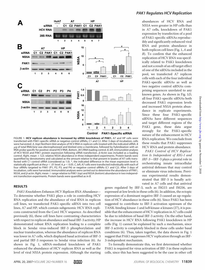
PAK1 Knockdown Enhances HCV Replicon RNA Abundance—To determine whether PAK1 plays a role in controlling HCVRNA replication and the abundance of viral RNA in repliconcell lines, we transfected PAK1-specific siRNA into two celllines, A7 and HP, which contain subgenomic HCV RNA repli-cons constructed from the Con1 HCV sequence. As describedpreviously (6), these cell lines have contrasting characteristicswith respect to replicon abundance and basal IRF-3 activity; HPdemonstrated robust RNA replication leading to a completeblock in Sendai virus-induced IRF-3 phosphorylation andnuclear translocation, whereas the abundance of replicon RNAwas lower in A7 cells, which displayed basal activation of IRF-3and partial IRF-3 responses to Sendai virus infection (6). Asshown in Fig. 1, siRNA-mediated knockdown of PAK1enhanced the abundance of HCV replicon RNA as well as thelevel of viral NS5A protein expression. Although the starting
abundances of HCV RNA andNS5A were greater in HP cells thanin A7 cells, knockdown of PAK1expression by transfection of a poolof PAK1-specific siRNAs reproduc-ibly and significantly enhanced viralRNA and protein abundance inboth replicon cell lines (Fig. 1,A andB). To confirm that the enhancedreplication of HCVRNAwas specif-ically related to PAK1 knockdownandnot a result of an off target effectof one of the siRNAs included in thepool, we transfected A7 repliconcells with each of the four individualPAK1-specific siRNAs as well astwo negative control siRNAs com-prising sequences unrelated to anyknown genes. As shown in Fig. 1D,all four PAK1-specific siRNAs bothdecreased PAK1 expression levelsand increased NS5A protein abun-dance in replicate experiments.Since these four PAK1-specificsiRNAs have different sequencesand target different regions of thePAK1 gene, these data arguestrongly for the PAK1-specificnature of the enhancement in HCVRNAreplication.We conclude fromthese results that PAK1 suppressesHCV RNA and protein abundance.PAK1 Control of HCV Replica-
tion Is Independent of RIG-I andIRF-3—IRF-3 plays a pivotal role inorchestrating innate intracellularantiviral defenses that may controlor eliminate virus infections. Previ-ous experimental results demon-strated that IRF-3 is basally acti-vated in A7 cells and that antiviral
genes regulated by IRF-3, such as ISG15 and ISG56, areexpressed at low levels in these cells (6). In addition, the ectopicexpression of a dominant negative IRF-3 caused an up-regula-tion of HCV abundance in these cells (6). Since PAK1 has beensuggested to contribute to IRF-3 activation upstream of theTANK-binding kinase-1 and I�B kinase-� kinases (27), it is pos-sible that the enhancement ofHCV replication inA7 cells couldbe due to inhibition of basal IRF-3 activity. On the other hand,the increase in HCV RNA following PAK1 knockdown in HPcells (Fig. 1) cannot be explained by such a mechanism, sinceIRF-3 activity is completely blocked in these cells under basalconditions (6). Thus, taken together, the data shown in Fig. 1suggest that PAK1 suppressesHCV replication through an IRF-3-independent mechanism.To formally demonstrate this, we first determined whether
PAK1 is required for virus activation of IRF-3 in these repliconcells, since this has been suggested to be the case in other cell
FIGURE 1. HCV replicon abundance is increased by siRNA knockdown of PAK1. A7 and HP cells weretransfected with PAK1-specific siRNA or negative control siRNAs, C1 and C2. After 3 days of incubation, cellswere harvested. A, (top) Northern blot analysis of HCV RNA in replicon cells treated with the indicated siRNA. 8�g of total RNA/lane was electrophoresed and blotted onto a membrane, followed by hybridization with anRNA probe specific for positive-strand HCV RNA. Bottom, 28 S RNA loading control. B, left, immunoblot analysisof HCV NS5A and PAK1 protein expression following siRNA transfection. �-Actin was included as a loadingcontrol. Right, mean � S.D. relative NS5A abundance in three independent experiments. Protein bands werequantified by densitometry and calculated as the amount relative to that present in lysates of A7 cells trans-fected with C1 control siRNA (considered as 1.0). *, the indicated difference in the mean expression level isstatistically significant at the p � .01 level. #, p � 0.05. C, left, A7 cells were transfected individually with each offour siRNAs targeted to PAK1 (P1, P2, P3, and P4) or negative control siRNAs (C1 and C2). After 3 days ofincubation, cells were harvested. Immunoblot analyses were carried out to determine the abundance of PAK1,NS5A, and �-actin. Right, mean � range relative to PAK1 (top) and NS5A (bottom) abundance in two independ-ent transfection experiments. Protein bands were quantified as in B.
PAK1 Regulates HCV Replication
APRIL 20, 2007 • VOLUME 282 • NUMBER 16 JOURNAL OF BIOLOGICAL CHEMISTRY 11839
by guest on Novem
ber 7, 2018http://w
ww
.jbc.org/D
ownloaded from
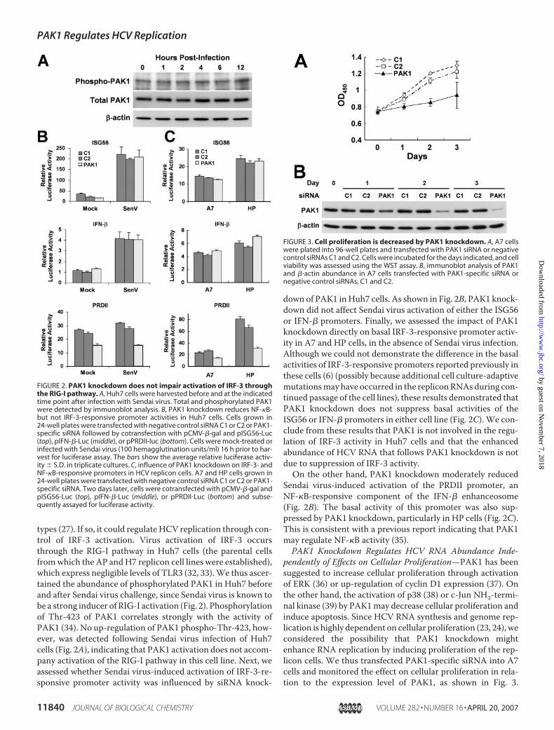
types (27). If so, it could regulate HCV replication through con-trol of IRF-3 activation. Virus activation of IRF-3 occursthrough the RIG-I pathway in Huh7 cells (the parental cellsfromwhich the AP andH7 replicon cell lines were established),which express negligible levels of TLR3 (32, 33).We thus ascer-tained the abundance of phosphorylated PAK1 in Huh7 beforeand after Sendai virus challenge, since Sendai virus is known tobe a strong inducer of RIG-I activation (Fig. 2). Phosphorylationof Thr-423 of PAK1 correlates strongly with the activity ofPAK1 (34). No up-regulation of PAK1 phospho-Thr-423, how-ever, was detected following Sendai virus infection of Huh7cells (Fig. 2A), indicating that PAK1 activation does not accom-pany activation of the RIG-I pathway in this cell line. Next, weassessed whether Sendai virus-induced activation of IRF-3-re-sponsive promoter activity was influenced by siRNA knock-
down of PAK1 inHuh7 cells. As shown in Fig. 2B, PAK1 knock-down did not affect Sendai virus activation of either the ISG56or IFN-� promoters. Finally, we assessed the impact of PAK1knockdown directly on basal IRF-3-responsive promoter activ-ity in A7 and HP cells, in the absence of Sendai virus infection.Although we could not demonstrate the difference in the basalactivities of IRF-3-responsive promoters reported previously inthese cells (6) (possibly because additional cell culture-adaptivemutationsmay have occurred in the repliconRNAsduring con-tinued passage of the cell lines), these results demonstrated thatPAK1 knockdown does not suppress basal activities of theISG56 or IFN-� promoters in either cell line (Fig. 2C). We con-clude from these results that PAK1 is not involved in the regu-lation of IRF-3 activity in Huh7 cells and that the enhancedabundance of HCV RNA that follows PAK1 knockdown is notdue to suppression of IRF-3 activity.On the other hand, PAK1 knockdown moderately reduced
Sendai virus-induced activation of the PRDII promoter, anNF-�B-responsive component of the IFN-� enhanceosome(Fig. 2B). The basal activity of this promoter was also sup-pressed by PAK1 knockdown, particularly in HP cells (Fig. 2C).This is consistent with a previous report indicating that PAK1may regulate NF-�B activity (35).PAK1 Knockdown Regulates HCV RNA Abundance Inde-
pendently of Effects on Cellular Proliferation—PAK1 has beensuggested to increase cellular proliferation through activationof ERK (36) or up-regulation of cyclin D1 expression (37). Onthe other hand, the activation of p38 (38) or c-Jun NH2-termi-nal kinase (39) by PAK1may decrease cellular proliferation andinduce apoptosis. Since HCV RNA synthesis and genome rep-lication is highly dependent on cellular proliferation (23, 24), weconsidered the possibility that PAK1 knockdown mightenhance RNA replication by inducing proliferation of the rep-licon cells. We thus transfected PAK1-specific siRNA into A7cells and monitored the effect on cellular proliferation in rela-tion to the expression level of PAK1, as shown in Fig. 3.
FIGURE 2. PAK1 knockdown does not impair activation of IRF-3 throughthe RIG-I pathway. A, Huh7 cells were harvested before and at the indicatedtime point after infection with Sendai virus. Total and phosphorylated PAK1were detected by immunoblot analysis. B, PAK1 knockdown reduces NF-�B-but not IRF-3-responsive promoter activities in Huh7 cells. Cells grown in24-well plates were transfected with negative control siRNA C1 or C2 or PAK1-specific siRNA followed by cotransfection with pCMV-�-gal and pISG56-Luc(top), pIFN-�-Luc (middle), or pPRDII-luc (bottom). Cells were mock-treated orinfected with Sendai virus (100 hemagglutination units/ml) 16 h prior to har-vest for luciferase assay. The bars show the average relative luciferase activ-ity � S.D. in triplicate cultures. C, influence of PAK1 knockdown on IRF-3- andNF-�B-responsive promoters in HCV replicon cells. A7 and HP cells grown in24-well plates were transfected with negative control siRNA C1 or C2 or PAK1-specific siRNA. Two days later, cells were cotransfected with pCMV-�-gal andpISG56-Luc (top), pIFN-�-Luc (middle), or pPRDII-Luc (bottom) and subse-quently assayed for luciferase activity.
FIGURE 3. Cell proliferation is decreased by PAK1 knockdown. A, A7 cellswere plated into 96-well plates and transfected with PAK1 siRNA or negativecontrol siRNAs C1 and C2. Cells were incubated for the days indicated, and cellviability was assessed using the WST assay. B, immunoblot analysis of PAK1and �-actin abundance in A7 cells transfected with PAK1-specific siRNA ornegative control siRNAs, C1 and C2.
PAK1 Regulates HCV Replication
11840 JOURNAL OF BIOLOGICAL CHEMISTRY VOLUME 282 • NUMBER 16 • APRIL 20, 2007
by guest on Novem
ber 7, 2018http://w
ww
.jbc.org/D
ownloaded from
Although transfection of two different negative control siRNAsresulted in no apparent change in the growth of the cells, asignificant decrease in cellular proliferation was observed incells transfected with PAK1 siRNA (Fig. 3A). This decreasein cellular proliferationwas associatedwith a slight reduction inthe PAK1 expression level 24 h post-transfection of PAK1-spe-cific siRNA with substantially greater suppression evident at48–72 h (Fig. 3B). Similar results were obtained in HP cells(data not shown). These results indicate that the up-regulationof HCV abundance following PAK1 knockdown is not due topleiotropic effects promoting cellular proliferation. Indeed,since viral RNA replication has been shown to be substantiallyreduced in slowly growing cells (23, 24), the fact that PAK1knockdown enhances viral RNA abundance while significantlyslowing cell proliferation suggests that PAK1 may have agreater effect on HCV RNA replication than suggested simplyby the magnitude of the increase in viral RNA abundance.These competing effects of PAK1 on cell proliferation and viralreplication may account for the varying increases in NS5Aabundance in relation to the magnitude of PAK1 knockdownthat we observed following transfection of the individualsiRNAs in Fig. 1C.PAK1 Is Basally Activated in Replicon Cells—Next, we asked
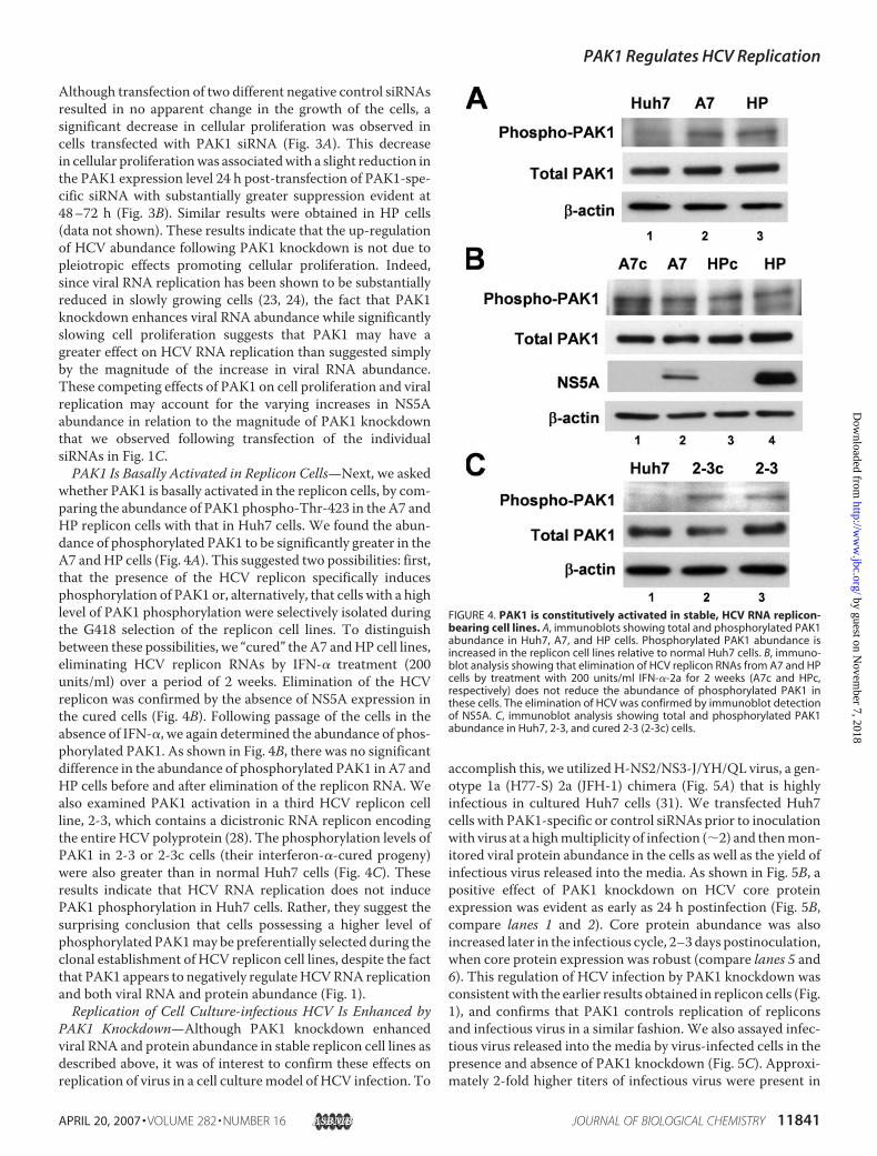
whether PAK1 is basally activated in the replicon cells, by com-paring the abundance of PAK1 phospho-Thr-423 in the A7 andHP replicon cells with that in Huh7 cells. We found the abun-dance of phosphorylated PAK1 to be significantly greater in theA7 andHP cells (Fig. 4A). This suggested two possibilities: first,that the presence of the HCV replicon specifically inducesphosphorylation of PAK1 or, alternatively, that cells with a highlevel of PAK1 phosphorylation were selectively isolated duringthe G418 selection of the replicon cell lines. To distinguishbetween these possibilities, we “cured” the A7 andHP cell lines,eliminating HCV replicon RNAs by IFN-� treatment (200units/ml) over a period of 2 weeks. Elimination of the HCVreplicon was confirmed by the absence of NS5A expression inthe cured cells (Fig. 4B). Following passage of the cells in theabsence of IFN-�, we again determined the abundance of phos-phorylated PAK1. As shown in Fig. 4B, there was no significantdifference in the abundance of phosphorylated PAK1 in A7 andHP cells before and after elimination of the replicon RNA. Wealso examined PAK1 activation in a third HCV replicon cellline, 2-3, which contains a dicistronic RNA replicon encodingthe entire HCV polyprotein (28). The phosphorylation levels ofPAK1 in 2-3 or 2-3c cells (their interferon-�-cured progeny)were also greater than in normal Huh7 cells (Fig. 4C). Theseresults indicate that HCV RNA replication does not inducePAK1 phosphorylation in Huh7 cells. Rather, they suggest thesurprising conclusion that cells possessing a higher level ofphosphorylated PAK1may be preferentially selected during theclonal establishment of HCV replicon cell lines, despite the factthat PAK1 appears to negatively regulateHCVRNA replicationand both viral RNA and protein abundance (Fig. 1).Replication of Cell Culture-infectious HCV Is Enhanced by
PAK1 Knockdown—Although PAK1 knockdown enhancedviral RNA and protein abundance in stable replicon cell lines asdescribed above, it was of interest to confirm these effects onreplication of virus in a cell culturemodel of HCV infection. To
accomplish this, we utilizedH-NS2/NS3-J/YH/QL virus, a gen-otype 1a (H77-S) 2a (JFH-1) chimera (Fig. 5A) that is highlyinfectious in cultured Huh7 cells (31). We transfected Huh7cells with PAK1-specific or control siRNAs prior to inoculationwith virus at a highmultiplicity of infection (�2) and thenmon-itored viral protein abundance in the cells as well as the yield ofinfectious virus released into the media. As shown in Fig. 5B, apositive effect of PAK1 knockdown on HCV core proteinexpression was evident as early as 24 h postinfection (Fig. 5B,compare lanes 1 and 2). Core protein abundance was alsoincreased later in the infectious cycle, 2–3 days postinoculation,when core protein expression was robust (compare lanes 5 and6). This regulation of HCV infection by PAK1 knockdown wasconsistentwith the earlier results obtained in replicon cells (Fig.1), and confirms that PAK1 controls replication of repliconsand infectious virus in a similar fashion. We also assayed infec-tious virus released into the media by virus-infected cells in thepresence and absence of PAK1 knockdown (Fig. 5C). Approxi-mately 2-fold higher titers of infectious virus were present in
FIGURE 4. PAK1 is constitutively activated in stable, HCV RNA replicon-bearing cell lines. A, immunoblots showing total and phosphorylated PAK1abundance in Huh7, A7, and HP cells. Phosphorylated PAK1 abundance isincreased in the replicon cell lines relative to normal Huh7 cells. B, immuno-blot analysis showing that elimination of HCV replicon RNAs from A7 and HPcells by treatment with 200 units/ml IFN-�-2a for 2 weeks (A7c and HPc,respectively) does not reduce the abundance of phosphorylated PAK1 inthese cells. The elimination of HCV was confirmed by immunoblot detectionof NS5A. C, immunoblot analysis showing total and phosphorylated PAK1abundance in Huh7, 2-3, and cured 2-3 (2-3c) cells.
PAK1 Regulates HCV Replication
APRIL 20, 2007 • VOLUME 282 • NUMBER 16 JOURNAL OF BIOLOGICAL CHEMISTRY 11841
by guest on Novem
ber 7, 2018http://w
ww
.jbc.org/D
ownloaded from
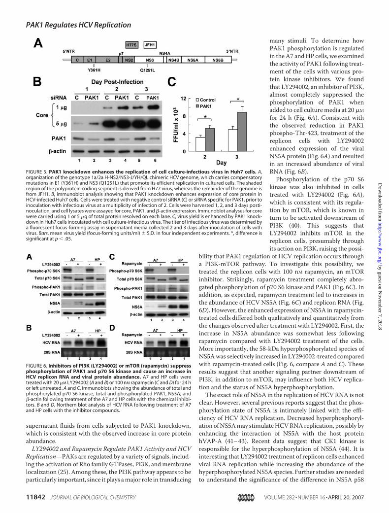
supernatant fluids from cells subjected to PAK1 knockdown,which is consistent with the observed increase in core proteinabundance.LY294002 and Rapamycin Regulate PAK1 Activity and HCV
Replication—PAKs are regulated by a variety of signals, includ-ing the activation of Rho family GTPases, PI3K, andmembranelocalization (25). Among these, the PI3K pathway appears to beparticularly important, since it plays amajor role in transducing
many stimuli. To determine howPAK1 phosphorylation is regulatedin the A7 andHP cells, we examinedthe activity of PAK1 following treat-ment of the cells with various pro-tein kinase inhibitors. We foundthat LY294002, an inhibitor of PI3K,almost completely suppressed thephosphorylation of PAK1 whenadded to cell culturemedia at 20�Mfor 24 h (Fig. 6A). Consistent withthe observed reduction in PAK1phospho-Thr-423, treatment of thereplicon cells with LY294002enhanced expression of the viralNS5A protein (Fig. 6A) and resultedin an increased abundance of viralRNA (Fig. 6B).Phosphorylation of the p70 S6
kinase was also inhibited in cellstreated with LY294002 (Fig. 6A),which is consistent with its regula-tion by mTOR, which is known inturn to be activated downstream ofPI3K (40). This suggests thatLY294002 inhibits mTOR in thereplicon cells, presumably throughits action on PI3K, raising the possi-
bility that PAK1 regulation of HCV replication occurs througha PI3K-mTOR pathway. To investigate this possibility, wetreated the replicon cells with 100 nM rapamycin, an mTORinhibitor. Strikingly, rapamycin treatment completely abro-gated phosphorylation of p70 S6 kinase and PAK1 (Fig. 6C). Inaddition, as expected, rapamycin treatment led to increases inthe abundance of HCV NS5A (Fig. 6C) and replicon RNA (Fig.6D). However, the enhanced expression ofNS5A in rapamycin-treated cells differed both qualitatively and quantitatively fromthe changes observed after treatment with LY294002. First, theincrease in NS5A abundance was somewhat less followingrapamycin compared with LY294002 treatment of the cells.More importantly, the 58-kDa hyperphosphorylated species ofNS5Awas selectively increased in LY294002-treated comparedwith rapamycin-treated cells (Fig. 6, compare A and C). Theseresults suggest that another signaling partner downstream ofPI3K, in addition to mTOR, may influence both HCV replica-tion and the status of NS5A hyperphosphorylation.The exact role of NS5A in the replication of HCVRNA is not
clear. However, several previous reports suggest that the phos-phorylation state of NS5A is intimately linked with the effi-ciency of HCV RNA replication. Decreased hyperphosphoryl-ation ofNS5Amay stimulateHCVRNAreplication, possibly byenhancing the interaction of NS5A with the host proteinhVAP-A (41–43). Recent data suggest that CK1 kinase isresponsible for the hyperphosphorylation of NS5A (44). It isinteresting that LY294002 treatment of replicon cells enhancedviral RNA replication while increasing the abundance of thehyperphosphorylatedNS5A species. Further studies are neededto understand the significance of the difference in NS5A p58
FIGURE 5. PAK1 knockdown enhances the replication of cell culture-infectious virus in Huh7 cells. A,organization of the genotype 1a/2a H-NS2/NS3-J/YH/QL chimeric HCV genome, which carries compensatorymutations in E1 (Y361H) and NS3 (Q1251L) that promote its efficient replication in cultured cells. The shadedregion of the polyprotein coding segment is derived from H77 virus, whereas the remainder of the genome isfrom JFH1. B, immunoblot analysis showing that PAK1 knockdown enhances expression of core protein inHCV-infected Huh7 cells. Cells were treated with negative control siRNA (C) or siRNA specific for PAK1, prior toinoculation with infectious virus at a multiplicity of infection of 2. Cells were harvested 1, 2, and 3 days posti-noculation, and cell lysates were assayed for core, PAK1, and �-actin expression. Immunoblot analyses for corewere carried using 1 or 5 �g of total protein resolved on each lane. C, virus yield is enhanced by PAK1 knock-down in Huh7 cells inoculated with cell culture-infectious virus. The titer of infectious virus was determined bya fluorescent focus-forming assay in supernatant media collected 2 and 3 days after inoculation of cells withvirus. Bars, mean virus yield (focus-forming units/ml) � S.D. in four independent experiments. *, difference issignificant at p � .05.
FIGURE 6. Inhibitors of PI3K (LY294002) or mTOR (rapamycin) suppressphosphorylation of PAK1 and p70 S6 kinase and cause an increase inHCV replicon RNA and viral protein abundance. A7 and HP cells weretreated with 20 �M LY294002 (A and B) or 100 nM rapamycin (C and D) for 24 hor left untreated. A and C, immunoblots showing the abundance of total andphosphorylated p70 S6 kinase, total and phosphorylated PAK1, NS5A, and�-actin following treatment of the A7 and HP cells with the chemical inhibi-tors. B and D, Northern blot analysis of HCV RNA following treatment of A7and HP cells with the inhibitor compounds.
PAK1 Regulates HCV Replication
11842 JOURNAL OF BIOLOGICAL CHEMISTRY VOLUME 282 • NUMBER 16 • APRIL 20, 2007
by guest on Novem
ber 7, 2018http://w
ww
.jbc.org/D
ownloaded from

expression, but, taken collectively, these data indicate thatPAK1 suppression of HCV replication is regulated, at least inpart, by mTOR, and upstream of mTOR by PI3K.
p70 S6 Kinase Activates PAK1and Regulates HCV Replication—mTOR has two recognized sub-strates, p70 S6 kinase and 4EBP1(45, 46). We carried out furtherexperiments to determine whethereither of these proteins might par-ticipate in PAK1 phosphorylationand regulation of HCV replicationin the replicon cell lines. First, wetransfected A7 and HP cells withsiRNA specific for p70 S6 kinase(S6K), a mutant siRNA (MutS6K)with two base substitutions fromthe S6 kinase-specific siRNA, ornegative control siRNAs (C1 andC2) with sequence unrelated to anyknown gene and assessed theimpact of p70 S6 kinase knockdownon the phosphorylation of PAK1and NS5A protein abundance (Fig.7A). Interestingly, the abundance ofPAK1 phospho-Thr-423 was mark-edly reduced by specific knockdownof the p70 S6 kinase. HCV NS5Aexpression was also enhanced in afashion similar to that observed incells transfected with PAK1 siRNA.Neither effect was observed follow-ing transfection of the MutS6KsiRNA, suggesting that these effectsderived specifically from knock-down of p70 S6 kinase. These dataare consistent with the hypothesisthat p70 S6 kinase activates PAK1and that this activation causes sup-pression of HCV replication.We next asked whether 4EBP1,
which is also known to be a sub-strate of mTOR, is also capable ofregulating PAK1 activity and HCVreplication. Activated mTOR phos-phorylates 4EBP1, resulting in itsdissociation from eukaryotic trans-lation initiation factor 4E, whichconsequently enables eukaryotictranslation initiation factor 4E toregulate translation initiation (47).Therefore, to mimic the effect ofrapamycin on 4EBP1, we ectopicallyexpressed a constitutively active4EBP1 mutant (4EBP1-T46A), inwhichThr-46, a critical phosphoryl-ation site, was mutated to Ala (21).As shown in Fig. 7B, overexpression
of 4EBP1-T46A did not result in changes in the abundance ofPAK1 phospho-Thr-423 in either A7 or HP cells. On the otherhand, the abundance of the NS5A protein and replicon RNA
FIGURE 7. p70 S6 kinase knockdown inhibits PAK1 phosphorylation and enhances HCV protein abun-dance. A, immunoblots showing the impact of siRNA knockdown of S6 kinase on the abundance of total andphosphorylated PAK1, NS5A, and �-actin. A7 and HP cells were transfected with negative control siRNA, C1 orC2, S6 kinase-specific siRNA (S6K), or mutant S6 kinase siRNA (MutS6K (Mut)). Three days later, cells wereharvested, and protein abundance was determined by immunoblot analysis. B, ectopic expression of 4EBP1-T46A enhances HCV protein abundance in replicon cell lines. A7 and HP cells were transfected with emptyvector or the 4EBP1-T46A expression vector. Immunoblot analyses were carried out to determine the abun-dance of total and phosphorylated PAK1, NS5A, 4EBP1-T46A (using anti-FLAG antibody), and �-actin. C, 4EBP1-T46A expression also enhances HCV RNA abundance in replicon cell lines. Northern blot analysis for HCV RNAusing total RNA extracted from A7 and HP cells transfected with empty vector or 4EBP1-T46A expressionvector. D, schematic showing the organization of dicistronic reporter plasmids containing the Renilla luciferaseand firefly luciferase sequences. E, impact of 4EBP1-T46A expression and PAK1 knockdown on HCV IRES-specific translation. Left, Huh7 cells were cotransfected with pRC22F and pCMV-4EBP1-T46A or empty vector.24 h later, cells were harvested and assayed for HCV IRES-dependent firefly luciferase activity (empty bars) andcap-dependent Renilla luciferase activity (shaded bars). Right, Huh7 cells were transfected with pRC22F fol-lowed by treatment with negative control siRNA C1 or C2 or PAK1-specific siRNA. Three days later, cells wereharvested and assayed for luciferase activities. The activity of each luciferase was calculated as the percentageof that present in lysates of cells transfected with empty vector and C1 control siRNA.
PAK1 Regulates HCV Replication
APRIL 20, 2007 • VOLUME 282 • NUMBER 16 JOURNAL OF BIOLOGICAL CHEMISTRY 11843
by guest on Novem
ber 7, 2018http://w
ww
.jbc.org/D
ownloaded from
was enhanced by the expression of 4EBP1-T46A (Fig. 7, B andC). Thus, both substrates of mTOR, p70 S6 kinase and 4EBP1,appear capable of independently regulating the abundance ofHCVproteins in the replicon cells. However, only p70 S6 kinaseregulates the activation of PAK1, suggesting an alternativemechanism for NS5A regulation by 4EBP1.Since 4EBP1 suppresses cap-dependent translation, we con-
sidered the possibility that it might positively regulate HCVprotein expression and/or replication by stimulating transla-tion of the viral polyprotein, which is initiated in a cap-indepen-dent process mediated by the encephalomyocarditis virus IRESplaced between the two cistrons of the replicon (9). To test thishypothesis, we co-transfected replicon cells with the 4EBP1-T46A expression vector and pRC22F, a dicistronic reportervector expressing Renilla luciferase by cap- dependent transla-tion, and a downstream firefly luciferase coding sequence undercap-independent control of the HCV IRES (Fig. 7D). Asexpected, cap-dependent translation was reduced by theectopic expression of 4EBP1-T46A (about 40%), whereas IRES-directed translation of the firefly luciferase was increased about2.5-fold (therefore leading to a 5-fold increase in viral transla-tion relative to cellular cap-dependent translation (Fig. 7E, left).This suggests that 4EBP1 expression might favorably influenceHCV replication by stimulating viral translation, independentof any effect on PAK1 activation. This is consistent with theresults we observed in the experiment shown in Fig. 7, B and C.We also assessed cap-dependent versus IRES-directed trans-
lation following siRNA knockdown of PAK1. Under these con-ditions, HCV IRES activity was not enhanced, and cap-depend-ent translation was only slightly decreased (about 30%) (Fig. 7E,right). These results suggest that PAK1 is unlikely to regulateHCV replication by directlymodulating the activity of theHCVIRES.PI3K and ERK Pathways Lead to Activation of p70 S6 Kinase—
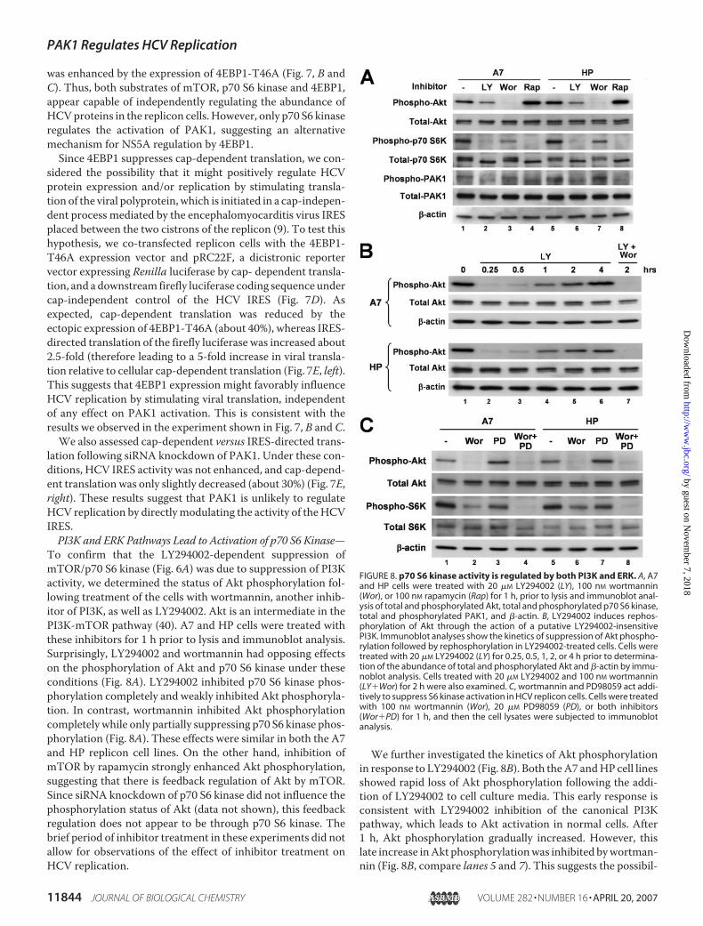
To confirm that the LY294002-dependent suppression ofmTOR/p70 S6 kinase (Fig. 6A) was due to suppression of PI3Kactivity, we determined the status of Akt phosphorylation fol-lowing treatment of the cells with wortmannin, another inhib-itor of PI3K, as well as LY294002. Akt is an intermediate in thePI3K-mTOR pathway (40). A7 and HP cells were treated withthese inhibitors for 1 h prior to lysis and immunoblot analysis.Surprisingly, LY294002 and wortmannin had opposing effectson the phosphorylation of Akt and p70 S6 kinase under theseconditions (Fig. 8A). LY294002 inhibited p70 S6 kinase phos-phorylation completely and weakly inhibited Akt phosphoryla-tion. In contrast, wortmannin inhibited Akt phosphorylationcompletely while only partially suppressing p70 S6 kinase phos-phorylation (Fig. 8A). These effects were similar in both the A7and HP replicon cell lines. On the other hand, inhibition ofmTOR by rapamycin strongly enhanced Akt phosphorylation,suggesting that there is feedback regulation of Akt by mTOR.Since siRNA knockdown of p70 S6 kinase did not influence thephosphorylation status of Akt (data not shown), this feedbackregulation does not appear to be through p70 S6 kinase. Thebrief period of inhibitor treatment in these experiments did notallow for observations of the effect of inhibitor treatment onHCV replication.
We further investigated the kinetics of Akt phosphorylationin response to LY294002 (Fig. 8B). Both theA7 andHP cell linesshowed rapid loss of Akt phosphorylation following the addi-tion of LY294002 to cell culture media. This early response isconsistent with LY294002 inhibition of the canonical PI3Kpathway, which leads to Akt activation in normal cells. After1 h, Akt phosphorylation gradually increased. However, thislate increase inAkt phosphorylationwas inhibited bywortman-nin (Fig. 8B, compare lanes 5 and 7). This suggests the possibil-
FIGURE 8. p70 S6 kinase activity is regulated by both PI3K and ERK. A, A7and HP cells were treated with 20 �M LY294002 (LY), 100 nM wortmannin(Wor), or 100 nM rapamycin (Rap) for 1 h, prior to lysis and immunoblot anal-ysis of total and phosphorylated Akt, total and phosphorylated p70 S6 kinase,total and phosphorylated PAK1, and �-actin. B, LY294002 induces rephos-phorylation of Akt through the action of a putative LY294002-insensitivePI3K. Immunoblot analyses show the kinetics of suppression of Akt phospho-rylation followed by rephosphorylation in LY294002-treated cells. Cells weretreated with 20 �M LY294002 (LY) for 0.25, 0.5, 1, 2, or 4 h prior to determina-tion of the abundance of total and phosphorylated Akt and �-actin by immu-noblot analysis. Cells treated with 20 �M LY294002 and 100 nM wortmannin(LY�Wor) for 2 h were also examined. C, wortmannin and PD98059 act addi-tively to suppress S6 kinase activation in HCV replicon cells. Cells were treatedwith 100 nM wortmannin (Wor), 20 �M PD98059 (PD), or both inhibitors(Wor�PD) for 1 h, and then the cell lysates were subjected to immunoblotanalysis.
PAK1 Regulates HCV Replication
11844 JOURNAL OF BIOLOGICAL CHEMISTRY VOLUME 282 • NUMBER 16 • APRIL 20, 2007
by guest on Novem
ber 7, 2018http://w
ww
.jbc.org/D
ownloaded from
ity of negative feedback in the regulation of PI3K, with therephosphorylation ofAkt involving awortmannin-sensitive butLY294002- insensitive PI3K-related kinase. However, sincewortmannin blocked Akt activation completely, the partialinhibition of p70 S6 kinase by wortmannin implies that p70 S6kinasemay be regulated by othermeans in addition to the PI3K/Akt pathway. On the other hand, LY294002 inhibited p70 S6kinase phosphorylation completely, even after rephosphoryla-tion of Akt. This raises the possibility that LY294002 mayinhibit the pathway at some point downstream of Akt, perhapsmTOR itself, as a previous report has suggested (48).Finally, we observed that inhibition of ERK by PD98059 par-
tially suppressed p70 S6 kinase (Fig. 8C). More importantly, acombination of PD98059 and wortmannin completely abol-ished the phosphorylation of p70 S6 kinase, suggesting thatPI3K and ERK are both likely to regulate mTOR activation,PAK1 phosphorylation, and thus cellular control of HCV RNAreplication.
DISCUSSION
We have shown here that HCV replication is suppressed byactivation of PAK1. Although our studies were initiated inresponse to a report that PAK1 may act upstream of TANK-binding kinase-1 and I�Bkinase-� in signaling pathways leadingto activation of IRF-3 (27), the PAK1 regulation of HCV repli-cation that we have demonstrated here appears to occur inde-pendently of IRF-3 activation. Virus activation of IRF-3 andattendant type I interferon responses is known to occurthrough several distinct pathways involving different patho-gen-associatedmolecular pattern receptors. TLR3 and its adap-tor TRIF (49) initiate signaling on binding extracellular double-stranded RNA, whereas the RIG-I and its homolog MDA5recognize viral RNAs within the cytoplasm and signal to IRF-3through the adaptor protein,MAVS (8). The twoHCV repliconcell lines we used in these studies, A7 and HP, are derived fromHuh7 cells and thus express negligible amounts of TLR3 (32).Furthermore, the RIG-I/MAVS pathway is completely dis-rupted in HP cells due to the expression of a high abundance oftheHCVNS3 protease, which efficiently targetsMAVS for pro-teolysis (50). Since neither the TLR3 or RIG-I pathway is activein HP cells, it is difficult to attribute the increase in HCV repli-con abundance observed with PAK1 knockdown (Fig. 1) in HPcells to inhibition of an IRF-3-mediated response.However, we did assess the possibility that PAK1 could play
an essential role in the RIG-I activation of IRF-3 in Huh7 cellsby determining whether siRNA knockdown of PAK1 interferedwith IRF-3 activation by Sendai virus, a potent stimulator of theRIG-I pathway. We found no differences in the activation ofIRF-3-responsive promoters following Sendai virus infection ofHuh7 cells in the presence or absence of PAK1 knockdown (Fig.2B). Moreover, Sendai virus infection was not accompanied byan increase in PAK1 phosphorylation (Fig. 2A). These data sug-gest that RIG-I-mediated activation of IRF-3 is not dependentupon PAK1 in Huh7 cells. Moreover, we also found thatpoly(I:C) stimulation of TLR3 did not lead to an increasedabundance of phosphorylated PAK1 in PH5CH8 cells (data notshown), a hepatocyte-derived cell line that does express abun-dant TLR3 (32). This suggests that the TLR3 pathway is also
likely to operate independently of PAK1. Our results are con-sistent with those reported recently by Noyce et al. (51), whodemonstrated that PAK1 is not essential for IRF-3 induction ofinterferon-stimulated genes mediated by Sendai virus.Importantly, we demonstrated that viral protein expres-
sion and the yield of cell culture-infectious HCV were bothenhanced by PAK1 knockdown in cells infected with anintergenotypic HCV chimera (Fig. 5). These data extend therange of HCV genotypes regulated by PAK1 beyond the gen-otype 1b replicons present in the HP and A7 cell lines, sincethe nonstructural protein-coding and nontranslated RNAsegments of the chimeric H-NS2/NS3-J/YH/QL virus arederived from the genotype 2a JFH1 virus (31). Patientsinfected with genotype 1 versus genotype 2 viruses areknown to respond differently to interferon treatment, with ahigh rate of sustained viral response in genotype 2-infectedpatients. Moreover, Ishii et al. (52) have demonstrated thatthe suppressive effect of cyclosporin on replicon RNA abun-dance is specific for genotype 1b. Cyclosporin has a lessereffect on JFH-1 replication, possibly because the cellular rep-lication cofactor, cyclophilin B, which stimulates the RNAbinding activity of genotype 1b NS5B, does not appear to berequired for replication of JFH1 (52). These data indicatethat there may be considerable diversity in the replicationphenotypes of different HCV genotypes. Although furtherstudies are needed, the results reported here suggest thatPAK1 knockdown enhances both replicon RNA abundanceas well as the replication of cell culture-infectious virus inde-pendent of genotype.PAK1 may be activated through multiple PI3K-dependent
pathways. In the cell lines we studied, we found that PI3Kand ERK both contributed to PAK1 activation. Rac1 (orCdc42) has been shown to bind to PAK1, leading to auto-phosphorylation and activation (26). However, the path-way(s) between PI3K and Rac1/PAK1 are not well delin-eated. PI3K is known to activate Akt, leading to theactivation of a number of cellular signaling partners and reg-ulation of proliferation and survival/apoptosis. In the celllines we studied, PAK1 was regulated by mTOR, which wefurther demonstrated to be activated by PI3K and ERK inexperiments involving the use of specific inhibitors (Fig. 9)(53, 54). Rapamycin binds to mTOR and inhibits its phos-phorylation, thus triggering suppression of the cap-depend-ent cellular protein synthesis machinery (55). Rapamycintreatment reduced PAK1 phosphorylation in replicon cellswhile enhancing HCV RNA replication (Fig. 6, C and D). Asimilar effect of mTOR on HCV replication was recentlydescribed by Mannova and Beretta (22).The potential roles of PI3K/Akt, ERK, and themTOR/p70 S6
kinase pathway, as defined in our studies, are depicted in Fig. 9.Our data suggest a model in which mTOR is regulated by bothAkt and ERK and in which there is negative feedback betweenmTOR and PI3K. This feedback is not mediated by p70 S6kinase, because the level of phosphorylated Akt did not changewhen p70 S6 kinase expression was disrupted by siRNA (datanot shown). Although mTOR is believed to be regulated byPI3K, Ma et al. (54) demonstrated that ERK phosphorylatestuberin (TSC2) and modulates mTOR signaling. They showed
PAK1 Regulates HCV Replication
APRIL 20, 2007 • VOLUME 282 • NUMBER 16 JOURNAL OF BIOLOGICAL CHEMISTRY 11845
by guest on Novem
ber 7, 2018http://w
ww
.jbc.org/D
ownloaded from

that either wortmannin orU0126 (an ERK inhibitor) could sup-press p70 S6 kinase phosphorylation and that these inhibitorshad a synergistic effect when used in combination. Consideringour results in the context of previous reports, it is possible thatthe relative contributions of PI3K and ERK to the activation ofmTOR may vary in different cell types. In addition, the mech-anism by which Akt rephosphorylation occurs following expo-sure to LY294002 remains unknown.We speculate that canon-ical (i.e. wortmannin- and LY294002-sensitive) PI3K activatesAkt under normal circumstances, whereas a putative nonca-nonical (wortmannin-sensitive, LY294002-insensitive) PI3K isactivated following LY294002 inhibition of the canonicalkinase. Consistent with this interpretation, Sharrard et al. (56)have suggested that rephosphorylation of Akt is mediated by aPI3K possessing a higher than usual IC50 for LY294002. Theseobservations would explain our results, although this PI3K isnot well characterized.mTOR has two known substrates, the p70 S6 kinase and
4EBP1, which together are responsible for mTOR-dependentregulation of cellular translation. Experiments inwhichweusedspecific siRNAs to knock down p70 S6 kinase demonstratedthat this kinase is upstream of PAK1 (Fig. 7A), whereas ectopicexpression of a constitutively active 4EBP1 did not influencethe abundance of phosphorylated PAK1 in the HCV repliconcells (Fig. 7B). Although disruption of p70 S6 kinase abolishedPAK1 phosphorylation (Fig. 7A), it is not likely that p70 S6kinase phosphorylates PAK1 directly, because the consensusphosphorylation motif of p70 S6 kinase ((K/R)XRXX(S/T)X)(57) is not present at sites of PAK1 phosphorylation. However,PAK1 has been recognized to form complexeswith the protein-serine/threonine phosphatase 2A and p70 S6 kinase (58), sug-gesting that theremay be an interaction between PAK1 and p70S6 kinase.
We also found that HCV replicon abundance was enhancedby expression of constitutively active 4EBP1, which binds toeukaryotic translation initiation factor 4E and blocks transla-tion in the same way as nonphosphorylated 4EBP1. Theseobservations confirm a recent report byMurata et al. (21). Thisenhancement seems likely to be due to increased efficiency ofIRES-directed translation in the context of decreased cap-de-pendent translation (Fig. 7E, left). In contrast, PAK1 knock-down had no effect on HCV IRES activity (Fig. 7E, right). Theseresults thus showed that both known mTOR substrates arecapable of suppressing HCV replication but that they do sothrough independent mechanisms. Our data indicate thatPAK1 plays a primary role in the p70 S6 kinase-mediated sup-pression of HCV replication.Several previous studies have investigated the relationship
between HCV RNA replication and PI3K/mTOR signaling.The PI3K has been shown to be activated by a direct inter-action between NS5A and the p85 regulatory subunit of PI3Kas well as by the enhanced expression of N-Ras in HCV rep-licon cells (22). N-Ras-mediated PI3K activation resulted inmTOR activation, and when mTOR was knocked down,HCV abundance was enhanced. These earlier findings areconsistent with our results, since we found HCV RNA repli-cation to be suppressed by either of the two mTOR sub-strates (Fig. 7). However, although we found PAK1 to beactivated in HCV replicon cells (Fig. 4, A and C), this did notappear to be due to the presence of HCV proteins, since itpersisted after the replicon RNAs were eliminated from theA7, HP, and 2-3 cell lines by interferon treatment (Fig. 4, Band C). Thus, PAK1 activation is constitutive in these repli-con cells lines. These results suggest that cells with highPI3K activity may have a selective advantage during the iso-lation of G418-resistant Huh7 cell colonies containing rep-licon RNA. Although this may seem contrary to the notionthat PAK1 (and PI3K) may negatively regulate the replica-tion of HCV, it may reflect other phenotypic properties ofcells with high PI3K, including enhanced proliferation (Fig.3) or cell survival.The ability of mTOR to suppress HCV replication has been
suggested to be due to the ability of the p70 S6 kinase to hyper-phosphorylate NS5A (57). However, this seems unlikely, sinceour results indicate that PAK1,which appears to be activated byit (Fig. 9), also suppresses HCV replicaton. Significantly, siRNAknockdown of either kinase did not result in a reduction of thehyperphosphorylated formofNS5A in the replicon cell lineswestudied (Figs. 1B and 7A).
The mechanism by which PAK1 influences HCV replica-tion is unclear. Since PAK1 may activate members of themitogen-activated protein kinase family (ERK, c-Jun NH2-terminal kinase, and p38) (36, 38, 39), it is possible that PAK1regulates HCV replication through these kinases. However,treatment of the replicon cells with rapamycin did notdecrease the phosphorylation of these kinases, implying thatthese pathways are irrelevant to PAK1-mediated regulationof HCV replication (data not shown). Similarly, inhibition ofNF-�B, which is also regulated by PAK1, by a chemical inhib-itor (BAY 11-7082) did not enhance but rather reduced rep-licon abundance (data not shown), suggesting that the
FIGURE 9. Schematic showing pathways by which PI3K and ERK/mTOR/p70 S6 kinase suppress HCV replication through PAK1. PAK1 is regulatedby mTOR downstream of PI3K and ERK. Among the two substrates of mTOR,4EBP1 and p70 S6 kinase, only the p70 S6 kinase activates PAK1, althoughphosphorylation of 4EBP1 by mTOR also suppresses HCV replication throughan alternative mechanism involving down-regulation of IRES-directedtranslation.
PAK1 Regulates HCV Replication
11846 JOURNAL OF BIOLOGICAL CHEMISTRY VOLUME 282 • NUMBER 16 • APRIL 20, 2007
by guest on Novem
ber 7, 2018http://w
ww
.jbc.org/D
ownloaded from
NF-�B signaling pathway is not involved either. Somecytoskeleton molecules have been demonstrated to be phos-phorylated or otherwise regulated by PAK1 (25), and it ispossible that they may play a role in HCV replicaton. Op18/stathmin normally associates with microtubules and dis-rupts microtubule dimerization (59). When phosphorylatedby PAK1, it no longer associates with microtubules, suggest-ing that PAK1 contributes to the stabilization of microtu-bules. Since microtubules appear to be required for HCVreplication (60), it is unlikely that this specific action ofPAK1 suppresses HCV replication. However, the influenceof PAK1 on other cytoskeleton molecules and their potentialrelationship to HCV replication is unclear.
Acknowledgments—We thank Michael Gale for A7 and HP cells andthe pISG56-Luc and pPRDII-Luc constructs, Charles Rice for Huh7.5cells, Craig Cameron for antibody to NS5A, and Rongtuan Lin forpIFN-�-Luc.We also thankQingDingWang andMark Evers for help-ful discussions and Emi Arimoto Ishida for excellent technicalsupport.
REFERENCES1. Seeff, L. B. (1997) Hepatology 26, 21S–28S2. Lauer, G. M., and Walker, B. D. (2001) N. Engl. J. Med. 345, 41–523. Choo, Q. L., Richman, K. H., Han, J. H., Berger, K., Lee, C., Dong, C.,
Gallegos, C., Coit, D., Medina-Selby, A., Barr, P. J., Weiner, A. J., Bradley,D.W., Kuo, G., andHoughton,M. (1991) Proc. Natl. Acad. Sci. U. S. A. 88,2451–2455
4. Major, M. E., and Feinstone, S. M. (1997) Hepatology 25, 1527–15385. Conzelmann, K. K. (2005) J. Virol. 79, 5241–52486. Foy, E., Li, K.,Wang, C., Sumpter, R., Jr., Ikeda,M., Lemon, S.M., andGale,
M., Jr. (2003) Science 300, 1145–11487. Li, K., Foy, E., Ferreon, J. C., Nakamura, M., Ferreon, A. C., Ikeda, M., Ray,
S. C., Gale, M., Jr., and Lemon, S. M. (2005) Proc. Natl. Acad. Sci. U. S. A.102, 2992–2997
8. Meylan, E., Curran, J., Hofmann, K., Moradpour, D., Binder, M., Barten-schlager, R., and Tschopp, J. (2005) Nature 437, 1167–1172
9. Lohmann, V., Korner, F., Koch, J.-O., Herian, U., Theilmann, L., and Bar-tenschlager, R. (1999) Science 285, 110–113
10. Wakita, T., Pietschmann, T., Kato, T., Date, T., Miyamoto, M., Zhao, Z.,Murthy, K., Habermann, A., Krausslich, H. G., Mizokami, M., Barten-schlager, R., and Liang, T. J. (2005) Nat. Med. 11, 791–796
11. Yi, M., Villanueva, R. A., Thomas, D. L., Wakita, T., and Lemon, S. M.(2006) Proc. Natl. Acad. Sci. U. S. A. 103, 2310–2315
12. Lindenbach, B. D., Evans, M. J., Syder, A. J., Wolk, B., Tellinghuisen, T. L.,Liu, C. C.,Maruyama, T., Hynes, R. O., Burton, D. R.,McKeating, J. A., andRice, C. M. (2005) Science 309, 623–626
13. Zhong, J., Gastaminza, P., Cheng, G., Kapadia, S., Kato, T., Burton, D. R.,Wieland, S. F., Uprichard, S. L., Wakita, T., and Chisari, F. V. (2005) Proc.Natl. Acad. Sci. U. S. A. 102, 9294–9299
14. Frese, M., Pietschmann, T., Moradpour, D., Haller, O., and Barten-schlager, R. (2001) J. Gen. Virol. 82, 723–733
15. Frese, M., Schwarzle, V., Barth, K., Krieger, N., Lohmann, V., Mihm, S.,Haller, O., and Bartenschlager, R. (2002) Hepatology 35, 694–703
16. Zhu, H., and Liu, C. (2003) J. Virol. 77, 5493–549817. Murata, T., Ohshima, T., Yamaji, M., Hosaka, M., Miyanari, Y., Hijikata,
M., and Shimotohno, K. (2005) Virology 331, 407–41718. Pause, A., Kukolj, G., Bailey, M., Brault, M., Do, F., Halmos, T., Lagace, L.,
Maurice, R.,Marquis,M.,McKercher, G., Pellerin, C., Pilote, L., Thibeault,D., and Lamarre, D. (2003) J. Biol. Chem. 278, 20374–20380
19. Dhanak, D., Duffy, K. J., Johnston, V. K., Lin-Goerke, J., Darcy, M., Shaw,A. N., Gu, B., Silverman, C., Gates, A. T., Nonnemacher, M. R., Earnshaw,D. L., Casper, D. J., Kaura, A., Baker, A., Greenwood, C., Gutshall, L. L.,Maley, D., DelVecchio, A., Macarron, R., Hofmann, G. A., Alnoah, Z.,
Cheng, H. Y., Chan, G., Khandekar, S., Keenan, R. M., and Sarisky, R. T.(2002) J. Biol. Chem. 277, 38322–38327
20. Randall, G., Grakoui, A., and Rice, C. M. (2003) Proc. Natl. Acad. Sci.U. S. A. 100, 235–240
21. Murata, T., Hijikata, M., and Shimotohno, K. (2005) Virology 340,105–115
22. Mannova, P., and Beretta, L. (2005) J. Virol. 79, 8742–874923. Scholle, F., Li, K., Bodola, F., Ikeda, M., Luxon, B. A., and Lemon, S. M.
(2004) J. Virol. 78, 1513–152424. Pietschmann, T., Lohmann, V., Rutter, G., Kurpanek, K., and Barten-
schlager, R. (2001) J. Virol. 75, 1252–126425. Bokoch, G. M. (2003) Annu. Rev. Biochem. 72, 743–78126. Parrini, M. C., Lei, M., Harrison, S. C., andMayer, B. J. (2002)Mol. Cell 9,
73–8327. Ehrhardt, C., Kardinal, C.,Wurzer,W. J.,Wolff, T., von Eichel-Streiber, C.,
Pleschka, S., Planz, O., and Ludwig, S. (2004) FEBS Lett. 567, 230–23828. Ikeda, M., Yi, M., Li, K., and Lemon, S. M. (2002) J. Virol. 76,
2997–300629. Fredericksen, B., Akkaraju, G. R., Foy, E., Wang, C., Pflugheber, J., Chen,
Z. J., and Gale, M., Jr. (2003) Viral Immunol. 15, 29–4030. Wang, T. H., Rijnbrand, R. C., and Lemon, S. M. (2000) J. Virol. 74,
11347–1135831. Yi, M., Ma, Y., Yates, J., and Lemon, S. M. (2007) J. Virol. 81, 629–63832. Li, K., Chen, Z., Kato, N., Gale, M., Jr., and Lemon, S. M. (2005) J. Biol.
Chem. 280, 16739–1674733. Sumpter, R., Jr., Loo,M. Y., Foy, E., Li, K., Yoneyama,M., Fujita, T., Lemon,
S. M., and Gale, M. J., Jr. (2005) J. Virol. 79, 2689–269934. Menard, R. E., and Mattingly, R. R. (2003) Cell. Signal. 15, 1099–110935. Frost, J. A., Swantek, J. L., Stippec, S., Yin, M. J., Gaynor, R., and Cobb,
M. H. (2000) J. Biol. Chem. 275, 19693–1969936. Frost, J. A., Steen, H., Shapiro, P., Lewis, T., Ahn,N., Shaw, P. E., andCobb,
M. H. (1997) EMBO J. 16, 6426–643837. Balasenthil, S., Sahin, A. A., Barnes, C. J., Wang, R. A., Pestell, R. G., Vad-
lamudi, R. K., and Kumar, R. (2004) J. Biol. Chem. 279, 1422–142838. Zhang, S., Han, J., Sells,M.A., Chernoff, J., Knaus, U.G., Ulevitch, R. J., and
Bokoch, G. M. (1995) J. Biol. Chem. 270, 23934–2393639. Brown, J. L., Stowers, L., Baer, M., Trejo, J., Coughlin, S., and Chant, J.
(1996) Curr. Biol. 6, 598–60540. Hay, N., and Sonenberg, N. (2004) Genes Dev. 18, 1926–194541. Evans,M. J., Rice, C.M., andGoff, S. P. (2004) Proc. Natl. Acad. Sci. U. S. A.
101, 13038–1304342. Blight, K. J., Kolykhalov, A. A., and Rice, C. M. (2000) Science 290,
1972–197443. Neddermann, P., Quintavalle, M., Di Pietro, C., Clementi, A., Cerretani,
M., Altamura, S., Bartholomew, L., andDe Francesco, R. (2004) J. Virol.78,13306–13314
44. Quintavalle, M., Sambucini, S., Di Pietro, C., De Francesco, R., and Ned-dermann, P. (2006) J. Virol. 80, 11305–11312
45. Brown, E. J., Beal, P. A., Keith, C. T., Chen, J., Shin, T. B., and Schreiber,S. L. (1995) Nature 377, 441–446
46. Hara, K., Yonezawa, K., Kozlowski, M. T., Sugimoto, T., Andrabi, K.,Weng,Q. P., Kasuga,M., Nishimoto, I., andAvruch, J. (1997) J. Biol. Chem.272, 26457–26463
47. Haghighat, A.,Mader, S., Pause, A., and Sonenberg, N. (1995) EMBO J. 14,5701–5709
48. Brunn, G. J., Williams, J., Sabers, C., Wiederrecht, G., Lawrence, J. C., Jr.,and Abraham, R. T. (1996) EMBO J. 15, 5256–5267
49. Yamamoto, M., Sato, S., Mori, K., Hoshino, K., Takeuchi, O., Takeda, K.,and Akira, S. (2002) J. Immunol. 169, 6668–6672
50. Loo, Y. M., Owen, D.M., Li, K., Erickson, A. K., Johnson, C. L., Fish, P. M.,Carney, D. S.,Wang, T., Ishida, H., Yoneyama,M., Fujita, T., Saito, T., Lee,W. M., Hagedorn, C. H., Lau, D. T., Weinman, S. A., Lemon, S. M., andGale, M., Jr. (2006) Proc. Natl. Acad. Sci. U. S. A. 103, 6001–6006
51. Noyce, R. S., Collins, S. E., and Mossman, K. L. (2006) J. Virol. 80,226–235
52. Ishii, N.,Watashi, K., Hishiki, T., Goto, K., Inoue, D., Hijikata, M.,Wakita,T., Kato, N., and Shimotohno, K. (2006) J. Virol. 80, 4510–4520
53. Inoki, K., Li, Y., Zhu, T., Wu, J., and Guan, K. L. (2002) Nat. Cell Biol. 4,
PAK1 Regulates HCV Replication
APRIL 20, 2007 • VOLUME 282 • NUMBER 16 JOURNAL OF BIOLOGICAL CHEMISTRY 11847
by guest on Novem
ber 7, 2018http://w
ww
.jbc.org/D
ownloaded from

648–65754. Ma, L., Chen, Z., Erdjument-Bromage, H., Tempst, P., and Pandolfi, P. P.
(2005) Cell 121, 179–19355. Gingras, A. C., Raught, B., and Sonenberg, N. (2001) Genes Dev. 15,
807–82656. Sharrard, R. M., and Maitland, N. J. (2007) Cell. Signal. 19, 129–13857. Coito, C., Diamond, D. L., Neddermann, P., Korth, M. J., and Katze, M. G.
(2004) J. Virol. 78, 3502–351358. Westphal, R. S., Coffee, R. L., Jr., Marotta, A., Pelech, S. L., andWadzinski,
B. E. (1999) J. Biol. Chem. 274, 687–69259. Wittmann, T., Bokoch, G. M., andWaterman-Storer, C. M. (2003) J. Biol.
Chem. 279, 6196–620360. Bost, A. G., Venable, D., Liu, L., and Heinz, B. A. (2003) J. Virol. 77,
4401–4408
PAK1 Regulates HCV Replication
11848 JOURNAL OF BIOLOGICAL CHEMISTRY VOLUME 282 • NUMBER 16 • APRIL 20, 2007
by guest on Novem
ber 7, 2018http://w
ww
.jbc.org/D
ownloaded from
Hisashi Ishida, Kui Li, MinKyung Yi and Stanley M. LemonVirus in Human Hepatoma Cells
Rapamycin/p70 S6 Kinase Pathway and Regulates the Replication of Hepatitis C p21-activated Kinase 1 Is Activated through the Mammalian Target of
doi: 10.1074/jbc.M610106200 originally published online January 25, 20072007, 282:11836-11848.J. Biol. Chem.
10.1074/jbc.M610106200Access the most updated version of this article at doi:
Alerts:
When a correction for this article is posted•
When this article is cited•
to choose from all of JBC's e-mail alertsClick here
http://www.jbc.org/content/282/16/11836.full.html#ref-list-1
This article cites 60 references, 38 of which can be accessed free at
by guest on Novem
ber 7, 2018http://w
ww
.jbc.org/D
ownloaded from