Embed Size (px)
Citation preview

Antifibrotic Effects of Noscapine through Activation ofProstaglandin E2 Receptors and Protein Kinase A*
Received for publication, December 29, 2013, and in revised form, January 31, 2014 Published, JBC Papers in Press, February 3, 2014, DOI 10.1074/jbc.M113.546812
Jacob Kach‡, Nathan Sandbo§, Jennifer La‡, Darcy Denner‡, Eleanor B. Reed‡, Olga Akimova¶, Svetlana Koltsova¶,Sergei N. Orlov¶, and Nickolai O. Dulin‡1
From the ‡Department of Medicine, University of Chicago, Chicago, Illinois 60637, the §Department of Medicine, University ofWisconsin-Madison, Madison, Wisconsin 52792, and ¶Faculty of Biology, Moscow State University, Moscow, Russia 119991
Background: Noscapine is a safe orally available anticough medicine also known to bind microtubules and induce cancercell death.Results: Noscapine inhibits myofibroblast differentiation and pulmonary fibrosis through prostaglandin receptors and activa-tion of PKA.Conclusion: Noscapine is an antifibrotic drug acting through PKA activation via EP2 prostaglandin receptors.Significance: This study describes a novel antifibrotic function and novel mechanism of action of noscapine.
Myofibroblast differentiation is a key process in the patho-genesis of fibrotic disease. We have shown previously that dif-ferentiation of myofibroblasts is regulated by microtubulepolymerization state. In this work, we examined the potentialantifibrotic effects of the antitussive drug, noscapine, recentlyfound to bind microtubules and affect microtubule dynamics.Noscapine inhibited TGF-�-induced differentiation of culturedhuman lung fibroblasts (HLFs). Therapeutic noscapine treat-ment resulted in a significant attenuation of pulmonary fibrosisin the bleomycin model of the disease. Noscapine did not affectgross microtubule content in HLFs, but inhibited TGF-�-in-duced stress fiber formation and activation of serum responsefactor without affecting Smad signaling. Furthermore, noscap-ine stimulated a rapid and profound activation of protein kinaseA (PKA), which mediated the antifibrotic effect of noscapine inHLFs, as assessed with the PKA inhibitor, PKI. In contrast,noscapine did not activate PKA in human bronchial or alve-olar epithelial cells. Finally, activation of PKA and the antifi-brotic effect of noscapine in HLFs were blocked by the EP2
prostaglandin E2 receptor antagonist, PF-04418948, but notby the antagonists of EP4, prostaglandin D2, or prostacyclinreceptors. Together, we demonstrate for the first time theantifibrotic effect of noscapine in vitro and in vivo, and wedescribe a novel mechanism of noscapine action through EP2prostaglandin E2 receptor-mediated activation of PKA in pul-monary fibroblasts.
Idiopathic pulmonary fibrosis is a progressive, fatal diseasecharacterized by parenchymal fibrosis and structural distortionof the lungs. Age-adjusted mortality due to pulmonary fibrosisis increasing (1), and it poses a vexing clinical challenge given
the lack of proven efficacious therapy. Idiopathic pulmonaryfibrosis is thought to be a disorder of abnormal wound healing(2, 3), in which the initial trigger to the fibrotic response isinjury to the alveolar epithelial cells, followed by an exuberant,nonresolving wound healing response (4 – 6) driven by myofi-broblasts, specialized fibroblasts with enhanced contractile andmatrix gene expression (7–9). Disrupting cellular mechanismsresponsible for the acquisition of the myofibroblast phenotypemay be a potential strategy to attenuate the ongoing fibroticresponse in pulmonary fibrosis.
Transforming Growth Factor-�1 (TGF-�1) is a key mediatorof myofibroblast differentiation and tissue fibrosis (10 –12). Wehave demonstrated previously that TGF-� utilizes stress fiber-dependent signaling driving nuclear accumulation of mega-karyoblastic leukemia-1 (MKL1)2 and activation of serumresponse factor (SRF) to fully induce the myofibroblast pheno-type, and that activators of protein kinase A (PKA) attenuatemyofibroblast differentiation through inhibition of SRF, butnot Smad signaling (13, 14). These data suggest that targetingthe MKL1/SRF pathway could be an attractive approach forpreventing myofibroblast differentiation and pulmonary fibro-sis without affecting the initial Smad signaling of TGF-�.
Previous studies using the bleomycin model of pulmonaryfibrosis have shown the protective effects of the prostacyclinanalog iloprost or of prostaglandin E2 (15, 16), both of which areknown to act through cAMP/PKA signaling. We have shown asimilar protective effect of adrenomedullin in a mouse modelwith genetic sensitization of adrenomedullin signaling in myo-fibroblasts (17). We also have shown recently that myofibro-blast differentiation is regulated by microtubule dynamicsthrough a control of actin stress fiber/SRF signaling, indepen-dent of Smads (18). Unfortunately, the prototypical microtubulestabilizer, paclitaxel (Taxol), has significant toxicity which lim-its its potential as a therapeutic in fibrotic disease. A search formodulators of microtubule dynamics with low toxicity in vivo
* This work was supported, in whole or in part, by National Institutes of HealthGrants R01 GM85058 (to N. O. D), K08 HL093367-01A1 (to N. S.), andT32HL007237 (to J. K.). This work was also supported by American HeartAssociation Fellowship 10PRE4190120 (to J. K.).
1 To whom correspondence should be addressed: Section of Pulmonary andCritical Care Medicine, Dept. of Medicine, the University of Chicago, 5841 S.Maryland Ave., MC6076, Chicago, IL 60637. Tel.: 773-702-5198; Fax: 773-702-6500; E-mail: [email protected].
2 The abbreviations used are: MKL1, megakaryoblastic leukemia-1; DP,D-prostanoid receptor; EP, E-prostanoid receptor; HLF, human lung fibro-blast; SM, smooth muscle; SMA, SM-�-actin; SRF, serum response factor;VASP, vasodilator-stimulated phosphoprotein.
THE JOURNAL OF BIOLOGICAL CHEMISTRY VOL. 289, NO. 11, pp. 7505–7513, March 14, 2014© 2014 by The American Society for Biochemistry and Molecular Biology, Inc. Published in the U.S.A.
MARCH 14, 2014 • VOLUME 289 • NUMBER 11 JOURNAL OF BIOLOGICAL CHEMISTRY 7505
by guest on February 3, 2019http://w
ww
.jbc.org/D
ownloaded from
identified noscapine as a potential candidate compound, andwe sought to investigate its antifibrotic potential.
Noscapine is a nonsedative, nonaddictive alkaloid found inopium latex, which has been used with low toxicity as an anti-tussive medication in humans (19 –21). The mechanism of theantitussive action of noscapine remains unclear but mayinvolve the central nervous system (22, 23) and more specifi-cally, sigma receptors, as this effect of noscapine was inhibitedby the sigma receptor antagonist, rimcazole (24). Morerecently, studies by Joshi’s group have shown that noscapinebinds to tubulin, changes its conformation, and arrests dividingmammalian cells in mitosis through its effects on microtubuleassembly (25). Therefore, noscapine has been studied exten-sively in cancer research, and its antitumor activity has beendemonstrated in multiple in vivo studies (25–28).
In this study, we show for the first time that noscapine is aneffective antifibrotic agent both in vitro and in vivo and describea novel mechanism of noscapine action through a rapid activa-tion of cAMP/PKA signaling mediated by EP2 prostaglandin E2receptors in pulmonary fibroblasts.
EXPERIMENTAL PROCEDURES
Primary Culture of Human and Mouse Lung Fibroblasts—Human lung fibroblasts were cultured as described previously(13). Briefly, tissue samples from explanted lungs from patientsundergoing lung transplantation were placed in DMEM withantibiotics. Lung tissue was minced to �1-mm3 pieces, washed,and plated on 10-cm plates in growth medium containingDMEM supplemented with 10% FBS and antibiotics. Themedium was changed twice a week. After �2 weeks, theexplanted and amplified fibroblasts were cleared from the tis-sue pieces, trypsinized, and further amplified as passage 1.Mouse lung fibroblasts were cultured in a similar manner. Forexperiments, cells were grown in 12-well plates at a density of1 � 105 cells/well in a growth medium for 24 h, starved inDMEM containing 0.1% bovine serum albumin (BSA) for 48 h,and treated with desired drugs for various times as indicated inthe figure legends. All primary cultures were used from passage3 to 10.
Cytotoxicity Assay—Cytotoxicity of drugs was examined bymeasuring the release of lactate dehydrogenase in the media,using the CytoTox 96 assay (Promega) following the manufa-cturer’s protocol.
Cell Lysis and Western Blotting—Cells were lysed in radioim-munoprecipitation assay buffer containing 25 mM HEPES (pH7.5), 150 mM NaCl, 0.1% sodium dodecyl sulfate (SDS), 1% Tri-ton X-100, 0.5% sodium deoxycholate, 2 mM EDTA, 2 mM
EGTA, 10% glycerol, 1 mM NaF, 200 �M sodium orthovanadate,and protease inhibitors (1 �g/ml leupeptin, 1 �g/ml aprotinin,1 mM PMSF), scraped, sonicated for 5 s, mixed with Laemmlibuffer, and boiled for 5 min. Samples were then subjected topolyacrylamide gel electrophoresis and Western blotting withdesired primary antibodies and corresponding horseradish per-oxidase (HRP)-conjugated secondary antibodies, and devel-oped by chemiluminescence reaction (Pierce). Digital chemilu-minescent images below the saturation level were obtainedwith a LAS-4000 analyzer, and the light intensity was quantifiedusing Multi Gauge software (Fujifilm).
Bleomycin-induced Pulmonary Fibrosis— 8 –10 week oldC57BL/6 were intratracheally instilled with 1 unit/kg bleomy-cin (Bleocip, Cipla Ltd.). Noscapine was administered intra-peritoneally daily at a dose of 100 mg/kg beginning on day 7after bleomycin treatment. Lungs were removed 21 days afterbleomycin administration. Left lungs were formalin-fixed andparaffin embedded, and immunohistochemistry was performedon lung sections. Images of the stained sections were obtained on aCRi Pannoramic whole slide scanner. Right lungs were used forhydroxyproline content measurements.
Hydroxyproline Assay—The hydroxyproline assay was per-formed as described previously (17). Briefly, right lungs werehomogenized in 6 N hydrochloric acid and hydrolyzed for 12 hat 110 °C. An aliquot was evaporated, resuspended in citrate-acetate buffer with chloramine T, and left at room temperaturefor 20 min. Ehrlich’s solution was then added, and samples wereheated at 65 °C for 15 min. After cooling to room temperature,absorbance was measured at 550 nm. Hydroxyproline contentwas determined against a standard curve generated from purehydroxyproline.
Transfection and Luciferase Assay—Subconfluent cells wereco-transfected with desired firefly luciferase reporter plasmid,and thymidine kinase (TK) promoter-driven Renilla luciferaseplasmid (TK-Rl). Cells were serum-starved followed by stimu-lation with the desired agonists. Cells were washed and thenlysed in protein extraction reagent. Lysates were assayed forfirefly and Renilla luciferase activity using the dual luciferaseassay kit (Promega). To account for differences in transfectionefficiency, firefly luciferase activity of each sample was normal-ized to Renilla luciferase activity.
In Vitro Isolation of Stress Fibers—Stress fibers were isolatedas described previously (14, 18). All of the procedures wereperformed on ice using buffers containing protease inhibitors(Sigma). After stimulation with desired agonists, cells werewashed and then extracted with buffer containing 2.5 mM tri-ethanolamine (pH 8.2), followed by extraction with 0.05% Non-idet P-40 (pH 7.2) and subsequent extraction with 0.5% TritonX-100 (pH 7.2). Cells were washed, PBS was added, and the cellswere scraped and centrifuged at 100,000 � g for 1 h. Superna-tant was removed, and the pellet was sonicated in PBS.
Isolation of nuclear and cytoplasmic fractions was performedusing NE-PER nuclear and cytoplasmic extraction kit (ThermoScientific) following the manufacturer’s protocol. The purity ofisolated fractions was confirmed by Western blotting with thenuclear lamin A/C and cytoplasmic 14-3-3�.
In Vitro Isolation of Soluble and Insoluble MicrotubuleFractions—Soluble and insoluble fractions of microtubuleswere isolated using a method described previously (29). Cellswere rinsed twice with warm Dulbecco’s phosphate-bufferedsaline and lysed at room temperature in microtubule-stabiliz-ing buffer containing 2 M glycerol, 20 mM Tris-HCl (pH 6.8),140 mM NaC1, 0.5% Nonidet P-40, 1 mM MgC12, 2 mM EGTA,10 �M Taxol, and protease inhibitors mixture. The plates werethen put on ice; cells were scraped and transferred to a micro-centrifuge tube. Each well was rinsed with a second aliquot ofstabilization buffer, combined with the first aliquot, and brieflyvortexed. Lysates were centrifuged at 20,000 � g for 10 min at4 °C. Supernatants containing soluble tubulin were removed,
Activation of PKA by Noscapine
7506 JOURNAL OF BIOLOGICAL CHEMISTRY VOLUME 289 • NUMBER 11 • MARCH 14, 2014
by guest on February 3, 2019http://w
ww
.jbc.org/D
ownloaded from

and the pellets with polymerized tubulin were lysed in radioim-munoprecipitation assay buffer and sonicated.
Indirect Immunofluorescence Microscopy—Cells were fixedin 4% paraformaldehyde in PBS followed by permeabilizationwith 0.2% Triton X-100 in PBS. Cells were then incubated in 2%BSA in PBS, followed by incubation with mouse �-tubulin anti-bodies at 4 °C overnight, washed, and incubated with anti-mouse FITC-conjugated secondary antibodies, washed, andcoverslips were mounted using Vectashield mounting mediumcontaining DAPI nuclear stain (Vector Laboratories, Burlin-game, CA). Immunofluorescence images were obtained usingan Olympus 1X71 fluorescent microscope.
Adenovirus-mediated DNA Transduction—Adenovirus (0.2 �109 viral particles/ml) was incubated with cells for 24 h withLipofectamine LTX reagent (Stratagene, La Jolla, CA) to facili-tate the efficiency of transduction. Cells were then serum-starved and stimulated with the desired agonists for the indi-cated times.
Reagents—Noscapine hydrochloride hydrate was from Sigma-Aldrich. TGF-� was from Calbiochem. Pharmaceutical gradebleomycin (Bleocip) was from Cipla LTD. SC-560 and NS-298were from Sigma-Aldrich. AH-6809, RO-1138452,PF-04418948, L-161,982 and BW A868C were from CaymanChemicals. Rimcazole was from Santa Cruz Biotechnology.Antibodies against SM �-actin, �-actin, and �-tubulin werefrom Sigma-Aldrich; collagen-1 antibodies used for Westernblotting were from Cedarlane; vasodilator-stimulated phos-phoprotein (VASP) antibodies were from Calbiochem; MKL1antibodies were from Bethyl Laboratories; PKA-substrate anti-bodies were from Cell Signaling Technologies. Antibodiesagainst collagen I (ab21286) and collagen III (ab7778) wereobtained from Abcam and were used for immunohistochemis-try at the dilution of 1:300 and 1:600, respectively. The fireflyluciferase reporter for SRF activity (SRF-Luc) was a gift fromTatyana Voyno-Yasenetskaya (University of Illinois at Chi-cago). The firefly luciferase reporter for the �125 base pair SM�-actin promoter was a kind gift of Dr. Joseph Miano (Univer-sity of Rochester). The plasmid for luciferase reporter driven bySmad-binding elements (SBE-Luc) was provided by Dr. BertVogelstein (The Johns Hopkins University). Ad-PKI and Ad-LacZ were described previously (30).
Statistical Analysis—Quantitative data from three indepen-dent experiments were analyzed by Student’s t test. Values ofp � 0.05 (*) were considered statistically significant.
RESULTS
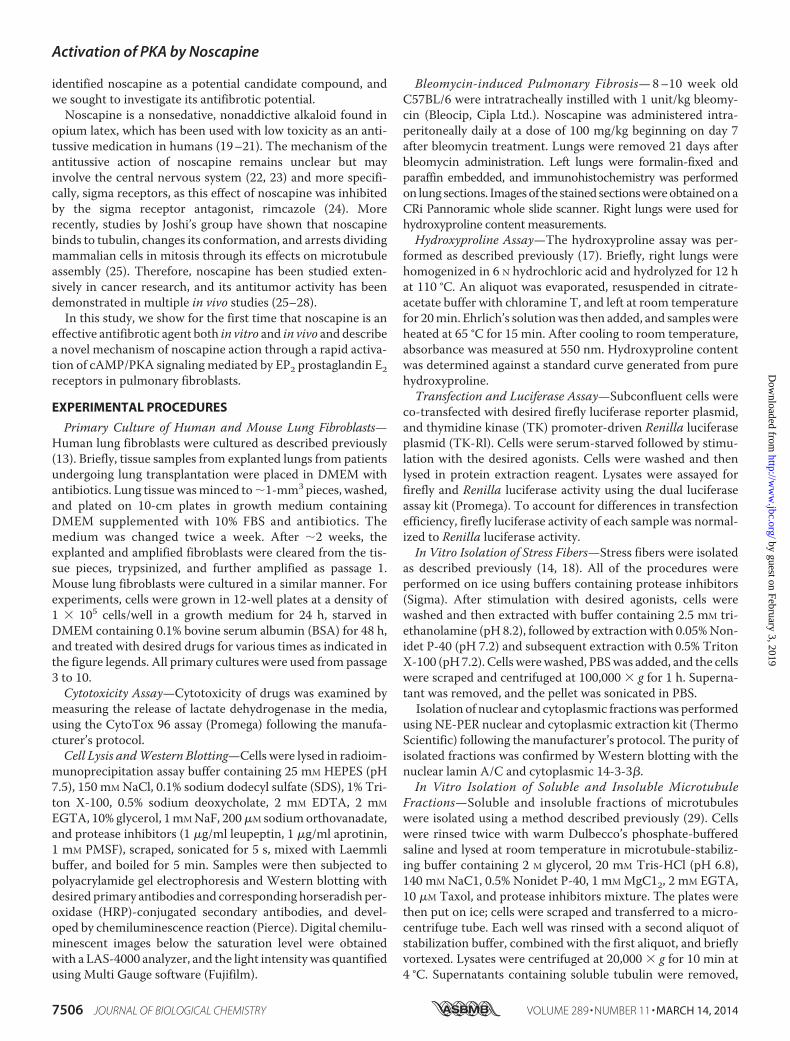
Antifibrotic Effects of Noscapine in Vitro and in Vivo—Wefirst examined the effect of noscapine on TGF-�-induced dif-ferentiation of cultured human lung fibroblasts (HLFs). Asshown in Fig. 1A, noscapine significantly attenuated TGF-�-induced expression of SM-�-actin and collagen-1 in culturedHLFs. Given the reported effect of noscapine on apoptosis ofcancer cells (25), we examined the cytotoxic effect of noscapineon HLF by measuring the release of lactate dehydrogenease. Asshown in Fig. 1B, noscapine treatment promoted a smallalthough statistically significant release of lactate dehydroge-nase compared with a profound effect of hydrogen peroxide.
To examine the effect of noscapine on fibrinogenesis in vivo,we used the bleomycin model of pulmonary fibrosis in mice. Inthis model, intratracheal administration of bleomycin results inthe initial injury of alveolar epithelial cells followed by aninflammatory response during the first week and developmentof pulmonary fibrosis at 2–3 weeks after bleomycin administra-tion. As shown in Fig. 1, intratracheal administration of bleo-mycin resulted in a significant pulmonary fibrosis accompaniedby accumulation myofibroblasts, as determined by hydroxy-proline assay for collagen content (Fig. 1C) and by histologicalassessment of lung sections stained with H&E, trichrome, orwith antibodies against collagen I, collagen III, or SM-�-actin(Fig. 1D). To avoid a potential effect of noscapine on the initialinjury and inflammatory response, we used a “therapeutic” pro-tocol, wherein noscapine was administered daily beginning onday 7 after bleomycin administration, and the lungs were ana-lyzed on day 21. As shown in Fig. 1, administration of noscapineresulted in a significant decrease of pulmonary fibrosis asassessed by the hydroxyproline assay of mouse lungs and byimmunohistochemical staining of mouse lung sections withantibodies against collagen I, collagen III, or SM-�-actin.Importantly, noscapine-treated lungs had a clearly decreasednumber of SM-�-actin-positive cells (Fig. 1D), which is consis-tent with the regulation of myofibroblast differentiation bynoscapine in vitro (Fig. 1A).
Noscapine Inhibits TGF-�1-induced SRF Activity withoutAffecting Smad-dependent Gene Transcription—We nextexamined the mechanism by which noscapine inhibits myofi-broblast differentiation in response to TGF-�. We and othershave established previously that this process is initiated bySmad-dependent expression of intermediate signaling mole-cules driving the activation of SRF that is required for theexpression of SM-�-actin, collagen 1, and other myofibroblastdifferentiation markers (13, 14, 31). We also showed that acti-vation of SRF by TGF-� in HLF is mediated by increasedexpression and nuclear accumulation of SRF co-activatorMKL1 and that this process is blocked by microtubule stabi-lizer, Taxol (14, 18). As shown in Fig. 2A, noscapine attenuatedTGF-�-induced expression and accumulation of MKL1 in thenuclear fraction. Furthermore, treatment of HLFs with noscap-ine resulted in a significantly decreased activation of SRF byTGF-�, as assessed by a SRF-driven luciferase reporter (Fig.2B). Finally, noscapine blocked TGF-�-induced activation of�125-base pair fragment of SM-�-actin promoter (Fig. 2C)known to contain two SRF binding sites (32). In contrast,noscapine did not affect TGF-�-induced Smad2 phosphoryla-tion (Fig. 2D), nuclear accumulation (Fig. 2E), or Smad-depen-dent gene transcription, as determined by a luciferase reporterdriven by Smad-binding elements (Fig. 2F).
Noscapine Does Not Affect the Gross Microtubule Content butInhibits Actin Polymerization in Response to TGF-�—In vitrostudies by others suggest that noscapine does not promote orinhibit the gross microtubule polymerization, but rather affectsthe steady-state dynamics of microtubule assembly, by increas-ing the ”pause” state of microtubules in cancer cells (33). Nev-ertheless, given our previously published data showing thatmicrotubule stabilization by Taxol inhibits myofibroblast dif-ferentiation (18), we examined whether noscapine affected the
Activation of PKA by Noscapine
MARCH 14, 2014 • VOLUME 289 • NUMBER 11 JOURNAL OF BIOLOGICAL CHEMISTRY 7507
by guest on February 3, 2019http://w
ww
.jbc.org/D
ownloaded from
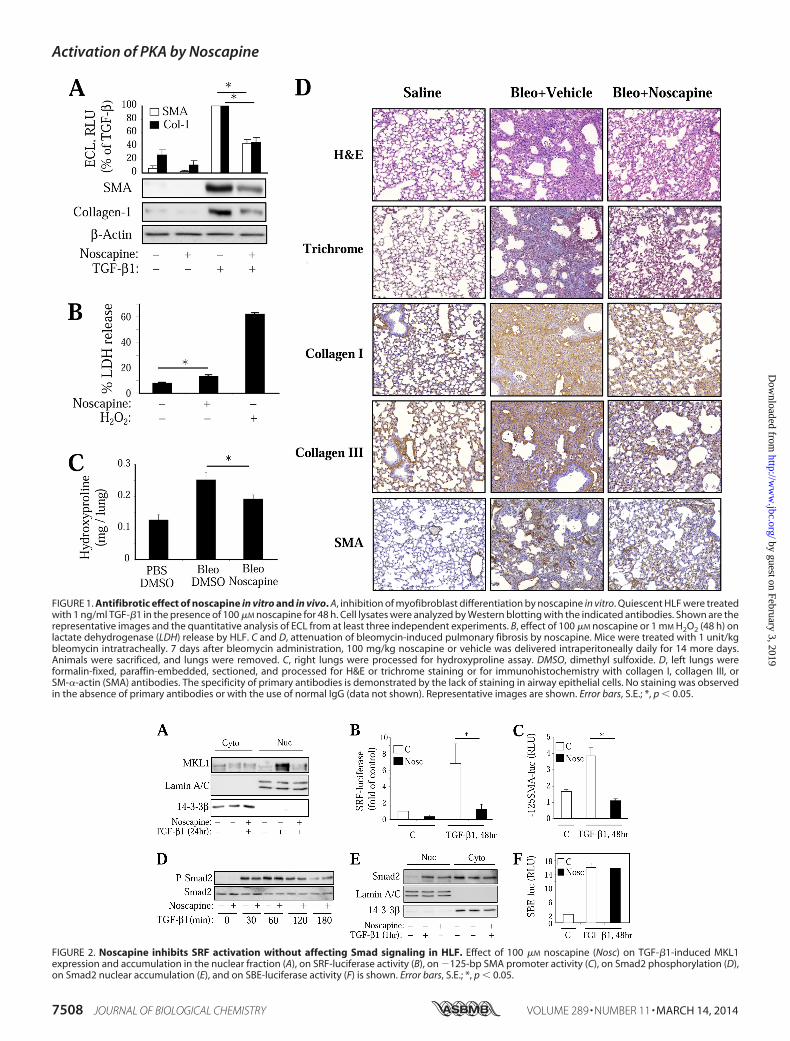
FIGURE 2. Noscapine inhibits SRF activation without affecting Smad signaling in HLF. Effect of 100 �M noscapine (Nosc) on TGF-�1-induced MKL1expression and accumulation in the nuclear fraction (A), on SRF-luciferase activity (B), on �125-bp SMA promoter activity (C), on Smad2 phosphorylation (D),on Smad2 nuclear accumulation (E), and on SBE-luciferase activity (F) is shown. Error bars, S.E.; *, p � 0.05.
FIGURE 1. Antifibrotic effect of noscapine in vitro and in vivo. A, inhibition of myofibroblast differentiation by noscapine in vitro. Quiescent HLF were treatedwith 1 ng/ml TGF-�1 in the presence of 100 �M noscapine for 48 h. Cell lysates were analyzed by Western blotting with the indicated antibodies. Shown are therepresentative images and the quantitative analysis of ECL from at least three independent experiments. B, effect of 100 �M noscapine or 1 mM H2O2 (48 h) onlactate dehydrogenase (LDH) release by HLF. C and D, attenuation of bleomycin-induced pulmonary fibrosis by noscapine. Mice were treated with 1 unit/kgbleomycin intratracheally. 7 days after bleomycin administration, 100 mg/kg noscapine or vehicle was delivered intraperitoneally daily for 14 more days.Animals were sacrificed, and lungs were removed. C, right lungs were processed for hydroxyproline assay. DMSO, dimethyl sulfoxide. D, left lungs wereformalin-fixed, paraffin-embedded, sectioned, and processed for H&E or trichrome staining or for immunohistochemistry with collagen I, collagen III, orSM-�-actin (SMA) antibodies. The specificity of primary antibodies is demonstrated by the lack of staining in airway epithelial cells. No staining was observedin the absence of primary antibodies or with the use of normal IgG (data not shown). Representative images are shown. Error bars, S.E.; *, p � 0.05.
Activation of PKA by Noscapine
7508 JOURNAL OF BIOLOGICAL CHEMISTRY VOLUME 289 • NUMBER 11 • MARCH 14, 2014
by guest on February 3, 2019http://w
ww
.jbc.org/D
ownloaded from

gross microtubule content of HLFs. Immunocytochemicalanalysis showed that noscapine clearly altered the architectureof microtubules, causing their concentration at the perinuclearspace (Fig. 3A). However, it was not evident from this analysiswhether noscapine promoted gross microtubule polymeriza-tion, whereas Taxol treatment showed a clear increase inimmunohistochemical staining of microtubules (Fig. 3A).Therefore, we performed in vitro microtubule fractionationfrom cultured HLFs as a quantitative assay for the gross micro-tubule content. As shown in Fig. 3B, the microtubule fractionwas not significantly enriched with �-tubulin by noscapine fol-lowing 24-h treatment, whereas Taxol treatment clearlyresulted in microtubule polymerization. We also failed todetect microtubule stabilization by noscapine at earlier timepoints (1–3 h) using this assay (data not shown), whereas Taxolwas effective in microtubule polymerization at these timepoints (14). These data suggests that noscapine and Taxolinhibit myofibroblast differentiation by distinct mechanisms.
Therefore, we then further examined the possible mecha-nism of regulation of myofibroblast differentiation by noscap-ine. Given that noscapine inhibited SRF activation but notSmad-dependent gene transcription in response to TGF-� (Fig.2), and given our previous studies pointing to the role of actinstress fiber formation in TGF-�-induced myofibroblast differ-entiation downstream of Smad signaling (14), we examined theeffect of noscapine on this process by in vitro isolation of stressfibers. As shown in Fig. 3C, noscapine appreciably attenuatedTGF-�-induced accumulation of the housekeeping �-actin aswell as of the newly expressed SM-�-actin in the stress fiberfraction.
Activation of PKA Mediates the Regulation of MyofibroblastDifferentiation by Noscapine—We and others have previouslyestablished that myofibroblast differentiation can be regulatedby activators of cAMP/PKA signaling through inhibition of SRFbut not of Smad-dependent gene transcription (13, 34). There-fore, we examined whether the effect of noscapine is dependenton PKA activation. Strikingly, we found that noscapine stimu-lated a rapid and prolonged stimulation of PKA activity inHLFs, as assessed by the electrophoretic mobility shift of PKAsubstrate, VASP (Fig. 4A). We have previously established thatsuch a shift of VASP is mediated by a PKA-dependent phos-
phorylation and serves as a sensitive reporter for PKA activity(35). Supporting this, noscapine induced phosphorylation ofmultiple proteins phosphorylated by PKA, as assessed by West-ern blotting with PKA-substrate antibodies (Fig. 4A), the spec-ificity of which was demonstrated by us previously (36). Impor-tantly, a 1-h stimulation with Taxol, which resulted in acomplete microtubule polymerization (18), had no effect onPKA activation (Fig. 4A), suggesting that the effect of noscapineon myofibroblast differentiation may be not related to micro-tubule stabilization.
Noscapine also induced a rapid VASP shift in primary cul-tured mouse lung fibroblasts, albeit to a lesser extent (Fig. 4B).Interestingly, noscapine failed to induce VASP shift in a humanbronchial epithelial cell line 16-HBE or in the alveolar epithelialcancer cell line A549 (Fig. 4B), suggesting a cell-specific effect ofnoscapine on PKA activation.
To examine the role of PKA in the regulation of myofibro-blast differentiation by noscapine, we used adenovirus-medi-ated expression of the PKA inhibitor protein, PKI (Ad-PKI),which we and others have shown to be an effective and specifictool for the assessment of the role of PKA (30, 37). As shown inFig. 4C, transduction of HLFs with Ad-PKI abolished thenoscapine-induced VASP shift, whereas the control Ad-LacZdid not. Importantly, Ad-PKI but not Ad-LacZ rescued HLFcells from the inhibition of TGF-�-induced expression ofSM-�-actin and collagen-1 by noscapine (Fig. 4D).
EP2 Prostaglandin Receptors Mediate Regulation of Myofi-broblast Differentiation by Noscapine in HLF—Previous studieshave demonstrated that human lung fibroblasts are highlyresponsive to prostaglandin E2 (PGE2), prostaglandin D2(PGD2), and prostacyclin (PGI2) in terms of cAMP activationand inhibition of myofibroblast activation (13, 34, 38). We alsoshowed that PKA can be rapidly activated by endothelin-1 or byATP through cyclooxygenase (COX)-mediated prostanoid syn-thesis in vascular smooth muscle cells (30, 35, 37). In addition,the effects of noscapine on the cough reflex may involve opioidsigma receptors (24), although there is no evidence for activa-tion of PKA through these receptors. To test the potentialinvolvement of these mechanisms in the action of noscapine,we used pharmacological inhibitors of the correspondingreceptors and enzymes. As shown in Fig. 5A, noscapine-in-
FIGURE 3. Noscapine has no effect on gross microtubule polymerization state but inhibits stress fiber formation in HLF. A, HLFs were treated with 100�M noscapine or 10 �M Taxol for 24 h, followed by immunostaining with �-tubulin antibodies (green) and counterstaining for nuclei with DAPI (blue). Shownare the representative images at �400 and �1000 magnification. B, HLFs were treated with 100 �M noscapine or 10 �M Taxol for 24 h. Soluble and polymerized�-tubulin were in vitro fractionated and detected by Western blotting. C, HLFs were treated with 1 ng/ml TGF-�1 with or without 100 �M noscapine for 24 h. Invitro stress fiber preparations or total cell lysates were analyzed by Western blotting for �-actin or SMA as indicated.
Activation of PKA by Noscapine
MARCH 14, 2014 • VOLUME 289 • NUMBER 11 JOURNAL OF BIOLOGICAL CHEMISTRY 7509
by guest on February 3, 2019http://w
ww
.jbc.org/D
ownloaded from

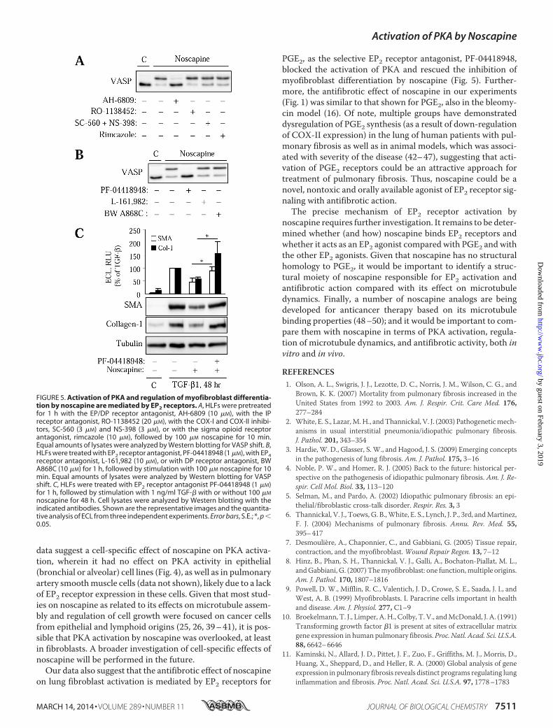
duced VASP phosphorylation was not affected by the sigmareceptor antagonist, rimcazole, by the specific antagonist ofprostacyclin receptors, RO-1138452, or by the combined inhi-bition of COX-I and COX-II with SC-560 and NS-398,respectively. In contrast the PGE2/PGD2 receptor antago-nist, AH6809, completely blocked the noscapine-inducedVASP shift (Fig. 5A).
We then examined the effect of more selective antagonists ofprostanoid receptors known to mediate cAMP production onthe regulation of VASP shift and on myofibroblast differentia-tion by noscapine. Fig. 5B shows that noscapine-induced VASPshift was blocked by the selective antagonist of EP2 receptorsfor PGE2, PF-04418948. In contrast, noscapine-induced VASPshift was unaffected by the antagonist of EP4 receptors for PGE2(L-161,982), or of DP receptors for PGD2 (BW A868C) (Fig.5B). Furthermore, PF-04418948 rescued cells from the inhibi-tion of TGF-�-induced expression of SM-�-actin and colla-gen-1 by noscapine (Fig. 5C). Together, these data suggest thatregulation of myofibroblast differentiation by noscapine is, atleast in part, mediated by activation of PKA through EP2receptors.
DISCUSSION
Noscapine has been studied extensively in cancer researchdue to its effects on microtubule dynamics, and its antitumoractivity has been demonstrated in multiple in vivo studies (25–28). Our study demonstrates for the first time that (i) noscapineattenuates fibrotic responses both in cultured human pulmo-nary fibroblasts and in an experimental model of pulmonaryfibrosis; (ii) noscapine activates PKA specifically in pulmonary
fibroblasts but not in bronchial or alveolar epithelial cells; (iii)PKA activation mediates the antifibrotic effects of noscapine;and (iv) the mechanism of PKA stimulation by noscapineinvolves activation of EP2 prostanoid receptors, which mediateregulation of myofibroblast differentiation by noscapine.
The antifibrotic effect of noscapine found in our study issimilar to the stable prostacyclin analog, iloprost, in the bleo-mycin model (15). However, the therapeutic potential of ilo-prost maybe limited due to the side effects associated with vaso-dilation of vascular pulmonary beds. Of note, noscapine had noeffect on PKA activation in cultured human pulmonary arterysmooth muscle cells (data not shown). Thus, noscapine mayhave a greater therapeutic potential for treatment of pulmonaryfibrosis, as it has been already used for decades as an antitussivedrug with low toxicity in several countries throughout Europe,Asia, and South America, and there are no reports on pulmo-nary vasodilation in response to noscapine treatment.
Our studies show for the first time that noscapine induces arapid and profound activation of PKA in human lung fibro-blasts, which we reproduced using noscapine from two inde-pendent sources (Sigma-Aldrich and R&D Systems). We alsoshow that PKA activation by noscapine is causatively associatedwith inhibition of myofibroblast differentiation. Given numer-ous studies on the mechanism of noscapine action, one wouldbe surprised that this effect of noscapine has not been describedbefore. In fact, one earlier study suggested that noscapine canpromote cAMP accumulation in response to the activator ofadenylyl cyclase, forskolin, in brain slices, while having no effecton cAMP accumulation in the absence of forskolin (23). Our
FIGURE 4. Activation of PKA mediates the regulation of myofibroblast differentiation by noscapine. A, quiescent HLFs were treated with 10 �M Taxol or100 �M noscapine for the indicated times. Lysates were analyzed by Western blotting with antibodies against VASP or against PKA-substrate (PKAS) antibodies.B, HLFs, mouse lung fibroblasts (MLF), human bronchial epithelial cells (16HBE), or human alveolar epithelial cells (A549) were treated with 100 �M noscapineor 10 �M forskolin for the indicated times. Lysates were analyzed by Western blotting for VASP shift. C and D, HLF were transduced with Ad-PKI or controlAd-LacZ adenoviruses. C, cells were then treated with 100 �M noscapine for 30 min, and equal amounts of protein were assessed by Western blotting for VASPshift. D, cells were treated with 100 �M noscapine followed by 1 ng/ml TGF-�1 for 48 h. Equal amounts of cell lysates were analyzed by Western blotting for thedesired proteins. Cell lysates were analyzed by Western blotting with the indicated antibodies. Shown are the representative images and the quantitativeanalysis of enhanced chemiluminescence from three independent experiments. Error bars, S.E.; *, p � 0.05.
Activation of PKA by Noscapine
7510 JOURNAL OF BIOLOGICAL CHEMISTRY VOLUME 289 • NUMBER 11 • MARCH 14, 2014
by guest on February 3, 2019http://w
ww
.jbc.org/D
ownloaded from
data suggest a cell-specific effect of noscapine on PKA activa-tion, wherein it had no effect on PKA activity in epithelial(bronchial or alveolar) cell lines (Fig. 4), as well as in pulmonaryartery smooth muscle cells (data not shown), likely due to a lackof EP2 receptor expression in these cells. Given that most stud-ies on noscapine as related to its effects on microtubule assem-bly and regulation of cell growth were focused on cancer cellsfrom epithelial and lymphoid origins (25, 26, 39 – 41), it is pos-sible that PKA activation by noscapine was overlooked, at leastin fibroblasts. A broader investigation of cell-specific effects ofnoscapine will be performed in the future.
Our data also suggest that the antifibrotic effect of noscapineon lung fibroblast activation is mediated by EP2 receptors for
PGE2, as the selective EP2 receptor antagonist, PF-04418948,blocked the activation of PKA and rescued the inhibition ofmyofibroblast differentiation by noscapine (Fig. 5). Further-more, the antifibrotic effect of noscapine in our experiments(Fig. 1) was similar to that shown for PGE2, also in the bleomy-cin model (16). Of note, multiple groups have demonstrateddysregulation of PGE2 synthesis (as a result of down-regulationof COX-II expression) in the lung of human patients with pul-monary fibrosis as well as in animal models, which was associ-ated with severity of the disease (42– 47), suggesting that acti-vation of PGE2 receptors could be an attractive approach fortreatment of pulmonary fibrosis. Thus, noscapine could be anovel, nontoxic and orally available agonist of EP2 receptor sig-naling with antifibrotic action.
The precise mechanism of EP2 receptor activation bynoscapine requires further investigation. It remains to be deter-mined whether (and how) noscapine binds EP2 receptors andwhether it acts as an EP2 agonist compared with PGE2 and withthe other EP2 agonists. Given that noscapine has no structuralhomology to PGE2, it would be important to identify a struc-tural moiety of noscapine responsible for EP2 activation andantifibrotic action compared with its effect on microtubuledynamics. Finally, a number of noscapine analogs are beingdeveloped for anticancer therapy based on its microtubulebinding properties (48 –50); and it would be important to com-pare them with noscapine in terms of PKA activation, regula-tion of microtubule dynamics, and antifibrotic activity, both invitro and in vivo.
REFERENCES1. Olson, A. L., Swigris, J. J., Lezotte, D. C., Norris, J. M., Wilson, C. G., and
Brown, K. K. (2007) Mortality from pulmonary fibrosis increased in theUnited States from 1992 to 2003. Am. J. Respir. Crit. Care Med. 176,277–284
2. White, E. S., Lazar, M. H., and Thannickal, V. J. (2003) Pathogenetic mech-anisms in usual interstitial pneumonia/idiopathic pulmonary fibrosis.J. Pathol. 201, 343–354
3. Hardie, W. D., Glasser, S. W., and Hagood, J. S. (2009) Emerging conceptsin the pathogenesis of lung fibrosis. Am. J. Pathol. 175, 3–16
4. Noble, P. W., and Homer, R. J. (2005) Back to the future: historical per-spective on the pathogenesis of idiopathic pulmonary fibrosis. Am. J. Re-spir. Cell Mol. Biol. 33, 113–120
5. Selman, M., and Pardo, A. (2002) Idiopathic pulmonary fibrosis: an epi-thelial/fibroblastic cross-talk disorder. Respir. Res. 3, 3
6. Thannickal, V. J., Toews, G. B., White, E. S., Lynch, J. P., 3rd, and Martinez,F. J. (2004) Mechanisms of pulmonary fibrosis. Annu. Rev. Med. 55,395– 417
7. Desmoulière, A., Chaponnier, C., and Gabbiani, G. (2005) Tissue repair,contraction, and the myofibroblast. Wound Repair Regen. 13, 7–12
8. Hinz, B., Phan, S. H., Thannickal, V. J., Galli, A., Bochaton-Piallat, M. L.,and Gabbiani, G. (2007) The myofibroblast: one function, multiple origins.Am. J. Pathol. 170, 1807–1816
9. Powell, D. W., Mifflin, R. C., Valentich, J. D., Crowe, S. E., Saada, J. I., andWest, A. B. (1999) Myofibroblasts. I. Paracrine cells important in healthand disease. Am. J. Physiol. 277, C1–9
10. Broekelmann, T. J., Limper, A. H., Colby, T. V., and McDonald, J. A. (1991)Transforming growth factor �1 is present at sites of extracellular matrixgene expression in human pulmonary fibrosis. Proc. Natl. Acad. Sci. U.S.A.88, 6642– 6646
11. Kaminski, N., Allard, J. D., Pittet, J. F., Zuo, F., Griffiths, M. J., Morris, D.,Huang, X., Sheppard, D., and Heller, R. A. (2000) Global analysis of geneexpression in pulmonary fibrosis reveals distinct programs regulating lunginflammation and fibrosis. Proc. Natl. Acad. Sci. U.S.A. 97, 1778 –1783
FIGURE 5. Activation of PKA and regulation of myofibroblast differentia-tion by noscapine are mediated by EP2 receptors. A, HLFs were pretreatedfor 1 h with the EP/DP receptor antagonist, AH-6809 (10 �M), with the IPreceptor antagonist, RO-1138452 (20 �M), with the COX-I and COX-II inhibi-tors, SC-560 (3 �M) and NS-398 (3 �M), or with the sigma opioid receptorantagonist, rimcazole (10 �M), followed by 100 �M noscapine for 10 min.Equal amounts of lysates were analyzed by Western blotting for VASP shift. B,HLFs were treated with EP2 receptor antagonist, PF-04418948 (1 �M), with EP4receptor antagonist, L-161,982 (10 �M), or with DP receptor antagonist, BWA868C (10 �M) for 1 h, followed by stimulation with 100 �M noscapine for 10min. Equal amounts of lysates were analyzed by Western blotting for VASPshift. C, HLFs were treated with EP2 receptor antagonist PF-04418948 (1 �M)for 1 h, followed by stimulation with 1 ng/ml TGF-� with or without 100 �M
noscapine for 48 h. Cell lysates were analyzed by Western blotting with theindicated antibodies. Shown are the representative images and the quantita-tive analysis of ECL from three independent experiments. Error bars, S.E.; *, p �0.05.
Activation of PKA by Noscapine
MARCH 14, 2014 • VOLUME 289 • NUMBER 11 JOURNAL OF BIOLOGICAL CHEMISTRY 7511
by guest on February 3, 2019http://w
ww
.jbc.org/D
ownloaded from
12. Sime, P. J., Xing, Z., Graham, F. L., Csaky, K. G., and Gauldie, J. (1997)Adenovector-mediated gene transfer of active transforming growth fac-tor-�1 induces prolonged severe fibrosis in rat lung. J. Clin. Invest. 100,768 –776
13. Sandbo, N., Kregel, S., Taurin, S., Bhorade, S., and Dulin, N. O. (2009)Critical role of serum response factor in pulmonary myofibroblast differ-entiation induced by TGF-�. Am. J. Respir. Cell Mol. Biol. 41, 332–338
14. Sandbo, N., Lau, A., Kach, J., Ngam, C., Yau, D., and Dulin, N. O. (2011)Delayed stress fiber formation mediates pulmonary myofibroblast differ-entiation in response to TGF-�. Am. J. Physiol. Lung Cell Mol. Physiol.301, L656 –L666
15. Zhu, Y., Liu, Y., Zhou, W., Xiang, R., Jiang, L., Huang, K., Xiao, Y., Guo, Z.,and Gao, J. (2010) A prostacyclin analogue, iloprost, protects from bleo-mycin-induced pulmonary fibrosis in mice. Respir. Res. 11, 34
16. Dackor, R. T., Cheng, J., Voltz, J. W., Card, J. W., Ferguson, C. D., Garrett,R. C., Bradbury, J. A., DeGraff, L. M., Lih, F. B., Tomer, K. B., Flake, G. P.,Travlos, G. S., Ramsey, R. W., Jr., Edin, M. L., Morgan, D. L., and Zeldin,D. C. (2011) Prostaglandin E2 protects murine lungs from bleomycin-induced pulmonary fibrosis and lung dysfunction. Am. J. Physiol. Lung CellMol. Physiol. 301, L645–L655
17. Kach, J., Sandbo, N., Sethakorn, N., Williams, J., Reed, E. B., La, J., Tian, X.,Brain, S. D., Rajendran, K., Krishnan, R., Sperling, A. I., Birukov, K., andDulin, N. O. (2013) Regulation of myofibroblast differentiation and bleo-mycin-induced pulmonary fibrosis by adrenomedullin. Am. J. Physiol.Lung Cell Mol. Physiol. 304, L757–L764
18. Sandbo, N., Ngam, C., Torr, E., Kregel, S., Kach, J., and Dulin, N. (2013)Control of Myofibroblast differentiation by microtubule dynamicsthrough a regulated localization of mDia2. J. Biol. Chem. 288,15466 –15473
19. Empey, D. W., Laitinen, L. A., Young, G. A., Bye, C. E., and Hughes, D. T.(1979) Comparison of the antitussive effects of codeine phosphate 20 mg,dextromethorphan 30 mg and noscapine 30 mg using citric acid-inducedcough in normal subjects. Eur. J. Clin. Pharmacol. 16, 393–397
20. Dahlström, B., Mellstrand, T., Löfdahl, C. G., and Johansson, M. (1982)Pharmacokinetic properties of noscapine. Eur. J. Clin. Pharmacol. 22,535–539
21. Karlsson, M. O., Dahlström, B., Eckernäs, S. A., Johansson, M., and Alm,A. T. (1990) Pharmacokinetics of oral noscapine. Eur. J. Clin. Pharmacol.39, 275–279
22. Karlsson, M. O., Dahlström, B., and Neil, A. (1988) Characterization ofhigh-affinity binding sites for the antitussive [3H]noscapine in guinea pigbrain tissue. Eur. J. Pharmacol. 145, 195–203
23. Mourey, R. J., Dawson, T. M., Barrow, R. K., Enna, A. E., and Snyder, S. H.(1992) [3H]noscapine binding sites in brain: relationship to indoleaminesand the phosphoinositide and adenylyl cyclase messenger systems. Mol.Pharmacol. 42, 619 – 626
24. Kamei, J., Iwamoto, Y., Misawa, M., and Kasuya, Y. (1993) Effects of rim-cazole, a specific antagonist of sigma sites, on the antitussive effects ofnonnarcotic antitussive drugs. Eur. J. Pharmacol. 242, 209 –211
25. Ye, K., Ke, Y., Keshava, N., Shanks, J., Kapp, J. A., Tekmal, R. R., Petros, J.,and Joshi, H. C. (1998) Opium alkaloid noscapine is an antitumor agentthat arrests metaphase and induces apoptosis in dividing cells. Proc. Natl.Acad. Sci. U.S.A. 95, 1601–1606
26. Landen, J. W., Lang, R., McMahon, S. J., Rusan, N. M., Yvon, A. M., Adams,A. W., Sorcinelli, M. D., Campbell, R., Bonaccorsi, P., Ansel, J. C., Archer,D. R., Wadsworth, P., Armstrong, C. A., and Joshi, H. C. (2002) Noscapinealters microtubule dynamics in living cells and inhibits the progression ofmelanoma. Cancer Res. 62, 4109 – 4114
27. Barken, I., Geller, J., and Rogosnitzky, M. (2008) Noscapine inhibits hu-man prostate cancer progression and metastasis in a mouse model. Anti-cancer Res. 28, 3701–3704
28. Jackson, T., Chougule, M. B., Ichite, N., Patlolla, R. R., and Singh, M. (2008)Antitumor activity of noscapine in human non-small cell lung cancerxenograft model. Cancer Chemother. Pharmacol. 63, 117–126
29. Minotti, A. M., Barlow, S. B., and Cabral, F. (1991) Resistance to antimi-totic drugs in Chinese hamster ovary cells correlates with changes in thelevel of polymerized tubulin. J. Biol. Chem. 266, 3987–3994
30. Hogarth, D. K., Sandbo, N., Taurin, S., Kolenko, V., Miano, J. M., and
Dulin, N. O. (2004) Dual role of PKA in phenotypic modulation of vascularsmooth muscle cells by extracellular ATP. Am. J. Physiol. Cell Physiol. 287,C449 – 456
31. Small, E. M., Thatcher, J. E., Sutherland, L. B., Kinoshita, H., Gerard, R. D.,Richardson, J. A., Dimaio, J. M., Sadek, H., Kuwahara, K., and Olson, E. N.(2010) Myocardin-related transcription factor-a controls myofibroblastactivation and fibrosis in response to myocardial infarction. Circ. Res. 107,294 –304
32. Mack, C. P., Thompson, M. M., Lawrenz-Smith, S., and Owens, G. K.(2000) Smooth muscle �-actin CArG elements coordinate formation of asmooth muscle cell-selective, serum response factor-containing activa-tion complex. Circ. Res. 86, 221–232
33. Zhou, J., Panda, D., Landen, J. W., Wilson, L., and Joshi, H. C. (2002) Minoralteration of microtubule dynamics causes loss of tension across kineto-chore pairs and activates the spindle checkpoint. J. Biol. Chem. 277,17200 –17208
34. Huang, S., Wettlaufer, S. H., Hogaboam, C., Aronoff, D. M., and Peters-Golden, M. (2007) Prostaglandin E2 inhibits collagen expression and pro-liferation in patient-derived normal lung fibroblasts via E prostanoid 2receptor and cAMP signaling. Am. J. Physiol. Lung Cell Mol. Physiol. 292,L405–L413
35. Davis, A., Hogarth, K., Fernandes, D., Solway, J., Niu, J., Kolenko, V.,Browning, D., Miano, J. M., Orlov, S. N., and Dulin, N. O. (2003) Func-tional significance of protein kinase A activation by endothelin-1 andATP: negative regulation of SRF-dependent gene expression by PKA. Cell.Signal. 15, 597– 604
36. Taurin, S., Sandbo, N., Qin, Y., Browning, D., and Dulin, N. O. (2006)Phosphorylation of �-catenin by cyclic AMP-dependent protein kinase.J. Biol. Chem. 281, 9971–9976
37. Taurin, S., Hogarth, K., Sandbo, N., Yau, D. M., and Dulin, N. O. (2007)G��-mediated prostacyclin production and cAMP-dependent protein ki-nase activation by endothelin-1 promotes vascular smooth muscle cellhypertrophy through inhibition of glycogen synthase kinase-3. J. Biol.Chem. 282, 19518 –19525
38. Ayabe, S., Kida, T., Hori, M., Ozaki, H., and Murata, T. (2013) Prostaglan-din D2 inhibits collagen secretion from lung fibroblasts by activating theDP receptor. J. Pharmacol. Sci. 121, 312–317
39. Landen, J. W., Hau, V., Wang, M., Davis, T., Ciliax, B., Wainer, B. H., VanMeir, E. G., Glass, J. D., Joshi, H. C., and Archer, D. R. (2004) Noscapinecrosses the blood-brain barrier and inhibits glioblastoma growth. Clin.Cancer Res. 10, 5187–5201
40. Zhou, J., Gupta, K., Yao, J., Ye, K., Panda, D., Giannakakou, P., and Joshi,H. C. (2002) Paclitaxel-resistant human ovarian cancer cells undergo c-Jun NH2-terminal kinase-mediated apoptosis in response to noscapine.J. Biol. Chem. 277, 39777–39785
41. Aneja, R., Zhou, J., Vangapandu, S. N., Zhou, B., Chandra, R., and Joshi,H. C. (2006) Drug-resistant T-lymphoid tumors undergo apoptosis selec-tively in response to an antimicrotubule agent, EM011. Blood 107,2486 –2492
42. Wilborn, J., Crofford, L. J., Burdick, M. D., Kunkel, S. L., Strieter, R. M., andPeters-Golden, M. (1995) Cultured lung fibroblasts isolated from patientswith idiopathic pulmonary fibrosis have a diminished capacity to synthe-size prostaglandin E2 and to express cyclooxygenase-2. J. Clin. Invest. 95,1861–1868
43. Keerthisingam, C. B., Jenkins, R. G., Harrison, N. K., Hernandez-Rodri-guez, N. A., Booth, H., Laurent, G. J., Hart, S. L., Foster, M. L., andMcAnulty, R. J. (2001) Cyclooxygenase-2 deficiency results in a loss of theanti-proliferative response to transforming growth factor-� in human fi-brotic lung fibroblasts and promotes bleomycin-induced pulmonary fi-brosis in mice. Am. J. Pathol. 158, 1411–1422
44. Bonner, J. C., Rice, A. B., Ingram, J. L., Moomaw, C. R., Nyska, A., Brad-bury, A., Sessoms, A. R., Chulada, P. C., Morgan, D. L., Zeldin, D. C., andLangenbach, R. (2002) Susceptibility of cyclooxygenase-2-deficient miceto pulmonary fibrogenesis. Am. J. Pathol. 161, 459 – 470
45. Petkova, D. K., Clelland, C. A., Ronan, J. E., Lewis, S., and Knox, A. J. (2003)Reduced expression of cyclooxygenase (COX) in idiopathic pulmonaryfibrosis and sarcoidosis. Histopathology 43, 381–386
Activation of PKA by Noscapine
7512 JOURNAL OF BIOLOGICAL CHEMISTRY VOLUME 289 • NUMBER 11 • MARCH 14, 2014
by guest on February 3, 2019http://w
ww
.jbc.org/D
ownloaded from
46. Xaubet, A., Roca-Ferrer, J., Pujols, L., Ramírez, J., Mullol, J., Marin-Argue-das, A., Torrego, A., Gimferrer, J. M., and Picado, C. (2004) Cyclooxyge-nase-2 is up-regulated in lung parenchyma of chronic obstructive pulmo-nary disease and down-regulated in idiopathic pulmonary fibrosis.Sarcoidosis Vasc. Diffuse Lung Dis. 21, 35– 42
47. Hodges, R. J., Jenkins, R. G., Wheeler-Jones, C. P., Copeman, D. M., Bot-toms, S. E., Bellingan, G. J., Nanthakumar, C. B., Laurent, G. J., Hart, S. L.,Foster, M. L., and McAnulty, R. J. (2004) Severity of lung injury in cy-clooxygenase-2-deficient mice is dependent on reduced prostaglandin E2
production. Am. J. Pathol. 165, 1663–167648. Aneja, R., Vangapandu, S. N., and Joshi, H. C. (2006) Synthesis and bio-
logical evaluation of a cyclic ether fluorinated noscapine analog. Bioorg.Med. Chem. 14, 8352– 8358
49. Aneja, R., Vangapandu, S. N., Lopus, M., Chandra, R., Panda, D., and Joshi,H. C. (2006) Development of a novel nitro-derivative of noscapine for thepotential treatment of drug-resistant ovarian cancer and T-cell lym-phoma. Mol. Pharmacol. 69, 1801–1809
50. Aneja, R., Vangapandu, S. N., Lopus, M., Viswesarappa, V. G., Dhiman, N.,Verma, A., Chandra, R., Panda, D., and Joshi, H. C. (2006) Synthesis ofmicrotubule-interfering halogenated noscapine analogs that perturb mi-tosis in cancer cells followed by cell death. Biochem. Pharmacol. 72,415– 426
Activation of PKA by Noscapine
MARCH 14, 2014 • VOLUME 289 • NUMBER 11 JOURNAL OF BIOLOGICAL CHEMISTRY 7513
by guest on February 3, 2019http://w
ww
.jbc.org/D
ownloaded from
Akimova, Svetlana Koltsova, Sergei N. Orlov and Nickolai O. DulinJacob Kach, Nathan Sandbo, Jennifer La, Darcy Denner, Eleanor B. Reed, Olga
Receptors and Protein Kinase A2Antifibrotic Effects of Noscapine through Activation of Prostaglandin E
doi: 10.1074/jbc.M113.546812 originally published online February 3, 20142014, 289:7505-7513.J. Biol. Chem.
10.1074/jbc.M113.546812Access the most updated version of this article at doi:
Alerts:
When a correction for this article is posted•
When this article is cited•
to choose from all of JBC's e-mail alertsClick here
http://www.jbc.org/content/289/11/7505.full.html#ref-list-1
This article cites 50 references, 17 of which can be accessed free at
by guest on February 3, 2019http://w
ww
.jbc.org/D
ownloaded from