Embed Size (px)
Citation preview
REVIEW Open Access
Oxidative toxicity in diabetes andAlzheimer’s disease: mechanisms behindROS/ RNS generationWaqar Ahmad1* , Bushra Ijaz2, Khadija Shabbiri1, Fayyaz Ahmed2 and Sidra Rehman3
Abstract
Reactive oxidative species (ROS) toxicity remains an undisputed cause and link between Alzheimer’s disease (AD)and Type-2 Diabetes Mellitus (T2DM). Patients with both AD and T2DM have damaged, oxidized DNA, RNA, proteinand lipid products that can be used as possible disease progression markers. Although the oxidative stress hasbeen anticipated as a main cause in promoting both AD and T2DM, multiple pathways could be involved in ROSproduction. The focus of this review is to summarize the mechanisms involved in ROS production and theirpossible association with AD and T2DM pathogenesis and progression. We have also highlighted the role of currenttreatments that can be linked with reduced oxidative stress and damage in AD and T2DM.
Keywords: Alzheimer’s disease, Type-2 diabetes mellitus, Oxidative stress, ROS production, Antioxidant treatments,Anti-diabetic drugs
BackgroundA set of chemical processes through which living bodiessustain their lives called as metabolism. This includes diges-tion of food, transport into the body cells and excretion ofwaste materials through well-conserved intermediary me-tabolism. The metabolic pathways are the bio-chemicalprocesses involving DNA replication, transcription andtranslation by enzyme catalysed reaction through whichfood or other chemicals from the body transformed intodifferent chemicals and produce energy for various lifefunctions [1]. In the living organism’s body cells and tissuesare always gone through the assembly, and disassemblyprocesses in a regular manner involving several metabolicpathways. Disturbs in metabolic process by any external orinternal factors may result in metabolic disorder followedby many types of life-threatening diseases. The understand-ing of the cellular and molecular mechanism for incurablediseases like Alzheimer’s disease (AD) and Type-2 DiabetesMellitus (T2DM) has been progressing rapidly, which alsoenhances the therapeutic approaches [2].
It has been noteworthy that the advancement in diagnos-tic and therapeutic approaches improved the disease man-agement. However, pathophysiology of many diseases is stillunder way. AD and T2DM, the two-utmost communaloverwhelming diseases caused by neurological and insulinfunction disorder, have become a major public health con-cern worldwide [3, 4], and needed to be address effectively.A large-scale clinico-epidemiological data indicates thatboth T2DM and AD are most common age-associated dis-eases around the globe. People withT2DM are prone to riskof AD. The first strong evidence regarding the correlationbetween AD and T2DM was reported in Rotterdam cohortstudy [3–5]. A number of clinical, epidemiological,biological, molecular and genetic data supports a patho-physiological link between T2DM and AD, includingobesity, impaired glucose, cholesterol metabolism, andhypertension [6–8]. Presence of these symptoms altogetherknown as metabolic syndrome (MetS) and could signify apathological connection between impaired metabolism andseveral neurological disorders [9, 10]. Uncontrolled in-creased blood glucose is a major cause of T2DM, which isassociated with injury of insulin-producing pancreatic β-cells or by insulin sensitivity in adipose or muscle tissues[11, 12]. Both T2DM and AD induce disease severity basedon same path-physiological mechanisms, including
* Correspondence: [email protected];[email protected] of Biological Sciences, University of Queensland, Brisbane 4072,AustraliaFull list of author information is available at the end of the article
© The Author(s). 2017 Open Access This article is distributed under the terms of the Creative Commons Attribution 4.0International License (http://creativecommons.org/licenses/by/4.0/), which permits unrestricted use, distribution, andreproduction in any medium, provided you give appropriate credit to the original author(s) and the source, provide a link tothe Creative Commons license, and indicate if changes were made. The Creative Commons Public Domain Dedication waiver(http://creativecommons.org/publicdomain/zero/1.0/) applies to the data made available in this article, unless otherwise stated.
Ahmad et al. Journal of Biomedical Science (2017) 24:76 DOI 10.1186/s12929-017-0379-z
mitochondrial damage and formation of advanced glycationproducts (AGEs). Both mitochondrial damage and AGEsare influenced by induced oxidative stress, which not onlyimpair mtDNA and RNA but also affect protein and lipids[13, 14]. Several studies found induced levels of DNA,RNA, protein and lipid oxidative products in T2DM andAD like 8-hydroxyguanosine, 8-hydroxydeoxyguanosine,protein carbonyls and nitrotyrosine; and lipid peroxidationmarkers, for example, 4-hydroxynonenal, F2-isoprostanes,and malondialdehyde [15–21].Oxidative stress has been proposed to play a signifi-
cant role in T2DM and AD progression. The present re-view highlights the complex mechanism involved in theproduction of reactive oxygen species (ROS), inducedoxidative stress, and their impact on T2DM and ADprogression. Moreover, we also highlight the possibletreatments to cope with the bad effects of oxidativestress in T2DM and AD.
ROS production and oxidative damageROS in living organisms was first described in 1954 [22,23]. In 1969, theory of oxygen toxicity was expressed in aer-obic organisms after the discovery of superoxide dismutase(SOD) by McCord and Fridovich. ROS production can beassociated with age-related diseases, their developmentalprocesses and cell singling pathways [24, 25]. Oxidative rad-icals have very short lifespan and react rapidly with othermolecules [26]. Presence of transition metals, especially Feand Cu can help to clarify and explain oxidative damage toliving cells [27]. Important oxidants in the living organismincludes ROS, reactive nitrogen species (RNS) and sulphur-centred radicals. Although not all of them are radicals butin many cases, these non-radicals can produce radical spe-cies by reacting cellular compounds and damaging them byoxidation [28]. The ROS can be classified into two groups;radicals and non-radicals. The radicals contain superoxide(O.
2−), alkoxyl (RO.), peroxyl (ROO.), hydroxyl (OH.), hydro-
peroxyl (HO.2) and nitric oxide (NO.). The non-radicals in-
clude hydrogen peroxide (H2O2), organic peroxides(ROOH), aldehydes (HCOR), hydrochlorous acid (HOCL),peroxynitrite (ONOOH/ ONOO−), ozone (O3) and singletoxygen (1O2) [29, 30].ROS and RNS can be generated through exogenous and
endogenous sources [28]. Exogenous sources may includeUV radiations (direct oxidation of cellular components)[31, 32], ultrasound, drugs (like narcotics, anaesthetizes,adreamicine, nitroglycerine and belomycinem) [33], food(containing oxidants such as transition metals, aldehydes,fatty acids and peroxides) [34], γ- radiations [35], pollutants,xenobiotics and toxic chemicals (alcohol, phosphine, mus-tard gas) [36, 37]. The endogenous sources may includeneutrophils, cytokines and other components of whiteblood cells [38, 39], direct ROS producing enzymes such asNO synthase, indirect ROS producing enzymes such as the
xanthin oxidase, mitochondrial, metals and side effects ofvarious diseases [40, 41]. These molecules ultimately targetthe macromolecules like proteins, lipids and nucleotidesthat result in genome instability and impaired organ func-tions [30–34]. These molecules are critical for neuronaland pancreatic beta cell stability and functions [42–44].ROS readily attacks and generates a variety of variety DNAlesions. These lesions could result in DNA base transver-sions (e.g. G:C to T:A) [35–37]. More than 200 clinical dis-orders have been associated with early initiation of ROS.These disorders may include T2DM, AD, cardiovasculardamage, inflammation, intestinal tract disease, eye diseases,brain degenerative impairments, aging, hemochromatosis,thalassemia, and Wilson disease [45, 46].In living organisms, Oxidants and antioxidants play a
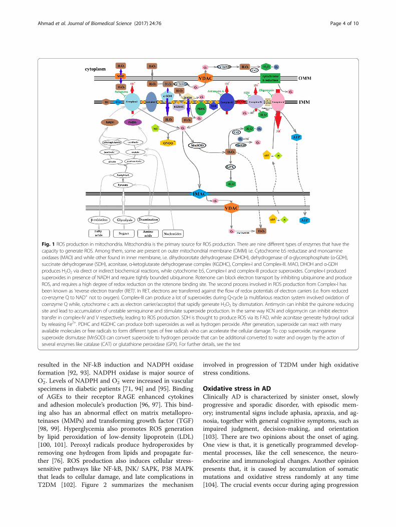
significant role in regulating free radical balance withinthe body produced during active metabolism. A dis-turbed endogenous antioxidant system favors shift to-wards more pro-oxidants production called as “oxidativestress.” If it shifts towards more production of antioxi-dants or reducing power termed as “reductive stress”[25, 30, 47–49]. As induced oxidative stress impairs nat-ural defense by unbalancing the oxidants and de-oxidantratio, balancing oxidative stress is an emerging therapyin various diseases. Figure 1 explains the detailed mech-anism involved in the ROS generation in mitochondria.
ROS as cellular defenceROS generally maintains the normal physiological func-tions and cellular defense of the body. Many living organ-isms survive below a specific homeostatic set point [24].Although ROS production is beneficial for cellular mech-anism, their excessive quantities are always toxic and leadto oxidative damage of many biological functions [25, 50].To reduce its toxicity, mammalian cells have evolveddefense mechanisms, including different DNA base exci-sions and strand repair enzymes [51, 52]. In this way, liv-ing organisms have not only adapted themselves todevelop self-protective mechanisms for ROS but also ableto use it constructively [24, 53]. Intracellular low level ofROS may act as signaling molecules in many physiologicalprocesses, including redox homeostasis and cellular signaltransduction [54]. The divergent effects of ROS on manycellular processes suggest that ROS is not merely detri-mental by-products, but also generated purposefully tomediate a variety of signaling pathways.
Oxidative stress in T2DMDM is a metabolic disorder categorized into two maingroups: Type I (Insulin dependent) that is due toimmune-mediated beta-cells destruction and lead to insu-lin deficiency, and Type 2 (Non-Insulin dependent) that isdue to insulin secretion defects and resulted in insulin re-sistance [55]. Prolonged period of high blood-glucose
Ahmad et al. Journal of Biomedical Science (2017) 24:76 Page 2 of 10
levels generally linked to both macro and micro vascularcomplications like CVDs, strokes, peripheral vascular dis-eases, neuropathy, retinopathy and nephropathy [56–58].In addition to elevated blood-glucose levels, other factorsinclude high-cholesterol level (hyperlipidemia) and oxida-tive stress leading to high risk of complications [59]. Ac-cording to epidemiological studies, diabetic mortalitiescan be explained by an increase in vascular diseases thatcould be a cause of oxidative damage [60]. Current re-search reported that apo-lipoprotein component of LDLinstead of lipid alone could be a cause of oxidative damagein DM [60].Production of free radicals and their high levels in dia-
betic patients could be non-enzymatic (i.e. glycated pro-teins, glucose oxidation and increased lipid peroxidation)or enzymatic (over/under-expressed levels of enzymes likecatalase (CAT), superoxide dismutase (SOD) and glutathi-one peroxidise (GSH–Px)). These abnormalities may leadto damage of enzymes, cellular machinery and increasedinsulin resistance due to oxidative stress [61, 62]. Recentstudies have provided a clear evidence that the mainsource of ROS/ RNS production in T2DM is mitochondria[63–65]. Abnormal mitochondrial functions and excessiveROS/RNS production play a primary role in onset T2DMand its complications. These studies also support the pos-sibility for mitochondrial-targeted antioxidant’s therapy ofT2DM complications [66].During cellular metabolism, insulin reacts with it re-
ceptors that lead to activation of Akt and translocationof GLUT4 to cell membrane. Impaired oxidative phos-phorylation, reduced NADH oxidoreductase and citratesynthase activities resulted in insulin resistance [6, 67].This insulin resistance could be the result from eitherimpaired fatty acid acetyl-CoA oxidation or from subse-quent accumulation of intracellular lipid and diacylglyc-erol with consequent activation of protein kinase C andROS production. This impaired fatty acid oxidation re-sulted in activation of serine kinases followed by phos-phorylation of insulin receptor substrates and interferinginsulin signal transduction [68].Multiple studies have observed the presence of oxidative
markers like F2-isoprostane and nitrotyrosine in urine,plasma and tissue levels of diabetic patients [69, 70]. ROSand NOS production in DM can be promoted by both en-zymatic and non-enzymatic sources. Main enzymaticsources may be endothelial and vascular smooth musclecells, NADPH oxidase, xanthine oxidase, cyclooxygenaseand uncoupled NOS whereas, non-enzymatic sources in-clude mitochondrial respiratory chain, AGES, glucose aut-oxidation process and activated polyol pathway [71].ROS production has become a fundamental part in the
T2DM pathogenesis and severity [72]. During the normalglucose oxidation process, the final product is NADH andpyruvate. NADH can reduce pyruvate to lactate or
donates its reducing equivalents to electron transportchain. On the other hand, in mitochondrial pyruvate en-ters into Krebs’s cycle, get oxidised and produce CO2,H2O, NADH and FADH2 [73]. In glucose autoxidation,glucose forms radical and converted to reactive ketoalde-hydes and superoxide, consequently, produced hydroxylradical in presence of transition metals via H2O2 [74, 75].Superoxide can also form peroxynitrite radicals by react-ing with nitric oxide [76, 77]. Hyperglycemia inducedsuperoxide formation in the mitochondrial electron trans-port chain by driving the inner mitochondrial membranepotential upward through the generation of excessive elec-tron donors in the Krebs’s cycle [78]. This situation re-sulted in hyperpolarization of mitochondrial membranepotential and increase in ATP/ADP ratio followed by aninhibition of complex-III and electron accumulation at co-enzyme Q. Consequently; this situation accelerates freeradical formation by partial reduction of O2 and reducesATP synthesis [79, 80].Superoxide presence decreases glyceraldehyde-3-
phosphate dehydrogenase (GAPDH) activity by 66% andresulted in PARP activation and NAD+ depletion [81]. Inhyperglycemia, glucose conversion to the polyalcoholsorbitol and fructose via the polyol pathway reduces NAD+ to NADH. Sorbitol oxidation through NAD+ escort toincreased cytosolic NADH: NAD+ ratio and inhibit theGAPDH activity, and consequently, increased productionof triose phosphate [80]. Increased triose phosphate in-duced formation of methylglyoxal and diacylglycerol(DAG), PKC and PARP activation [82, 83]. Hyperglycemiaalso increases hexosamine pathway flux because of in-creased bio-availability of nutrients and enhancesfructose-6-phosphate levels by inhibiting GAPDH by ROS[84, 85]. The outcome of the hexosamine pathway isUDP-N-acetyl glucosamine that triggers many transcrip-tion factors and pathways, and lead to microvascular com-plications of T2DM [86, 87].Overproduction of superoxidase radicals is countered
by superoxide dismutase’s (SODs) and by uncouplingproteins (UCPs). In hyperglycemia, over expression ofUCPs reduce mitochondrial hyperpolarization and ROSformation, and block the glucose induced cell death.Superoxide radical generation was enhanced in patientswith diabetic endothelial cells that promote oxidativestress toxicity [88, 89]. A study by Nishikawa et al. ob-served the excessive generation of pyruvate via acceler-ated glycolysis and production of superoxides radicals atthe Complex-II level under hyperglycemia [79, 90]. Al-though glucose is least reactive reducing sugar, it maylead to Amadori product through Schiff base formationby reacting free amino acids. These Aamdori productsaccumulate on proteins and start the production ofAGEs [79, 91] that in turn increase ROS productionthrough binding to RAGE (receptors of AGEs) and
Ahmad et al. Journal of Biomedical Science (2017) 24:76 Page 3 of 10
resulted in the NF-kB induction and NADPH oxidaseformation [92, 93]. NADPH oxidase is major source ofO2
−. Levels of NADPH and O2− were increased in vascular
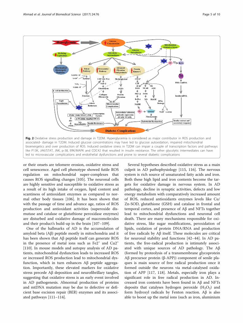
specimens in diabetic patients [71, 94] and [95]. Bindingof AGEs to their receptor RAGE enhanced cytokinesand adhesion molecule’s production [96, 97]. This bind-ing also has an abnormal effect on matrix metallopro-teinases (MMPs) and transforming growth factor (TGF)[98, 99]. Hyperglycemia also promotes ROS generationby lipid peroxidation of low-density lipoprotein (LDL)[100, 101]. Peroxyl radicals produce hydroperoxides byremoving one hydrogen from lipids and propagate fur-ther [76]. ROS production also induces cellular stress-sensitive pathways like NF-kB, JNK/ SAPK, P38 MAPKthat leads to cellular damage, and late complications inT2DM [102]. Figure 2 summarizes the mechanism
involved in progression of T2DM under high oxidativestress conditions.
Oxidative stress in ADClinically AD is characterized by sinister onset, slowlyprogressive and sporadic disorder, with episodic mem-ory; instrumental signs include aphasia, apraxia, and ag-nosia, together with general cognitive symptoms, such asimpaired judgment, decision-making, and orientation[103]. There are two opinions about the onset of aging.One view is that, it is genetically programmed develop-mental processes, like the cell senescence, the neuro-endocrine and immunological changes. Another opinionpresents that, it is caused by accumulation of somaticmutations and oxidative stress randomly at any time[104]. The crucial events occur during aging progression
Fig. 1 ROS production in mitochondria. Mitochondria is the primary source for ROS production. There are nine different types of enzymes that have thecapacity to generate ROS. Among them, some are present on outer mitochondrial membrane (OMM) i.e. Cytochrome b5 reductase and monoamineoxidases (MAO) and while other found in inner membrane, i.e. dihydroorotate dehydrogenase (DHOH), dehydrogenase of α-glycerophosphate (α-GDH),succinate dehydrogenase (SDH), aconitase, α-ketoglutarate dehydrogenase complex (KGDHC), Complex-I and Complex-III. MAO, DHOH and α-GDHproduces H2O2 via direct or indirect biochemical reactions, while cytochrome b5, Complex-I and complex-III produce superoxides. Complex-I producedsuperoxides in presence of NADH and require tightly bounded ubiquinone. Rotenone can block electron transport by inhibiting ubiquinone and produceROS, and requires a high degree of redox reduction on the rotenone binding site. The second process involved in ROS production from Complex-I hasbeen known as ‘reverse electron transfer (RET)’. In RET, electrons are transferred against the flow of redox potentials of electron carriers (i.e. from reducedco-enzyme Q to NAD+ not to oxygen). Complex-III can produce a lot of superoxides during Q-cycle (a multifarious reaction system involved oxidation ofcoenzyme Q while, cytochrome c acts as electron carrier/acceptor) that rapidly generate H2O2 by dismutation. Antimycin can inhibit the quinone reducingsite and lead to accumulation of unstable semiquinone and stimulate superoxide production. In the same way KCN and oligomycin can inhibit electrontransfer in complex-IV and V respectively, leading to ROS production. SDH is thought to produce ROS via its FAD, while aconitase generate hydroxyl radicalby releasing Fe2+. PDHC and KGDHC can produce both superoxides as well as hydrogen peroxide. After generation, superoxide can react with manyavailable molecules or free radicals to form different types of free radicals who can accelerate the cellular damage. To cop superoxide, manganesesuperoxide dismutase (MnSOD) can convert superoxide to hydrogen peroxide that can be additional converted to water and oxygen by the action ofseveral enzymes like catalase (CAT) or glutathione peroxidase (GPX). For further details, see the text
Ahmad et al. Journal of Biomedical Science (2017) 24:76 Page 4 of 10
or their onsets are telomere erosion, oxidative stress andcell senescence. Aged cell phenotype showed futile ROSregulation on mitochondrial super-complexes thatcauses ROS signalling changes [105]. The neuronal cellsare highly sensitive and susceptible to oxidative stress asa result of its high intake of oxygen, lipid content andscantiness of antioxidant enzymes as compared to nor-mal other body tissues [106]. It has been shown thatwith the passage of time and advance age, ratios of ROSproduction and antioxidant activities (superoxide dis-mutase and catalase or glutathione peroxidase enzymes)are disturbed and oxidative damage of macromoleculesand their product’s build-up in the brain [107–109].One of the hallmarks of AD is the accumulation of
amyloid beta (Aβ) peptide mostly in mitochondria and ithas been shown that Aβ peptide itself can generate ROSin the presence of metal ions such as Fe2+ and Cu2+
[110]. In mouse models and autopsy analysis of AD pa-tients, mitochondrial dysfunction leads to increased ROSor increased ROS production lead to mitochondrial dys-function, which in turn enhances Aβ peptide aggrega-tion. Importantly, these elevated markers for oxidativestress precede Aβ deposition and neurofibrillary tangles,suggesting that oxidative stress is an early event involvedin AD pathogenesis. Abnormal production of proteinsand mtDNA mutation may be due to defective or defi-cient base excision repair (BER) enzymes and its associ-ated pathways [111–114].
Several hypotheses described oxidative stress as a mainculprit in AD pathophysiology [115, 116]. The nervoussystem is rich source of unsaturated fatty acids and iron.Both these high lipid and iron contents become the tar-gets for oxidative damage in nervous system. In ADpathology, decline in synaptic activities, defects and lowenergy metabolism with comparatively increased amountof ROS, reduced antioxidants enzymes levels like Cu/Zn-SOD, glutathione (GSH) and catalase in frontal andtemporal cortex, and presence of Aβ and NFTs togetherlead to mitochondrial dysfunctions and neuronal celldeath. There are many mechanisms responsible for oxi-dative stress, like sugar modifications, peroxidation oflipids, oxidation of protein DNA/RNA and productionof free radicals by Aβ itself. These molecules are criticalfor neuronal stability and functions [42–44]. In AD pa-tients, the free-radical production is intimately associ-ated with unique sources of AD pathology. The Aβ(formed by proteolysis of a transmembrane glycoproteinAβ precursor protein (β-APP)) component of senile pla-ques is main source of free radical production once itformed outside the neurons via metal-catalysed oxida-tion of APP [117, 118]. Metals, especially iron plays asignificant role in free radical production in AD. In-creased iron contents have been found in Aβ and NFTsdeposits that catalyses hydrogen peroxide (H2O2) andform hydroxyl radicals by Fenton reaction. Aβ is alsoable to boost up the metal ions (such as iron, aluminium
Fig. 2 Oxidative stress production and damage in T2DM. Hyperglycemia is considered as major contributor in ROS production andassociated- damage in T2DM. Induced glucose concentrations may have led to glucose autoxidation, impaired mitochondrialbioenergetics and over production of ROS. Induced oxidative stress in T2DM can impair a couple of transcription factors and pathwayslike P13K, JAK/STAT, JNK, p-38, ERK/MAPK and CDC42 that resulted in insulin resistance. The other glycolytic intermediates can haveled to microvascular complications and endothelial dysfunctions and prone to several diabetic complications
Ahmad et al. Journal of Biomedical Science (2017) 24:76 Page 5 of 10
and copper) capacity to generate free radicals. Aβ hasbeen shown to produce (H2O2) and releasing thiobabitu-ric acid reactive substances (TBARS) mainly associatedwith hydroxyl radicals (OH) via metal ion reduction. Aβalso induce neurodegeneration by targeting microglialNADPH oxidase however, mechanism behind this de-struction is poorly understood [119].AGEs that are present in the senile plaques also produce
free radicals by chemical oxidation and degradation, bybinding to their receptors (RAGE) or interacting withmicroglia that surrounds the senile plaques. It results inrespiratory blast and production of superoxides and NO[120, 121]. The membranes from the brain are composed ofproteins and phospholipids. Presence of aluminium in NFTsstimulates iron-induced lipid peroxidation of oxidisablepolyunsaturated fatty acids (PUFAs) that contain weakdouble bond hydrogen atoms. These PUFAs (like arachi-donic acid, docosahexaenoic acid) resulted in multiple alde-hydes like acrolein and 4-hydroxy-2-nonenal (HNE). HNEaccumulation was shown in NFTs may cause tau phosphor-ylation, damage or kill primary hippocampus neurons, geneinduction, crosslinking of cytoskeletal proteins, cytotoxicityand inhibition of cyclins D1 and D2. HNE also disrupts thebinding of histones to DNA and increases chances of DNAoxidation in AD brain [122]. F2-isoprostanes a lipid reliableperoxidation marker is also produced from non-enzymaticperoxidation of arachidonic acid [123].The oxidation of amino acids like lysine, arginine, pro-
line and histidine via peroxynitrite generates protein car-bonyls and nitrile that were increased in AD [124, 125].Increased levels of protein carbonyls may decrease ATPavailability in synaptic terminals and disrupt the cyto-skeletal protein assembly [125]. The protein oxidationvia nitric oxide produce ONOO radical and nitro-tyrosine that are important non-invasive marker forprotein oxidation in AD [125, 126]. The other protein’soxidation such as ubiquitin, methionine and cysteine isassociated with NFTs and the number of tangles has in-verse relation with soluble proteins. [127].The oxidation of DNA and RNA especially mtDNA in
AD results in hydroxylated base’s products, DNA-proteincrosslinking, strand breakage and impairment of DNA re-pair system. The levels of 8OHdG were high in AD whencompared to the age-matched controls [128, 129]. RNAoxidation is a primary target in AD as RNA is less securethan DNA due to single stranded and specific proteins likehistones. The non-coding RNAs are also involved in synap-sis, neuronal specification and differentiation, and regula-tion of dendritic spine development. So their damage dueto oxidative stress contributes in development of neurode-generative diseases specially AD [130, 131]. Nunamara etal., extensively reviewed the RNA oxidation in neurodegen-erative diseases and discussed the biological significanceand cellular mechanism against RNA oxidation [132].
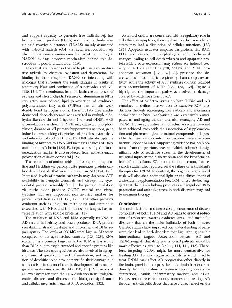
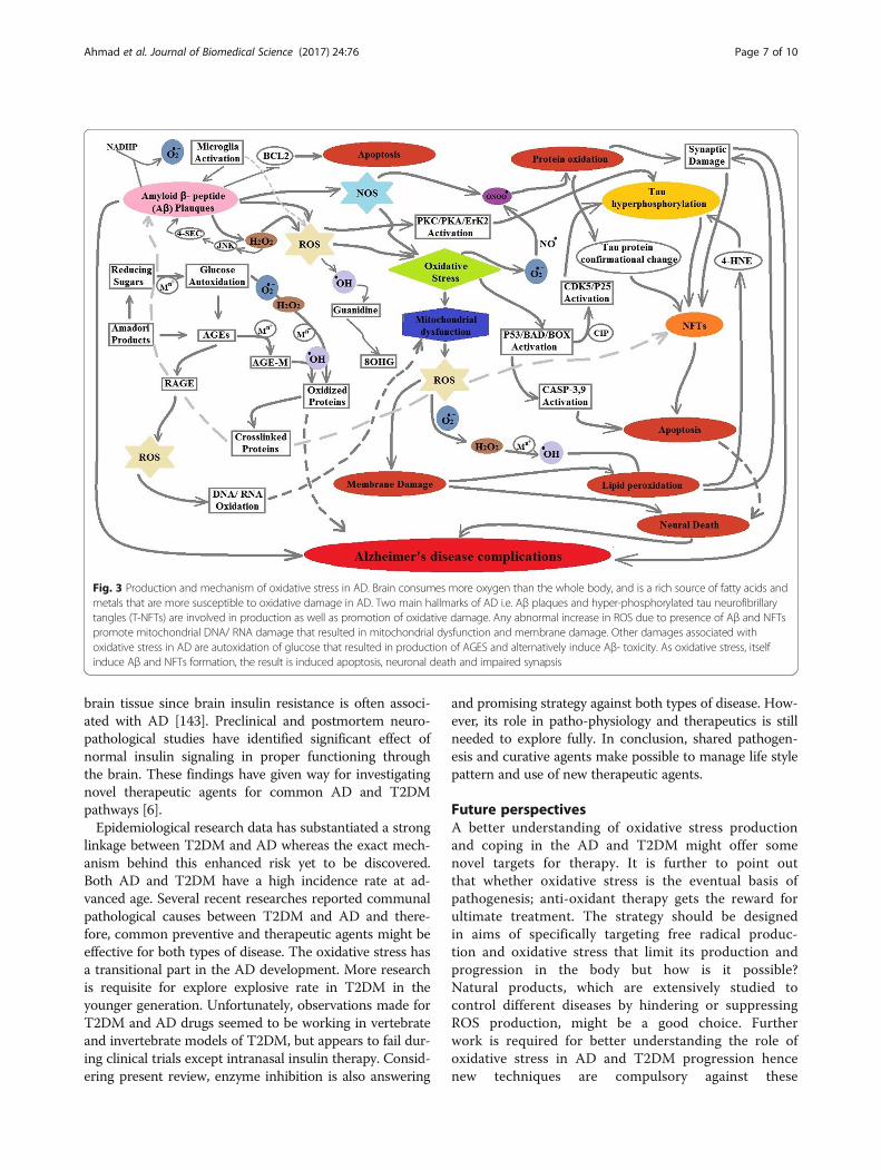
As mitochondria are concerned with a regulatory role incells through apoptosis, their dysfunction due to oxidativestress may lead a disruption of cellular functions [133,134]. Apoptosis activates caspases via proteins like BAD,BOX and results in morphological and biochemicalchanges leading to cell death whereas anti-apoptotic pro-tein BCL-2 over expression may reduce Aβ-induced tox-icity in AD via inhibiting p38, MAPK and NFkB pro-apoptotic activation [135–137]. Aβ presence also de-creased the mitochondrial respiratory chain complexes ac-tivity, while the activity of ATP synthase α-chain reducedwith accumulation of NFTs [129, 138, 139]. Figure 3highlighted the important pathways involved in damagecreated by oxidative stress in AD.The effect of oxidative stress on both T2DM and AD
remained to define. Intervention to excessive ROS pro-duction through scavenging free radicals and increasingantioxidant defence mechanisms are extensively antici-pated as anti-aging therapy and also managing AD andT2DM. However, positive and conclusive results have notbeen achieved even with the association of supplementa-tion and pharmacological or natural compounds. It is pos-sible that few antioxidants may become useless or evenharmful sooner or later. Supporting evidence has been ob-tained from the previous research, which indicates the sig-nificant role of oxidative stress in the development ofneuronal injury in the diabetic brain and the beneficial ef-fects of antioxidants. We must take into account, that re-search studies also reported on the failure of antioxidant’stherapies for T2DM. In contrast, the ongoing large clinicaltrials will also shed additional light on the clinical merit ofantioxidant supplementation [66, 140]. These studies sug-gest that the clearly linking products i.e. deregulated ROSproduction and oxidative stress in both disorders may leadto common therapy.
ConclusionsThe multi-factorial and inexorable phenomenon of diseasecomplexity of both T2DM and AD leads to gradual reduc-tion of resistance towards oxidative stress, and metabolicdisorders that are the major hallmarks of both illnesses.Genetic studies have improved our understanding of path-ways that lead to both disorders that highlighting possibleinterventional targets. Association between AD andT2DM suggests that drug givens to AD patients would bemore effective as given to DM [6, 114, 141, 142]. There-fore, targeting T2DM might be more constructive fortreating AD. It is also suggested that drugs which used totreat T2DM may affect AD progression ether directly inthe brain, provided they pass the blood-brain barrier or in-directly, by modification of systemic blood-glucose con-centrations, insulin, inflammatory markers and AGEs.Hence, recent research mostly focuses on treating ADthrough anti-diabetic drugs that have a direct effect on the
Ahmad et al. Journal of Biomedical Science (2017) 24:76 Page 6 of 10
brain tissue since brain insulin resistance is often associ-ated with AD [143]. Preclinical and postmortem neuro-pathological studies have identified significant effect ofnormal insulin signaling in proper functioning throughthe brain. These findings have given way for investigatingnovel therapeutic agents for common AD and T2DMpathways [6].Epidemiological research data has substantiated a strong
linkage between T2DM and AD whereas the exact mech-anism behind this enhanced risk yet to be discovered.Both AD and T2DM have a high incidence rate at ad-vanced age. Several recent researches reported communalpathological causes between T2DM and AD and there-fore, common preventive and therapeutic agents might beeffective for both types of disease. The oxidative stress hasa transitional part in the AD development. More researchis requisite for explore explosive rate in T2DM in theyounger generation. Unfortunately, observations made forT2DM and AD drugs seemed to be working in vertebrateand invertebrate models of T2DM, but appears to fail dur-ing clinical trials except intranasal insulin therapy. Consid-ering present review, enzyme inhibition is also answering
and promising strategy against both types of disease. How-ever, its role in patho-physiology and therapeutics is stillneeded to explore fully. In conclusion, shared pathogen-esis and curative agents make possible to manage life stylepattern and use of new therapeutic agents.
Future perspectivesA better understanding of oxidative stress productionand coping in the AD and T2DM might offer somenovel targets for therapy. It is further to point outthat whether oxidative stress is the eventual basis ofpathogenesis; anti-oxidant therapy gets the reward forultimate treatment. The strategy should be designedin aims of specifically targeting free radical produc-tion and oxidative stress that limit its production andprogression in the body but how is it possible?Natural products, which are extensively studied tocontrol different diseases by hindering or suppressingROS production, might be a good choice. Furtherwork is required for better understanding the role ofoxidative stress in AD and T2DM progression hencenew techniques are compulsory against these
Fig. 3 Production and mechanism of oxidative stress in AD. Brain consumes more oxygen than the whole body, and is a rich source of fatty acids andmetals that are more susceptible to oxidative damage in AD. Two main hallmarks of AD i.e. Aβ plaques and hyper-phosphorylated tau neurofibrillarytangles (T-NFTs) are involved in production as well as promotion of oxidative damage. Any abnormal increase in ROS due to presence of Aβ and NFTspromote mitochondrial DNA/ RNA damage that resulted in mitochondrial dysfunction and membrane damage. Other damages associated withoxidative stress in AD are autoxidation of glucose that resulted in production of AGES and alternatively induce Aβ- toxicity. As oxidative stress, itselfinduce Aβ and NFTs formation, the result is induced apoptosis, neuronal death and impaired synapsis
Ahmad et al. Journal of Biomedical Science (2017) 24:76 Page 7 of 10
substances. Poor knowledge of basic mechanisms in-volved in aging process, which might interfere to pre-vent or delay age-related pathologies, like T2DM,cardiovascular disorders, neurodegenerative disorders,and cancer. More investigations are clearly needed toclarify the discrepancy in the role of ROS and anti-oxidant enzymes in aging process and age-related dis-eases and to understand the precise role of freeradicals play in that processes.
AbbreviationsAD: Alzheimer’s disease; AGEs: Advanced glycation end products; Aβ: Beta-amyloid; BBB: Blood brain barrier; DAG: Diacylglycerol; GAPDH: Glyceraldehyde-3-phosphate dehydrogenase; GSH: Glutathione peroxidise; GSK3: Glycogen synthasekinase3; H2O2: hydrogen peroxide; HNE: 4-hydroxy-2-nonenal; KGDHC: α-Ketoglutarate dehydrogenase complex; LDL: Low-density lipoprotein; LMWA: Lowmolecular weight antioxidants; MAO: Monoamine oxidases; MetS: Metabolicsyndrome; MMPs: Matrix metalloproteinases; MPO: Myeloperoxidase;NADH: Nicotinamide dinucleotide; OMM: Outer mitochondrial membrane;RET: Reverse electron transfer; RNS: Reactive nitrogen species; ROS: Reactiveoxygen species; SOD: Superoxide dismutase; T2DM: Type-2 diabetes mellitus;TZDs: Thiazolidinediones; α –GDH: α-glycerophosphate
AcknowledgementsNot Applicable.
FundingNot Applicable.
Availability of data and materialsNot Applicable.
Authors’ contributionsWA, BI, KS, FA, and SR wrote the different parts of the manuscript. WA andKS draw the pictures included in this manuscript. WA and BI edited themanuscript. All authors read and approved the final manuscript.
Authors informationWaqar Ahmad is working as Research Officer while Khadija Shabbiri ispost-doctoral Research fellow at School of Biological Sciences, University ofQueensland, Australia. Bushra Ijaz is Assistant professor at CEMB, University ofthe Punjab, Lahore, Sidra Rehman is Assistant professor at COMSATS; whileFayyaz Ahmed is PhD a student.
Ethics approval and consent to participateNot Applicable.
Consent for publicationThe authors declare that this article is original and never been publishedbefore and not submitted to any other journal.
Competing interestsThe authors have no competing interests.
Publisher’s NoteSpringer Nature remains neutral with regard to jurisdictional claims inpublished maps and institutional affiliations.
Author details1School of Biological Sciences, University of Queensland, Brisbane 4072,Australia. 2Centre of Excellence in Molecular Biology, University of thePunjab, Thokar Niaz Baig, Lahore 54000, Pakistan. 3COMSATS Institute ofInformation Technology Abbottabad, Abbottabad 22010, Pakistan.
Received: 5 January 2017 Accepted: 5 September 2017
References1. Mulukutla BC, et al. Regulation of Glucose Metabolism - A Perspective From
Cell Bioprocessing. Trends Biotechnol. 2016;34(8):638-51.2. McNamara JT, Morgan JL, Zimmer J. A molecular description of cellulose
biosynthesis. Annu Rev Biochem. 2015;84:895–921.3. Berchtold NC, Cotman CW. Evolution in the conceptualization of dementia
and Alzheimer’s disease: Greco-roman period to the 1960s. NeurobiolAging. 1998;19(3):173–89.
4. Münch G, et al. Alzheimer's Disease–synergistic effects of glucose deficit,oxidative stress and advanced glycation endproducts. J Neural Transm.1998;105(4-5):439–61.
5. Hofman A, et al. The Rotterdam Study: 2016 objectives and design update.Eur J Epidemiol. 2015;30(8):661-708. PMID:26386597; PMCID:PMC4579264.doi:10.1007/s10654-015-0082-x.
6. Ahmad W. Overlapped metabolic and therapeutic links between Alzheimerand diabetes. Mol Neurobiol. 2013;47(1):399–424.
7. Mittal K, Katare DP. Shared links between type 2 diabetes mellitus andAlzheimer's disease: A review. Diabetes Metab Syndr. 2016;10(2 Suppl 1):S144-9.
8. Rosales-Corral S, et al. Diabetes and Alzheimer disease, two overlappingpathologies with the same background: oxidative stress. Oxidative Med CellLongev. 2015;2015:985845.
9. Biessels G-J, et al. Cerebral function in diabetes mellitus. Diabetologia. 1994;37(7):643–50.
10. Takeuchi M, Yamagishi S-i. Possible involvement of advanced glycation end-products (AGEs) in the pathogenesis of Alzheimer's disease. Curr PharmDes. 2008;14(10):973–8.
11. Biessels G, Kappelle L. Increased risk of Alzheimer's disease in type IIdiabetes: insulin resistance of the brain or insulin-induced amyloidpathology? Biochem Soc Trans. 2005;33(5):1041–4.
12. Butterfield DA, Di Domenico F, Barone E. Elevated risk of type 2 diabetes fordevelopment of Alzheimer disease: a key role for oxidative stress in brain.Biochim Biophys Acta. 2014;1842(9):1693–706.
13. Montecalvo MA, et al. Natural history of colonization with vancomycin-resistantEnterococcus faecium. Infect Control Hosp Epidemiol. 1995;16(12):680–5.
14. Reser JE. Alzheimer's Disease and natural cognitive aging may representadaptive metabolism reduction programs. Behav Brain Funct. 2009;5(1):1.
15. Wang J, Yang X, Zhang J. Bridges between mitochondrial oxidative stress, ER stressand mTOR signaling in pancreatic beta cells. Cell Signal. 2016;28(8):1099–104.
16. Sadeghi A, et al. The effect of diabetes mellitus on apoptosis inhippocampus: cellular and molecular aspects. Int J Prev Med. 2016;7:57.
17. Zheng H, et al. Protein modifications as manifestations of hyperglycemicGlucotoxicity in diabetes and its complications. Biochem Insights. 2016;9:1–9.
18. Ceriello A, Testa R, Genovese S. Clinical implications of oxidative stress andpotential role of natural antioxidants in diabetic vascular complications. NutrMetab Cardiovasc Dis. 2016;26(4):285–92.
19. Tramutola A, et al. Oxidative stress, protein modification and Alzheimerdisease. Brain Res Bull. 2017;133:88-96.
20. Grimm A, Mensah-Nyagan AG, Eckert A. Alzheimer, mitochondria andgender. Neurosci Biobehav Rev. 2016;67:89-101.
21. Cervellati C, et al. Oxidative challenge in Alzheimer's disease: state ofknowledge and future needs. J Investig Med. 2016;64(1):21–32.
22. Commoner B, Townsend J, Pake GE. Free radicals in biological materials.Nature. 1954;174(4432):689–91.
23. Newsholme P, et al. Molecular mechanisms of ROS production andoxidative stress in diabetes. Biochem J. 2016;473(24):4527–50.
24. Finkel T, Holbrook NJ. Oxidants, oxidative stress and the biology of ageing.Nature. 2000;408(6809):239–47.
25. Gracy R, et al. Reactive oxygen species: the unavoidable environmentalinsult? Mutat Res Fundam Mol Mech Mutagen. 1999;428(1):17–22.
26. Bansal AK, Bilaspuri G. Impacts of oxidative stress and antioxidants onsemen functions. Vet Med Int. 2010;2011
27. Halliwell B, Gutteridge J. Biologically relevant metal ion-dependent hydroxylradical generation an update. FEBS Lett. 1992;307(1):108–12.
28. Kohen R, Gati I. Skin low molecular weight antioxidants and their role inaging and in oxidative stress. Toxicology. 2000;148(2):149–57.
29. Phaniendra A, Jestadi DB, Periyasamy L. Free radicals: properties, sources,targets, and their implication in various diseases. Indian J Clin Biochem.2015;30(1);11-26.
Ahmad et al. Journal of Biomedical Science (2017) 24:76 Page 8 of 10
30. Kohen R, Moor E, Oron M. Measurements of biological reducing power inhealth and diseases by voltammetric methods. Redox Genome InteractionHealth Dis. 2004:13–42.
31. Victorin K. Review of the genotoxicity of nitrogen oxides. Mutat Res. 1994;317(1):43–55.
32. Koren HS. Associations between criteria air pollutants and asthma. EnvironHealth Perspect. 1995;103(Suppl 6):235.
33. Chinev S, et al. Lipid peroxidation in rat lung induced by neuroleptanalgesiaand its components. Eur J Anaesthesiol. 1998;15(6):686–94.
34. Sen C, Packer L, Hänninen O. Biological thiols and redox regulation ofcellular signal transduction pathways. Handbook of oxidants andantioxidants in exercise. Amsterdam: Elsevier; 2000. p. 375–402.
35. Shadyro O, Yurkova I, Kisel M. Radiation-induced peroxidation and fragmentationof lipids in a model membrane. Int J Radiat Biol. 2002;78(3):211–7.
36. Elsayed NM, et al. Free radical-mediated lung response to themonofunctional sulfur mustard butyl 2-chloroethyl sulfide aftersubcutaneous injection. Toxicology. 1992;72(2):153–65.
37. Obata T, et al. Release of dopamine by perfusion with 1-methyl-4-phenylpyridinium ion (MPP+) into the striatum is associated with hydroxylfree radical generation. Brain Res. 2001;906(1):170–5.
38. Dhalla NS, Temsah RM, Netticadan T. Role of oxidative stress incardiovascular diseases. J Hypertens. 2000;18(6):655–73.
39. Forman HJ, Torres M. Reactive oxygen species and cell signaling: respiratoryburst in macrophage signaling. Am J Respir Crit Care Med. 2002;166(supplement_1):S4–8.
40. Fleury C, Mignotte B, Vayssière J-L. Mitochondrial reactive oxygen species incell death signaling. Biochimie. 2002;84(2):131–41.
41. Gutteridge JM, Halliwell B. Invited review free radicals in disease processes:a compilation of cause and consequence. Free Radic Res Commun. 1993;19(3):141–58.
42. Dong J, et al. Metal binding and oxidation of amyloid-β within isolatedsenile plaque cores: Raman microscopic evidence. Biochemistry. 2003;42(10):2768–73.
43. Hoyer S. Causes and consequences of disturbances of cerebral glucosemetabolism in sporadic Alzheimer disease: therapeutic implications. AdvExp Med Biol. 2004;541:135-52.
44. Butterfield DA, Pocernich CB. The glutamatergic system and Alzheimer’sdisease. CNS drugs. 2003;17(9):641–52.
45. Kaul M, Garden GA, Lipton SA. Pathways to neuronal injury and apoptosis inHIV-associated dementia. Nature. 2001;410(6831):988–94.
46. Bielski BH, et al. Reactivity of HO2/O− 2 radicals in aqueous solution. J PhysChem Ref Data. 1985;14(4):1041–100.
47. Schafer FQ, Buettner GR. Redox environment of the cell as viewed throughthe redox state of the glutathione disulfide/glutathione couple. Free RadicBiol Med. 2001;30(11):1191–212.
48. Kohen R, Nyska A. Oxidation of biological systems: oxidative stressphenomena, antioxidants, redox reactions, and methods for theirquantification. Toxicol Pathol. 2002;30(6):620-50.
49. Hrbac J, Kohen R. Biological redox activity: its importance, methods for itsquantification and implication for health and disease. Drug Dev Res. 2000;50(3-4):516–27.
50. Comporti M. Three models of free radical-induced cell injury. Chem BiolInteract. 1989;72(1):1–56.
51. Priora R, et al. Measurement of mixed disulfides including glutathionylatedproteins. Methods Enzymol. 2010;473:149–59.
52. Muralidharan P, et al. Evidence for redox sensing by a human cardiaccalcium channel. Sci Rep. 2016;6
53. Dröge W. Free radicals in the physiological control of cell function. PhysiolRev. 2002;82(1):47–95.
54. Newsholme P, et al. Nutrient regulation of insulin secretion and action. JEndocrinol. 2014;221(3):R105–20.
55. Salsali A, Nathan M. A review of types 1 and 2 diabetes mellitus and theirtreatment with insulin. Am J Ther. 2006;13(4):349-61.
56. Wallace T, Matthews D. Recent advances in the monitoring andmanagement of diabetic ketoacidosis. QJM. 2004;97(12):773–80.
57. Asmat U, Abad K, Ismail K. Diabetes mellitus and oxidative stress—a concisereview. Saudi Pharm J. 2016;24(5):547–53.
58. Loghmani E. Diabetes mellitus: type 1 and type 2. Guidelines for adolescentnutrition services; 2005. p. 167–82.
59. Kangralkar V, Patil SD, Bandivadekar R. Oxidative stress and diabetes: areview. Int J Pharm Appl. 2010;1(1):38–45.
60. Pham-Huy LA, He H, Pham-Huy C. Free radicals, antioxidants in disease andhealth. Int J Biomed Sci. 2008;4(2):89–96.
61. Maritim A, Sanders a, Watkins r J. Diabetes, oxidative stress, and antioxidants: areview. J Biochem Mol Toxicol. 2003;17(1):24–38.
62. Lipinski B. Pathophysiology of oxidative stress in diabetes mellitus. J DiabetesComplicat. 2001;15(4):203–10.
63. Moussa S. Oxidative stress in diabetes mellitus. Romanian J Biophys. 2008;18(3):225–36.
64. Erejuwa OO. Oxidative stress in diabetes mellitus: is there a role for hypoglycemicdrugs and/or antioxidants. Oxidative stress and diseases; 2012. p. 217–46.
65. Ceriello PA. Oxidative stress and diabetes-associated complications. EndocrPract. 2006;12(Supplement 1):60–2.
66. Calkins MJ, Manczak M, Reddy PH. Mitochondria-targeted antioxidant SS31prevents amyloid beta-induced mitochondrial abnormalities and synapticdegeneration in Alzheimer’s disease. Pharmaceuticals. 2012;5(10):1103–19.
67. Jahangir Z, Ahmad W, Shabbiri K. Alternate Phosphorylation/O-GlcNAcmodification on human insulin IRSs: a road towards impaired insulinsignaling in Alzheimer and diabetes. Adv Bioinforma. 2014;2014:324753.
68. Sorriento D, et al. Targeting mitochondria as therapeutic strategy formetabolic disorders. ScientificWorldJournal. 2014;2014:604685.
69. Guzik TJ, et al. Vascular superoxide production by NAD (P) H oxidaseassociation with endothelial dysfunction and clinical risk factors. Circ Res.2000;86(9):e85–90.
70. Ceriello A, et al. Detection of nitrotyrosine in the diabetic plasma: evidenceof oxidative stress. Diabetologia. 2001;44(7):834–8.
71. Guzik TJ, et al. Mechanisms of increased vascular superoxide production inhuman diabetes mellitus role of NAD (P) H oxidase and endothelial nitricoxide synthase. Circulation. 2002;105(14):1656–62.
72. Pazdro R, Burgess JR. The role of vitamin E and oxidative stress in diabetescomplications. Mech Ageing Dev. 2010;131(4):276–86.
73. Brownlee M. Internationale Fachliteratur. Nature. 2001;414(6865):813–20.74. Jiang Z-Y, Woollard A, Wolff SP. Hydrogen peroxide production during
experimental protein glycation. FEBS Lett. 1990;268(1):69–71.75. Wolff SP, Dean R. Glucose autoxidation and protein modification. The
potential role of ‘autoxidative glycosylation’in diabetes. Biochem J. 1987;245(1):243–50.
76. Halliwell B, Gutteridge JM. [1] Role of free radicals and catalytic metal ionsin human disease: an overview. Methods Enzymol. 1990;186:1–85.
77. Hogg N, et al. Inhibition of low-density lipoprotein oxidation by nitric oxidepotential role in atherogenesis. FEBS Lett. 1993;334(2):170–4.
78. Korshunov SS, Skulachev VP, Starkov AA. High protonic potential actuates amechanism of production of reactive oxygen species in mitochondria. FEBSLett. 1997;416(1):15–8.
79. Brownlee M. Biochemistry and molecular cell biology of diabeticcomplications. Nature. 2001;414(6865):813–20.
80. Nishikawa T, Edelstein D, Brownlee M. The missing link: a single unifyingmechanism for diabetic complications. Kidney Int. 2000;58:S26–30.
81. Pacher P, Szabó C. Role of poly (ADP-ribose) polymerase-1 activation in thepathogenesis of diabetic complications: endothelial dysfunction, as acommon underlying theme. Antioxid Redox Signal. 2005;7(11-12):1568–80.
82. Williamson JR, et al. Hyperglycemic pseudohypoxia and diabeticcomplications. Diabetes. 1993;42(6):801–13.
83. Choudhury, M., Involvement of Caspase-7 in photoreceptor and retinalganglion cell death. 2014.
84. Zhang Z, et al. High glucose inhibits glucose-6-phosphate dehydrogenasevia cAMP in aortic endothelial cells. J Biol Chem. 2000;275(51):40042–7.
85. James LR, et al. Flux through the Hexosamine pathway is a determinant ofnuclear factor κB–dependent promoter activation. Diabetes. 2002;51(4):1146–56.
86. McClain DA, Crook ED. Hexosamines and insulin resistance. Diabetes. 1996;45(8):1003–9.
87. Goldberg HJ, Whiteside CI, Fantus IG. The hexosamine pathway regulatesthe plasminogen activator inhibitor-1 gene promoter and Sp1transcriptional activation through protein kinase C-βI and-δ. J Biol Chem.2002;277(37):33833–41.
88. Du X-L, et al. Hyperglycemia-induced mitochondrial superoxideoverproduction activates the hexosamine pathway and inducesplasminogen activator inhibitor-1 expression by increasing Sp1glycosylation. Proc Natl Acad Sci. 2000;97(22):12222–6.
89. Li L-X, et al. Uncoupling protein-2 participates in cellular defense againstoxidative stress in clonal β-cells. Biochem Biophys Res Commun. 2001;282(1):273–7.
Ahmad et al. Journal of Biomedical Science (2017) 24:76 Page 9 of 10
90. Nishikawa T, et al. Normalizing mitochondrial superoxide production blocksthree pathways of hyperglycaemic damage. Nature. 2000;404(6779):787–90.
91. Ulrich P, Cerami A. Protein glycation, diabetes, and aging. Recent ProgHorm Res. 2000;56:1–21.
92. Baynes JW, Thorpe SR. Role of oxidative stress in diabetic complications: anew perspective on an old paradigm. Diabetes. 1999;48(1):1–9.
93. McCarthy A, Etcheverry S, Cortizo A. Effect of advanced glycationendproducts on the secretion of insulin-like growth factor-I and its bindingproteins: role in osteoblast development. Acta Diabetol. 2001;38(3):113–22.
94. Ergul A, et al. Vascular dysfunction of venous bypass conduits is mediatedby reactive oxygen species in diabetes: role of endothelin-1. J PharmacolExp Ther. 2005;313(1):70–7.
95. Amiri F, et al. Angiotensin II activation of the JAK/STAT pathway inmesangial cells is altered by high glucose. Kidney Int. 2002;61(5):1605–16.
96. Wake H, et al. Histamine inhibits advanced glycation end products-inducedadhesion molecule expression on human monocytes. J Pharmacol Exp Ther.2009;330(3):826–33.
97. Takahashi HK, et al. Advanced glycation end products subspecies-selectivelyinduce adhesion molecule expression and cytokine production in humanperipheral blood mononuclear cells. J Pharmacol Exp Ther. 2009;330(1):89–98.
98. Lohwasser C, et al. The receptor for advanced glycation end products is highlyexpressed in the skin and upregulated by advanced glycation end productsand tumor necrosis factor-alpha. J Investig Dermatol. 2006;126(2):291–9.
99. Hergrueter AH, Nguyen K, Owen CA. Matrix metalloproteinases: all the RAGEin the acute respiratory distress syndrome. Am J Physiol Lung Cell MolPhysiol. 2011;300(4):L512-5.
100. Tsai EC, et al. Reduced plasma peroxyl radical trapping capacity andincreased susceptibility of LDL to oxidation in poorly controlled IDDM.Diabetes. 1994;43(8):1010–4.
101. Kawamura M, Heinecke JW, Chait A. Pathophysiological concentrations ofglucose promote oxidative modification of low density lipoprotein by asuperoxide-dependent pathway. J Clin Investig. 1994;94(2):771.
102. Evans JL, et al. Are oxidative stress− activated signaling pathways mediatorsof insulin resistance and β-cell dysfunction? Diabetes. 2003;52(1):1–8.
103. Roth M. The association of clinical and neurological findings and its bearingon the classification and aetiology of Alzheimer's disease. Br Med Bull. 1986;42(1):42–50.
104. Juhaszova M, et al. Protection in the aged heart: preventing the heart-breakof old age? Cardiovasc Res. 2005;66(2):233–44.
105. Tani M, et al. Direct activation of mitochondrial KATP channels mimicspreconditioning but protein kinase C activation is less effective in middle-aged rat hearts. Cardiovasc Res. 2001;49(1):56–68.
106. Jang YC, et al. Increased superoxide in vivo accelerates age-associatedmuscle atrophy through mitochondrial dysfunction and neuromuscularjunction degeneration. FASEB J. 2010;24(5):1376–90.
107. Li F-J, Shen L, Ji H-F. Dietary intakes of vitamin E, vitamin C, and β-carotene andrisk of Alzheimer's disease: a meta-analysis. J Alzheimers Dis. 2012;31(2):253–8.
108. Migliaccio E, et al. The p66shc adaptor protein controls oxidative stressresponse and life span in mammals. Nature. 1999;402(6759):309–13.
109. Giorgio M, et al. Electron transfer between cytochrome c and p66 Shcgenerates reactive oxygen species that trigger mitochondrial apoptosis. Cell.2005;122(2):221–33.
110. Cha M-Y, et al. Mitochondria-specific accumulation of amyloid β inducesmitochondrial dysfunction leading to apoptotic cell death. PLoS One. 2012;7(4):e34929.
111. Reddy PH. Amyloid beta, mitochondrial structural and functional dynamicsin Alzheimer's disease. Exp Neurol. 2009;218(2):286–92.
112. Canugovi C, et al. Base excision DNA repair levels in mitochondrial lysatesof Alzheimer's disease. Neurobiol Aging. 2014;35(6):1293–300.
113. Yan MH, Wang X, Zhu X. Mitochondrial defects and oxidative stress inAlzheimer disease and Parkinson disease. Free Radic Biol Med. 2013;62:90–101.
114. Ahmad W, Ebert PR. Metformin Attenuates Abeta Pathology MediatedThrough Levamisole Sensitive Nicotinic Acetylcholine Receptors in a C.elegans Model of Alzheimer's Disease. Mol Neurobiol. 2017;54(7):5427-439.
115. Praticò D, Delanty N. Oxidative injury in diseases of the central nervoussystem: focus on Alzheimer’s disease. Am J Med. 2000;109(7):577–85.
116. Floyd RA. Antioxidants, oxidative stress, and degenerative neurologicaldisorders. Exp Biol Med. 1999;222(3):236–45.
117. Zhu X, et al. Alzheimer disease, the two-hit hypothesis: an update. BiochimBiophys Acta. 2007;1772(4):494–502.
118. Wang X, et al. Amyloid-β overproduction causes abnormal mitochondrialdynamics via differential modulation of mitochondrial fission/fusionproteins. Proc Natl Acad Sci. 2008;105(49):19318–23.
119. Park KW, Baik HH, Jin BK. IL-13-induced oxidative stress via microglialNADPH oxidase contributes to death of hippocampal neurons in vivo. JImmunol. 2009;183(7):4666–74.
120. Baynes JW. Role of oxidative stress in development of complications indiabetes. Diabetes. 1991;40(4):405–12.
121. Hazel JR, Williams EE. The role of alterations in membrane lipid compositionin enabling physiological adaptation of organisms to their physicalenvironment. Prog Lipid Res. 1990;29(3):167–227.
122. Prasad MR, et al. Regional membrane phospholipid alterations inAlzheimer's disease. Neurochem Res. 1998;23(1):81–8.
123. Roberts LJ, Morrow JD. Measurement of F 2-isoprostanes as an index ofoxidative stress in vivo. Free Radic Biol Med. 2000;28(4):505–13.
124. Stadtman ER. Metal ion-catalyzed oxidation of proteins: biochemical mechanismand biological consequences. Free Radic Biol Med. 1990;9(4):315–25.
125. Smith MA, et al. Widespread peroxynitrite-mediated damage in Alzheimer’sdisease. J Neurosci. 1997;17(8):2653–7.
126. Good PF, et al. Evidence of neuronal oxidative damage in Alzheimer'sdisease. Am J Pathol. 1996;149(1):21.
127. Moreira PI, et al. Alzheimer's Disease and oxidative stress: the old problemremains unsolved. Curr Med Chem Cent Nerv Syst Agents. 2005;5(1):51–62.
128. Aksenov M, et al. Protein oxidation in the brain in Alzheimer's disease.Neuroscience. 2001;103(2):373–83.
129. Bubber P, et al. Mitochondrial abnormalities in Alzheimer brain: mechanisticimplications. Ann Neurol. 2005;57(5):695–703.
130. Cao X, et al. Noncoding RNAs in the mammalian central nervous system.Annu Rev Neurosci. 2006;29:77–103.
131. Mehler MF, Mattick JS. Noncoding RNAs and RNA editing in braindevelopment, functional diversification, and neurological disease. PhysiolRev. 2007;87(3):799–823.
132. Nunomura A, et al. RNA oxidation in Alzheimer disease and relatedneurodegenerative disorders. Acta Neuropathol. 2009;118(1):151–66.
133. Bonda DJ, et al. Mitochondrial dynamics in Alzheimer’s disease. DrugsAging. 2010;27(3):181–92.
134. Zhu X, et al. Abnormal mitochondrial dynamics in the pathogenesis ofAlzheimer's disease. J Alzheimer's Dis. 2013;33(0 1):S253.
135. Zaid H, et al. The voltage-dependent anion channel-1 modulates apoptoticcell death. Cell Death Differ. 2005;12(7):751–60.
136. Ferri KF, Kroemer G. Organelle-specific initiation of cell death pathways. NatCell Biol. 2001;3(11):E255–63.
137. Pereira C, et al. Cell degeneration induced by amyloid-β peptides. J MolNeurosci. 2004;23(1-2):97–104.
138. Sergeant N, et al. Association of ATP synthase α-chain with neurofibrillarydegeneration in Alzheimer’s disease. Neuroscience. 2003;117(2):293–303.
139. Poon SR, B-K D, Klein JB, Merchant M, Markesbery WR, Butterfield DA. Redoxproteomics identification of oxidized proteins in Alzheimer's diseasehippocampus and cerebellum: an approach to understand pathological andbiochemical alterations in AD. Neurobiol Aging. 2006;27:1564–76.
140. Yan L-J. Positive oxidative stress in aging and aging-related diseasetolerance. Redox Biol. 2014;2:165–9.
141. Akter K, et al. Diabetes mellitus and Alzheimer's disease: shared pathologyand treatment? Br J Clin Pharmacol. 2011;71(3):365–76.
142. Bomfim TR, et al. An anti-diabetes agent protects the mouse brain fromdefective insulin signaling caused by Alzheimer’s disease–associated Aβoligomers. J Clin Invest. 2012;122(4):1339–53.
143. Chiti F, Dobson CM. Protein misfolding, functional amyloid, and humandisease. Annu Rev Biochem. 2006;75:333–66.
Ahmad et al. Journal of Biomedical Science (2017) 24:76 Page 10 of 10