Embed Size (px)
Citation preview
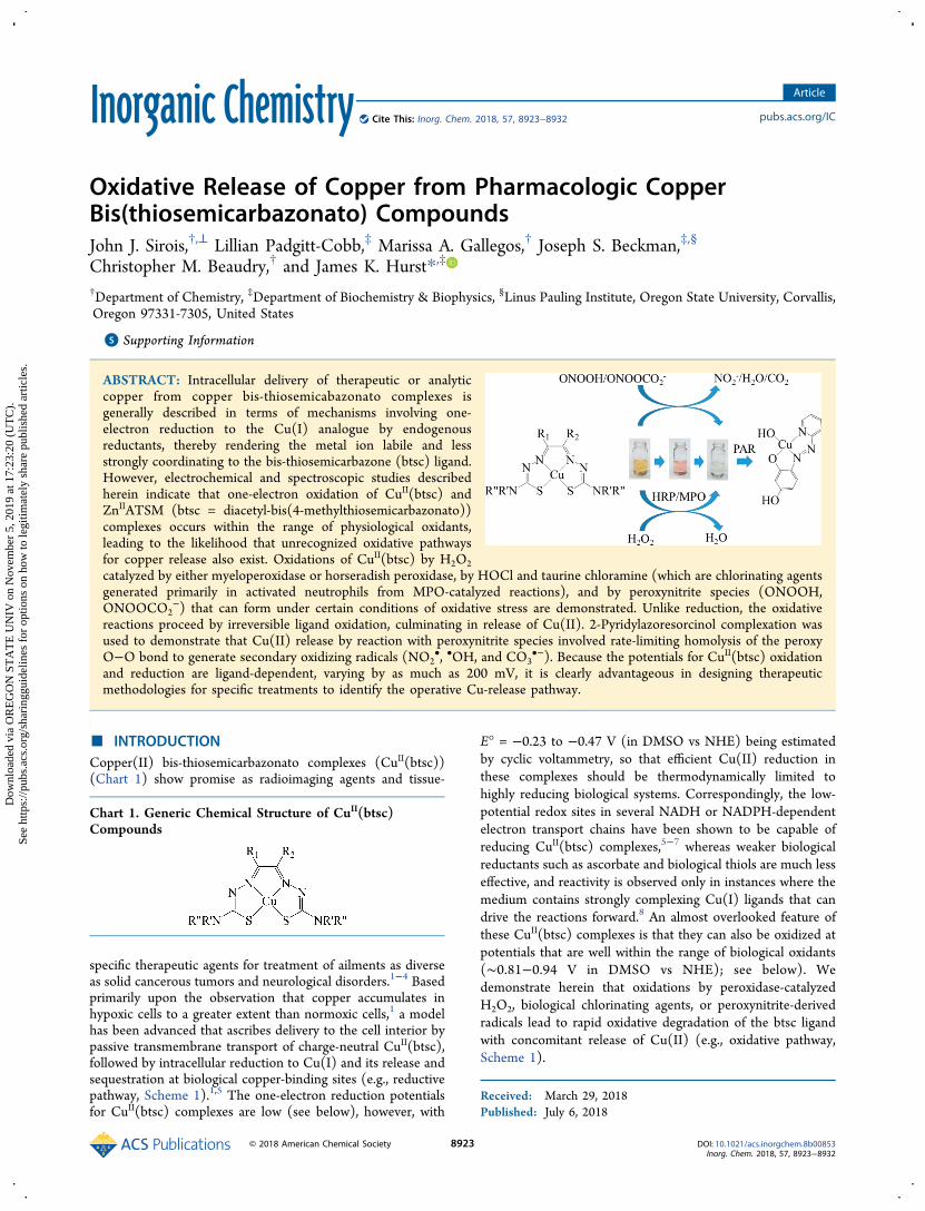
Oxidative Release of Copper from Pharmacologic CopperBis(thiosemicarbazonato) CompoundsJohn J. Sirois,†,⊥ Lillian Padgitt-Cobb,‡ Marissa A. Gallegos,† Joseph S. Beckman,‡,§
Christopher M. Beaudry,† and James K. Hurst*,‡
†Department of Chemistry, ‡Department of Biochemistry & Biophysics, §Linus Pauling Institute, Oregon State University, Corvallis,Oregon 97331-7305, United States
*S Supporting Information
ABSTRACT: Intracellular delivery of therapeutic or analyticcopper from copper bis-thiosemicabazonato complexes isgenerally described in terms of mechanisms involving one-electron reduction to the Cu(I) analogue by endogenousreductants, thereby rendering the metal ion labile and lessstrongly coordinating to the bis-thiosemicarbazone (btsc) ligand.However, electrochemical and spectroscopic studies describedherein indicate that one-electron oxidation of CuII(btsc) andZnIIATSM (btsc = diacetyl-bis(4-methylthiosemicarbazonato))complexes occurs within the range of physiological oxidants,leading to the likelihood that unrecognized oxidative pathwaysfor copper release also exist. Oxidations of CuII(btsc) by H2O2catalyzed by either myeloperoxidase or horseradish peroxidase, by HOCl and taurine chloramine (which are chlorinating agentsgenerated primarily in activated neutrophils from MPO-catalyzed reactions), and by peroxynitrite species (ONOOH,ONOOCO2
−) that can form under certain conditions of oxidative stress are demonstrated. Unlike reduction, the oxidativereactions proceed by irreversible ligand oxidation, culminating in release of Cu(II). 2-Pyridylazoresorcinol complexation wasused to demonstrate that Cu(II) release by reaction with peroxynitrite species involved rate-limiting homolysis of the peroxyO−O bond to generate secondary oxidizing radicals (NO2
•, •OH, and CO3•−). Because the potentials for CuII(btsc) oxidation
and reduction are ligand-dependent, varying by as much as 200 mV, it is clearly advantageous in designing therapeuticmethodologies for specific treatments to identify the operative Cu-release pathway.
■ INTRODUCTIONCopper(II) bis-thiosemicarbazonato complexes (CuII(btsc))(Chart 1) show promise as radioimaging agents and tissue-
specific therapeutic agents for treatment of ailments as diverseas solid cancerous tumors and neurological disorders.1−4 Basedprimarily upon the observation that copper accumulates inhypoxic cells to a greater extent than normoxic cells,1 a modelhas been advanced that ascribes delivery to the cell interior bypassive transmembrane transport of charge-neutral CuII(btsc),followed by intracellular reduction to Cu(I) and its release andsequestration at biological copper-binding sites (e.g., reductivepathway, Scheme 1).1,5 The one-electron reduction potentialsfor CuII(btsc) complexes are low (see below), however, with
E° = −0.23 to −0.47 V (in DMSO vs NHE) being estimatedby cyclic voltammetry, so that efficient Cu(II) reduction inthese complexes should be thermodynamically limited tohighly reducing biological systems. Correspondingly, the low-potential redox sites in several NADH or NADPH-dependentelectron transport chains have been shown to be capable ofreducing CuII(btsc) complexes,5−7 whereas weaker biologicalreductants such as ascorbate and biological thiols are much lesseffective, and reactivity is observed only in instances where themedium contains strongly complexing Cu(I) ligands that candrive the reactions forward.8 An almost overlooked feature ofthese CuII(btsc) complexes is that they can also be oxidized atpotentials that are well within the range of biological oxidants(∼0.81−0.94 V in DMSO vs NHE); see below). Wedemonstrate herein that oxidations by peroxidase-catalyzedH2O2, biological chlorinating agents, or peroxynitrite-derivedradicals lead to rapid oxidative degradation of the btsc ligandwith concomitant release of Cu(II) (e.g., oxidative pathway,Scheme 1).
Received: March 29, 2018Published: July 6, 2018
Chart 1. Generic Chemical Structure of CuII(btsc)Compounds
Article
pubs.acs.org/ICCite This: Inorg. Chem. 2018, 57, 8923−8932
© 2018 American Chemical Society 8923 DOI: 10.1021/acs.inorgchem.8b00853Inorg. Chem. 2018, 57, 8923−8932
Dow
nloa
ded
via
OR
EG
ON
ST
AT
E U
NIV
on
Nov
embe
r 5,
201
9 at
17:
23:2
0 (U
TC
).Se
e ht
tps:
//pub
s.ac
s.or
g/sh
arin
ggui
delin
es f
or o
ptio
ns o
n ho
w to
legi
timat
ely
shar
e pu
blis
hed
artic
les.
■ RESULTS AND DISCUSSIONElectrochemistry. The electrochemical redox properties of
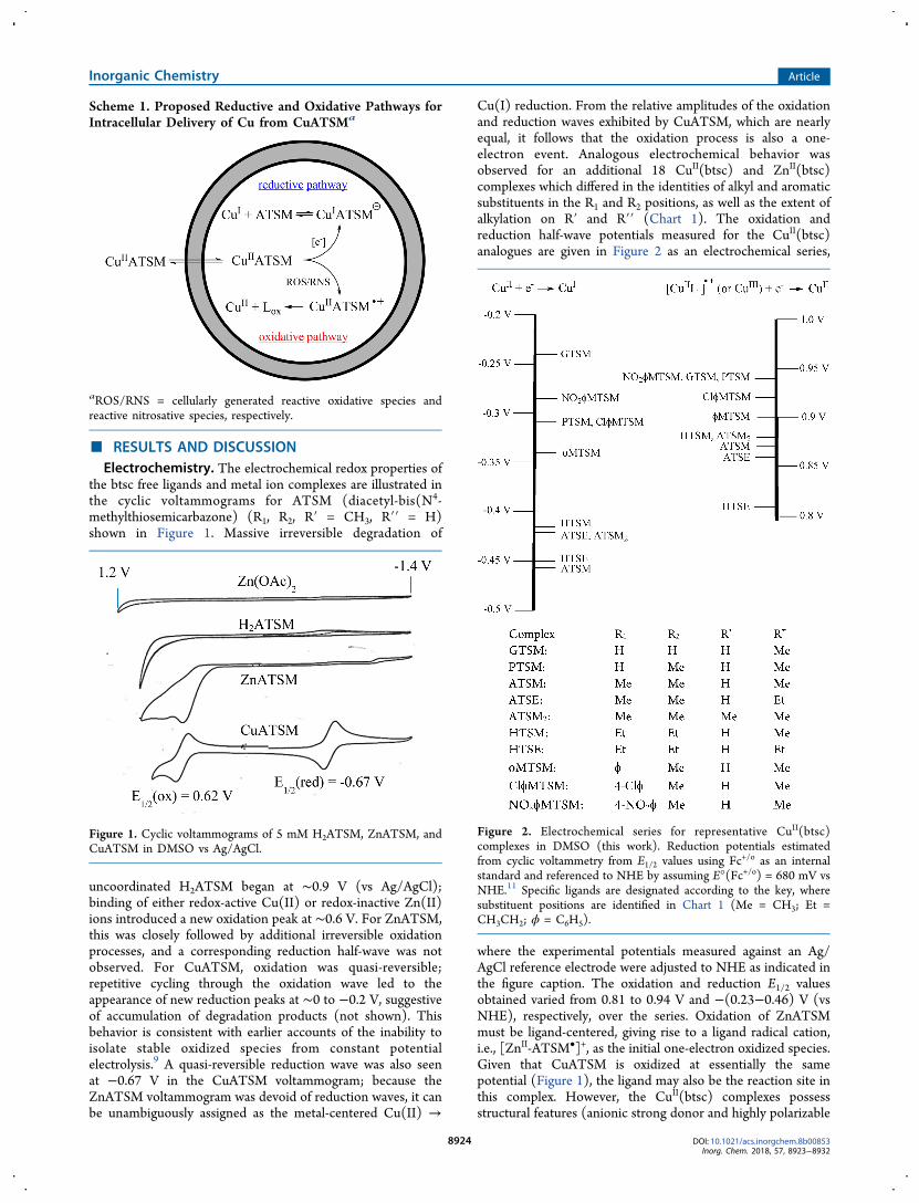
the btsc free ligands and metal ion complexes are illustrated inthe cyclic voltammograms for ATSM (diacetyl-bis(N4-methylthiosemicarbazone) (R1, R2, R′ = CH3, R′′ = H)shown in Figure 1. Massive irreversible degradation of
uncoordinated H2ATSM began at ∼0.9 V (vs Ag/AgCl);binding of either redox-active Cu(II) or redox-inactive Zn(II)ions introduced a new oxidation peak at ∼0.6 V. For ZnATSM,this was closely followed by additional irreversible oxidationprocesses, and a corresponding reduction half-wave was notobserved. For CuATSM, oxidation was quasi-reversible;repetitive cycling through the oxidation wave led to theappearance of new reduction peaks at ∼0 to −0.2 V, suggestiveof accumulation of degradation products (not shown). Thisbehavior is consistent with earlier accounts of the inability toisolate stable oxidized species from constant potentialelectrolysis.9 A quasi-reversible reduction wave was also seenat −0.67 V in the CuATSM voltammogram; because theZnATSM voltammogram was devoid of reduction waves, it canbe unambiguously assigned as the metal-centered Cu(II) →
Cu(I) reduction. From the relative amplitudes of the oxidationand reduction waves exhibited by CuATSM, which are nearlyequal, it follows that the oxidation process is also a one-electron event. Analogous electrochemical behavior wasobserved for an additional 18 CuII(btsc) and ZnII(btsc)complexes which differed in the identities of alkyl and aromaticsubstituents in the R1 and R2 positions, as well as the extent ofalkylation on R′ and R′′ (Chart 1). The oxidation andreduction half-wave potentials measured for the CuII(btsc)analogues are given in Figure 2 as an electrochemical series,
where the experimental potentials measured against an Ag/AgCl reference electrode were adjusted to NHE as indicated inthe figure caption. The oxidation and reduction E1/2 valuesobtained varied from 0.81 to 0.94 V and −(0.23−0.46) V (vsNHE), respectively, over the series. Oxidation of ZnATSMmust be ligand-centered, giving rise to a ligand radical cation,i.e., [ZnII-ATSM•]+, as the initial one-electron oxidized species.Given that CuATSM is oxidized at essentially the samepotential (Figure 1), the ligand may also be the reaction site inthis complex. However, the CuII(btsc) complexes possessstructural features (anionic strong donor and highly polarizable
Scheme 1. Proposed Reductive and Oxidative Pathways forIntracellular Delivery of Cu from CuATSMa
aROS/RNS = cellularly generated reactive oxidative species andreactive nitrosative species, respectively.
Figure 1. Cyclic voltammograms of 5 mM H2ATSM, ZnATSM, andCuATSM in DMSO vs Ag/AgCl.
Figure 2. Electrochemical series for representative CuII(btsc)complexes in DMSO (this work). Reduction potentials estimatedfrom cyclic voltammetry from E1/2 values using Fc+/o as an internalstandard and referenced to NHE by assuming E°(Fc+/o) = 680 mV vsNHE.11 Specific ligands are designated according to the key, wheresubstituent positions are identified in Chart 1 (Me = CH3; Et =CH3CH2; ϕ = C6H5).
Inorganic Chemistry Article
DOI: 10.1021/acs.inorgchem.8b00853Inorg. Chem. 2018, 57, 8923−8932
8924
lead-in atoms in a tetradentate square planar geometry) thatwould stabilize the d8 Cu(III) center and could thereby lowerits reduction potential into the observed range. Numeroussimilarly structured cupric complexes have in fact beendescribed for which E°(CuIII/II) ∼ 0.7−0.9 V (NHE).10 Theexperiments described herein do not allow discriminationbetween metal-centered and ligand-centered oxidation ofCuII(btsc), although one might speculate that the greaterstability of its oxidized state evident in the CuATSM andZnATSM voltammograms (Figure 1) relates to its capacity toundergo metal-centered oxidation.Chemical Oxidations: General Considerations. The
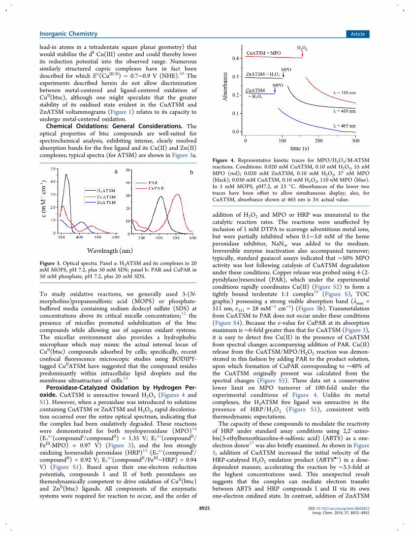
optical properties of btsc compounds are well-suited forspectrochemical analysis, exhibiting intense, clearly resolvedabsorption bands for the free ligand and its Cu(II) and Zn(II)complexes; typical spectra (for ATSM) are shown in Figure 3a.
To study oxidative reactions, we generally used 3-(N-morpholino)propanesulfonic acid (MOPS) or phosphate-buffered media containing sodium dodecyl sulfate (SDS) atconcentrations above its critical micelle concentration;12 thepresence of micelles promoted solubilization of the btsccompounds while allowing use of aqueous oxidant systems.The micellar environment also provides a hydrophobicmicrophase which may mimic the actual internal locus ofCuII(btsc) compounds adsorbed by cells; specifically, recentconfocal fluorescence microscopic studies using BODIPY-tagged CuIIATSM have suggested that the compound residespredominantly within intracellular lipid droplets and themembrane ultrastructure of cells.13
Peroxidase-Catalyzed Oxidation by Hydrogen Per-oxide. CuATSM is unreactive toward H2O2 (Figures 4 andS1). However, when a peroxidase was introduced to solutionscontaining CuATSM or ZnATSM and H2O2, rapid decoloriza-tion occurred over the entire optical spectrum, indicating thatthe complex had been oxidatively degraded. These reactionswere demonstrated for both myeloperoxidase (MPO)14
(E7°′(compoundI/compoundII) = 1.35 V; E7°′(compoundII/FeIII-MPO) = 0.97 V) (Figure 3), and the less stronglyoxidizing horseradish peroxidase (HRP)15 (E7°′(compoundI/compoundII) = 0.92 V; E7°′(compoundII/FeIII−HRP) = 0.94V) (Figure S1). Based upon their one-electron reductionpotentials, compounds I and II of both peroxidases arethemodynamically competent to drive oxidation of CuII(btsc)and ZnII(btsc) ligands. All components of the enzymaticsystems were required for reaction to occur, and the order of
addition of H2O2 and MPO or HRP was immaterial to thecatalytic reaction rates. The reactions were unaffected byinclusion of 1 mM DTPA to scavenge adventitious metal ions,but were partially inhibited when 0.1−3.0 mM of the hemeperoxidase inhibitor, NaN3, was added to the medium.Irreversible enzyme inactivation also accompanied turnover;typically, standard guaiacol assays indicated that ∼50% MPOactivity was lost following catalysis of CuATSM degradationunder these conditions. Copper release was probed using 4-(2-pyridylazo)resorcinol (PAR), which under the experimentalconditions rapidly coordinates Cu(II) (Figure S2) to form atightly bound terdentate 1:1 complex16 (Figure S3, TOCgraphic) possessing a strong visible absorption band (λmax =511 nm, ε511 = 28 mM−1 cm−1) (Figure 3b). Transmetalationfrom CuATSM to PAR does not occur under these conditions(Figure S4). Because the ε-value for CuPAR at its absorptionmaximum is ∼6-fold greater than that for CuATSM (Figure 3),it is easy to detect free Cu(II) in the presence of CuATSMfrom spectral changes accompanying addition of PAR. Cu(II)release from the CuATSM/MPO/H2O2 reaction was demon-strated in this fashion by adding PAR to the product solution,upon which formation of CuPAR corresponding to ∼40% ofthe CuATSM originally present was calculated from thespectral changes (Figure S5). These data set a conservativelower limit on MPO turnover of 100-fold under theexperimental conditions of Figure 4. Unlike its metalcomplexes, the H2ATSM free ligand was unreactive in thepresence of HRP/H2O2 (Figure S1), consistent withthermodynamic expectations.The capacity of these compounds to modulate the reactivity
of HRP under standard assay conditions using 2,2′-azino-bis(3-ethylbenzothiazoline-6-sulfonic acid) (ABTS) as a one-electron donor17 was also briefly examined. As shown in Figure5, addition of CuATSM increased the initial velocity of theHRP-catalyzed H2O2 oxidation product (ABTS•+) in a dose-dependent manner, accelerating the reaction by ∼3.5-fold atthe highest concentrations used. This unexpected resultsuggests that the complex can mediate electron transferbetween ABTS and HRP compounds I and II via its ownone-electron oxidized state. In contrast, addition of ZnATSM
Figure 3. Optical spectra. Panel a: H2ATSM and its complexes in 20mM MOPS, pH 7.2, plus 50 mM SDS; panel b: PAR and CuPAR in50 mM phosphate, pH 7.2, plus 20 mM SDS.
Figure 4. Representative kinetic traces for MPO/H2O2/M-ATSMreactions. Conditions: 0.020 mM CuATSM, 0.10 mM H2O2, 55 nMMPO (red); 0.020 mM ZnATSM, 0.10 mM H2O2, 37 nM MPO(black); 0.030 mM CuATSM, 0.10 mM H2O2, 110 nM MPO (blue).In 5 mM MOPS, pH7.2, at 23 °C. Absorbances of the lower twotraces have been offset to allow simultaneous display; also, forCuATSM, absorbance shown at 465 nm is 3× actual value.
Inorganic Chemistry Article
DOI: 10.1021/acs.inorgchem.8b00853Inorg. Chem. 2018, 57, 8923−8932
8925
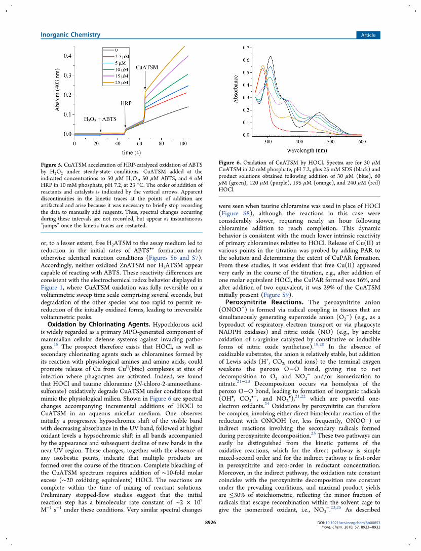
or, to a lesser extent, free H2ATSM to the assay medium led toreduction in the initial rates of ABTS•+ formation underotherwise identical reaction conditions (Figures S6 and S7).Accordingly, neither oxidized ZnATSM nor H2ATSM appearcapable of reacting with ABTS. These reactivity differences areconsistent with the electrochemical redox behavior displayed inFigure 1, where CuATSM oxidation was fully reversible on avoltammetric sweep time scale comprising several seconds, butdegradation of the other species was too rapid to permit re-reduction of the initially oxidized forms, leading to irreversiblevoltammetric peaks.Oxidation by Chlorinating Agents. Hypochlorous acid
is widely regarded as a primary MPO-generated component ofmammalian cellular defense systems against invading patho-gens.18 The prospect therefore exists that HOCl, as well assecondary chlorinating agents such as chloramines formed byits reaction with physiological amines and amino acids, couldpromote release of Cu from CuII(btsc) complexes at sites ofinfection where phagocytes are activated. Indeed, we foundthat HOCl and taurine chloramine (N-chloro-2-aminoethane-sulfonate) oxidatively degrade CuATSM under conditions thatmimic the physiological milieu. Shown in Figure 6 are spectralchanges accompanying incremental additions of HOCl toCuATSM in an aqueous micellar medium. One observesinitially a progressive hypsochromic shift of the visible bandwith decreasing absorbance in the UV band, followed at higheroxidant levels a hypsochromic shift in all bands accompaniedby the appearance and subsequent decline of new bands in thenear-UV region. These changes, together with the absence ofany isosbestic points, indicate that multiple products areformed over the course of the titration. Complete bleaching ofthe CuATSM spectrum requires addition of ∼10-fold molarexcess (∼20 oxidizing equivalents) HOCl. The reactions arecomplete within the time of mixing of reactant solutions.Preliminary stopped-flow studies suggest that the initialreaction step has a bimolecular rate constant of ∼2 × 10⁷
M−1 s−1 under these conditions. Very similar spectral changes
were seen when taurine chloramine was used in place of HOCl(Figure S8), although the reactions in this case wereconsiderably slower, requiring nearly an hour followingchloramine addition to reach completion. This dynamicbehavior is consistent with the much lower intrinsic reactivityof primary chloramines relative to HOCl. Release of Cu(II) atvarious points in the titration was probed by adding PAR tothe solution and determining the extent of CuPAR formation.From these studies, it was evident that free Cu(II) appearedvery early in the course of the titration, e.g., after addition ofone molar equivalent HOCl, the CuPAR formed was 16%, andafter addition of two equivalent, it was 29% of the CuATSMinitially present (Figure S9).
Peroxynitrite Reactions. The peroxynitrite anion(ONOO−) is formed via radical coupling in tissues that aresimultaneously generating superoxide anion (O2
−) (e.g., as abyproduct of respiratory electron transport or via phagocyteNADPH oxidases) and nitric oxide (NO) (e.g., by aerobicoxidation of L-arginine catalyzed by constitutive or inducibleforms of nitric oxide synthetase).19,20 In the absence ofoxidizable substrates, the anion is relatively stable, but additionof Lewis acids (H+, CO2, metal ions) to the terminal oxygenweakens the peroxo O−O bond, giving rise to netdecomposition to O2 and NO2
− and/or isomerization tonitrate.21−23 Decomposition occurs via homolysis of theperoxo O−O bond, leading to formation of inorganic radicals(OH•, CO3
•−, and NO2•),21,22 which are powerful one-
electron oxidants.24 Oxidations by peroxynitrite can thereforebe complex, involving either direct bimolecular reaction of thereductant with ONOOH (or, less frequently, ONOO−) orindirect reactions involving the secondary radicals formedduring peroxynitrite decomposition.25 These two pathways caneasily be distinguished from the kinetic patterns of theoxidative reactions, which for the direct pathway is simplemixed-second order and for the indirect pathway is first-orderin peroxynitrite and zero-order in reductant concentration.Moreover, in the indirect pathway, the oxidation rate constantcoincides with the peroxynitrite decomposition rate constantunder the prevailing conditions, and maximal product yieldsare ≤30% of stoichiometric, reflecting the minor fraction ofradicals that escape recombination within the solvent cage togive the isomerized oxidant, i.e., NO3
−.23,25 As described
Figure 5. CuATSM acceleration of HRP-catalyzed oxidation of ABTSby H2O2 under steady-state conditions. CuATSM added at theindicated concentrations to 50 μM H2O2, 50 μM ABTS, and 4 nMHRP in 10 mM phosphate, pH 7.2, at 23 °C. The order of addition ofreactants and catalysts is indicated by the vertical arrows. Apparentdiscontinuities in the kinetic traces at the points of addition areartifactual and arise because it was necessary to briefly stop recordingthe data to manually add reagents. Thus, spectral changes occurringduring these intervals are not recorded, but appear as instantaneous“jumps” once the kinetic traces are restarted.
Figure 6. Oxidation of CuATSM by HOCl. Spectra are for 30 μMCuATSM in 20 mM phosphate, pH 7.2, plus 25 mM SDS (black) andproduct solutions obtained following addition of 30 μM (blue), 60μM (green), 120 μM (purple), 195 μM (orange), and 240 μM (red)HOCl.
Inorganic Chemistry Article
DOI: 10.1021/acs.inorgchem.8b00853Inorg. Chem. 2018, 57, 8923−8932
8926
below, all of the CuII(btsc) complexes examined reacted withperoxynitrite via the indirect pathway. Additionally, for thosecomplexes that are relatively easily oxidized, evidence for acompeting direct reaction between ONOOH and thecomplexes was found.Peroxynitrous acid (ONOOH) and the peroxynitrite-CO2
adduct (ONOOCO2−) rapidly degraded ZnATSM and
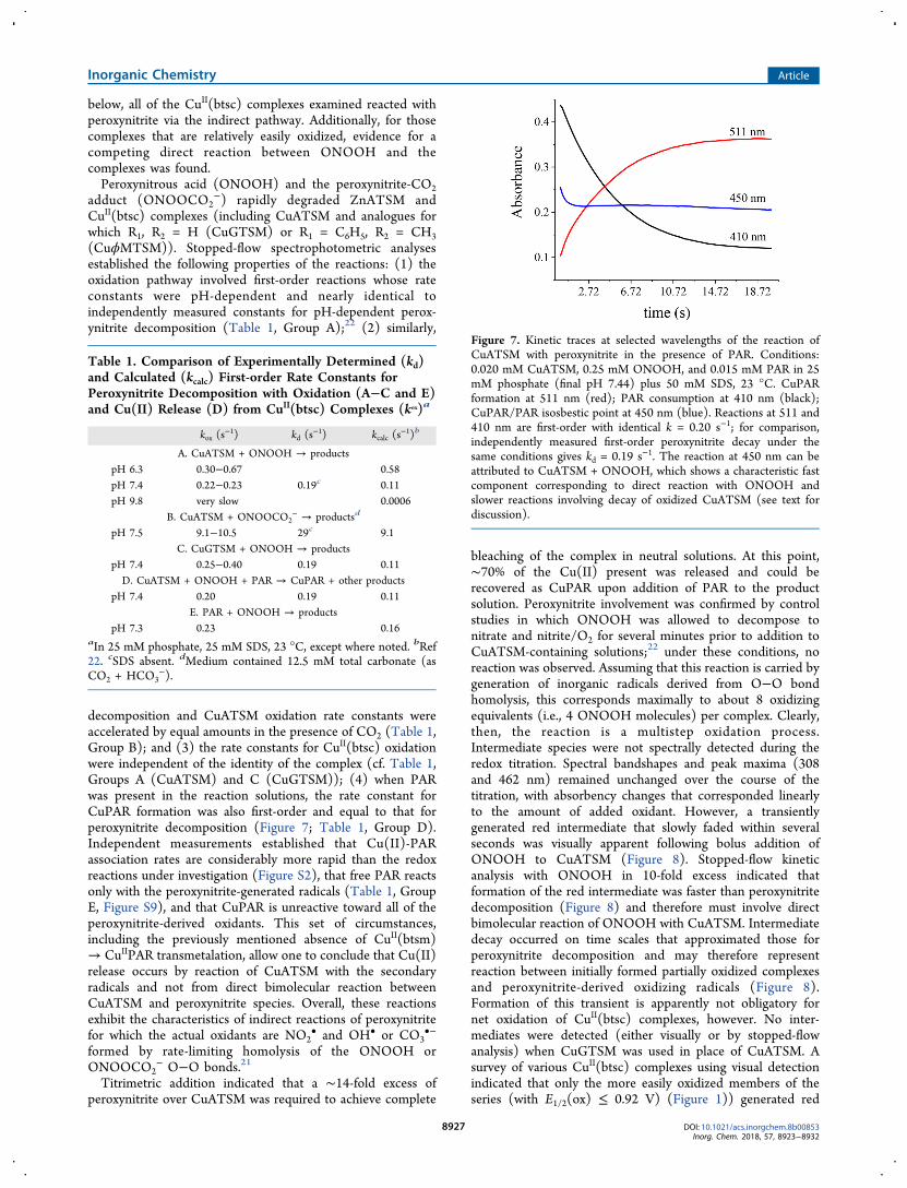
CuII(btsc) complexes (including CuATSM and analogues forwhich R1, R2 = H (CuGTSM) or R1 = C6H5, R2 = CH3(CuϕMTSM)). Stopped-flow spectrophotometric analysesestablished the following properties of the reactions: (1) theoxidation pathway involved first-order reactions whose rateconstants were pH-dependent and nearly identical toindependently measured constants for pH-dependent perox-ynitrite decomposition (Table 1, Group A);22 (2) similarly,
decomposition and CuATSM oxidation rate constants wereaccelerated by equal amounts in the presence of CO2 (Table 1,Group B); and (3) the rate constants for CuII(btsc) oxidationwere independent of the identity of the complex (cf. Table 1,Groups A (CuATSM) and C (CuGTSM)); (4) when PARwas present in the reaction solutions, the rate constant forCuPAR formation was also first-order and equal to that forperoxynitrite decomposition (Figure 7; Table 1, Group D).Independent measurements established that Cu(II)-PARassociation rates are considerably more rapid than the redoxreactions under investigation (Figure S2), that free PAR reactsonly with the peroxynitrite-generated radicals (Table 1, GroupE, Figure S9), and that CuPAR is unreactive toward all of theperoxynitrite-derived oxidants. This set of circumstances,including the previously mentioned absence of CuII(btsm)→ CuIIPAR transmetalation, allow one to conclude that Cu(II)release occurs by reaction of CuATSM with the secondaryradicals and not from direct bimolecular reaction betweenCuATSM and peroxynitrite species. Overall, these reactionsexhibit the characteristics of indirect reactions of peroxynitritefor which the actual oxidants are NO2
• and OH• or CO3•−
formed by rate-limiting homolysis of the ONOOH orONOOCO2
− O−O bonds.21
Titrimetric addition indicated that a ∼14-fold excess ofperoxynitrite over CuATSM was required to achieve complete
bleaching of the complex in neutral solutions. At this point,∼70% of the Cu(II) present was released and could berecovered as CuPAR upon addition of PAR to the productsolution. Peroxynitrite involvement was confirmed by controlstudies in which ONOOH was allowed to decompose tonitrate and nitrite/O2 for several minutes prior to addition toCuATSM-containing solutions;22 under these conditions, noreaction was observed. Assuming that this reaction is carried bygeneration of inorganic radicals derived from O−O bondhomolysis, this corresponds maximally to about 8 oxidizingequivalents (i.e., 4 ONOOH molecules) per complex. Clearly,then, the reaction is a multistep oxidation process.Intermediate species were not spectrally detected during theredox titration. Spectral bandshapes and peak maxima (308and 462 nm) remained unchanged over the course of thetitration, with absorbency changes that corresponded linearlyto the amount of added oxidant. However, a transientlygenerated red intermediate that slowly faded within severalseconds was visually apparent following bolus addition ofONOOH to CuATSM (Figure 8). Stopped-flow kineticanalysis with ONOOH in 10-fold excess indicated thatformation of the red intermediate was faster than peroxynitritedecomposition (Figure 8) and therefore must involve directbimolecular reaction of ONOOH with CuATSM. Intermediatedecay occurred on time scales that approximated those forperoxynitrite decomposition and may therefore representreaction between initially formed partially oxidized complexesand peroxynitrite-derived oxidizing radicals (Figure 8).Formation of this transient is apparently not obligatory fornet oxidation of CuII(btsc) complexes, however. No inter-mediates were detected (either visually or by stopped-flowanalysis) when CuGTSM was used in place of CuATSM. Asurvey of various CuII(btsc) complexes using visual detectionindicated that only the more easily oxidized members of theseries (with E1/2(ox) ≤ 0.92 V) (Figure 1)) generated red
Table 1. Comparison of Experimentally Determined (kd)and Calculated (kcalc) First-order Rate Constants forPeroxynitrite Decomposition with Oxidation (A−C and E)and Cu(II) Release (D) from CuII(btsc) Complexes (kox)a
kox (s−1) kd (s
−1) kcalc (s−1)b
A. CuATSM + ONOOH → productspH 6.3 0.30−0.67 0.58pH 7.4 0.22−0.23 0.19c 0.11pH 9.8 very slow 0.0006
B. CuATSM + ONOOCO2− → productsd
pH 7.5 9.1−10.5 29c 9.1C. CuGTSM + ONOOH → products
pH 7.4 0.25−0.40 0.19 0.11D. CuATSM + ONOOH + PAR → CuPAR + other products
pH 7.4 0.20 0.19 0.11E. PAR + ONOOH → products
pH 7.3 0.23 0.16aIn 25 mM phosphate, 25 mM SDS, 23 °C, except where noted. bRef22. cSDS absent. dMedium contained 12.5 mM total carbonate (asCO2 + HCO3
−).
Figure 7. Kinetic traces at selected wavelengths of the reaction ofCuATSM with peroxynitrite in the presence of PAR. Conditions:0.020 mM CuATSM, 0.25 mM ONOOH, and 0.015 mM PAR in 25mM phosphate (final pH 7.44) plus 50 mM SDS, 23 °C. CuPARformation at 511 nm (red); PAR consumption at 410 nm (black);CuPAR/PAR isosbestic point at 450 nm (blue). Reactions at 511 and410 nm are first-order with identical k = 0.20 s−1; for comparison,independently measured first-order peroxynitrite decay under thesame conditions gives kd = 0.19 s−1. The reaction at 450 nm can beattributed to CuATSM + ONOOH, which shows a characteristic fastcomponent corresponding to direct reaction with ONOOH andslower reactions involving decay of oxidized CuATSM (see text fordiscussion).
Inorganic Chemistry Article
DOI: 10.1021/acs.inorgchem.8b00853Inorg. Chem. 2018, 57, 8923−8932
8927

intermediates upon exposure to peroxynitrite; moreover, theextent of formation of the CuATSM intermediate was highlypH-dependent, decreasing with decreasing acidity from pH 6.3to 9.8, where it was barely detectable by stopped-flow analysis.The kinetic waveforms obtained at pH 7.2 were accuratelyreproduced by fitting to a double exponential form, suggestingthat the reactions proceeded by concurrent pathways. Overall,these data are consistent with simultaneous expression of twopathways in which (1) thermodynamically limited directreaction of the more easily oxidized complexes within theCuII(btsc) series with ONOOH, but not with the less stronglyoxidizing ONOO− (pKa = 6.6), runs in parallel with (2)indirect reactions of peroxynitrite-derived oxidizing radicalsthat can rapidly oxidize all complexes within the series. Finally,transient diode array spectrophotometry of CuATSMindicated that the spectrum of the intermediate has a relativelyintense near UV band at 340 nm, and very broad weakabsorption above ∼500 nm that accounts for the redcoloration. These spectral features are consistent with eitherligand radical formation (e.g., CuII(ATSM)•+) or oxidation toCu(III)ATSM. Specifically, strikingly similar spectra have beenobserved for multidentate Cu(II) and other metal complexescontaining phenoxyl and N-heterocyclic ligand radicals,26,27
but many tetradentate square-planar Cu(III) complexes alsoexhibit intense LMCT absorption bands in the near-UVregion.10 Stopped-flow analyses also indicated that several
intermediates could be temporally resolved in reactions ofCuϕMTSM with peroxynitrite and, although ZnATSM wasextensively degraded by peroxynitrite, no discrete intermedi-ates could be detected in that reaction.
Biomedical Implications. Mechanisms for intracellularrelease of copper are yet to be identified. However, it is likelythat both reductive and oxidative pathways (Scheme 1) canprevail under different physiological conditions. Thus, forexample, the reductive pathway seems well-established inpositron-emission tomography (PET) applications to detecthypoxia, where the extent of intracellular copper accumulationparallels the reduction potentials of CuII(btsc) complexes.1 Insimilar PET imaging studies to detect regions of oxidativestress in tissues from patients suffering from neurodegenerativedisorders, enhanced copper release has been attributed toincreased reducing capacity in cells as a consequence ofNADH accumulation caused by impaired mitochondrialelectron transport,28,29 although these conditions also promoteformation of reactive oxygen species (ROS) that arethermodynamically competent to oxidize the complexes.Moreover, disease progression in neurological disorders forwhich CuII(btsc) treatment may have therapeutic benefit ischaracterized by oxidative damage by ROS and reactivenitrosative species, the latter possibly including ONOOH. Theantineoplastic activity of certain CuII(btsc) complexes (notablyCuGTSM) has recently been attributed to their capacity forintracellular generation of ROS via redox cycling between theCu(II) and Cu(I) states of the complexes; notably, in thesestudies, a marked dependence of cellular toxicity upon theCuII/I(btsc) reduction potential was observed.30 The accel-eration of HRP-catalyzed ABTS oxidation by H2O2 observedin this study when CuATSM was introduced into the assaymedium (Figure 5) suggests that cycling between CuII(btsc)and oxidized Cu(btsc) states might also occur in biologicalsettings. Biological precedents for this type of reactivity can befound in the reactions of galactose oxidase (GAO), a proteincontaining a single active site copper atom that catalyzesaerobic oxidation of primary alcohols to the correspondingaldehydes with stoichiometric formation of H2O2,
31 andcopper complexes that have been developed as functionalmodels of the catalytic reaction center.26 A pertinent exampleis the study by Wieghardt and co-workers on the oxidativechemistry of N,N′-bis(3,5-di-tert-butyl-2-hydroxyphenyl)-1,2-phenylenediamine.32 This ligand forms monomeric tetraden-tate square-planar complexes with Cu(II) and Zn(II) withN2O2 coordination sets that are structurally quite similar to theMII(btsc) complexes studied here. Five different oxidationstates are accessible in the GAO model complex; catalyticaerobic oxidation of primary alcohols to aldehydes, i.e.,RCH2OH + O2 → RCHO + H2O2, is proposed to occur bynet two-electron cycling between two ligand radical states ofthe complexes, namely, a reduced state containing adi(hydroxyphenyl)-diiminosemiquinone ligand and an oxi-dized state containing a hydroxyphenyl(phenoxyl)-diiminoqui-none ligand. Notably, both the Cu(II) and Zn(II) complexesare effective catalysts. Collectively, the studies on GAOcatalysis and CuATSM cocatalysis of the HRP/H2O2/ABTSreaction suggest a scenario in which intracellular release ofcopper might occur via a reaction sequence in whichtransmembrane diffusion of CuII(btsc) is followed bycomplex-catalyzed generation of the oxidant (H2O2) thatleads to its own oxidative destruction. Upregulation of MPO indamaged neuronal tissues has also been documented, opening
Figure 8. Stopped-flow traces of the CuATSM-ONOOH reaction.Conditions: 0.025 mM CuATSM + 0.25 mM ONOOH in 25 mMSDS plus 25 mM phosphate, pH 7.5, at 23 °C. Overlay of biphasictraces for CuATSM decay at 465 nm (black) and intermediateformation and decay at 590 nm (red, 4× actual absorbance).Photographs above the kinetic traces give a visual record ofintermediate formation and decay in the reaction. Conditions: 0.03mM CuATSM with 0.5 mM peroxynitrite in 50 mM phosphate plus20 mM SDS, pH 7.2, 23 °C.
Inorganic Chemistry Article
DOI: 10.1021/acs.inorgchem.8b00853Inorg. Chem. 2018, 57, 8923−8932
8928

pathways for formation of additional oxidizing species as partof the neurodegenerative processes.33−36 In this context, thepresent studies have clearly demonstrated facile cupric ionrelease following oxidation by either ONOOH/ONOOCO2
−
or MPO-catalyzed peroxide systems, including HOCl, thephysiological product of neutrophil MPO-catalyzed reactions.Our demonstration that reactions of major ROS and RNSoxidants effect efficient Cu(II) release from CuII(btsc)complexes establish the plausibility of similar reactionsoccurring in biological tissues, particularly in regions under-going oxidative stress. Distinguishing between oxidative andreductive pathways in biological environments may provedifficult, but we note that the fate of the btsc ligand isfundamentally different in the two general pathways outlinedin Scheme 1, where it remains intact following reduction of thecomplex to continually cycle metal ions, but is modified withloss of metal-binding capacity in the oxidative pathway. Thus,the pathways could presumably be distinguished in specificinstances by quantitative determination of the fate of theligand. Finally, we note that cupric ion in the presence of H2O2is potently bactericidal.37 Use of CuII(btsc) complexes ascopper delivery systems offers potential for developing newantibiotics, as has been demonstrated in recent studies.38,39
Interestingly, one proposed bactericidal mechanism isinhibition of respiration through direct binding of intactCuII(btsc) to the bacterial electron transport chain. Presum-ably, for this application, one would seek to identify CuII(btsc)complexes that underwent minimal intracellular degradation.
■ EXPERIMENTAL SECTIONMaterials. All reagents and solvents used in preparation and
characterization of synthetic compounds were purchased through TCIAmerica or Sigma-Aldrich and used directly without purification.Bisthiosemicarbazone ligands used in these studies were preparedfollowing the general procedure outlined below:30
Typically, 2.0 equiv of 4-methyl-thiosemicarbazide was added to anoven-dried 250 mL round-bottom flask containing 50 mL anhydrousEtOH, and the mixture was heated to 65 °C while stirring until thereactant completely dissolved. One equivalent of the appropriatedicarbonyl compound was then added dropwise to the stirringsolution, followed by 5 drops of conc H2SO4. Within 5 min aprecipitate formed; following overnight stirring, the mixture wasfiltered and sequentially washed with deionized water, MeOH andEtOH. Product analyses included (1) 1H NMR and 13C NMRspectra, recorded in d6-DMSO on a Bruker 700 MHz Avance IIIspectrometer and a Bruker 400 MHz DPX-400 spectrometer,respectively and (2) mass spectra acquired on a Thermo LTQ-FTUltra hybrid linear ion trap-Fourier transform ion cyclotron resonancemass spectrometer with a Finnigan Ion Max API source configured forESI in positive ion mode. NMR and mass spectra for each compoundagreed with those previously reported in the literature.30 Specifically,for diacetyl-bis(4-methylthiosemicarbazone) (H2ATSM), 1H NMR(700 MHz, d6-DMSO): 10.215 (s, 2H), 8.372 (q, 2H, J = 5, J = 13),3.023 (d, 6H, J = 5), 2.209 (s, 6H); 13C NMR (176 MHz, d6-DMSO): 178.9587, 148.4411, 31.6750, 12.1330; MS: calcd forCuC8H15N6S2 [(M+H)+] 322.01, found 322.08; for glyoxal-bis(4-methylthiosemicarbazone) (H2GTSM), 1H NMR (700 MHz, d6-DMSO): 11.7317 (s, 2H), 8.4736 (q, 2H, J = 5, J = 13), 7.7182 (s,2H), 2.9576 (d, 6H, J = 5); 13C NMR (176 MHz, d6-DMSO):178.0312, 140.5007, 31.3725; MS: calcd for CuC6H11N6S2 [(M+H)+]293.90, found 294.00; for 1-phenyl-1,2-propanedione bis(4-methyl-3-
thiosemicarbazone)40 (H2ϕMTSM), 1H NMR (700 MHz, d6-DMSO): 10.704 (s, 1H), 8.7402 (m, 1H), 8.6505 (s, 1H), 7.592−7.541 (m, 3H), 7.295−7.265 (m, 2H), 6.979 (m, 1H), 3.043 (d, 3H, J= 5), 2.819 (d, 3H, J= 5), 2.373 (s, 3H); M.S.: calcd forCuC13H17N6S2 [(M + H+)] 384.03, found 384.08. Cupric and zinccomplexes were generally prepared as stock solutions obtained byadding a slight excess of a concentrated solution of Cu(OAc)2 orZn(OAc)2 to the free ligand dissolved in DMSO; these weremonitored spectrally using a Shimadzu UV-2041PC recordingspectrophotometer to confirm complete formation of 1:1 complexes.During the course of subsequent experimentation, it was noted thatthe presence of even small amounts (∼2%) of DMSO partiallyinhibited release of Cu(II) (measured as formation of CuIIPAR inexperiments using peroxynitrite as oxidant). This protection almostcertainly arose from scavenging of secondary peroxynitrite-derivedoxidizing radicals by the DMSO. Additional experiments withperoxynitrite were therefore done using solid CuATSM that wassolubilized in aqueous-based media devoid of DMSO; these studiesindicated that the presence of DMSO at concentration levels presentin the reaction medium caused only minor alteration of the reactioncourse. Solid CuATSM was obtained following a general procedure41
that involved dropwise addition of aqueous CuSO4 to a suspensionsof H2ATSM in methanol and isolating the CuATSM that precipitated.A more critical issue was the highly effective scavenging of HOCl byDMSO, which completely masked its reaction with CuATSM at eventhe millimolar concentration levels present following dilution ofconcentrated DMSO stock solutions of CuATSM into the reactionmedium. This problem was circumvented by preparing CuATSM inN-methyl-2-pyrrolidone, which control studies showed did not reduceHOCl when diluted into the reaction medium for periods exceeding 1h.
Myeloperoxidase (MPO) was isolated from bovine spleens bycolumn chromatography as previously described;42 the purifiedenzyme had an RZ value of 0.70 and a specific activity of 345units/mg protein, as measured by the guaiacol assay.43 Type VIhorseradish peroxidase (HRP) was obtained from Sigma, and itsactivity was determined by HRP-catalyzed oxidation of 2,2′-azino-bis(3-ethylbenzothiazoline-6-sulfonic acid) (ABTS) with H2O2.
17
Reactants. To investigate oxidative reactions, we used wherepossible aqueous buffered media containing 20−50 mM SDS. Underthese conditions, most of the SDS is present as microphase-separatedmicelles. Specifically, the SDS critical micelle concentration is ∼8mM, and its aggregation number in aqueous solutions is ∼60,12 sothat the effective concentration of micelles in these mixtures was200−700 μM. Because the MII(btsc) concentrations never exceeded50 μM in these experiments, the average occupancy number, i.e.,MII(btsc)/micelle, was always less than 1. We believe that thismedium effectively mimics the biological milieu inside eukaryoticcells, which contain numerous phase-separated microdomains.13
Inclusion of SDS also serves to increase the effective solubility ofthe neutral CuII(btsc) complexes in aqueous media. As noted above,some of the reactions studied are incompatible with all systemcomponents. Most prominently, SDS inactivated the peroxidases(HRP, MPO) used in studying catalyzed oxidation by H2O2 andcould not be included in the reaction medium. These studies utilizedthe Good’s buffer, 3-(N-morpholino)propanesulfonic acid (MOPS), aneutral tertiary amine that does not strongly coordinate Cu(II).However, MOPS also protected CuATSM from reaction withperoxynitrite, so that it was necessary to substitute phosphate ion asthe buffer component in reactions involving ONOOH andONOOCO2
− as oxidants.Reactant concentrations of free and metalated bis-thiosemicarba-
zones were determined spectrophotometrically in buffered aqueous/SDS micellar suspensions using independently measured molarabsorptivities from serially diluted samples in the same medium.These standard solutions followed Beer’s law over the entireconcentration range encompassed in subsequent experiments. Theextinction coefficients (in mM−1 cm−1) at the peak maxima are asfollows: H2ATSM (ε326 = 45); CuATSM (ε309 = 21, ε465 = 5.6);ZnATSM (ε413 = 8.56); H2GTSM (ε335 = ε346 = 41); CuGTSM (ε314
Inorganic Chemistry Article
DOI: 10.1021/acs.inorgchem.8b00853Inorg. Chem. 2018, 57, 8923−8932
8929
= 16.7, ε485 = 6.4); H2ϕMTSM (ε333 = 45); CuϕMTSM (ε311 = 22,ε479 = 6.4); PAR (ε413 = 33); CuPAR (ε511 = 34). Reactant solutionsof ONOOH were prepared by diluting into alkaline (pH > 12) waterportions of frozen concentrated stocks (∼0.1 M) that had been madeby flow-mixing sodium nitrite with hydrogen peroxide;44 concen-trations were determined spectrophotometrically using ε302 = 1.67mM−1 cm−1.45 Reactant solutions of hypochlorous acid were preparedby dilution of commercial bleach into H2O and determining theirconcentrations spectrophotometrically in alkaline (pH > 12) solutionsusing ε292(OCl
−) = 350 M−1 cm−1.46 Reactant solutions of taurinechloramine were prepared by dropwise addition of HOCl to a finalconcentration of 10 mM into a rapidly stirred aqueous solutioncontaining 2-fold excess taurine; spectrophotometric analysisconfirmed the complete conversion of HOCl to the monochloraminewithout formation of detectable amounts of taurine dichloramine.Instrumental Methods. Cyclic voltammograms were measured
in DMSO with an EG&G Model 273 potentiostat/galvanostat usingan electrochemical cell in a standard 3-electrode configuration withglassy carbon, Pt wire, and Ag/AgCl/KNO3 working, counter andreference electrodes, respectively; 0.1 M tetrabutylammoniumtetrafluoroborate was the inert electrolyte and ferrocene (Fc) wasadded internally as a reference standard to determine peak potentials.The Fc+/o potential in DMSO was taken to be 0.68 V vs NHE.11
Sweep rates were typically 50 mV/s. Peroxidase-catalyzed oxidationsof MII(btsc) complexes were monitored spectroscopically byfollowing the time course of bleaching of their optical bands usinga Shimadzu UV-2041PC recording spectrophotometer interfaced toShimadzu UVProbe software. This instrument was also used to makeroutine spectrophotometric analyses. Rapid reactions, includingONOOH and ONOOCO2
− decompositions, peroxynitrite oxidationof both MII(btsc) complexes and PAR, and PAR complexation byCu(II) were followed using an Applied Photophysics SX20 stopped-flow apparatus operated in the single-mixing absorption detectionmode; kinetic traces were analyzed using the associated Pro-Data SXsoftware. Transient spectra formed during reactions of CuATSM withperoxynitrite were recorded from 230 to 600 nm by using a ShimadzuMultispec 1500 instrument. Reactions were initiated by bolus additionof the peroxynitrite to buffered SDS micellar solutions containingCuATSM, followed by repetitive spectral determinations at 1−3 sintervals.
■ ASSOCIATED CONTENT
*S Supporting InformationThe Supporting Information is available free of charge on theACS Publications website at DOI: 10.1021/acs.inorg-chem.8b00853.
Additional electrochemical, spectroscopic and kinetic
information involving CuII(btsc) oxidations, Cu-PAR
association dynamics, and ATSM inhibition of the HRP-
catalyzed ABTS-H2O2 reaction (PDF)
■ AUTHOR INFORMATION
Corresponding Author*E-mail: [email protected].
ORCIDJames K. Hurst: 0000-0003-1070-2690Present Address⊥J.J.S.: Department of Chemistry, Matanuska-Susitna CollegeUniversity of Alaska, Anchorage, Palmer, Alaska 99645, UnitedStates.
NotesThe authors declare no competing financial interest.
■ ACKNOWLEDGMENTSThis work was supported by the Office of the AssistantSecretary of Defense for Health Affairs through the Congres-sionally Directed Medical Research Program on ALS(AL140108, Award Number W81XWH-15-1-0289) andfunding from the Oregon Chapter of the ALS Association(ALSA 16-320).
■ REFERENCES(1) Vavere, A. L.; Lewis, J. S. Cu-ATSM: A Radiopharmaceutcal forthe PET Imaging of Hypoxia. Dalton Trans. 2007, 4893−4902.(2) Crouch, P. J.; Hung, L. W.; Adlard, P. A.; Cortes, M.; Lal, V.;Filiz, G.; Perez, K. A.; Nurjono, M.; Caragounis, A.; Du, T.; Laughton,K.; Volitakis, I.; Bush, A. I.; Li, Q.; Masters, C. L.; Cappai, R.; Cherny,R. A.; Donnelly, P. S.; White, A. R.; Barnham, K. Increasing CuBioavailability Inhibits Aβ Oligomers and Tau Phosphorylation. Proc.Natl. Acad. Sci. U. S. A. 2009, 106, 381−386.(3) Hung, L. W.; Villemagne, V. L.; Cheng, L.; Sherratt, N. A.;Ayton, S.; White, A. R.; Crouch, P. J.; Lim, S.; Leong, S. L.; Wilkins,S.; George, J.; Roberts, B. R.; Pham, C. L. L.; Liu, X.; Chiu, F. C. K.;Shackleford, D. M.; Powell, A. K.; Masters, C. L.; Bush, A. I.; O'Keefe,G.; Culvenor, J. G.; Cappai, R.; Cherny, R. A.; Donnelly, P. S.; Hill, A.F.; Finkelstein, D. I.; Barnham, K. J. The Hypoxia Imaging AgentCuII(atsm) is Neuroprotective and Improves Motor and CognitiveFunctions in Multiple Animal Models of Parkinson’s Disease. J. Exp.Med. 2012, 209, 837−854.(4) Soon, C. P. W.; Donnelly, P. S.; Turner, B. J.; Hung, L. W.;Crouch, P. J.; Sherratt, N. A.; Tan, J.-L.; Lim, N. K-H.; Lam, L.; Bica,L.; Lim, S.; Hickey, J. L.; Morizzi, J.; Powell, A.; Finkelstein, D. I.;Culvenor, J. G.; Masters, C. L.; Duce, J.; White, A. R.; Barnham, K. J.;Li, Q.-X. Diacetylbis N(4)-methylthiosemicarbazonato) Copper(II)(CuII(atsm)) Protects against Peroxynitrite-induced NitrosativeDamage and Prolongs Survivial in Amyotrophic Lateral SclerosisMouse Model. J. Biol. Chem. 2011, 286, 44035−44044.(5) Holland, J. P.; Giansiracusa, J. H.; Bell, S. G.; Wong, L.-L.;Dilworth, J. R. In vitro Kinetic Studies on the Mechanism of Oxygen-dependent Cellular Uptake of Copper Radiopharmaceuticals. Phys.Med. Biol. 2009, 54, 2103−2119.(6) Fubayashi, Y.; Taniuchi, H.; Yoshihara, Y.; Ohtani, H.; Konishi,J.; Yokoyama, A. Copper-62-ATSM: A New Hypoxia Imaging Agentwith High Membrane Permeability and Low Redox Potential. J. Nucl.Med. 1997, 38, 1155−1160.(7) Obata, A.; Yoshimi, E.; Waki, A.; Lewis, J. S.; Oyama, N.; Welch,M. J.; Saji, H.; Yonekura, Y.; Fujibayashi, Y. Retention Mechanism ofHypoxia Selective Nuclear Imaging/Radiotherapeutic Agent Cu-diacetyl-bis(N4-methylthiosemicarbazone) (Cu-ATSM) in TumorCells. Ann. Nucl. Med. 2001, 15, 499−504.(8) Xiao, Z.; Donnelly, P. S.; Zimmermann, M.; Wedd, A. G.Transfer of Copper between Bis(thiosemicarbazone) Ligands andIntracellular Copper-Binding Proteins. Insights into Mechanisms ofCopper Uptake and Hypoxia Selectivity. Inorg. Chem. 2008, 47,4338−4347.(9) Holland, J. P.; Barnard, P. J.; Collison, D.; Dilworth, J. R.; Edge,R.; Green, J. C.; McInnes, E. J. L. Spectroelectrochemical andComputational Studies on the Mechanism of Hypoxia Selectivity ofCopper Radiopharmaceuticals. Chem. - Eur. J. 2008, 14, 5890−5907.(10) McDonald, M. R.; Fredericks, F. C.; Margerum, D. W.Characterization of Copper(III)-Tetrapeptide Complexes withHistidine as the Third Residue. Inorg. Chem. 1997, 36, 3119−3124.Hanss, J.; Beckmann, A.; Kruger, H.-J. Stabilization of Copper(III)Ions with Deprotonated Hydroxyiminoamide Ligands: Syntheses,Structures, and Electronic Properties of Copper(II) and Copper(III)Complexes. Eur. J. Inorg. Chem. 1999, 1999, 163−172. Fritsky, I. O.;Kozlowski, H.; Kanderal, O. M.; Haukka, M.; Swiatek-Kozlowska, J.;Gumienna-Kontecka, E.; Meyer, F. Efficient Stabilization of Copper-(III) in Tetraaza Pseudo-macrocyclic Oxime- and Hydrazide Ligandswith Adjustable Cavity Size. Chem. Commun. 2006, 4125−4127.Pratesi, A.; Zanello, P.; Giorgi, G.; Messori, L.; Laschi, F.; Casini, A.;
Inorganic Chemistry Article
DOI: 10.1021/acs.inorgchem.8b00853Inorg. Chem. 2018, 57, 8923−8932
8930

Corsini, M.; Gabbiani, C.; Orfei, M.; Rosani, C.; Ginanneschi, M.New Copper(II)/Cyclic Tetrapeptide System that Easily Oxidizes toCopper(III) under Atmospheric Oxygen. Inorg. Chem. 2007, 46,10038−10040. Keown, W.; Gary, J. B.; Stack, T. D. P. High-valentCopper in Biomimetic and Biological Oxidations. JBIC, J. Biol. Inorg.Chem. 2017, 22, 289−305.(11) Barrette, W. C., Jr.; Johnson, H. W.; Sawyer, D. T.Voltammetric Evaluation of the Effective Acidities (pKa’) forBroensted Acids in Aprotic Solvents. Anal. Chem. 1984, 56, 1890−1898.(12) Dominguez, A.; Fernandez, A.; Gonzalez, N.; Iglesias, E.;Montenegro, L. Determination of the Critical Micelle Concentrationof Some Surfactants by Three Techniques. J. Chem. Educ. 1997, 74,1227−1231.(13) Hickey, J. L.; James, J. L.; Henderson, C. A.; Price, K. A.; Mot,A. I.; Buncic, G.; Crouch, P. J.; White, J. M.; White, A. R.; Smith, T.A.; Donnelly, P. S. Intracellular Distribution of Fluorescent Copperand Zinc Bis(thiosemicarbazonato) Complexes Measured withFluorescence Lifetime Spectroscopy. Inorg. Chem. 2015, 54, 9556−9567.(14) Furtmuller, P. G.; Arnhold, J.; Jantschko, W.; Pichler, H.;Obinger, C. Redox Properties of the Couples Compound I/Compound II and Compound II/Native Enzyme of HumanMyeloperoxidase. Biochem. Biophys. Res. Commun. 2003, 301, 551−557.(15) Hayashi, Y.; Yamazaki, I. The Oxidation-Reduction Potentialsof Compound I/Compound II and Compound II/Ferric Couples inHorseradish Peroxidases A2 and C. J. Biol. Chem. 1979, 254, 9101−9106.(16) Ooi, S.; Carter, D.; Fernando, Q. The Structure of a Chelate ofCopper(II) with 1-(2-Pyridylazo)-2-Napthol. Chem. Commun. 1967,1301−1302.(17) Childs, R. E.; Bardsley, W. G. The Steady-state Kinetics ofPeroxidase with 2,2′-Azino-di-(3-ethyl-benzthiazoline-6-sulphonicacid) as Chromagen. Biochem. J. 1975, 145, 93−103.(18) Klebanoff, S. J.; Kettle, A.; Rosen, H.; Winterbourn, C. C.;Nauseef, W. M. Myeloperoxidase: A Front-line Defender AgainstPhagocytosed Microorganisms. J. Leukocyte Biol. 2013, 93, 1−14.Hurst, J. K. What Really Happens in the Neutrophil Phagosome? FreeRadical Biol. Med. 2012, 53, 508−520.(19) Pacher, P.; Beckman, J. S.; Liaudet, L. Nitric Oxide andPeroxynitrite in Health and Disease. Physiol. Rev. 2007, 87, 315−424.(20) Ischiropoulos, H.; Zhu, L.; Beckman, J. S. PeroxynitriteFormation from Macrophage-derived Nitric Oxide. Arch. Biochem.Biophys. 1992, 298, 446−451.(21) Lymar, S. V.; Hurst, J. K. Rapid Reaction between PeroxonitriteIon and Carbon Dioxide: Implications for Biological Reactivity. J. Am.Chem. Soc. 1995, 117, 8867−8868.(22) Coddington, J. S.; Hurst, J. K.; Lymar, S. V. Hydroxyl RadicalFormation during Peroxynitrous Acid Decomposition. J. Am. Chem.Soc. 1999, 121, 2438−2444.(23) Goldstein, S.; Lind, J.; Merenyi, G. Chemistry of Peroxynitritesas Compared to Peroxynitrates. Chem. Rev. 2005, 105, 2457−2470.(24) Stanbury, D. A. Reduction Potentials involving InorganicRadicals in Aqueous Solution. Adv. Inorg. Chem. 1989, 33, 69−138.(25) Ferrer-Sueta, G.; Campolo, N.; Trujillo, M.; Bartesaghi, S.;Carballal, S.; Romero, N.; Alvarez, B.; Radi, R. Biochemistry ofPeroxynitrite and Protein Tyrosine Nitration. Chem. Rev. 2018, 118,1338−1408.(26) Jazdzewski, B. A.; Tolman, W. B. Understanding the Copper-Phenoxyl Radical Array in Galactose Oxidase: Contributions fromSynthetic Modeling Studies. Coord. Chem. Rev. 2000, 200−202, 633−685.(27) Roy, N.; Sproules, S.; Weyhermuller; Wieghardt, K. TrivalentIron and Ruthenium Complexes with a Redox Noninnocent (2-Mercaptophenylimino)-methyl-4,6-di-tert-butylphenolate(2-) Ligand.Inorg. Chem. 2009, 48, 3783−3791.(28) Neishi, H.; Ikawa, M.; Okazawa, H.; Tsujikawa, T.; Arishima,H.; Kikuta, K. I.; Yoneda, M. Precise Evaluation of Striatal Oxidative
Stress Corrected for Severity of Dopaminergic Neural Degenerationin Patientis with Parkinson’s Disease: A Study with 62Cu-ATSM PETand 123I-FP-CIT SPECT. Eur. Neurol. 2017, 78, 161−168.(29) Donnelly, P. S.; Liddell, J. R.; Lim, S.-C.; Paterson, B. M.;Cater, M. A.; Savva, M. S.; Mot, A. I.; James, J. L.; Trounce, I. A.;White, A. R.; Crouch, P. J. An Impaired Mitochondrial ElectronTransport Chain Increases Retention of the Hypoxia Imaging AgentDiacetylbis(4-methylthiosemicarbazonato)copperII. Proc. Natl. Acad.Sci. U. S. A. 2012, 109, 47−52.(30) Stefani, C.; Al-Eisawi, Z.; Jansson, P. J.; Kalinowski, D. S.;Richardson, D. R. Identification of Differential Anti-neoplasticActivity of Copper bis(thiosemicarbazones) that is Mediated byIntracellular Reactive Oxygen Species Generation and LysosomalMembrane Permeation. J. Inorg. Biochem. 2015, 152, 20−37.(31) Whittaker, J. W. The Free Radical-Coupled Copper OxidativeSite in Galactose Oxidase. InMetal Ions in Biological Systems; Sigel, H.,Sigel, A., Eds.; Marcel Dekker: New York, 1994; Vol. 30, pp 315−360.(32) Chaudhuri, P.; Hess, M.; Muller, J.; Hildenbrand, K.; Bill, E.;Weyhermuller, T.; Wieghardt, K. Aerobic Oxidation of PrimaryAlcohols (Including Methanol) by Copper(II)- and Zinc(II)-Phenoxyl Radical Catalysts. J. Am. Chem. Soc. 1999, 121, 9599−9610.(33) Choi, D.-K.; Pennathur, S.; Perier, C.; Tieu, K.; Teismann, P.;Wu, D.-C.; Jackson-Lewis, V.; Vila, M.; Vonsattel, J.-P.; Heinecke, J.W.; Przedborski, S. J. Ablation of the Inflammatory EnzymeMyeloperoxidase Mitigates Features of Parkinson’s Disease in Mice.Neuroscience 2005, 25, 6594−6600.(34) Maki, R. A.; Tyurin, V. A.; Lyon, R. C.; Hamilton, R. L.;DeKosky, S. T.; Kagan, V. E.; Reynolds, W. F. Aberrant Expression ofMyeloperoxidase in Astrocytes Promotes Phospholipid Oxidation andMemory Deficits in a Mouse Model of Alzheimer Disease. J. Biol.Chem. 2009, 284, 3158−3169.(35) Green, P. S.; Mendez, A. J.; Jacob, J. S.; Crowley, J. R.;Growdon, W.; Hyman, B. T.; Heinecke, J. W. Neuronal Expression ofMyeloperoxidase is Increased in Alzheimer’s Disease. J. Neurochem.2004, 90, 724−733.(36) Jeitner, T. M.; Kalogiannis, M.; Krasnikov, B. F.; Gomlin, I.;Peltier, M. R.; Moran, G. R. Linking Inflammation and ParkinsonDisease: Hypochlorous Acid Generates Parkinsonian Poisons. Toxicol.Sci. 2016, 151, 388−402.(37) Elzanowska, H.; Wolcott, R. G.; Hannum, D. M.; Hurst, J. K.Bactericidal Properties of Hydrogen Peroxide and Copper or Iron-containing Complex Ions in Relation to Leukocyte Function. FreeRadical Biol. Med. 1995, 18, 437−449.(38) Djoko, K. Y.; Paterson, B. M.; Donnelly, P. S.; McEwan, A. G.Antimicrobial Effects of Copper(II) Bis(thiosemicarbazonato) Com-plexes Provide New Insight into their Biochemical Mode of Action.Metallomics 2014, 6, 854−863.(39) Djoko, K. Y.; Goytia, M. M.; Donnelly, P. S.; Schembri, M. A.;Shafer, W. M.; McEwan, A. G. Copper(II)-Bis(Thiosemicarbazonato)Complexes as Antibacterial Agents: Insights into Their Mode ofAction and Potential as Therapeutics. Antimicrob. Agents Chemother.2015, 59, 6444−6453.(40) Beraldo, H.; Kaisner, S. B.; Turner, J. D.; Billeh, I. S.; Ives, J. S.;West, D. X. Copper (II) and Nickel(II) Complexes of the Bis{N(3)-Substituted Thiosemicarbazones} of Phenylglyoxyl and 1-Phenyl-propane-1,2-Dione. Transition Met. Chem. 1997, 22, 459−464.(41) Hueting, R.; Christlieb, M.; Dilworth, J. R.; Garayoa, E. G.;Gouverneur, V.; Jones, M. W.; Maes, V.; Schibli, R.; Sun, X.; Tourwe,D. Bis(thiosemicarbazones) as bifunctional chelators for the roomtemperature 64-copper labeling of peptides. Dalton Trans. 2010, 39,3620−3632.(42) Ikeda-Saito, M. Spectroscopic, Ligand Binding, and EnzymaticProperties of Spleen Green Hemeprotein. A Comparison withMyeloperoxidase. J. Biol. Chem. 1985, 260, 11688−11695.(43) Klebanoff, S. J.; Waltersdorph, A. M.; Rosen, H. AntimicrobialActivity of Myeloperoxidase. Methods Enzymol. 1984, 105, 399−403.(44) Saha, A.; Goldstein, S.; Cabelli, D.; Czapski, G. Determinationof Optimal Conditions for Synthesis of Peroxynitrite by Mixing
Inorganic Chemistry Article
DOI: 10.1021/acs.inorgchem.8b00853Inorg. Chem. 2018, 57, 8923−8932
8931

Hydrogen Peroxide with Nitrite. Free Radical Biol. Med. 1998, 24,653−659.(45) Hughes, M. N.; Nicklin, H. G. The Chemistry of Pernitrites.Part I. Kinetics of Decomposition of Pernitrous Acid. J. Chem. Soc. A1968, 450−452.(46) Morris, J. C. The acid ionization constant of HOCl from 5 to35 °C. J. Phys. Chem. 1966, 70, 3798−3805.
Inorganic Chemistry Article
DOI: 10.1021/acs.inorgchem.8b00853Inorg. Chem. 2018, 57, 8923−8932
8932