Embed Size (px)
Citation preview

Purdue UniversityPurdue e-PubsWeldon School of Biomedical Engineering FacultyWorking Papers Weldon School of Biomedical Engineering
11-29-1992
Oxidative Mechanisms in Arterial DistensionInjury: Observations Relevant to Restenosis afterAngioplastyCharles F. BabbsPurdue University, [email protected]
Yuan Zhong
Follow this and additional works at: http://docs.lib.purdue.edu/bmewp
Part of the Biomedical Engineering and Bioengineering Commons
This document has been made available through Purdue e-Pubs, a service of the Purdue University Libraries. Please contact [email protected] foradditional information.
Recommended CitationBabbs, Charles F. and Zhong, Yuan, "Oxidative Mechanisms in Arterial Distension Injury: Observations Relevant to Restenosis afterAngioplasty" (1992). Weldon School of Biomedical Engineering Faculty Working Papers. Paper 11.http://docs.lib.purdue.edu/bmewp/11

1 BABBS & ZHONG OXIDANTS IN VASCULAR INJURY
Oxidative Mechanisms in Arterial Distension Injury:
Observations Relevant to Restenosis after Angioplasty
CHARLES F. BABBS AND YUAN ZHONG
W. A. Hillenbrand Biomedical Engineering Center and School of Veterinary
Medicine, Purdue University, West Lafayette, Indiana, USA
November 29, 1992
ABSTRACT
Background: To explore the hypothesis that tissue iron and reactive oxygen species, including
superoxide (O2), mediate acute inflammatory and late hyperplastic responses to vascular injury,
we studied experimental overdistension of normal carotid and femoral arteries in dogs.
Experimental design: Arterial segments isolated in situ were distended with Ringer solution at
2 atmospheres pressure. In initial experiments the arteries were excised immediately after
distension and immersed in diaminobenzidine solutions containing Mn++
ions to initiate
histochemical reactions for O2. In other experiments distended arterial segments were
reperfused with arterial blood in the presence or absence of the iron chelator, deferoxamine, or
the low-cost superoxide dismutase mimic, manganese chloride, and examined microscopically at
3 hours, 7 days, or 30 days. The degree of acute inflammation or smooth muscle hyperplasia was
determined by quantitative morphometry; free iron capable of redox cycling was determined
histochemically.
Results: (1) O2 was produced by injured endothelial cells; (2) free iron is present on the
surfaces of endothelial cells in the distended arterial segments; (3) an acute subintimal
inflammatory response occurs at 3 hours after distension injury, followed by proliferation of
mural smooth muscle cells at 7 days, and neointima formation at 30 days; (4) single doses of
either the low molecular weight superoxide dismutase mimic, manganese chloride, or the strong
iron chelator, deferoxamine, administered during and immediately after arterial distension,
prevent the early inflammatory and late hyperplastic responses to distension injury.
Conclusions: Tissue iron and oxidants may participate importantly in the over-reaction to
arterial injury that leads to restenosis.
Key words: Endothelial cells, Fenton reaction, Histochemistry, Hydrogen peroxide,
Hyperplastic smooth muscle, Iron, Neointima, Neutrophils, Smooth muscle, Superoxide.

2 BABBS & ZHONG OXIDANTS IN VASCULAR INJURY
INTRODUCTION
Since its introduction in 1977 balloon angioplasty has become a widely used and successful
treatment for patients with atherosclerotic coronary artery disease. Initially restricted to patients
with single vessel disease and stable angina pectoris, the use of coronary angioplasty has been
expanded to unstable angina, acute myocardial infarction, and multiple vessel coronary artery
disease.1 The technique, however, has yet to achieve its full potential as a non-surgical remedy
that produces ideal, lasting results, largely because of late restenosis in about 35% of cases,
occurring within one to six months after initially successful angioplasty.2
The mechanisms by which balloon angioplasty works therapeutically are now well documented.1,
3, 4 These include creation of tears, fractures, splits, and cracks in the stenotic plaque; dissection
through the intima into the media; stretching of the adventitia of the treated artery; compression
of lipid and thrombotic material; and--especially in asymmetrical or eccentric lesions--stretching
of the disease-free arc of normal arterial wall with little or no damage to the plaque itself. All of
these mechanisms result in deep arterial injury or trauma affecting the residual normal elements
of the vascular wall. In turn, restenosis after angioplasty appears to result from an inappropriate
over-reaction of the vessel wall to distension injury, characterized by migration and excessive
proliferation of smooth muscle cells at sites of tears or lacerations that were created by balloon
expansion.1, 5, 6
Rather than being an abnormal trait that occurs in only a small number of
identifiable hyper-responders, the propensity for restenosis seems to represent an expression of
normal tissue biology that is widespread in the human population7 and demonstrable in a wide
variety of animal models.8-11
In the present paper we propose that a neglected early key to the inflammatory and proliferative
responses to angioplasty may be the release of oxidants in the distended arterial segment, first by
activated endothelial cells and subsequently by activated leukocytes. In particular, we suggest
that injury to endothelial cells caused by distension activates them to produce superoxide ions
(O2). In turn, O2
ions interact with redox cycling iron chelates present on the endothelial
surface to create chemotactic substances for leukocytes, including lipid hydroperoxides and their
breakdown products.12-14
Neutrophils, drawn into the injured area, become activated and produce
additional O2. We hypothesize further that the transmigration of neutrophils, and subsequently
monocytes, into the subintimal space after injury leads to the production of mitogenic factors that
stimulate the smooth muscle cells to divide.15
Such factors likely provide a link between initial
arterial wall injury and subsequent smooth muscle hyperplasia. According to this proposed
mechanism, reactive oxygen species released by activated endothelial cells and inflammatory
cells, acting in conjunction with iron complexes, may be critical initiators of the pathophysiology
of restenosis.

3 BABBS & ZHONG OXIDANTS IN VASCULAR INJURY
One frequently quoted chemical mechanism explaining the effects of superoxide radicals in
biological systems16-19
is that O2 ions are converted in the presence of low molecular weight iron
chelates to highly toxic hydroxyl radicals (HO) via the superoxide driven Fenton reaction:
(1)
(2)
(3)
in which iron is shown complexed to a chelator anion An
. Chelation of iron in biological
systems is required for its catalytic action in reactions (1) and (3), because ferric iron is very
sparingly soluble at a pH of 7.4 (Ksp for Fe(OH)3 = 1 x 1036
).20
The hydroxyl radicals (or
perhaps closely related oxo-iron complexes in the absence of free hydroxyl radicals)21, 22
are then
capable of inducing lipid peroxidation leading to deleterious modifications of membrane
structures and creation of toxic and/or chemotactic compounds.14, 23-25
Accordingly, the objectives of the presently reported experiments were (1) to determine if O2,
could be detected histochemically in arterial segments after distension injury; (2) to determine if
iron complexes, capable of redox cycling at physiologic pH are present in such vessels near
sources of O2; and (3) to determine if either an O2
scavenger or a strong iron chelator that
prevents redox cycling can attenuate or modify the tissue response to distension injury. The
results reported herein provide evidence that oxidative processes, occurring minutes to hours
after angioplasty, are be biologically important mediators of both the early inflammatory and the
late proliferative responses of arteries to acute distension injury.
EXPERIMENTAL DESIGN
Approach
Because it is highly likely that the residual normal tissue in atherosclerotic lesions is the source
of undesired amounts of reactive smooth muscle following clinical angioplasty;4, 5
we over-
distended segments of normal arteries in dogs to study certain fundamental aspects of the tissue
response. Four histochemical techniques were used to observe arterial reactions acutely and
chronically after distension. These included (1) the manganese/diaminobenzidine method for
superoxide generation, (2) a new and sensitive pseudoperoxidase method for free iron
accumulation, (3) routine hematoxylin/eosin staining for proliferative and inflammatory changes,
and (4) immunocytochemical staining for smooth muscle actin. Quantitative morphometric
techniques were used to measure the density of histochemically positive endothelial cells, the
area of acute subintimal inflammation, the area of subsequent myointimal hyperplasia, as well as
the number of medial smooth muscle cell nuclei in distended experimental segments vs.
undistended control segments of the same artery.

4 BABBS & ZHONG OXIDANTS IN VASCULAR INJURY
In initial histochemical studies canine common carotid segments, isolated by ligatures in vivo
and in situ, were distended with Ringer solution at 2 atmospheres pressure, excised immediately,
and immersed in histochemical reagents at 37 °C prior to aldehyde fixation and tissue processing
for light microscopy. In subsequent experiments carotid or femoral artery segments were
distended in situ with Ringer solution, with or without the addition of antioxidant drugs, and
reperfused by antegrade blood flow for periods ranging from 3 hr to 30 days before excision and
microscopic analysis.
Animal preparation
According to protocols approved by the Purdue Animal Care and Use Committee, beagle dogs
weighing 10 to 20 kg and fed a standard laboratory chow, without lipid or cholesterol
supplementation, were anesthetized with intravenous thiopental sodium (20 mg/kg to effect)
followed by intubation and maintenance of inhalation anesthesia with 0.5% to 1.5%
methoxyfluorane in oxygen. The femoral and/or carotid arteries were isolated using sterile
technique. A 1 mm I.D. Intramedic polyethylene (or Cook, Inc. Teflon) catheter was inserted
into the artery via a side branch, and an unbranched test segment of the artery, including the
catheter tip, was isolated with loops of #2 silk suture such that the segment could be distended by
fluid injected through the catheter. Five cm long segments of common carotid arteries or femoral
arteries were cannulated via the external carotid artery or the profunda femoris artery,
respectively, and subjected to static or pulsatile distension, as described subsequently, either with
various histochemical reagents directly or with Ringer solution. In most experiments the catheter
was open-ended without side holes.
Experimental distension injury
To create controlled, arterial distension injury, blood was withdrawn from the isolated arterial
segment with a syringe and replaced with sterile lactated Ringer solution (130 mM Na+, 4 mM
K+, 1.5 mM Ca
++, 109 mM Cl
, 28 mM lactate, from Abbott Laboratories) which in a typical
experiment was injected to produce a distending pressure of two atmospheres (30 PSI). In some
experiments distending pressure was applied in pulses at 0.5 Hz for 15 minutes. In other
experiments distending pressure was held constant at 30 PSI for 1 min. During distension the
vessel lumen contained Ringer solution only.
Pressure pulses were monitored visually with reference to an aneroid manometer (Weiss Gauge,
0 to 50 PSIG) connected to the catheter and syringe via a 3-way stopcock. In initial experiments
vessel distension was also monitored by measurement of the increase in vessel diameter,
determined from the length of a double loop of suture material passed around the isolated
segment by an assistant, first during baseline normotension with blood flowing freely through the
artery and then during maximal distension. For each measurement the double loop ("cow hitch")
of suture was cut flush, and vessel circumference was computed from the suture length, using a
rearrangement of the expression, Length = 2(D + d) + 8d, where D is the outer diameter of the
vessel, and d is the diameter of the suture. (In typical experiments with 30 PSI pressure, median
expansion was 64 percent of basal normotensive diameter, from 2.78 to 4.55 mm.)

5 BABBS & ZHONG OXIDANTS IN VASCULAR INJURY
Experimental pressure jet injury
In other experiments the catheter tip was heat sealed and rows of diametrically opposed pin-holes
were made near the end of a catheter with a 22 gauge needle to deliver the infused solutions in
the form of high velocity jets. This alternative pressure jet technique was used specifically to
introduce histochemical reagents deep into the substance of the tunica media at the time of acute
injury. In the jet experiments near maximal manual effort was used to inject solution using a 5 cc
or a 3 cc plastic syringe, and the power per unit of cross sectional area of a typical jet was
computed from the vertical height of a typical jet, h, in open air, when the solution was infused
through the catheter at the same rate as used experimentally, according to the formula gh,
where is the solution density, and g is the gravitational constant. This expression was derived
by equating the kinetic energy of the jet fluid exiting a side hole with the potential energy of the
fluid at height, h, and was used to characterize and control the intensity of the jets.
Histochemistry for superoxide ion
The arteries were exposed to histochemical reagents which, in the presence of superoxide ion,
react to form insoluble precipitates that can be observed by light microscopy. The method is a
modification26
of Karnovsky's manganese/diaminobenzidine technique,27
which we have used to
demonstrate O2 generation by activated endothelial cells in situ.
26, 28, 29 In principle, the
manganese/diaminobenzidine method for superoxide works as follows. Cells stimulated to
produce superoxide are exposed to a physiologic solution containing added divalent manganese
ions and diaminobenzidine (DAB). Then,
Superoxide, which can act as either an oxidant or a reductant,
18 in the present case oxidizes
divalent manganese quite rapidly (k = 6 x 10⁶ M
lsec
1)30
to the trivalent state, with concomitant
generation of hydrogen peroxide. This reaction is readily demonstrated in simple spot tests by
the rapid color change visible upon addition of granular potassium superoxide to solutions of
manganous chloride. Similarly, the ability of manganic ions to oxidize DAB may be readily
observed by the addition of manganic acetate (in ethanol) to test solutions of DAB, in which
reaction (5) probably proceeds by a radical chain mechanism.31, 32
The DAB polymers, created
by reaction (5) are widely exploited as sensitive histochemical markers.32, 33
They are insoluble
in aqueous and in organic solvents and do not migrate from their sites of original deposition.
The specificity of this histochemical reaction for superoxide is readily checked by inclusion of
negative control experiments, in which sodium ions are substituted for manganese ions (Table 1).
TABLE 1. COMPOSITIONS (mM) AND OSMOLARITIES (mOsm)

6 BABBS & ZHONG OXIDANTS IN VASCULAR INJURY
OF BUFFER SOLUTIONS*
* Values in parentheses are osmolarities at pH 7.4. Tris (trishydroxymethylaminomethane) was
used in lieu of phosphate buffer to avoid precipitation of Mn++
ions by phosphate.
To detect formation of reactive oxygen species histochemically, arterial segments were distended
for 60 sec with Ringer solution and immediately excised, cut into 5 mm length rings, and
submerged in histochemical reagents at 37 °C for 15 min. This intravital approach to
histochemistry is necessary because superoxide ions are short-lived, reactive species that are
made by viable cells only. The preparation buffers containing manganese/DAB was as described
previously.26
In brief, buffers containing 40 mM divalent manganese in combination with 40 mM
trisodium citrate, were prepared as indicated in Table 1. In the manganese/diaminobenzidine
experiments sodium azide (1 mM) was added, as specified by Karnovsky27
to minimize the
spurious formation of DAB reaction product both by peroxidase34, 35
and by mitochondrial
cytochrome oxidase.32

7 BABBS & ZHONG OXIDANTS IN VASCULAR INJURY
Histochemical detection of redox cyclable iron using hydrogen peroxide and
diaminobenzidine (DAB)
To localize redox cyclable iron at neutral pH with greater sensitivity than is provided by
conventional iron stains, we modified the method of Kuo and Fridovich,36
originally developed
for staining iron-containing proteins in electrophoretic gels. As a histochemical procedure, this
method exploits the peroxidatic activity of free iron to initiate the diaminobenzidine
polymerization reaction:
Low molecular weight chelates of ferric iron oxidize DAB to form the DAB radical and ferrous.
The excess hydrogen peroxide quickly oxidizes the ferrous iron chelate back to the ferric state,
continuing the cycle of initiation, which is followed by chain propagation in the usual manner,
until chain termination occurs
The result is a brown polymer that persists through further tissue processing and is readily visible
by light microscopy. Using this method, free iron was detected in paraffin embedded sections
after dewaxing in xylene and rehydration in graded ethanol solutions. Tissues selected for iron
staining had been reperfused in vivo for 3 hr after distention injury. Sections were exposed for
60 min at room temperature to Ringer solution containing 2.5 mM DAB, 120 mM H2O2, and 4.0
mM Tris buffer, pH 7.4. This buffer was used because it is compatible with DAB27
and because
we were particularly interested in forms of iron that redox cycle at physiologic pH. At the end of
the incubation period, the tissues were rinsed and counterstained lightly with methyl green,
dehydrated, and mounted in Permount. To confirm the specificity of the histochemical technique
for free iron, we performed hydrogen peroxide/DAB histochemical staining in the presence of
the iron chelator CP-94 (1,2 diethyl-3-hydroxypirid-4-one, from CIBA-GEIGY), 10 mM, a
concentration that inhibited the hydrogen peroxide/DAB reaction with up to 1 mM iron-EDTA in
pilot test tube experiments.

8 BABBS & ZHONG OXIDANTS IN VASCULAR INJURY
Selection of antioxidant drugs
In 46 dogs drugs were administered to probe for participation of superoxide and iron in the
development of tissue responses to distention injury. Manganese chloride was selected as a low
cost and relatively specific scavenger of superoxide.37
Manganese chloride and the enzyme,
superoxide dismutase, are about equally effective O2 scavengers on a weight basis.
38 However,
penetration of Mn++
ions (atomic weight 55) into the subintimal space is likely to be faster than
penetration of superoxide dismutase (molecular weight 32,000). Deferoxamine mesylate
(Desferal®
, CIBA) was selected as a prototypic and highly specific iron chelator (binding
constant 1031
)39
that is known to block both the superoxide driven Fenton reaction40, 41
and iron
dependent lipid peroxidation.42
Desferal is relatively non-toxic and was well tolerated in prior
studies.43
These agents served as pharmacologic probes for functionally important superoxide
and free iron.
Drug administration
In the present studies of fundamental mechanisms, we sought to produce a "stepfunction" of
drug concentration as a function of time in the subendothelial space of distended arteries using a
two stage, divided dosage regimen. Test drugs were added to the lactated Ringer solution used
for distension and also infused intravenously, immediately after distension. Untreated positive
control arteries were distended with Ringer solution only, followed by an intravenous drip of
Ringer solution. Formulations of test drugs for infusion were made in 250 ml bottles, as
described in Table 2. A syringe filled with 50 ml of the drug formulation was reserved for use in
distension of the artery to saturate the endothelium and subendothelial space with the drug. In
practice 10 to 20 ml were required to fill dead space in the catheter-gauge apparatus and to
replenish leaks. The balance of the 50 ml was given intravenously as a bolus after experimental
distension, and the remaining 200 ml of stock solution was hung as an intravenous drip to be
administered during the first hour after distension.
TABLE 2. FORMULATIONS OF TEST DRUGS
Stock Solution
Ringer only
Preparation Method
250 ml Ringer solution
Total Dose/10 kg
---
Deferoxamine 15 mM
250 mg in 250 ml Ringer
solution
250 mg
MnCl2 10 mM 0.495 g in 20 ml water
+ 230 ml Ringer solution
495 mg

9 BABBS & ZHONG OXIDANTS IN VASCULAR INJURY
After experimental distension injury, the ligatures isolating the distended arterial segment were
loosened and the catheter withdrawn to restore normal blood flow through the segment. The side
branch was ligated, hemostasis was secured, and the surrounding tissues were approximated, as
necessary, with towel clips or silk sutures. Tissue responses to experimental distension injury, in
the presence or absence of introduced manganese or deferoxamine, were allowed to develop in
situ for periods of 3 hours, 7 days, or 30 days.
RESULTS
Production of superoxide after acute arterial injury
When canine arterial segments were distended with Ringer solution and immediately immersed
in the histochemical reagents, reaction product was generated in an around traumatized
endothelial cells. FIG. 1(a) illustrates Mn++
/DAB reaction product (arrow) in a fragmented, but
partially detached endothelial cell. FIG. 1(b) illustrates Mn++
/DAB reaction product in two
endothelial cells more firmly attached to the underlying internal elastic lamina, but showing
extrusion of eosinophilic cytoplasmic material into the lumen, indicative of severe cellular
injury. Typically the reaction product was deposited in and around such sites of cytoplasmic
disruption. In visibly disrupted cells, reaction product was seen in the perinuclear cytoplasm as
well as on the cell surface (FIG. 1 (a)). Further, lesions produced by high pressure jets of
Mn++
/DAB induced tears of the internal elastic lamina and more abundant endothelial reaction
product than was produced by simple distension for 1 min at 2 atmospheres (FIG. 2). Vascular
smooth muscle cells did not appear to produce O2, even in areas where jet induced breaks in the
internal elastic lamina were present.
(a)

10 BABBS & ZHONG OXIDANTS IN VASCULAR INJURY
(b)
FIG. 1. Cross section of distended canine carotid artery showing positive reaction
product produced by the Mn++/DAB histochemical reaction for superoxide (arrows) in
physically disrupted endothelial cells. (a) Detached endothelial cell (arrow). (b)
Attached endothelial cells (vertical arrows). At the light microscope, the reaction product
appeared as dense amber-brown granules against the pink background of eosin
counterstain. Original magnification 1000X, oil immersion. Hematoxylin-eosin stain.
Horizontal arrow indicates internal elastic lamina.

11 BABBS & ZHONG OXIDANTS IN VASCULAR INJURY
FIG. 2. Positive reaction product produced by jets of histochemical reagents in a cross
section of canine carotid artery. The jet power at the orifice, computed from the formula
gh2gh for 35 cm high jets was 0.9 Watts/cm2. Reaction product appears on the
luminal surfaces of endothelial cells, for example at vertical arrow. Original
magnification 1000X, oil immersion. Hematoxylin-eosin stain. Horizontal arrow
indicates internal elastic lamina.
Light microscopically uninjured endothelial cells showed some occasional reaction product on
their luminal surfaces, as previously reported26
, but to a much lesser extent than was present in
and around injured cells. Incubations in the absence of manganese served as negative controls to
test the possibility that reaction product formed by the cytochrome oxidase activity of
mitochondria44
following the selectively rapid penetration of DAB into injured cells. In these
experiments (n=4), however, reaction product did not form when arteries were injured in the
presence of non-manganese containing solutions of DAB.
Quantitatively, it was insightful to analyze the results of these histochemical experiments in
terms of the number of all positively staining cells that fell into a particular category, based on
their light microscopic appearance after staining with hematoxylin and eosin (Table 3). This
analysis, suggests that three quarters of the Mn++
/DAB positive cells showed light
microscopically observable mechanical injury.
12 BABBS & ZHONG OXIDANTS IN VASCULAR INJURY
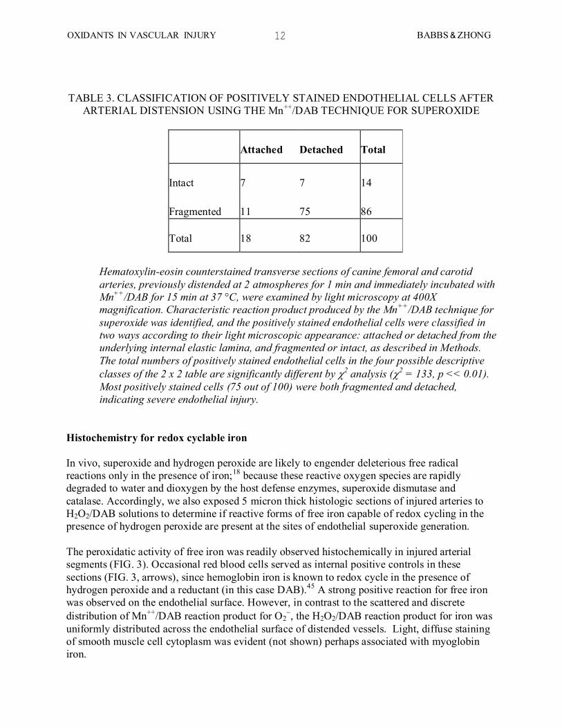
TABLE 3. CLASSIFICATION OF POSITIVELY STAINED ENDOTHELIAL CELLS AFTER
ARTERIAL DISTENSION USING THE Mn++
/DAB TECHNIQUE FOR SUPEROXIDE
Attached
Detached
Total
Intact
7
7
14
Fragmented
11
75
86
Total
18
82
100
Hematoxylin-eosin counterstained transverse sections of canine femoral and carotid
arteries, previously distended at 2 atmospheres for 1 min and immediately incubated with
Mn++/DAB for 15 min at 37 °C, were examined by light microscopy at 400X
magnification. Characteristic reaction product produced by the Mn++/DAB technique for
superoxide was identified, and the positively stained endothelial cells were classified in
two ways according to their light microscopic appearance: attached or detached from the
underlying internal elastic lamina, and fragmented or intact, as described in Methods.
The total numbers of positively stained endothelial cells in the four possible descriptive
classes of the 2 x 2 table are significantly different by 2 analysis (2 = 133, p << 0.01).
Most positively stained cells (75 out of 100) were both fragmented and detached,
indicating severe endothelial injury.
Histochemistry for redox cyclable iron
In vivo, superoxide and hydrogen peroxide are likely to engender deleterious free radical
reactions only in the presence of iron;18
because these reactive oxygen species are rapidly
degraded to water and dioxygen by the host defense enzymes, superoxide dismutase and
catalase. Accordingly, we also exposed 5 micron thick histologic sections of injured arteries to
H2O2/DAB solutions to determine if reactive forms of free iron capable of redox cycling in the
presence of hydrogen peroxide are present at the sites of endothelial superoxide generation.
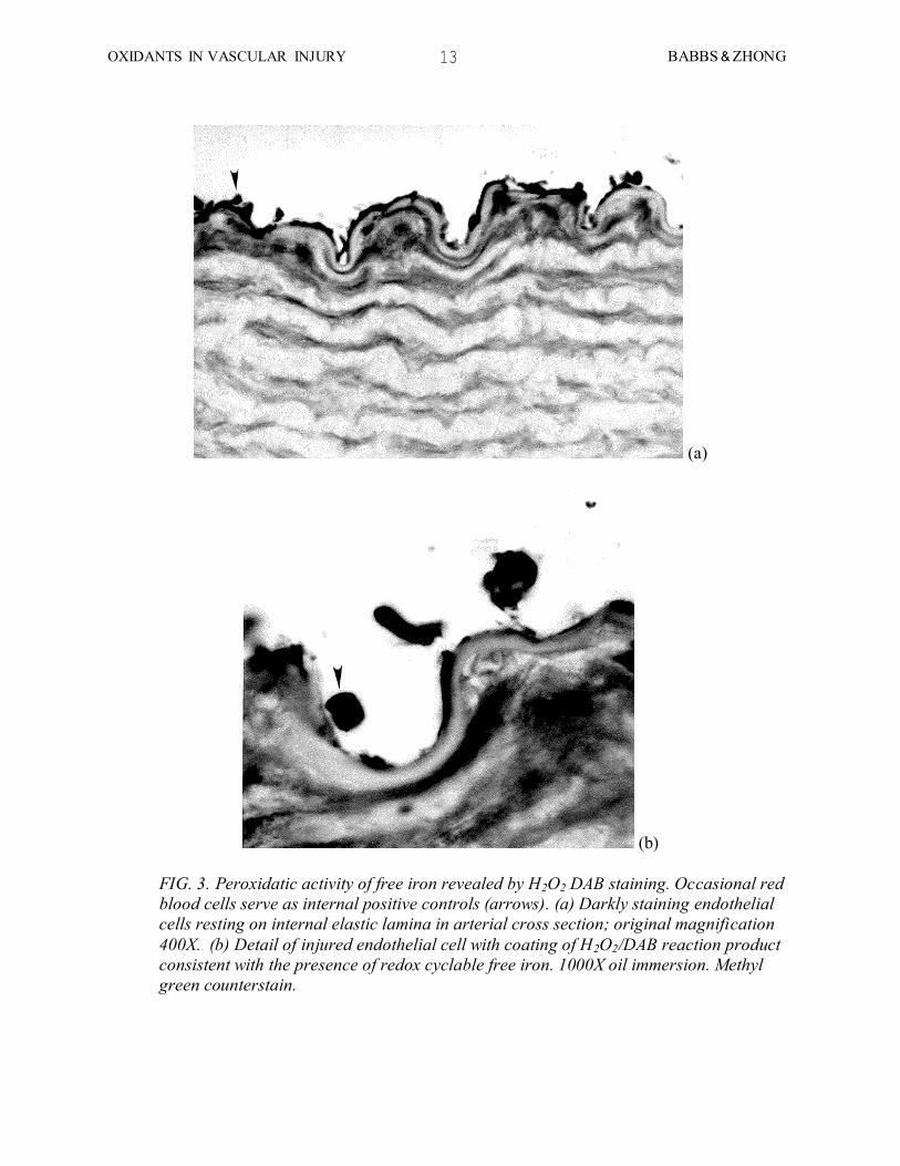
The peroxidatic activity of free iron was readily observed histochemically in injured arterial
segments (FIG. 3). Occasional red blood cells served as internal positive controls in these
sections (FIG. 3, arrows), since hemoglobin iron is known to redox cycle in the presence of
hydrogen peroxide and a reductant (in this case DAB).45
A strong positive reaction for free iron
was observed on the endothelial surface. However, in contrast to the scattered and discrete
distribution of Mn++
/DAB reaction product for O2, the H2O2/DAB reaction product for iron was
uniformly distributed across the endothelial surface of distended vessels. Light, diffuse staining
of smooth muscle cell cytoplasm was evident (not shown) perhaps associated with myoglobin
iron.
13 BABBS & ZHONG OXIDANTS IN VASCULAR INJURY
(a)
(b)
FIG. 3. Peroxidatic activity of free iron revealed by H2O2 DAB staining. Occasional red
blood cells serve as internal positive controls (arrows). (a) Darkly staining endothelial
cells resting on internal elastic lamina in arterial cross section; original magnification
400X. (b) Detail of injured endothelial cell with coating of H2O2/DAB reaction product
consistent with the presence of redox cyclable free iron. 1000X oil immersion. Methyl
green counterstain.

14 BABBS & ZHONG OXIDANTS IN VASCULAR INJURY
To confirm the specificity of the histochemical technique for free iron, especially at the
endothelial cell surface, and to control for the possibility that endothelial staining could represent
a form of surface artifact, we stained sections with H2O2/DAB in the presence of the iron
chelator CP-94 at concentrations shown to inhibit the hydrogen peroxide/DAB reaction in test
tube experiments. There was clearly a much lighter staining in this negative control (FIG. 4).
These results provide evidence that redox cyclable iron is associated with endothelial cells in
vivo and in situ.
FIG. 4. Endothelial surface of distended control vessel, stained with the H2O2/DAB for
free iron in the presence of the iron chelator CP-94 (10 mM). The rapidly penetrating
hydroxypyridone iron chelator abolished reaction product formation, as would be
expected if the reaction in FIG. 3 were caused by tissue iron. Ghost like red blood cells
are barely visible above the endothelial surface. Original magnification 400X. Methyl
green counterstain.

15 BABBS & ZHONG OXIDANTS IN VASCULAR INJURY
Effects of antioxidant drugs
Taken together, the forgoing histochemical experiments demonstrate that the compounds
required to produce oxidative stress via the superoxide driven Fenton reaction, namely
superoxide and iron, are indeed present in arteries shortly after acute distension injury. To
determine if these potentially deleterious species have an effect upon the biology of restenosis,
we observed the time course of tissue reaction in the absence or presence of two selected
antioxidant drugs: manganese chloride, a low molecular weight and low-cost superoxide
dismutase mimic,37
and deferoxamine, a strong iron chelator.39
The time course of the
histopathologic response to distension injury in the absence and presence of drug intervention is
shown qualitatively in Figures 5, 6, and 7, which illustrate microscopic findings at 3 hours, 7
days, and 30 days, respectively.
Three hours after injury there is an intense subintimal inflammatory response, comprised mostly
of neutrophils distributed in arcuate sheets. In untreated positive controls the initial and transient
inflammatory response to arterial distension is similar to that described earlier by Haudenschild
and Studer8 and by Rasmussen et al.
46 The acute vasculitis was predominantly
polymorphonuclear and confined to the subintimal space, comprising about 4 percent of the
arterial wall area in untreated, positive controls. There were numerous Corkscrew nuclei, a light
microscopic feature indicative of acute distension injury.9 (Figure 5(a))
The inflammatory response to the same mechanical injury was dramatically attenuated when the
dogs were pre-treated with either 25 mg/kg (0.04 mmoles/kg) deferoxamine (FIG. 5 (b)) or 0.25
mmoles/kg manganese chloride (FIG. 5 (c)), as described in Methods. The number of corkscrew
nuclei in drug treated arteries was similar to that in untreated controls, as expected, indicating
comparable initial traumatic elongation of the vascular smooth muscle cells.

16 BABBS & ZHONG OXIDANTS IN VASCULAR INJURY
(a)
(b)

17 BABBS & ZHONG OXIDANTS IN VASCULAR INJURY
(c)
FIG. 5. Response to distension injury at 3 hours in the absence or presence of single dose
iron chelator/anti-free radical therapy at the time of distension injury. (a) control in the
absence of drug treatment; (b) after treatment with deferoxamine 25 mg/kg (0.04
mmoles/kg); (c) after treatment with manganese chloride (0.25 mmoles/kg). Original
magnification 160X. Hematoxylin-eosin stain. Note greatly reduced subendothelial
infiltrates in (b) and (c).
In the arteries that were injured without drug treatment and observed after 7 days, the subintimal
inflammatory cells seen at hour 3 were no longer present; however the morphometric density of
spindle cell nuclei in all layers of the tunica media was increased by roughly 50% in these
untreated positive control segments, compared to that of proximal undistended segments of the
same artery (FIG. 6(a)). This medial hyperplasia was accounted for by cells that had the light
microscopic appearance of smooth muscle cells (i.e. spindle cells with elongated nuclei) with the
added feature of increased cytoplasmic basophilia. Such cytoplasmic basophilia is perhaps
indicative of the increased endoplasmic reticulum, described by Haudenschild, in the
organellerich hyperplastic smooth muscle phenotype that is expressed after angioplasty-type
ballooning.4 Intriguingly, the smooth muscle response at day 7 was greatly reduced when the
original distension was performed in the presence of deferoxamine (FIG. 6(b)) or manganese
chloride (FIG. 6(c)). In formerly distended, drug treated arteries bands of residual smooth muscle
cells, which are intermediate in appearance between normal and hyperplastic phenotypes, are
separated by layers of fibrotic extracellular matrix material in many sections. We interpret the
pattern to represent loss of cellular elements as a result of injury, with preservation of residual
fibroelastic matrix material.

18 BABBS & ZHONG OXIDANTS IN VASCULAR INJURY
(a)
(b)

19 BABBS & ZHONG OXIDANTS IN VASCULAR INJURY
(c)
FIG. 6. Response to distension injury at 7 days. (a) Control in the absence of drug
treatment; (b) after treatment with deferoxamine 25 mg/kg (0.04 mmoles/kg); (c) after
treatment with manganese chloride (0.25 mmoles/kg). Original magnification 250X.
Hematoxylin-eosin stain. Arrows indicate internal elastic lamina.
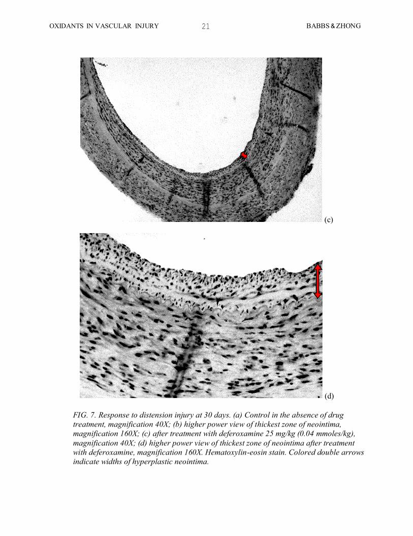
At 30 days after distension injury the classical neointimal hyperplastic response emerged in
untreated, positive control vessels (FIG. 7(a), FIG 7(b)). There was medial hyperplasia,
accompanied by neointima formation. Colored double arrows indicate widths of hyperplastic
neointima. The majority of the neointimal cells stained positively for smooth muscle actin (not
shown). However in animals given a single dose of deferoxamine at the time of injury, 30 days
before, the degree of medial hyperplasia and neointima formation was markedly less (FIG. 7(c),
FIG 7(d)). Manganese chloride was not evaluated at 30 days because animals succumbed to its
toxicity (hepatocellular necrosis and jaundice) in pilot studies.

20 BABBS & ZHONG OXIDANTS IN VASCULAR INJURY
(a)
(b)
21 BABBS & ZHONG OXIDANTS IN VASCULAR INJURY
(c)
(d)
FIG. 7. Response to distension injury at 30 days. (a) Control in the absence of drug
treatment, magnification 40X; (b) higher power view of thickest zone of neointima,
magnification 160X; (c) after treatment with deferoxamine 25 mg/kg (0.04 mmoles/kg),
magnification 40X; (d) higher power view of thickest zone of neointima after treatment
with deferoxamine, magnification 160X. Hematoxylin-eosin stain. Colored double arrows
indicate widths of hyperplastic neointima.

22 BABBS & ZHONG OXIDANTS IN VASCULAR INJURY
Quantitatively, the abnormalities at each stage in the time course of tissue responses to distension
were diminished by scavengers of either free iron or O2, administered during and shortly after
injury. FIG. 8(a) summarizes the morphometry of the subintimal inflammatory response at hour
3 after experimental distension injury, created using sterile technique, in terms of the fractional
area of the arterial wall occupied by inflammatory infiltrate. FIG. 8(b) quantifies the
hyperplastic smooth muscle response at day 7 after distension in terms of the density of smooth
muscle cell nuclei observed in a complete light microscopic cross section of the artery. FIG. 8(c)
summarizes morphometry of neointima formation at 30 days. At all sampling times, drug
interventions expected to inhibit the superoxide driven Fenton reaction, either by scavenging O2
or by chelating iron, significantly suppressed morphometric measures of the tissue responses to
distension injury.
FIG. 8. Results of morphometric analyses. (a) Morphometry of subintimal inflammatory
response at hour 3 after experimental distension injury, created using sterile technique,
in terms of the fractional area of the arterial wall occupied by inflammatory infiltrate;
involved areas after either deferoxamine and manganese treatment are significantly less
than after vehicle treatment (p < 0.01). (b) Hyperplastic smooth muscle responses at day
7 after distension in terms of the number of smooth muscle cell nuclei observed in a
arterial cross sections; counts of medial vascular smooth muscle nuclei after either
deferoxamine or manganese treatment are significantly less than after vehicle treatment
(p < 0.05). (c) Morphometry of neointima formation at 30 days; neointimal area after
deferoxamine treatment is significantly less than after vehicle treatment (p < 0.01).
23 BABBS & ZHONG OXIDANTS IN VASCULAR INJURY
DISCUSSION
In the present investigation we employed a high-yield, non-diseased animal model to study
inflammatory and proliferative aspects of the biology of restenosis. The model exhibits vigorous
medial smooth muscle hyperplasia at 7 days and fibromuscular intimal hyperplasia at 30 days
similar to that in human clinical cases.47
The results obtained support the evolving hypothesis
that iron-dependent oxidation reactions produced after traumatic activation of endothelial cells
interact with tissue iron to create chemoattractants for leukocytes, which in turn may produce
more oxidants in a positive feedback loop. These early events lead to an exuberant, site specific
biological response to vascular injury, culminating in medial and neointimal smooth muscle
hyperplasia, probably in response to endothelial cell and leukocyte derived growth factors, which
were not measured in the experiments reported here. Early intervention with the iron chelator,
deferoxamine, during and shortly after injury, appears to block critical initial steps in this
cascade in a manner that specifically inhibits the proliferation of vascular smooth muscle cells at
the site of injury for the duration of the repair process. Accordingly, we propose that the oxidant-
dependent pathways involving superoxide, low molecular weight chelate iron, and leukocyte
derived growth factors may play an important role in the stimulation of smooth muscle
proliferation and neointima formation.
Oxidative mechanisms such as we propose are well precedented in the literature. The production
of O2 by arterial endothelial cells has been shown by Rosen, Freeman, Ryan, and their
coworkers48, 49
for cultured cells using electron spin resonance and biochemical techniques and
by workers in our own laboratory26, 28
for pulmonary and systemic vascular endothelial cells in
situ using histochemical techniques. The production of abundant O2 by activated neutrophils has
been well shown both biochemically50
and histochemically.27
Species derived from the
interaction of superoxide and hydrogen peroxide with iron can elicit granulocyte infiltration in
the ischemically injured cat intestine.51
In particular, Esterbauer and coworkers52, 53
have shown
that several specific breakdown products of unsaturated lipid hydroperoxides, including 4-
hydroxynonenal (HNE), are highly potent chemotactic substances for neutrophils, effective at
nanomolar to picomolar concentrations. Such hydroperoxides are well known products of the
lipid oxidation induced in vitro by O2 in the presence of free iron.
24, 54,55 Further, the potential
role of biological oxidants, including O2 and H2O2, in the chemotaxis of macrophages has been
highlighted by the extensive studies of Ross, Steinberg, Steinbrecher, and others, related to foam
cell formation in early atherogenesis.56-59
The possible involvement of oxygen free radicals in triggering proliferative responses of vascular
smooth muscle to injury is supported by the in vitro studies of Berk and coworkers,60
who found
that oxygen-derived free radicals produced after injury may initiate cell cycle progression and
growth in cultured smooth muscle cells. This same group has also found that hydrogen peroxide
alone can be 70 percent as effective as calf serum in stimulating growth of cultured rat aortic
smooth muscle cells, and that the proto-oncogenes c-myc and c-fos are activated by H2O2,61
which can be produced by either spontaneous or enzyme catalyzed dismutation of superoxide
(2 O2 + 2H
+ H2O2 + O2).

24 BABBS & ZHONG OXIDANTS IN VASCULAR INJURY
Although the present study focused on oxidant production by endothelial cells, activated
neutrophils and monocytes are major sources of biological oxidants that could possibly stimulate
cell proliferation within injured arteries. The presence of neutrophils in injured arterial segments
has been noted previously10, 46, 62
and emphasized by Cole and coworkers,63
who showed that
neutrophils accumulate rapidly at sites of catheter-induced endothelial injury in rabbits.
Neutrophils in coronary sinus blood sampled immediately after angioplasty in humans showed
decreased ability to liberate superoxide, compared with neutrophils in aortic blood, providing
indirect evidence of their activation on-site as a result of angioplasty.64
Further, activated
neutrophils are known to produce a factor or factors that cause vascular smooth muscle cells in
culture to increase [3H]thymidine incorporation into DNA.
65 Accordingly, it is not unexpected
that oxidant production by inflammatory cells (Figure 5(a)), as well as by endothelial cells,66, 67
may be involved in the pathophysiology of restenosis.
Several caveats should be expressed regarding the present work. Although we found it
productive to focus on fundamental aspects of normal vascular wall biology in vivo, we agree
with others68-70
that an animal model of restenosis capable of simulating the human process in
full has not yet been found. In the present studies, for example, animals were studied in the
absence of hypercholesterolemia, hyperglycemia, or hypertension, which are present in many
human clinical cases. Technically, morphologic identification of sites of acute injury are limited
by tears and other artifacts introduced in preparation of the material that can mimic vascular
injury induced at the time of the experiment. For this reason we could not quantify the
percentage of tears in which oxidants were detected histochemically, since histochemically
negative tears might have been introduced during tissue processing. However, when reaction
product was observed, it was associated with sites of vascular injury much more so than with
uninjured endothelial cells. One possible interpretation of this observation is that cells must be
injured for Mn++
/DAB to penetrate the cell membranes and produce a positive histochemical
reaction. However, earlier work has shown that microscopically intact endothelial cells that are
not mechanically injured also are capable of producing positive histochemical reactions of this
type, either in response to the stimulus of ischemia and reperfusion or to a lesser degree in
response to buffer perfusion without ischemia.26, 28
Another important difference between our animal model of arterial stretching and clinical
angioplasty, is that a surgically isolated arterial segment is distended with Ringer solution, rather
than with a balloon catheter. This technique is intended to maximize reproducibility, since the
pressure "seen" by the vascular wall is known, controlled, and measurable. Because clinical
angioplasty balloons are volume limited, not pressure limited devices, the actual wall pressure
and strain experienced by the artery, and the associated hyperplastic response, depend critically
on the size match between balloon and artery.71
Thus the actual wall stress would be difficult to
measure and to reproduce from animal to animal if clinical angioplasty balloons were used,
because the pressure inside the noncompliant angioplasty balloon would be vastly different from
the pressure just outside the balloon at full expansion. For these reasons we adopted the simple
system of fluid inflation to produce experimental distension injury. Fortunately, the
histopathologic features of proliferative response were similar to those observed in human cases
of restenosis after angioplasty.

25 BABBS & ZHONG OXIDANTS IN VASCULAR INJURY
Using this model the present studies re-emphasize the importance of smooth muscle cell
proliferation after vascular injury in the pathogenesis of restenosis, with the addition of the
mechanistic idea that superoxide and free iron, embedded in positive feedback loops, provide
crucial initial triggers of at least one iron and oxidant-dependent chemical and cytological
cascade leading to smooth muscle cell proliferation and migration. In turn, drug interventions
that block oxidative cell signaling may allow a degree of traumatic smooth muscle cell loss
without replacement, so as to decrease the absolute thickness of media and neointima after
angioplasty.
Clinically, late restenosis has proved to be especially vexatious and refractory to prevention by a
host of drugs,68, 73,74
including antiplatelet agents, anticoagulants, serum cholesterol lowering
agents, vasodilators, calcium entry blockers, a serotonin antagonist, prostacyclin, colchicine, and
corticosteroids, as well as by alternative mechanical techniques for angioplasty, including stents,
lasers, and atherectomy devices.2, 68
Accordingly, authorities reviewing this field have concluded
that fresh biological insights into the pathophysiology of vascular injury are needed in order to
provide clues to more effective preventive therapies.1, 2
Iron chelators and antioxidants may
prove to be attractive candidate drugs to inhibit restenosis, because they are exceedingly non-
toxic (compared, for example, to high dose anticoagulants or corticosteroids) and unlikely to
interfere with the function of healthy cells in general, or with endothelial regrowth in particular.
The intrinsically low toxicity of these compounds is further enhanced by the apparent need, in
our study, for only a single dose, given either during angioplasty or immediately thereafter, to
suppress initial triggering events. Perhaps with proper administration of iron chelators or
antioxidant drugs, it may be possible to inhibit exuberant smooth muscle cell growth after
angioplasty, while permitting limited wound healing and tissue remodeling sufficient to stabilize
the viable parts of the treated vessel in a dilated state.
METHODS
Tissue sampling for drug studies
In the 3 hour duration experiments the animals were maintained under inhalation anesthesia prior
to excision of the test segment for histopathologic examination. In the longer experiments the
length of the test segment was marked with sutures in the perivascular tissues and the wound
closed in layers using sterile technique. Prophylactic antibiotics, sulfadiazine 200 mg and
trimethoprim 40 mg (Tribrissin®
), p.o., b.i.d., were administered, and the animal returned to the
holding area. Morphine sulfate (5 mg i.m.) was given to control discomfort, as necessary, in the
immediate postoperative period. In sampling tissues at 7 days or 30 days the animal was
positioned, under anesthesia, identically as during prior vascular distension. The distended
segment and a proximal, control segment were excised, transversely cut into 5 mm length rings,
fixed in vials of Trump's solution (buffered 1% glutaraldehyde, 4% formaldehyde, pH 7.4), and
submitted for routine tissue processing and staining with hematoxylin and eosin. Animals were
killed using intravenous barbiturate and KCl.

26 BABBS & ZHONG OXIDANTS IN VASCULAR INJURY
Histologic sections were evaluated to identify distension-related vascular changes. Qualitative
histopathological findings were documented with abundant photomicrographs. In some sections
immunohistochemical stains for smooth muscle actin were performed to characterize
hyperplastic neointimal cells. The technique of immunocytochemistry for smooth muscle actin
was as described by Sandusky and coworkers.75
Morphometry for acute subintimal inflammation and chronic neointima formation
To quantify the degree of vascular wall involvement with the acute inflammatory response at 3
hours, we developed a measurement formula applicable to light microscopic observations. The
goal was to compute the percentage or fractional area of the blood vessel wall infiltrated with
leukocytes as function of variables that can be easily estimated by a human observer at the light
microscope. The pattern of acute subintimal inflammation was that of a sleeve of nearly
continuous subintimal infiltrate of varying thickness, but coaxial with the other layers of the
vessel wall. In transverse sections, dense arcs of closely packed neutrophils appeared in the
subendothelial space and did not involve the media. At the microscope it was easy for a human
observer to estimate accurately, for each arcuate region of involvement, the proportion of the
vessel circumference, p, and the mean fractional wall thickness, , occupied by inflammatory
cells, where total wall thickness is taken as the radial distance from the surface of the
endothelium to the outer border of medial smooth muscle.
It is easily shown for thin walled vessels, in which the inner and outer radii are approximately
equal, that the involved fraction of vessel wall area, Ai/Atot, is simply p. For thicker walled
vessels, however, in which the difference in curvature between the inner and outer walls comes
into play,
where is the ratio of the wall thickness to the outer radius, i.e. the fraction of the diameter
occupied by wall rather than lumen. The term in braces represents the necessary corrections for
the curvature and wall thickness of the vessel, typically about 15% for canine femoral and
carotid arteries. In the present studies all three dimensionless parameters, p, , and were
readily estimated by a human observer through the light microscope using a micrometer eyepiece
to evaluate the fractional area included in each arc of inflammatory involvement, and in turn the
sum the fractional areas of involvement when more than one was present. The result, Ai/Atot,
represents the fraction of the arterial wall occupied by inflammatory cells in a particular
histologic section. This process was repeated for all technically satisfactory sections on the
microscope slide, and an average value for the test artery was obtained.
The same morphometric procedure was applied both to acute neutophilic infiltration of the
subintimal space at day 3 and to chronic neointima formation at day 30. The pattern of neointima
formation at day 30 was similar to the pattern of acute inflammation at 3 hours in that arcuate

27 BABBS & ZHONG OXIDANTS IN VASCULAR INJURY
sections of neointima are easily identified in transverse sections between the endothelium and the
internal elastic lamina. The micrometer eyepiece was used to measure the intimal thickness along
equally spaced radii around the whole circumference of the vessel wall. The following formula
was used to calculate the area of neointima present in a cross-section:
where, hi is the distance between endothelial surface and internal elastic lamina along the i-th
radius, n, the number of radii, and w, the distance between radii along the endothelial surface.
Morphometry for histochemical studies of superoxide generation
Cross sectional profiles of the artery segments on 1" by 3" glass slides were subjected to
morphometric analysis as follows. Positively stained endothelial cells containing amber-brown
polymer were identified, counted, and classified according to their light microscopic appearance.
Counts of positively stained cells were assigned to the cells of a 2 x 2 table according to the
descriptors "intact" vs. "fragmented" and "attached" vs. "detached" to characterize the type of
distension injury associated with reaction product formation. Intact cells were defined as having
a normal, flattened appearance without ballooning or disruption of the cell surface, as seen under
the light microscope. Fragmented cells were defined as being clearly torn or shredded with
disruption of surface integrity and frequently ballooning of the cytoplasm. Attached cells were
uniformly adherent to the underlying internal elastic lamina, whereas detached cells were
separated from the internal elastic lamina in whole or in part, but still present within 10 microns
of the endothelial surface.
Morphometry for smooth muscle hyperplasia
In contrast to the acute inflammatory response and to chronic neointima formation, the
hyperplastic response observed at day 7 after distension was a phenomenon of the tunica media.
To quantify the degree of medial smooth muscle hyperplasia morphometrically, we measured the
density of smooth muscle cell nuclei in 4 test regions of the artery wall, at 0, 90, 180, and 270
degrees of the arc, by standard cytometry, using a calibrated test grid embedded in the ocular
lens of the light microscope. Counts of smooth muscle cell nuclei were be made within 200 x
200 2 test areas (400X high power field), delimited by the micrometer eyepiece. One or two
such sampling grids approximately spanned the thickness of the tunica media. Nuclei intersected
by the left and upper margins of the counting frame were included and those intersected by the
right and lower margins were excluded to avoid systematic bias.76
The densities from the four
test grids, centered at the 12, 3, 6, and 9 o'clock positions around the circumference of the tunica
media, halfway between the internal elastic lamina and the adventitia, were averaged to estimate
nuclear density generally. Non-distended, control segments of the same artery at least 1 cm
axially distant from the distended segment served as a convenient reference to evaluate the
presence of smooth muscle hyperplasia.

28 BABBS & ZHONG OXIDANTS IN VASCULAR INJURY
Data analysis
For each sampling time after distension (3 hr, 7 days, or 30 days) a one-way analysis of variance
(ANOVA) was performed to test the null hypothesis that the morphometric index of either
inflammation, smooth muscle proliferation, or neointima formation is the same in both drug
treated and control artery segments. The ANOVA's were preceded by a Bartlett's chi-square test
for homogeneity of variance.77
If the variances of the morphometric indices for the treatment
groups were not similar, a suitable transformation was found, such as a square root
transformation,78
and the ANOVA performed on the transformed data. Specific comparisons of
lumped or individual treatment groups were made using a Scheffe multiple comparison test. (In
the case of only 2 groups, the Scheffe test is mathematically equivalent to the Student-t test). A
chi-square analysis was performed to test the null hypothesis that endothelial cells staining
positive for O2 production were found with equal frequency to be intact or fragmented and
attached or detached from the underlying internal elastic lamina. A p-value of 0.05 was
considered significant.
This work was supported in part by Grant HL-42015 from the National Heart, Lung, and Blood
Institute, U.S. Public Health Service, Bethesda, Maryland, and by a Focused Giving Grant from
Johnson & Johnson.
REFERENCES
1. Ip, JH, Fuster, V, Israel, D, Badmon, L, Badmon, J, and Chesebro, JH. The role of
platelets, thrombin, and hyperplasia in restenosis after coronary angioplasty. J Am Coll
Cardiol 1991; 17: 77B-88B.
2. Holmes, DR, Schwartz, RS, and Webster, MWI. Coronary restenosis: what have we
learned from angiography? J Am Coll Cardiol 1991; 17: 14B-22B.
3. Kohchi, K, Takebayashi, S, Block, PC, Hiroki, T, and Nobuyoshi, M. Arterial changes
after percutaneous transluminal coronary angioplasty. J Am Coll Cardiol 1987; 10: 592-
599.
4. Haudenschild, CC. Restenosis: Basic considerations. In: EJ Topol, Ed. Textbook of
Interventional Cardiology. Philadelphia; WB Saunders Company, 1990: 344-362.
5. Austin, GE, Ratliff, NB, Hollman, J, Tabei, S, and Phillips, DF. Initmal proliferation of
smooth muscle cells as an explanation for recurrent coronary artery stenosis after
percutaneous transluminal coronary angioplasty. J Am Coll Cardiol 1985; 6: 369-375.
6. Liu, MW, Roubin, GS, and King, SB. Restenosis after coronary angioplasty - potential
biologic determinants and role of hyperplasia. Circulation 1989; 79: 1374-1387.
7. Serruys, PW, Rensing, BJ, Hermans, WRM, and Deckers, JW. Luminal narrowing after
angioplasty follows a near Gaussian distribution: a quantitative angiographic study of
1452 lesions. Circulation 1991; 84 (Suppl II): 364.
8. Haudenschild, C and Studer, A. Early interactions between blood cells and severely
damaged rabbit aorta. Eur J Clin Invest 1971; 12: 1-7.

29 BABBS & ZHONG OXIDANTS IN VASCULAR INJURY
9. Steele, PM, Chesebro, JH, Stanson, AW, Jr, DR Homes, Dewanjee, MK, and, L
Badimon, and Fuster, V. Balloon angioplasty: natural history of the pathophysiological
response to injury in the pig model. Circ Res 1985; 57: 105-112.
10. Guyton, JR and Karnovsky, MJ. Smooth muscle cell proliferation in the occluded rat
carotid artery. Am J Pathol 1979; 94: 585-602.
11. Fingerle, J, Johnson, R, Couser, W, Clowes, AW, and Reidy, MA. Effects of
thrombocytopenia on smooth muscle proliferation and intima formation in injured rat
carotid. FASEB J 1988; 2: A1077.
12. Slater, TF. Free Radical Mechanisms in Tissue Injury. London; Pion Limited, 1972,
13. Aust, SD and Svingen, BA. The role of iron in enzymatic lipid peroxidation. In: WA
Pryor, Ed. Free Radicals in Biology. New York; Academic Press, 1982: 1-28.
14. Curzio, M, Esterbauer, H, DiMauro, C, Cecchini, G, and Dianzani, MU. Chemotactic
activity of the lipid peroxidation product 4-hydroxynonenal and homologous
hydroxyalkenals. Biol Chem Hoppe Seyler 1986; 367: 321-329.
15. Martin, BM, Gimbrone, MA, Unanue, IR, and Cotran, RS. Stimulation of nonlymphoid
mesenchymal cell proliferation by a macrophage-derived growth factor. Immunol
1981;126: 1510-1515.
16. Hamers, MN and Roos, D. Oxidative stress in human neutrophilic granulocytes. In:
Helmut Sies, Ed. Oxidative Stress. London; Academic Press, 1985: 351-381.
17. Fridovich, I. Superoxide radical: an endogenous toxicant. Ann Rev Pharmacol Toxicol
1983 ; 23: 239-257.
18. Aust, SD, Morehouse, LA, and Thomas, CE. Hypothesis paper -- role of metals in
oxygen radical reactions. J Free Radicals in Biology & Medicine 1985; 1: 3-25.
19. McCord, JM. Oxygen-derived free radicals in postischemic tissue injury. New England
Journal of Medicine 1985; 312: 159-163.
20. CRC Handbook of Chemistry and Physics, 60th Ed. CRC Press, Inc., Boca Raton,
Florida 33431, 1979: B-220.
21. Aust, SD, Bucher, JR, and Tien, M. Evidence for the initiation of lipid peroxidation by a
ferrous-dioxygen-ferric chelate complex. In: W Bors, M Saran, D Tait, Ed. Oxygen
Radicals in Chemistry and Biology. Berlin; Walter de Gruyter & Co., 1984: 147-154.
22. Rush, JD and Koppenol, WH. Oxidizing intermediates in the reaction of ferrous EDTA
with hydrogen peroxide. J Biol Chem 1986; 261: 6730-6733.
23. Meerson, FZ, Kagan, VE, Kozlov, YP, Belkina, LM, and Arkhipenko, YV. The role of
lipid peroxidation in pathogenesis of ischemic damage and the antioxidant protection of
the heart. Basic Res Cardiol 1982; 77: 465-485.
24. Porter, NE. Chemistry of lipid peroxidation. Methods in Enzymology 1984; 105: 273-
282.
25. Hochstein, P and Jain, SK. Association of lipid peroxidation and polymerization of
membrane proteins with erythrocyte aging. Federation Proc 1981; 40: 183-188.
26. Babbs, CF, Cregor, MD, Turek, JJ, and Badylak, SF. Endothelial superoxide production
in buffer perfused rat lungs, demonstrated by a new histochemical technique. Laboratory
Investigation 1991; 65: 484-496.
27. Briggs, RT, Robinson, JM, Karnovsky, ML, and Karnovsky, MJ. Superoxide production
by polymorphonuclear leukocytes. Histochemistry 1986; 84: 371-378.
28. Babbs, CF, Cregor, MD, Turek, JJ, and Badylak, SF. Endothelial superoxide production
in the isolated rat heart during early reperfusion after ischemia, a histochemical study.

30 BABBS & ZHONG OXIDANTS IN VASCULAR INJURY
Am J Pathol 1991; 139: 1069-1080.
29. Babbs, CF, Cregor, MD, and Badylak, SF. Histochemical demonstration of endothelial
superoxide and hydrogen peroxide generation in ischaemic and reoxygenated rat tissues.
Cardiovascular Research 1992; 26: 593-602.
30. Kono, Y, Takahashi, M, and Asada, K. Oxidation of manganous pyrophosphate by
superoxide radicals and illuminated spinach chloroplasts. Arch Biochem Biophys 1976;
174: 454-462.
31. Walling, C. Free Radicals in Solution. New York; John Wiley & Sons, Inc, 1957.
32. Seligman, AM, Karnovsky, MJ, Wasserkrug, HL, and Hanker, JS. Nondroplet
ultrastructural demonstration of cytochrome oxidase activity with a polymerizing
osmiophilic reagent, diaminobenzidine (DAB). J Biol Chem 1968; 38: 1-14.
33. Burstone, MS. New histochemical techniques for the demonstration of tissue oxidase
(cytochrome oxidase). J Histochem Cytochem 1959; 7: 112-122.
34. Graham Jr, RC and Karnovsky, MJ. The early stages of absorption of injected
horseradish peroxidase in the proximal tubules of mouse kidney: ultrastructural
cytochemistry by a new technique. The Journal of Histochemistry and Cytochemistry
1966; 14: 291-301.
35. Fahimi, HD. Cytochemical localization of peroxidase activity in rat hepatic micro
bodies (peroxisomes). J Histochem Cytochem 1968; 16: 547-550.
36. Kuo, C and Fridovich, I. A stain for iron containing proteins sensitive to nanogram levels
of iron. Anal Biochem 1988; 170: 183-185.
37. Archibald, FS and Fridovich, I. The scavenging of superoxide radical by manganous
complexes in vitro. Arch Biochem Biophys 1982; 214: 452-463.
38. Singh, RK, Kooreman, KM, Babbs, CF, and Fessler, JM. Potential use of simple
manganese salts as antioxidant drugs. Am J Vet Res 1992; 53: 1822-1829.
39. Keberle, H. The biochemistry of desferrioxamine and its relation to iron metabolism. Ann
NY Acad Sci 1974; 119: 758-768.
40. Gutteridge, JMC, Richmond, R, and Halliwell, B. Inhibition of the iron-catalyzed
formation of hydroxyl radicals from superoxide and lipid peroxidation by
desferrioxamine. Biochem J 1979; 184: 469-472.
41. Smith, JB, Cusumano, JC, and Babbs, CF. Quantitative effects of iron chelators on
hydroxyl radical production by the superoxide-driven Fenton reaction. Free Radical
Research Communications 1990; 8: 101-106.
42. Salaris, SC and Babbs, CF. A rapid, widely applicable screen for drugs that suppress free
radical formation in ischemia/reperfusion. Journal of Pharmacological Methods 1988; 20:
335-345.
43. Kompala, SD, Babbs, CF, and Blaho, KE. Effect of deferoxamine on late deaths
following cardiopulmonary resuscitation in rats. Annals Emerg Med 1986; 15: 405- 407.
44. Burstone, MS. Histochemical Demonstration of Cytochrome Oxidase with New Amine
Reagents. J Histochem Cytochem 1960; 8: 63-70.
45. Caughey, WS and Watkins, JA. Oxy radical and peroxide formation by hemoglobin and
myoglobin. In: RA Greenwald, Ed. Handbook of Methods for Oxygen Radical
Research. Boca Raton; CRC Press, 1985: 95-104.
46. Rasmussen, LH, Garbarsch, C, and Lorenzen, I. Injury and repair of smaller muscular
and elastic arteries--A light microcopical study on the different healing patterns of rabbit
femoral and carotid arteries following dilatation injuries by a balloon catheter. Virchows
31 BABBS & ZHONG OXIDANTS IN VASCULAR INJURY
Arch A 1987; 411: 87-92.
47. Waller, BF, Pinkerton, CA, Orr, CM, Slack, JD, VanTassel, JW, and Peters, T.
Restenosis 1 to 24 months after clinically successful coronary angioplasty: a necropsy
study of 20 patients. J Am Coll Cardiol 1991; 17: 58B-70B.
48. Rosen, GM and Freeman, BA. Detection of superoxide generated by endothelial cells.
Proc Natl Acad Sci USA 1984; 81: 7269-7273.
49. Ryan, US and Vann, JM. Endothelial Cells: A Source and Target of Oxidant Damage. In:
MG Simic, Ed. Oxygen Radicals in Biology and Medicine. New York; Plenum Press,
1988: 963-974.
50. Markert, M, Andrews, PC, and Babior, BM. Measurement of superoxide production by
human neutrophils. The preparation and assay of NADPH oxidasecontaining particles
from human neutrophils. Methods in Enzymology 1984; 105:358-365.
51. Zimmerman, BJ, Grisham, MB, and Granger, DN. Mechanisms of oxidantmediated
microvascular injury following reperfusion of the ischemic intestine. In: C von Sonntag,
Ed. Oxygen Radicals in Biology and Medicine. New York; Plenum Press, 1988: 881-
886.
52. Esterbauer, H, Zollner, H, and Schaur, RJ. Hydroxyalkenals: cytotoxic products of lipid
peroxidation. ISI Atlas of Science - Biochemistry, Volume 11988; 311-317.
53. Esterbauer, H, Quehenberger, O, and Jurgens, G. Oxidation of human low density
lipoprotein with special attention to aldehydic lipid peroxidation products. In: C Rice-
Evans and B Halliwell, Ed. Free Radicals Methodology And Concepts. London;
Richelieu Press, 1988: 243-268.
54. Thomas, CE, Morehouse, LA, and Aust, SD. Ferritin and superoxide-dependent lipid
peroxidation. J Biol Chem 1985; 260: 3275-3280.
55. Tien, M, Svingen, BA, and Aust, SD. Superoxide dependent lipid peroxidation.
Federation Proc 1981; 40: 179-182.
56. Ross, R, Faggiotto, A, Bowen-Pope, D, and Raines, E. The role of endothelial injury and
platelet and macrophage interactions in atherosclerosis. Circulation 1984; 70 (Suppl III):
III-77-82.
57. Carew, TE, Schwenke, DC, and Steinberg, D. Antiatherogenic effect of probucol
unrelated to its hypocholesterolemic effect: Evidence that antioxidants in vivo can
selectively inhibit low density lipoprotein degradation in atherosclerosis in the Watanabe
heritable hyperlipidimic rabbit. Proc Natl Acad Sci USA 1987; 84: 7725-7729.
58. Steinberg, D, Parthasarathy, S, Carew, TE, Khoo, JC, and Witztum, JL. Beyond
cholesterol: modifications of low-density lipoprotein that increase its athrogenicity. New
Engl J Med 1989; 320: 915-924.
59. Steinbrecher, UP, Zhang, H, and Lougheed, M. Role of oxidatively modified LDL in
atherosclerosis. Free Radical Biology & Medicine 1990; 9: 155-168.
60. Rao, GN and Berk, BC. Oxygen-driven free radicals stimulate c-myc mRNA expression
and vascular smooth muscle cell growth. Clinical Research 1990; 38: 501A.
61. Rao, GN and Berk, BC. Active oxygen species stimulate vascular smooth muscle cell
growth and proto-oncogene expression. Circulation Research 1992; 70: 593-599.
62. Kohchi, K, Takebayashi, S, Block, PC, Hirosumi, J, Nomoto, A, Ohkubo, Y, Sekiguchi,
C, Mutho, S, Yamaguchi, I, and Aoki, H. Inflammatory responses in cuff induced
atherosclerosis in rabbits. Atherosclerosis 1987; 64: 243-254.
63. Cole, CW, Hagen, PO, Lucas, JF, Mikat, EM, O'Mallery, MK, Radic, ZS, Makhoul, RG,

32 BABBS & ZHONG OXIDANTS IN VASCULAR INJURY
and McCann, RL. Association of polymorphonuclear leukocytes with sites of aortic
catheter-induced injury in rabbits. Atherosclerosis 1987; 67: 229-236.
64. DeServi, S, Ricevuti, G, Mazzone, A, Ghio, S, Ardissino, D, Bramucci, E, and Specchia,
G. Neutrophil activation after PCTA in humans. Circulation 1989; 80(II): 260.
65. Cole, CW, Makhoul, RG, McCann, RL, O'Malley, MK, and Hagen, PO. A neutrophil
derived factor(s) stimulates [3H]-thymidine incorporation by vascular smooth muscle
cells in vitro. Clin & Invest Med 1988; 11: 62-67.
66. Ryan, US. Pulmonary endothelium: a dynamic interface. Clinical and Investigative
Medicine 1986; 9: 124-132.
67. Ryan, US. Phagocytic properties of endothelial cells. In: US Ryan, Ed. Endothelial Cells,
Volume III. Boca Raton, Florida; CRC Press, Inc, 1988: 33-49.
68. Califf, RM, Fortin, DF, Frid, DJ, Harlan, WR, Ohman, M, Bengtson, JR, Nelson, CL,
Tcheng, JE, Mark, DB, and Stack, RS. Restenosis after coronary angioplasty: an
overview. J Am Coll Cardiol 1991; 17: 2B-13B.
69. White, CJ, Ramee, SR, Mesa, JE, and Collins, TJ. Percutaneous coronary angioscopy in
patients with restenosis after coronary angioplasty. J Am Coll Cardiol 1991; 17: 46B-
49B.
70. Currier, JW, Pow, TK, Haudenschild, CH, Minihan, AC, and Faxon, DP. Low molecular
weight heparin (Enoxaparin) reduces restenosis after iliac angioplasty in the
hypercholesterolemic rabbit. J Am Coll Cardiol 1991; 17: 118B-125B.
71. Sarembock, IJ, LaVeau, PJ, Sigal, SL, Timms, I, Sussman, J, Haudenschild, C, and
Ezekowitz, MD. Influence of inflation pressure and balloon size on the development of
intimal hyperplasia after balloon angioplasty. Circulation 1989; 80: 1029-1040.
72. DeMaio, SJ, King, SB, Lembo, NJ, Roubin, GS, Hearn, JA, Bhagavan, HN, and Sgoutas,
DS. Vitamin E supplementation, plasma lipids and incidence of restenosis after
percutaneous transluminal coronary angioplasty (PTCA). Journal of the American
College of Nutrition 1992; 11: 68-73.
73. Muller, DWM, Ellis, SG, and Topol, EJ. Colchicine and antineoplastic therapy for the
prevention of restenosis after percutaneous coronary interventions. J Am Coll Cardiol
1991; 17: 126B-131B.
74. Berk, BC, Gordon, JB, and Alexander, W. Pharmacologic roles of heparin and
glucocorticoids to prevent restenosis after coronary angioplasty. J Am Coll Cardiol 1991;
17: 111B-117B.
75. Sandusky, GE, Badylak, SF, Morff, RJ, Johnson, WD, and Lantz, G. Histologic findings
after in vivo placement of small intestine submucosal vascular grafts and saphenous vein
grafts in the carotid artery in dogs. Am J Pathol 1992; 140: 317-324.
76. Weibel, ER. Stereological Methods. Academic Press London, 1979
77. Cooper, BE. Statistics For Experimentalists. Headington Hill Hall, Oxford UK;
Permagon Press Ltd, 1969.
78. Anderson, VL and McLean, RA. Transformations of Y. In: Design of Experiments: A
Realistic Approach. New York; Marcel Dekker, Inc., 1974: 23.