Embed Size (px)
Citation preview
Ox-PAPC activation of PMET system increasesexpression of heme oxygenase-1 in human aorticendothelial cell
Sangderk Lee,* Rongsong Li,* Brandon Kim,* Roland Palvolgyi,* Tiffany Ho,* Qian-Zhou Yang,*Jason Xu,* Wan Lam Szeto,* Henry Honda,† and Judith A. Berliner1,*,†
Department of Pathology and Laboratory Medicine,* and Division of Cardiology, Department of Medicine,†
University of California-Los Angeles, Los Angeles, CA 90095
Abstract Oxidized-1-palmitoyl-2-arachidonyl-sn -glycerol-3-phosphocholine (Ox-PAPC) has been demonstrated to ac-cumulate in atherosclerotic lesions and regulates expressionof more than 1,000 genes in human aortic endothelial cell(HAEC). Among the most highly induced is heme oxygenase-1 (HO-1), a cell-protective antioxidant enzyme, which is sensi-tively induced by oxidative stress. To identify the pathway bywhich Ox-PAPC induces HO-1, we focused on the plasmamembrane electron transport (PMET) complex, which con-tains ecto-NADH oxidase 1 (eNOX1) and NADPH:quinoneoxidoreductase 1 (NQO1) and affects cellular redox statusby regulating levels of NAD(P)H. We demonstrated thatOx-PAPC and its active components stimulated electrontransfer through the PMET complex in HAECs from insideto outside [as determined by extracellular 2-(4-iodophenyl)-3-(44-nitrophenyl)-5-(2,4-disulfophenyl)-2H-tetrazolium (WST-1)reduction] and from outside to inside of the cell (as deter-mined by intracellular NBT reduction). Chemical inhibitorsof PMET system and siRNAs to PMET components (NQO1and eNOX1) significantly decreased HO-1 induction by Ox-PAPC. We present evidence that Ox-PAPC activation of nu-clear factor (erythroid-derived 2)-like 2 (Nrf2) in HAEC playsan important role in the induction of HO-1 and PMET in-hibitors blocked Nrf2 activation by Ox-PAPC. We hypothe-sized that PMET activation by Ox-PAPC causes intracellularNAD(P)H depletion, which leads to the increased oxidativestress and HO-1 induction. Supporting this hypothesis, cotreat-ment of cells with exogenous NAD(P)H and Ox-PAPC sig-nificantly decreased oxidative stress and HO-1 induction byOx-PAPC. Taken together, we demonstrated that the PMETsystem in HAEC plays an important role in the regulation ofcellular redox status and HO-1 expression by Ox-PAPC.—Lee, S., R. Li, B. Kim, R. Palvolgyi, T. Ho, Q-Z. Yang, J. Xu,W. L. Szeto, H. Honda, and J. A. Berliner. Ox-PAPC activationof PMET system increases expression of heme oxygenase-1 inhuman aortic endothelial cell. J. Lipid Res. 2009. 50: 265–274.
Supplementary key words oxidized-1-palmitoyl-2-arachidonyl-sn -glycerol-3-phosphocholine • plasma membrane electron transport •
Nrf2 • atherosclerosis • endothelium
Oxidation product of phospholipids with arachidonicacid at the sn2 position accumulate in the atheroscleroticlesions and other chronic inflammatory sites (1, 2). Mini-mally modified low-density lipoprotein whose bioactivecomponent is oxidized-1-palmitoyl-2-arachidonyl-sn-glycerol-3-phosphocholine (Ox-PAPC) was shown to activatemonocyte-endothelial cell interaction (2, 3). We recentlydemonstrated that a 4 h treatment with Ox-PAPC regu-lates more than 1,000 pro- and antiatherogenic genes inhuman aortic endothelial cell (HAEC) (4, 5). These in-clude genes shown to control inflammation, sterol synthe-sis, unfolded protein response, thrombosis, and redoxstatus (4, 5). Previously, we identified active componentsof Ox-PAPC namely 1-palmitoyl-2-(5,6-epoxyisoprostaneE2-sn-glycerol-3-phosphocholine) (PEIPC), 1-palmitoyl-2-glutaroyl-sn-glycerol-3-phosphocholine (PGPC), and POVPC(1-palmitoyl-2-oxovaleroyl-sn-glycerol-3-phosphocholine)with PEIPC being the most active component (2, 3, 6). Weand others have shown that heme oxygenase-1 (HO-1) isstrongly induced by Ox-PAPC and PEIPC in endothelialcells (5, 7, 8). HO-1 is sensitively induced by oxidative stressand has been shown to be cell-protective and antiathero-genic enzyme in endothelial cells (9, 10). The expressionof HO-1 is regulated by several transcriptional factors in-
This study was supported by National Institute of Health (NIH) grantsHL30568 and HL064731 (J.A.B.) and postdoctoral fellowship from AmericanHeart Association (S.L.).
Manuscript received 17 June 2008 and in revised form 26 August 2008.
Published, JLR Papers in Press, August 29, 2008.DOI 10.1194/jlr.M800317-JLR200
Abbreviations: DC, dicoumarol; DPI, diphenyleneiodonium; eNOX1,ecto-NADH oxidase 1; HAEC, human aortic endothelial cell; HO-1, hemeoxygenase-1; IL-8, interleukin-8; mPMS, 1-methoxy-5-methylphenazinium,methylsulfate; NBT, nitrobluetetrazolium; NQO1, NAD(P)H:quinoneoxidoreductase 1; Nrf2, nuclear factor (erythroid-derived 2)-like 2; Ox-PAPC, oxidized-1-palmitoyl-2-arachidonyl-sn-glycerol-3-phosphocholine;PEIPC, 1-palmitoyl-2-(5,6-epoxyisoprostane E2]-sn -glycerol-3-phospho-choline; PGPC, 1-palmitoyl-2-glutaroyl-sn -glycerol-3-phosphocholine;PMET, plasma membrane electron transport; WST-1, 2-(4-iodophenyl)-3-(44-nitrophenyl)-5-(2,4-disulfophenyl)-2H-tetrazolium.
1 To whom correspondence should be addressed.e-mail: [email protected]
Copyright © 2009 by the American Society for Biochemistry and Molecular Biology, Inc.
This article is available online at http://www.jlr.org Journal of Lipid Research Volume 50, 2009 265
by guest, on June 15, 2018w
ww
.jlr.orgD
ownloaded from
cluding nuclear factor (erythroid-derived 2)-like 2 (Nrf2)(7, 11). In the absence of stimulation, Nrf2 is associatedwith kelch-like ECH-associated protein 1 (Keap1), whichis a negative regulator of Nrf2; however increased oxidativestress induces Nrf2 to be released from keap1 and trans-located into the nucleus (12, 13). However the mechanismfor the induction of oxidative stress in the cells and sub-sequent Nrf2 activation by Ox-PAPC in HAEC is notknown. Our studies present evidence that plasma mem-brane electron transport (PMET) system plays a role inNrf2 activation by Ox-PAPC.
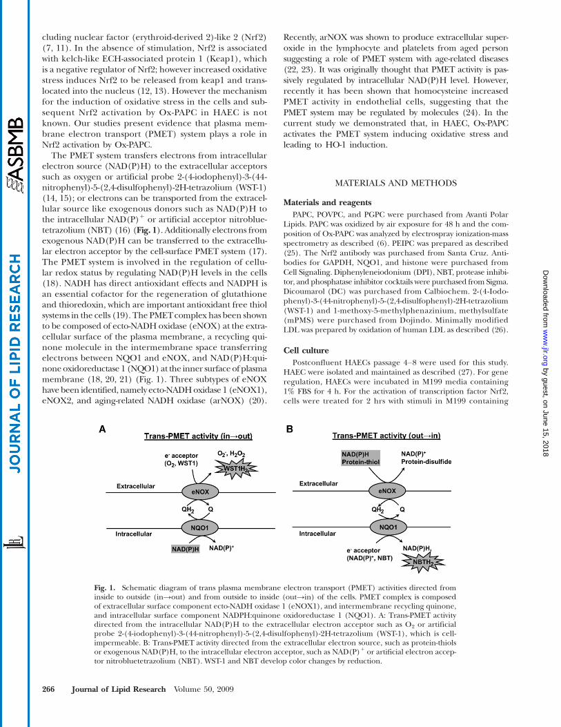
The PMET system transfers electrons from intracellularelectron source (NAD(P)H) to the extracellular acceptorssuch as oxygen or artificial probe 2-(4-iodophenyl)-3-(44-nitrophenyl)-5-(2,4-disulfophenyl)-2H-tetrazolium (WST-1)(14, 15); or electrons can be transported from the extracel-lular source like exogenous donors such as NAD(P)H tothe intracellular NAD(P)1 or artificial acceptor nitroblue-tetrazolium (NBT) (16) (Fig. 1). Additionally electrons fromexogenous NAD(P)H can be transferred to the extracellu-lar electron acceptor by the cell-surface PMET system (17).The PMET system is involved in the regulation of cellu-lar redox status by regulating NAD(P)H levels in the cells(18). NADH has direct antioxidant effects and NADPH isan essential cofactor for the regeneration of glutathioneand thioredoxin, which are important antioxidant free thiolsystems in the cells (19). The PMETcomplex has been shownto be composed of ecto-NADH oxidase (eNOX) at the extra-cellular surface of the plasma membrane, a recycling qui-none molecule in the intermembrane space transferringelectrons between NQO1 and eNOX, and NAD(P)H:qui-noneoxidoreductase 1 (NQO1) at the inner surface of plasmamembrane (18, 20, 21) (Fig. 1). Three subtypes of eNOXhave been identified, namely ecto-NADHoxidase 1 (eNOX1),eNOX2, and aging-related NADH oxidase (arNOX) (20).
Recently, arNOX was shown to produce extracellular super-oxide in the lymphocyte and platelets from aged personsuggesting a role of PMET system with age-related diseases(22, 23). It was originally thought that PMET activity is pas-sively regulated by intracellular NAD(P)H level. However,recently it has been shown that homocysteine increasedPMET activity in endothelial cells, suggesting that thePMET system may be regulated by molecules (24). In thecurrent study we demonstrated that, in HAEC, Ox-PAPCactivates the PMET system inducing oxidative stress andleading to HO-1 induction.
MATERIALS AND METHODS
Materials and reagentsPAPC, POVPC, and PGPC were purchased from Avanti Polar
Lipids. PAPC was oxidized by air exposure for 48 h and the com-position of Ox-PAPC was analyzed by electrospray ionization-massspectrometry as described (6). PEIPC was prepared as described(25). The Nrf2 antibody was purchased from Santa Cruz. Anti-bodies for GAPDH, NQO1, and histone were purchased fromCell Signaling. Diphenyleneiodonium (DPI), NBT, protease inhibi-tor, and phosphatase inhibitor cocktails were purchased from Sigma.Dicoumarol (DC) was purchased from Calbiochem. 2-(4-Iodo-phenyl)-3-(44-nitrophenyl)-5-(2,4-disulfophenyl)-2H-tetrazolium(WST-1) and 1-methoxy-5-methylphenazinium, methylsulfate(mPMS) were purchased from Dojindo. Minimally modifiedLDL was prepared by oxidation of human LDL as described (26).
Cell culturePostconfluent HAECs passage 4–8 were used for this study.
HAEC were isolated and maintained as described (27). For generegulation, HAECs were incubated in M199 media containing1% FBS for 4 h. For the activation of transcription factor Nrf2,cells were treated for 2 hrs with stimuli in M199 containing
Fig. 1. Schematic diagram of trans plasma membrane electron transport (PMET) activities directed frominside to outside (in→out) and from outside to inside (out→in) of the cells. PMET complex is composedof extracellular surface component ecto-NADH oxidase 1 (eNOX1), and intermembrane recycling quinone,and intracellular surface component NADPH:quinone oxidoreductase 1 (NQO1). A: Trans-PMET activitydirected from the intracellular NAD(P)H to the extracellular electron acceptor such as O2 or artificialprobe 2-(4-iodophenyl)-3-(44-nitrophenyl)-5-(2,4-disulfophenyl)-2H-tetrazolium (WST-1), which is cell-impermeable. B: Trans-PMET activity directed from the extracellular electron source, such as protein-thiolsor exogenous NAD(P)H, to the intracellular electron acceptor, such as NAD(P)1 or artificial electron accep-tor nitrobluetetrazolium (NBT). WST-1 and NBT develop color changes by reduction.
266 Journal of Lipid Research Volume 50, 2009
by guest, on June 15, 2018w
ww
.jlr.orgD
ownloaded from
0.2% FBS. Unless otherwise specified, cells were pretreated withinhibitors for 1 h and cotreated with stimuli for specified times.
Gene silencing by siRNA-transfectionScrambled control and siRNAs specific to eNOX1 (Hs_
FLJ10094_1_HP, Hs_FLJ10094_4_HP), NQO1 (Hs_NQO1_1_HP,Hs_NQO1_8_HP), and Nrf2 (Hs_NFE2L2_4_HP) were pur-chased from Qiagen. HAECs were transfected with siRNA usingLipofectamine 2000 (Invitrogen) following the recommendedprotocol by the manufacturer (28). After 2–6 days of culture,silencing of target genes was confirmed by QRT-PCR and West-ern blotting. Two siRNAs were used for each gene to control off-target effects.
QRT-PCRTotal RNA and cDNAwere prepared using RNA extraction and
cDNA synthesis kit (Bio-Rad). SYBR: green master mixture andPCR amplification system from Applied Biosystems were used forquantitative PCR. The primer sequences used for QuantitativeRT-PCR (QRT-PCR) were: GAPDH, F:5′-CCT CAA GAT CATCAG CAA TGC CTC CT-3′, R:3′-GGT CAT GAG TCC TTC CACGAT ACC AA-5′; HO-1, F:5′-ATA GAT GTG GTA CAG GGA GGCCAT CA-3′, R:5′-GGC AGA GAATGC TGA GTT CAT GAG GA-3′;IL-8: F:5′-ACC ACA CTG CGC CAA CAC AGA AAT-3′, R:5′-TCCAGA CAG AGC TCT CTT CCA TCA GA-3′; eNOX1, F: 5′-TGAGCA AGA GAT GGA GGA AGC CAA -3′, R:5′-ATT CCG GAGCTC CTC AGA ATG CTT-3′; eNOX2, F:5′-AGT ATT TGT GGGTGG TCT GCC TGA-3′, R: 5′-ACC ATG TAC TCC TCA GCAAAG CGA-3′; NQO1, F: 5′-AAG GAT GGA AGA AAC GCC TGGAGA -3′, R:5′-GGC CCA CAG AAA GGC CAA ATT TCT-3′;NQO2: F:5′-AGT GGA AAC CCA CGA AGC CTA CAA-3′, R:5′-TGA ACC AGT ACA GCG GGA ACT GAA-3′; Nrf2: F: 5′-AGCATG CCC TCA CCT GCT ACT TTA-3′, R: 5′-ACT GAG TGTTCT GGT GAT GCC ACA-3′.
Immunoblotting (Western blot)Laemmli buffer (Bio-Rad) containing protease and phospha-
tase inhibitor cocktails and PMSF (1 mM) was used for total celllysate preparation. Nuclear fraction was isolated as described inthe previous report (29). The protein samples were separated on4–20% Tris-glycine SDS-PAGE gel (ISC Bioexpress) using stan-dard protocol for gel running and blotting (28, 30). Blots wereincubated with antibodies in 5% fat-free milk or 3% BSA in TBST(Tris-buffered saline, 0.1% Tween 20) for 1 hr at room tempera-ture or overnight at 4°C. Blots were developed and the imagewas acquired using chemiluminescence kit (Amersham) andVersaDoc Imaging System-Model 5000 (Bio-Rad). Quantity One:program was used to measure band densities in the gel image.
Assay for trans-PMET activityTo measure the trans-PMET activity (in→out), which transfer
electrons from the intracellular electron source NAD(P)H tothe extracellular acceptor, the method described in the previous
report was used (17). Briefly, HAECs in 96-well formats were in-cubated with stimuli in Hankʼs Balance Salt Solution containingWST-1 (500 mM) and mPMS (1 mM) in total volume of 100 ml.WST-1 accepts electrons from PMET complex only in the pres-ence of an electron mediator mPMS and develops a yellow color(17). After incubation for the specified time, the absorbance at440 nm was measured as an indicator of trans-PMET activity(in→out). A blank test was performed in the absence of cellsbut in the presence of stimuli, and the blank values were sub-tracted from the values with cells. For the measurement oftrans-PMET activity directed from the exogenous NAD(P)H tothe intracellular electron acceptor (out→in), a cell-permeableartificial electron acceptor NBT was employed. Briefly, afterwashing twice with PBS confluent HAECs in 96-well dishes wereincubated with stimuli in Hankʼs Balance Salt Solution contain-ing NBT (400 mM) and NADH (100 mM) or NADPH (500 mM) intotal volume of 100 ml. NBT forms blue formazan upon reductionin the cells and NAD(P)H is generally regarded as cell-impermeable(18, 31). After washing twice with PBS, unincorporated NBTwas discarded by washing with methanol (150 ml). Formazan inthe cells was dissolved in 60 ml of 2M KOH and 90 ml of DMSO,and the absorbance at 690 nm was measured as an indicator offormazan formation in the cells.
Detection of intracellular reactive oxygen species formationFor the detection of reactive oxygen species in the cells, a
DCF assay was employed. Briefly cells were grown in 4 well-glasschamber slides to confluence. Cells were preincubated with DCF(5 mM, molecular probes) for 30 min in assay media (M199 plus1% FBS) at 37°C in CO2 incubator. After removing DCF solu-tion, cells were treated with stimuli (e.g., Ox-PAPC) in assaymedia. The green fluorescence (excitation: 488 nm, detection:505 nm) was measured using an inverted confocal microscope.Readings were analyzed at the times shown. The PASCAL programwas used for imaging and analysis of the signal. Minimum laser in-tensity and exposure time were used to prevent nonspecific bleach-ing of the cells during the observation. For the quantification ofthe signal, the mean signal intensity of 10 cells randomly chosenwas used. The same intensity of laser and settings for image acquir-ing were used for the comparison of multiple observations.
Statistical analysisTwo-tailed unpaired Studentʼs t -test was used to evaluate the
difference between two groups. The P value ,0.05 was regardedas a statistically significant.
RESULTS
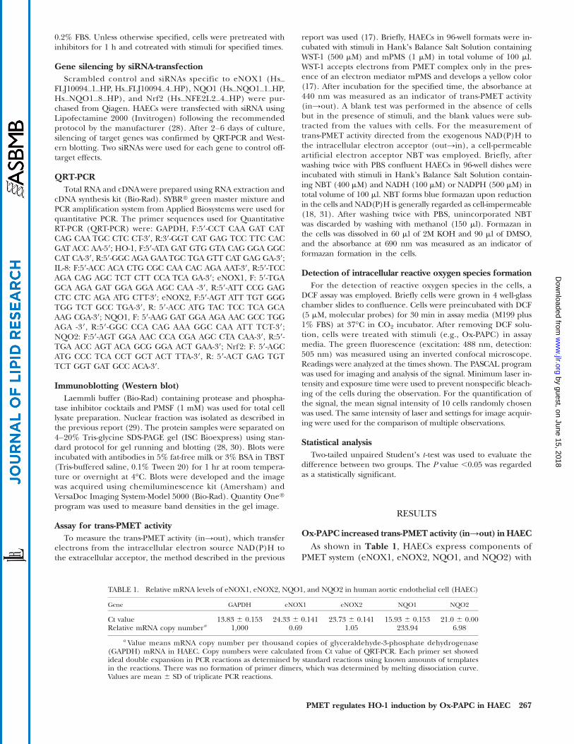
Ox-PAPC increased trans-PMET activity (in→out) in HAECAs shown in Table 1, HAECs express components of
PMET system (eNOX1, eNOX2, NQO1, and NQO2) with
TABLE 1. Relative mRNA levels of eNOX1, eNOX2, NQO1, and NQO2 in human aortic endothelial cell (HAEC)
Gene GAPDH eNOX1 eNOX2 NQO1 NQO2
Ct value 13.83 6 0.153 24.33 6 0.141 23.73 6 0.141 15.93 6 0.153 21.0 6 0.00Relative mRNA copy numbera 1,000 0.69 1.05 233.94 6.98
a Value means mRNA copy number per thousand copies of glyceraldehyde-3-phosphate dehydrogenase(GAPDH) mRNA in HAEC. Copy numbers were calculated from Ct value of QRT-PCR. Each primer set showedideal double expansion in PCR reactions as determined by standard reactions using known amounts of templatesin the reactions. There was no formation of primer dimers, which was determined by melting dissociation curve.Values are mean 6 SD of triplicate PCR reactions.
PMET regulates HO-1 induction by Ox-PAPC in HAEC 267
by guest, on June 15, 2018w
ww
.jlr.orgD
ownloaded from
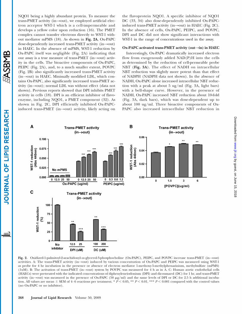
NQO1 being a highly abundant protein. To measure thetrans-PMET activity (in→out), we employed artificial elec-tron acceptor WST-1 which is a cell-impermeable anddevelops a yellow color upon reduction (16). The PMETcomplex cannot transfer electrons directly to WST-1 with-out mediator mPMS (18). As shown in Fig. 2A, Ox-PAPCdose-dependently increased trans-PMET activity (in→out)in HAEC. In the absence of mPMS, WST-1 reduction byOx-PAPC itself was negligible (Fig. 2A) indicating thatour assay is a true measure of trans-PMET (in→out) activ-ity in the cells. The bioactive components of Ox-PAPC,PEIPC (Fig. 2A), and, to a much smaller extent, POVPC(Fig. 2B) also significantly increased trans-PMET activity(in→out) in HAEC. Minimally modified LDL, which con-tains Ox-PAPC, also significantly increased trans-PMET ac-tivity (in→out); normal LDL was without effect (data notshown). Previous reports showed that DPI inhibits PMETactivity in cells (18). DPI is an efficient inhibitor of flavo-enzyme, including NQO1, a PMET component (32). Asshown in Fig. 2C, DPI efficiently inhibited Ox-PAPC-induced trans-PMET (in→out) activity, likely acting on
the flavoprotein NQO1. A specific inhibitor of NQO1DC (33, 34) also dose-dependently inhibited Ox-PAPC-induced trans-PMET activity (in→out) in HAEC (Fig. 2C).In the absence of cells, Ox-PAPC, PEIPC, and POVPC,DPI and DC did not show significant interactions withWST-1 in the range of concentrations used in the assay.
Ox-PAPC activated trans-PMET activity (out→in) in HAECInterestingly, Ox-PAPC dramatically increased electron
flow from exogenously added NAD(P)H into the cellsas determined by the reduction of cell-permeable probeNBT (Fig. 3A). The effect of NADH on intracellularNBT reduction was slightly more potent than that effectof NADPH (NADPH data not shown). In the absence ofNADH, Ox-PAPC alone increased intracellular NBT reduc-tion with a peak at about 5 ug/ml (Fig. 3A, light bars)with a bell-shape curve. However, in the presence ofNADH, Ox-PAPC increased NBT reduction about 10-fold(Fig. 3A, dark bars), which was dose-dependent up toabout 100 ug/ml. Three bioactive components of Ox-PAPC also increased intracellular NBT reduction in
Fig. 2. Oxidized-1-palmitoyl-2-arachidonyl-sn -glycerol-3-phosphocholine (Ox-PAPC), PEIPC, and POVPC increase trans-PMET (in→out)activities. A: The trans-PMET activity (in→out) induced by various concentration of Ox-PAPC and PEIPC was measured using WST-1as probe for 4 hr incubation in the presence or absence of electron mediator 1-methoxy-5-methylphenazinium, methylsulfate (mPMS)(1uM). B: The activation of trans-PMET (in→out) system by POVPC was measured for 4 h as in A. C: Human aortic endothelial cells(HAECs) were pretreated with the indicated concentrations of diphenyleneiodonium (DPI) and dicoumarol (DC) for 1 hr, and trans-PMETactivity (in→out) was measured in the presence of Ox-PAPC (50 mg/ml) and the same levels of DPI or DC for 2.5 h additional incuba-tion. All values are mean 6 SEM of 4–6 reactions per treatment. * P , 0.05, ** P , 0.01, *** P , 0.001 compared with the control values(no Ox-PAPC or no inhibitor).
268 Journal of Lipid Research Volume 50, 2009
by guest, on June 15, 2018w
ww
.jlr.orgD
ownloaded from
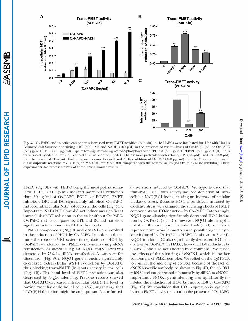
HAEC (Fig. 3B) with PEIPC being the most potent stimu-lator. PEIPC (0.1 ug/ml) induced more NBT reductionthan 50 ug/ml of Ox-PAPC, PGPC, or POVPC. PMETinhibitors DPI and DC significantly inhibited Ox-PAPC-induced intracellular NBT reduction in the cells (Fig. 3C).Importantly NAD(P)H alone did not induce any significantintracellular NBT reduction in the cells without Ox-PAPC.Ox-PAPC and its components, DPI, and DC did not showsignificant interactions with NBT without cells.
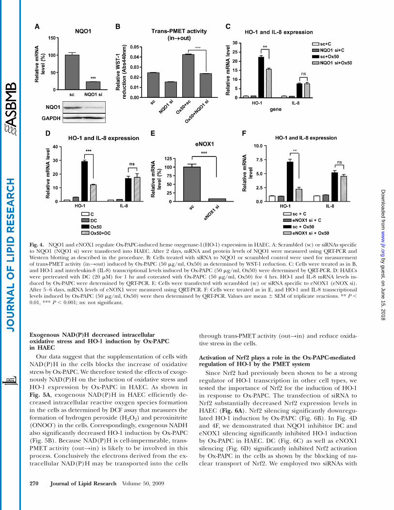
PMET components (NQO1 and eNOX1) are involvedin the induction of HO-1 by Ox-PAPC. In order to deter-mine the role of PMET system in regulation of HO-1 byOx-PAPC, we silenced two PMET components using siRNAtransfection. As shown in Fig. 4A, NQO1 mRNA level wasdecreased by 75% by siRNA transfection. As was seen fordicumarol (Fig. 3C), NQO1 gene silencing significantlydecreased extracellular WST-1 reduction by Ox-PAPCthus blocking trans-PMET (in→out) activity in the cells(Fig. 4B). The basal level of WST-1 reduction was alsodecreased by NQO1 silencing. Previous reports showedthat Ox-PAPC decreased intracellular NAD(P)H level inbovine vascular endothelial cells (35), suggesting thatNAD(P)H depletion might be an important factor for oxi-
dative stress induced by Ox-PAPC. We hypothesized thattrans-PMET (in→out) activity induced depletion of intra-cellular NAD(P)H levels, causing an increase of cellularoxidative stress. Because HO-1 is sensitively induced byoxidative stress, we examined the silencing effects of PMETcomponents on HO-induction by Ox-PAPC. Interestingly,NQO1 gene silencing significantly decreased HO-1 induc-tion by Ox-PAPC (Fig. 4C); however, NQO1 silencing didnot affect the induction of interleukin-8 (IL-8), which is arepresentative proinflammatory and proatherogenic cyto-kine induced by Ox-PAPC in HAEC. As shown in Fig. 4D,NQO1 inhibitor DC also significantly decreased HO-1 in-duction by Ox-PAPC in HAEC; however, IL-8 induction byOx-PAPC was also not affected by dicoumarol. We testedthe effects of the silencing of eNOX1, which is anothercomponent of PMET complex. We relied on the QRT-PCRto confirm the silencing of eNOX1 because of the lack ofeNOX1-specific antibody. As shown in Fig. 4D, the eNOX1mRNA level was decreased substantially by siRNA to eNOX1.Importantly eNOX1 gene silencing also significantly in-hibited the induction of HO-1 but not of IL-8 by Ox-PAPC(Fig. 4E). We concluded that HO-1 expression is regulatedby trans-PMETactivity (in→out) in the presence of Ox-PAPC.
Fig. 3. Ox-PAPC and its active components increased trans-PMET activities (out→in). A, B: HAECs were incubated for 1 hr with HankʼsBalanced Salt Solution containing NBT (400 mM) and NADH (100 mM) in the presence of various levels of Ox-PAPC (A), or Ox-PAPC(50 mg/ml), PEIPC (0.1mg/ml), 1-palmitoyl-2-glutaroyl-sn -glycerol-3-phosphocholine (PGPC) (50 mg/ml), POVPC (50 mg/ml) (B). Cellswere rinsed, lysed, and levels of reduced NBT were determined. C: HAECs were pretreated with vehicle, DPI (6.5 mM), and DC (200 mM)for 1 hr. Trans-PMET activity (out→in) was measured as in A and B after addition of Ox-PAPC (50 mg/ml) for 1 hr. Values were mean 6SD of duplicate reactions. * P , 0.05, ** P , 0.01, *** P , 0.001 compared with the control values (no Ox-PAPC or no inhibitor). Theseexperiments are representatives of three giving similar results.
PMET regulates HO-1 induction by Ox-PAPC in HAEC 269
by guest, on June 15, 2018w
ww
.jlr.orgD
ownloaded from
Exogenous NAD(P)H decreased intracellularoxidative stress and HO-1 induction by Ox-PAPCin HAEC
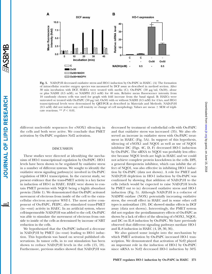
Our data suggest that the supplementation of cells withNAD(P)H in the cells blocks the increase of oxidativestress by Ox-PAPC. We therefore tested the effects of exoge-nously NAD(P)H on the induction of oxidative stress andHO-1 expression by Ox-PAPC in HAEC. As shown inFig. 5A, exogenous NAD(P)H in HAEC efficiently de-creased intracellular reactive oxygen species formationin the cells as determined by DCF assay that measures theformation of hydrogen peroxide (H2O2) and peroxinitrite(ONOO-) in the cells. Correspondingly, exogenous NADHalso significantly decreased HO-1 induction by Ox-PAPC(Fig. 5B). Because NAD(P)H is cell-impermeable, trans-PMET activity (out→in) is likely to be involved in thisprocess. Conclusively the electrons derived from the ex-tracellular NAD(P)H may be transported into the cells
through trans-PMET activity (out→in) and reduce oxida-tive stress in the cells.
Activation of Nrf2 plays a role in the Ox-PAPC-mediatedregulation of HO-1 by the PMET system
Since Nrf2 had previously been shown to be a strongregulator of HO-1 transcription in other cell types, wetested the importance of Nrf2 for the induction of HO-1in response to Ox-PAPC. The transfection of siRNA toNrf2 substantially decreased Nrf2 expression levels inHAEC (Fig. 6A). Nrf2 silencing significantly downregu-lated HO-1 induction by Ox-PAPC (Fig. 6B). In Fig. 4Dand 4F, we demonstrated that NQO1 inhibitor DC andeNOX1 silencing significantly inhibited HO-1 inductionby Ox-PAPC in HAEC. DC (Fig. 6C) as well as eNOX1silencing (Fig. 6D) significantly inhibited Nrf2 activationby Ox-PAPC in the cells as shown by the blocking of nu-clear transport of Nrf2. We employed two siRNAs with
Fig. 4. NQO1 and eNOX1 regulate Ox-PAPC-induced heme oxygenase-1(HO-1) expression in HAEC. A: Scrambled (sc) or siRNAs specificto NQO1 (NQO1 si) were transfected into HAEC. After 2 days, mRNA and protein levels of NQO1 were measured using QRT-PCR andWestern blotting as described in the procedure. B: Cells treated with siRNA to NQO1 or scrambled control were used for measurementof trans-PMET activity (in→out) induced by Ox-PAPC (50 mg/ml, Ox50) as determined by WST-1 reduction. C: Cells were treated as in B,and HO-1 and interleukin-8 (IL-8) transcriptional levels induced by Ox-PAPC (50 mg/ml, Ox50) were determined by QRT-PCR. D: HAECswere pretreated with DC (20 mM) for 1 hr and cotreated with Ox-PAPC (50 mg/ml, Ox50) for 4 hrs. HO-1 and IL-8 mRNA levels in-duced by Ox-PAPC were determined by QRT-PCR. E: Cells were transfected with scrambled (sc) or siRNA specific to eNOX1 (eNOX si).After 5–6 days, mRNA levels of eNOX1 were measured using QRT-PCR. F: Cells were treated as in E, and HO-1 and IL-8 transcriptionallevels induced by Ox-PAPC (50 mg/ml, Ox50) were then determined by QRT-PCR. Values are mean 6 SEM of triplicate reactions. ** P ,0.01, *** P , 0.001; ns: not significant.
270 Journal of Lipid Research Volume 50, 2009
by guest, on June 15, 2018w
ww
.jlr.orgD
ownloaded from
different nucleotide sequences for eNOX1 silencing inthe cells and both were active. We conclude that PMETactivation by Ox-PAPC regulates Nrf2 activation.
DISCUSSION
These studies were directed at identifying the mecha-nism of HO-1 transcriptional regulation by Ox-PAPC. HO-1levels have been shown to be regulated by oxidative stressin a number of different systems. We sought to identify theoxidative stress signaling pathway(s) involved in Ox-PAPCregulation of HO-1 transcription. In the current study, wepresent evidence that the trans-PMET activity is a key factorin induction of HO-1 in HAEC. HAEC were shown to con-tain PMET proteins with NQO1 being a highly abundantprotein (Table 1). We demonstrated that Ox-PAPC activatedmovement of electrons from inside of the cell to the extra-cellular electron acceptor WST-1. The most active com-ponent of Ox-PAPC, PEIPC, also stimulated trans-PMET(in→out) activity in HAEC. In an artificial system, wherecell-impermeable NAD(P)H was added to the cell, Ox-PAPCwas able to stimulate the movement of electrons from out-side to inside of the cells as determined by increase of NBTreduction in the cells.
We hypothesized that the Ox-PAPC induced a decreasein NAD(P)H by PMET (in→out) leading to HO-1 induc-tion. This hypothesis was based on several published ob-servations. In tumor cells, in to out stimulation has beenshown to reduce NAD(P)H levels in the cells (15, 18).Furthermore, previous studies showed that NAD(P)H was
decreased by treatment of endothelial cells with Ox-PAPCand that oxidative stress was increased (35). We also ob-served an increase in oxidative stress with Ox-PAPC treat-ment in HAEC (Fig. 5A). In support of this hypothesis,silencing of eNOX1 and NQO1 as well as use of NQO1inhibitor DC (Figs. 4C, D, F) decreased HO-1 inductionby Ox-PAPC. The siRNA to NQO1 was probably less effec-tive because NQO1 levels are high in HAEC and we couldnot achieve complete protein knockdown in the cells. DPI,a general flavoprotein inhibitor, which can inhibit the ef-fect of NQO1, was also effective in inhibiting HO-1 induc-tion by Ox-PAPC (data not shown). A role for PMET andNAD(P)H depletion in HO-1 induction by Ox-PAPC wasconfirmed by showing that addition of NAD(P)H to thecells (which would be expected to raise NAD(P)H levelsby PMET out to in) decreased oxidative stress and HO-1induction (Fig. 5). Although NAD(P)H is a cofactor ofNADPH oxidase (NOX) potentially increasing oxidativestress, the overall effect in HAEC and in some other celltypes is antioxidant (19). DC showed similar effects in DCFassay (data not shown). Interestingly, the PMET systemdid not regulate the proinflammatory effects of Ox-PAPC asshown by a lack of effect of the silencing of eNOX1, NQO1,and DC on IL-8 induction by Ox-PAPC. We have previouslyobserved that different signaling pathways mediate HO-1and IL-8 induction in HAEC (4, 28, 30, 36).
We also gained some insight into the mechanism bywhich PMET activation by Ox-PAPC increased HO-1 tran-scription. We demonstrated that activation of Nrf2 playedan important role in the induction of HO-1 by Ox-PAPCsince siRNA to Nrf2 decreased HO-1 induction by 50%
Fig. 5. NAD(P)H decreased oxidative stress and HO-1 induction by Ox-PAPC in HAEC. (A) The formationof intracellular reactive oxygen species was measured by DCF assay as described in method section. After30 min incubation with DCF, HAECs were treated with media (C); Ox-PAPC (50 mg/ml, Ox50), aloneor plus NADH (0.5 mM); or NADPH (0.5 mM) for 40 min. Relative mean fluorescence intensity from10 randomly chosen cells was used for graph with fold increase from the basal signal. B: HAECs wereuntreated or treated with Ox-PAPC (50 mg/ml, Ox50) with or without NADH (0.5 mM) for 4 hrs, and HO-1transcriptional levels were determined by QRT-PCR as described in Materials and Methods. NAD(P)H(0.5 mM) did not induce any cell toxicity or change of cell morphology. Values are mean 6 SEM of tripli-cate reactions. ** P , 0.01.
PMET regulates HO-1 induction by Ox-PAPC in HAEC 271
by guest, on June 15, 2018w
ww
.jlr.orgD
ownloaded from
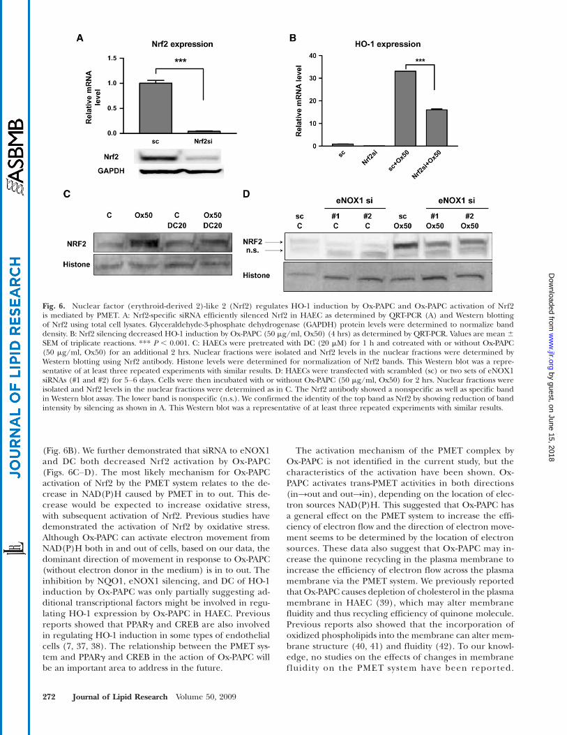
(Fig. 6B). We further demonstrated that siRNA to eNOX1and DC both decreased Nrf2 activation by Ox-PAPC(Figs. 6C–D). The most likely mechanism for Ox-PAPCactivation of Nrf2 by the PMET system relates to the de-crease in NAD(P)H caused by PMET in to out. This de-crease would be expected to increase oxidative stress,with subsequent activation of Nrf2. Previous studies havedemonstrated the activation of Nrf2 by oxidative stress.Although Ox-PAPC can activate electron movement fromNAD(P)H both in and out of cells, based on our data, thedominant direction of movement in response to Ox-PAPC(without electron donor in the medium) is in to out. Theinhibition by NQO1, eNOX1 silencing, and DC of HO-1induction by Ox-PAPC was only partially suggesting ad-ditional transcriptional factors might be involved in regu-lating HO-1 expression by Ox-PAPC in HAEC. Previousreports showed that PPARg and CREB are also involvedin regulating HO-1 induction in some types of endothelialcells (7, 37, 38). The relationship between the PMET sys-tem and PPARg and CREB in the action of Ox-PAPC willbe an important area to address in the future.
The activation mechanism of the PMET complex byOx-PAPC is not identified in the current study, but thecharacteristics of the activation have been shown. Ox-PAPC activates trans-PMET activities in both directions(in→out and out→in), depending on the location of elec-tron sources NAD(P)H. This suggested that Ox-PAPC hasa general effect on the PMET system to increase the effi-ciency of electron flow and the direction of electron move-ment seems to be determined by the location of electronsources. These data also suggest that Ox-PAPC may in-crease the quinone recycling in the plasma membrane toincrease the efficiency of electron flow across the plasmamembrane via the PMET system. We previously reportedthat Ox-PAPC causes depletion of cholesterol in the plasmamembrane in HAEC (39), which may alter membranefluidity and thus recycling efficiency of quinone molecule.Previous reports also showed that the incorporation ofoxidized phospholipids into the membrane can alter mem-brane structure (40, 41) and fluidity (42). To our knowl-edge, no studies on the effects of changes in membranefluidity on the PMET system have been reported.
Fig. 6. Nuclear factor (erythroid-derived 2)-like 2 (Nrf2) regulates HO-1 induction by Ox-PAPC and Ox-PAPC activation of Nrf2is mediated by PMET. A: Nrf2-specific siRNA efficiently silenced Nrf2 in HAEC as determined by QRT-PCR (A) and Western blottingof Nrf2 using total cell lysates. Glyceraldehyde-3-phosphate dehydrogenase (GAPDH) protein levels were determined to normalize banddensity. B: Nrf2 silencing decreased HO-1 induction by Ox-PAPC (50 mg/ml, Ox50) (4 hrs) as determined by QRT-PCR. Values are mean 6SEM of triplicate reactions. *** P , 0.001. C: HAECs were pretreated with DC (20 mM) for 1 h and cotreated with or without Ox-PAPC(50 mg/ml, Ox50) for an additional 2 hrs. Nuclear fractions were isolated and Nrf2 levels in the nuclear fractions were determined byWestern blotting using Nrf2 antibody. Histone levels were determined for normalization of Nrf2 bands. This Western blot was a repre-sentative of at least three repeated experiments with similar results. D: HAECs were transfected with scrambled (sc) or two sets of eNOX1siRNAs (#1 and #2) for 5–6 days. Cells were then incubated with or without Ox-PAPC (50 mg/ml, Ox50) for 2 hrs. Nuclear fractions wereisolated and Nrf2 levels in the nuclear fractions were determined as in C. The Nrf2 antibody showed a nonspecific as well as specific bandin Western blot assay. The lower band is nonspecific (n.s.). We confirmed the identity of the top band as Nrf2 by showing reduction of bandintensity by silencing as shown in A. This Western blot was a representative of at least three repeated experiments with similar results.
272 Journal of Lipid Research Volume 50, 2009
by guest, on June 15, 2018w
ww
.jlr.orgD
ownloaded from
However, several groups showed that membrane fluidity isan important factor for the efficiency of electron transportby quinone recycling as shown in mitochondrial electrontransport system (43, 44).
In summary our studies present evidence that the PMETsystem is activated by Ox-PAPC and involved in the regu-lation of HO-1 induction by Ox-PAPC through activationof Nrf2. These studies suggest that PMET system is animportant regulator of redox status in endothelial cellsand a potential regulator of atherogenesis in the vascu-lar system.
REFERENCES
1. Yla-Herttuala, S., W. Palinski, M. E. Rosenfeld, S. Parthasarathy,T. E. Carew, S. Butler, J. L. Witztum, and D. Steinberg. 1989. Evi-dence for the presence of oxidatively modified low density lipo-protein in atherosclerotic lesions of rabbit and man. J. Clin. Invest.84: 1086–1095.
2. Subbanagounder, G., N. Leitinger, D. C. Schwenke, J. W. Wong,H. Lee, C. Rizza, A. D. Watson, K. F. Faull, A. M. Fogelman, andJ. A. Berliner. 2000. Determinants of bioactivity of oxidized phos-pholipids. Specific oxidized fatty acyl groups at the sn-2 position.Arterioscler. Thromb. Vasc. Biol. 20: 2248–2254.
3. Watson, A. D., G. Subbanagounder, D. S. Welsbie, K. F. Faull, M.Navab, M. E. Jung, A. M. Fogelman, and J. A. Berliner. 1999. Struc-tural identification of a novel pro-inflammatory epoxyisoprostanephospholipid in mildly oxidized low density lipoprotein. J. Biol.Chem. 274: 24787–24798.
4. Gargalovic, P. S., M. Imura, B. Zhang, N. M. Gharavi, M. J. Clark,J. Pagnon, W. P. Yang, A. He, A. Truong, S. Patel, et al. 2006. Iden-tification of inflammatory gene modules based on variations ofhuman endothelial cell responses to oxidized lipids. Proc. Natl.Acad. Sci. USA. 103: 12741–12746.
5. Gargalovic, P. S., N. M. Gharavi, M. J. Clark, J. Pagnon, W. P. Yang, A.He, A. Truong, T. Baruch-Oren, J. A. Berliner, T. G. Kirchgessner,et al. 2006. The unfolded protein response is an important regula-tor of inflammatory genes in endothelial cells. Arterioscler. Thromb.Vasc. Biol. 26: 2490–2496.
6. Subbanagounder, G., J. W. Wong, H. Lee, K. F. Faull, E. Miller,J. L. Witztum, and J. A. Berliner. 2002. Epoxyisoprostane andepoxycyclopentenone phospholipids regulate monocyte chemo-tactic protein-1 and interleukin-8 synthesis. Formation of theseoxidized phospholipids in response to interleukin-1beta. J. Biol.Chem. 277: 7271–7281.
7. Kronke, G., V. N. Bochkov, J. Huber, F. Gruber, S. Bluml, A.Furnkranz, A. Kadl, B. R. Binder, and N. Leitinger. 2003. Oxi-dized phospholipids induce expression of human heme oxygenase-1 involving activation of cAMP-responsive element-binding protein.J. Biol. Chem. 278: 51006–51014.
8. Jyrkkanen, H. K., E. Kansanen, M. Inkala, A. M. Kivela, H. Hurttila,S. E. Heinonen, G. Goldsteins, S. Jauhiainen, S. Tiainen, H.Makkonen,et al. 2008. Nrf2 regulates antioxidant gene expression evokedby oxidized phospholipids in endothelial cells and murine arteriesin vivo. Circ. Res. 103: e1–e9.
9. Morse, D., and A. M. Choi. 2005. Heme oxygenase-1: from bench tobedside. Am. J. Respir. Crit. Care Med. 172: 660–670.
10. Gharavi, N. M., P. S. Gargalovic, I. Chang, J. A. Araujo, M. J.Clark, W. L. Szeto, A. D. Watson, A. J. Lusis, and J. A. Berliner.2007. High-density lipoprotein modulates oxidized phospholipidsignaling in human endothelial cells from proinflammatory toanti-inflammatory. Arterioscler. Thromb. Vasc. Biol. 27: 1346–1353.
11. Srisook, K., C. Kim, and Y. N. Cha. 2005. Molecular mechanismsinvolved in enhancing HO-1 expression: de-repression by hemeand activation by Nrf2, the “one-two” punch. Antioxid. Redox Signal.7: 1674–1687.
12. Kang, K. W., S. J. Lee, and S. G. Kim. 2005. Molecular mechanismof nrf2 activation by oxidative stress. Antioxid. Redox Signal. 7:1664–1673.
13. Sun, Z., S. Zhang, J. Y. Chan, and D. D. Zhang. 2007. Keap1 controlspostinduction repression of the Nrf2-mediated antioxidant re-
sponse by escorting nuclear export of Nrf2. Mol. Cell. Biol. 27:6334–6349.
14. Herst, P. M., A. S. Tan, D. J. Scarlett, and M. V. Berridge. 2004. Cellsurface oxygen consumption by mitochondrial gene knockout cells.Biochim. Biophys. Acta. 1656: 79–87.
15. Herst, P. M., and M. V. Berridge. 2007. Cell surface oxygen con-sumption: a major contributor to cellular oxygen consumption inglycolytic cancer cell lines. Biochim. Biophys. Acta. 1767: 170–177.
16. Berridge, M. V., P. M. Herst, and A. S. Tan. 2005. Tetrazolium dyesas tools in cell biology: new insights into their cellular reduction.Biotechnol. Annu. Rev. 11: 127–152.
17. Scarlett, D. J., P. M. Herst, and M. V. Berridge. 2005. Multiple pro-teins with single activities or a single protein with multiple activi-ties: the conundrum of cell surface NADH oxidoreductases. Biochim.Biophys. Acta. 1708: 108–119.
18. Herst, P. M., and M. V. Berridge. 2006. Plasma membrane electrontransport: a new target for cancer drug development. Curr. Mol.Med. 6: 895–904.
19. Ying, W. 2008. NAD1/NADH and NADP1/NADPH in cellularfunctions and cell death: regulation and biological consequences.Antioxid. Redox Signal. 10: 179–206.
20. Morre, D. J. 2004. Quinone oxidoreductases of the plasma mem-brane. Methods Enzymol. 378: 179–199.
21. Sun, I. L., E. E. Sun, F. L. Crane, D. J. Morre, A. Lindgren, and H.Low. 1992. Requirement for coenzyme Q in plasma membrane elec-tron transport. Proc. Natl. Acad. Sci. USA. 89: 11126–11130.
22. Morre, D. J., and D. M. Morre. 2006. Aging-related cell surfaceECTO-NOX protein, arNOX, a preventive target to reduce athero-genic risk in the elderly. Rejuvenation Res. 9: 231–236.
23. Morre, D. M., F. Guo, and D. J. Morre. 2003. An aging-related cellsurface NADH oxidase (arNOX) generates superoxide and is in-hibited by coenzyme Q. Mol. Cell. Biochem. 254: 101–109.
24. Rodriguez-Alonso, J., R. Montanez, L. Rodriguez-Caso, and M. A.Medina. 2008. Homocysteine is a potent modulator of plasmamembrane electron transport systems. J. Bioenerg. Biomembr. 40:45–51.
25. Jung, M. E., J. A. Berliner, D. Angst, D. Yue, L. Koroniak, A. D.Watson, and R. Li. 2005. Total synthesis of the epoxy isoprostanephospholipids PEIPC and PECPC. Org. Lett. 7: 3933–3935.
26. Liao, F., J. A. Berliner, M. Mehrabian, M. Navab, L. L. Demer, A. J.Lusis, and A. M. Fogelman. 1991. Minimally modified low densitylipoprotein is biologically active in vivo in mice. J. Clin. Invest. 87:2253–2257.
27. Navab, M., G. P. Hough, L. W. Stevenson, D. C. Drinkwater, H.Laks, and A. M. Fogelman. 1988. Monocyte migration into the sub-endothelial space of a coculture of adult human aortic endothelialand smooth muscle cells. J. Clin. Invest. 82: 1853–1863.
28. Zimman, A., K. P. Mouillesseaux, T. Le, N. M. Gharavi, A. Ryvkin,T. G. Graeber, T. T. Chen, A. D. Watson, and J. A. Berliner. 2007.Vascular endothelial growth factor receptor 2 plays a role in theactivation of aortic endothelial cells by oxidized phospholipids.Arterioscler. Thromb. Vasc. Biol. 27: 332–338.
29. Li, R., W. Chen, R. Yanes, S. Lee, and J. A. Berliner. 2007. OKL38 isan oxidative stress response gene stimulated by oxidized phospho-lipids. J. Lipid Res. 48: 709–715.
30. Gharavi, N. M., N. A. Baker, K. P. Mouillesseaux, W. Yeung, H. M.Honda, X. Hsieh, M. Yeh, E. J. Smart, and J. A. Berliner. 2006. Roleof endothelial nitric oxide synthase in the regulation of SREBP ac-tivation by oxidized phospholipids. Circ. Res. 98: 768–776.
31. Dikalov, S., K. K. Griendling, and D. G. Harrison. 2007. Measure-ment of reactive oxygen species in cardiovascular studies. Hyperten-sion. 49: 717–727.
32. Chakraborty, S., and V. Massey. 2002. Reaction of reduced flavinsand flavoproteins with diphenyliodonium chloride. J. Biol. Chem.277: 41507–41516.
33. Asher, G., O. Dym, P. Tsvetkov, J. Adler, and Y. Shaul. 2006.The crystal structure of NAD(P)H quinone oxidoreductase 1 incomplex with its potent inhibitor dicoumarol. Biochemistry. 45:6372–6378.
34. Forthoffer, N., C. Gomez-Diaz, R. I. Bello, M. I. Buron, S. F. Martin,J. C. Rodriguez-Aguilera, P. Navas, and J. M. Villalba. 2002. A novelplasma membrane quinone reductase and NAD(P)H:quinone oxi-doreductase 1 are upregulated by serum withdrawal in human pro-myelocytic HL-60 cells. J. Bioenerg. Biomembr. 34: 209–219.
35. Rouhanizadeh, M., J. Hwang, R. E. Clempus, L. Marcu, B.Lassegue, A. Sevanian, and T. K. Hsiai. 2005. Oxidized-1-palmitoyl-2-arachidonoyl-sn-glycero-3-phosphorylcholine induces vascular
PMET regulates HO-1 induction by Ox-PAPC in HAEC 273
by guest, on June 15, 2018w
ww
.jlr.orgD
ownloaded from
endothelial superoxide production: implication of NADPH oxi-dase. Free Radic. Biol. Med. 39: 1512–1522.
36. Yeh, M., N. M. Gharavi, J. Choi, X. Hsieh, E. Reed, K. P.Mouillesseaux, A. L. Cole, S. T. Reddy, and J. A. Berliner. 2004.Oxidized phospholipids increase interleukin 8 (IL-8) synthesis byactivation of the c-src/signal transducers and activators of tran-scription (STAT)3 pathway. J. Biol. Chem. 279: 30175–30181.
37. Kronke, G., A. Kadl, E. Ikonomu, S. Bluml, A. Furnkranz, I. J.Sarembock, V. N. Bochkov, M. Exner, B. R. Binder, and N.Leitinger. 2007. Expression of heme oxygenase-1 in human vascu-lar cells is regulated by peroxisome proliferator-activated receptors.Arterioscler. Thromb. Vasc. Biol. 27: 1276–1282.
38. Leitinger, N. 2003. Oxidized phospholipids as modulators of in-flammation in atherosclerosis. Curr. Opin. Lipidol. 14: 421–430.
39. Yeh, M., A. L. Cole, J. Choi, Y. Liu, D. Tulchinsky, J. H. Qiao, M. C.Fishbein, A. N. Dooley, T. Hovnanian, K. Mouilleseaux, et al. 2004.Role for sterol regulatory element-binding protein in activation ofendothelial cells by phospholipid oxidation products. Circ. Res. 95:780–788.
40. Hazen, S. L. 2008. Oxidized phospholipids as endogenous pat-tern recognition ligands in innate immunity. J. Biol. Chem. 283:15527–15531.
41. Greenberg, M. E., X. M. Li, B. G. Gugiu, X. Gu, J. Qin,R. G. Salomon, and S. L. Hazen. 2008. The lipid whisker modelof the structure of oxidized cell membranes. J. Biol. Chem. 283:2385–2396.
42. Jain, S., M. Thomas, G. P. Kumar, and M. Laloraya. 1993. Pro-grammed lipid peroxidation of biomembranes generating kinkedphospholipids permitting local molecular mobility: a peroxidativetheory of fluidity management. Biochem. Biophys. Res. Commun.195: 574–580.
43. Kim, S. M., T. Yamamoto, Y. Todokoro, Y. Takayama, T. Fujiwara, J. S.Park, and H. Akutsu. 2006. Phospholipid-dependent regulationof cytochrome c3-mediated electron transport across membranes.Biophys. J. 90: 506–513.
44. Dhanasekaran, M., and J. Ren. 2005. The emerging role of coen-zyme Q-10 in aging, neurodegeneration, cardiovascular disease,cancer and diabetes mellitus. Curr Neurovasc Res. 2: 447–459.
274 Journal of Lipid Research Volume 50, 2009
by guest, on June 15, 2018w
ww
.jlr.orgD
ownloaded from


![ERK Activation by Fucoidan Leads to Inhibition of ... · 30 YS Song, et al it is generally accepted that ERK activation increases cell proliferation in a variety of cell types [14]](https://img.pdfslide.us/doc/110x75/5b94e7d709d3f27f5b8bb64f/erk-activation-by-fucoidan-leads-to-inhibition-of-30-ys-song-et-al-it-is.jpg)